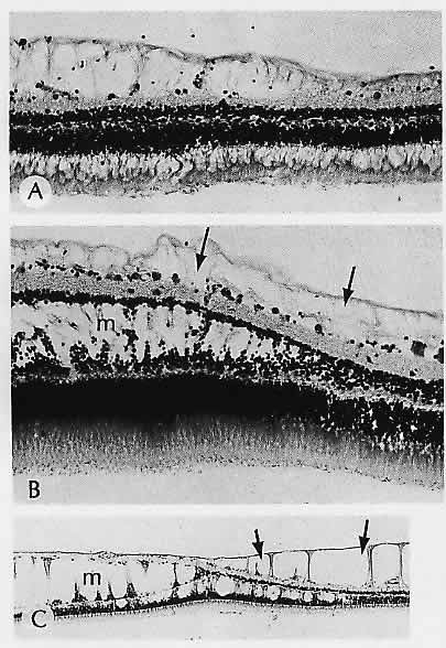
Fig. 14.
Reticular cystoid degeneration. A. Limited to the innermost layers of the retina. B and C. Reticular cystoid
(arrows)
and typical microcystoid degeneration (m) in the same regions of the retina. (A, H&E, × 165; B, H&E, × 165; C, H&E, ×40)