|
|
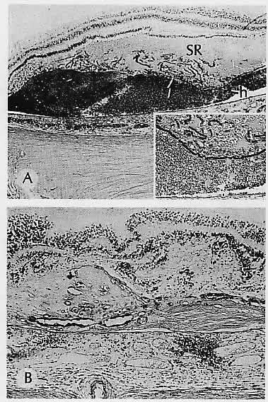
| Fig. 21. Senile disciform macular degeneration. A. Left eye shows a hemorrhage (h) in the subretinal pigment epithelial (RPE) space and eosinophilic coagulum in the subsensory retinal space (SR) in the macular area. The RPE (arrow) has undergone postmortem autolytic changes and is present artifactitiously as short segments in the SR (see higher magnification in inset). B, Other eye from the same patient shows RPE proliferation, disciform fibrous scar, and chronic nongranulomatous choroiditis in the macular area. (A, H&E, × 16; inset, H&E, ×40; B, PAS, ×40) |