|
|
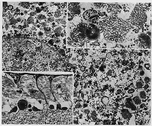
| Fig. 23. Chloroquine retinal degeneration. inset A. A retinal ganglion cell from the ganglion cell layer is shown in light micrograph. The cytoplasm of the ganglion cell, as seen by electron microscopy in the main figure contains myriad clusters of curvilinear structures (CT) and membranous cytoplasmic bodies (MCB). The bodies are better seen in inset B; the suggestion of the continuity of the curvilinear structure with the membranous body is seen at the free arrows. (NUC, nucleus of the ganglion cell.) (Main figure, × 10,200; inset A, 1.5 μm section, toluidine blue, ×380; inset B, ×25,800) |