|
|
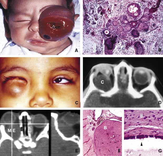
| Fig. 1 Congenital lesions. A very large cystic teratoma of a 1-month old child (A). B. Histology of this lesion that contains a variety of tissues, endodermal, ectodermal, and mesenchymal. An orbital cyst (C) in an orbit containing micro-ophthalmic globe. C, D. The protrusion of the cyst inferiorly creates a mechanical lower lid ptosis that narrows the right maldeveloped conjunctival sac even further. Axial and sagittal CT scan showing a meningoencephalocele (ME) occupying the entire orbit (E). The histopathologic examination of the lesion revealed both meningeal (M) and brain (B) tissues (F). The high-power histopathology reveals ciliated ependymal cells lining some of the cystic spaces (arrowhead) (G). |