|
|
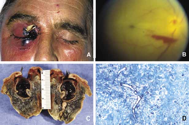
| Fig. 8 Mucormycosis. The appareance of mucormycosis in the right orbit, periorbital skin and maxillary sinuses of a 60-year-old man with diabetic ketoacidosis (A). B shows the funduscopic apperance of a central retinal artery thromboembolism with resultant “cherry-red spot” from another case of orbital mucormycosis. C shows the extensively necrotic, bloodless cut surface of the exenteration specimen from the patient shown in A. Histopathologic examination of this specimen revealed numerous nonseptated mucormycosis hyphae with 90-degree branching (D) in Gomori methanamine silver (GMS) stain. |