|
|
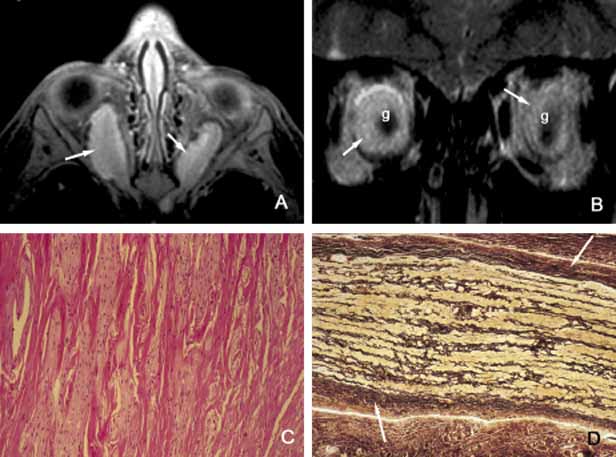
| Fig. 21 Glioma. T1-weighted axial (A) and coronal (B) MRI images showing bilateral optic nerve gliomas (g). Meningoendothelial hyperplasia surround the glioma as depicted in MRI (A, B) and histopathologically with Bodian stain (D). Note the somewhat hyperintense signal of the gliomas in comparison to meningoendothelial hyperplasia depicted in frame A. Histopathologic appearance of the glioma (C) consists of haphazard proliferation of the glial cells with distortion of the pial septae. Glial cells do not display significant atypia because of the low grade of the tumor. |