|
|
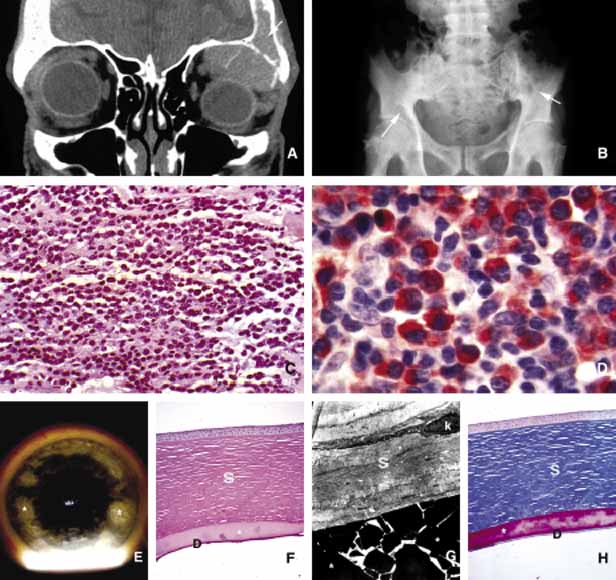
| Fig. 30 Multiple myeloma. Multiple lytic lesions of the bone are depicted in the coronal CT scan (A) and the plain film of the pelvis (B) (arrows). The excisional biopsy sample obtained from the orbital mass contains diffuse infiltrates of plasmocytoid cells (C) that were stained positively for kappa light chain. Immunoglobulin deposits may be seen in other parts of the eye and adnexae in multiple myeloma cases. Frame E depicts light-chain lambda deposits within the cornea in the slit lamp photograph as well as with light and electron microscopy (star) (F, G, H) (S, stroma; k, keratocyte; D, Descemet membrane). (Frame E courtesy of Delmar R. Caldwell, MD of New Orleans, LA) |