
|
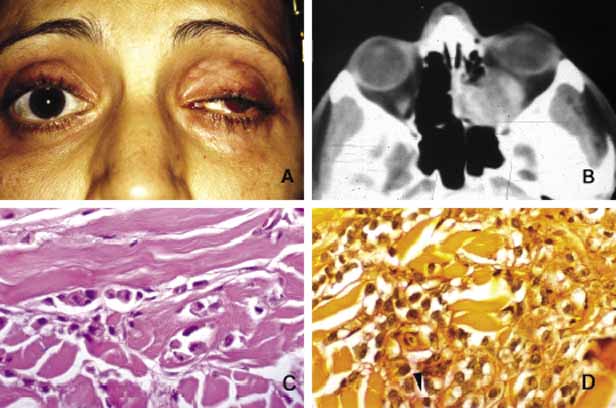
| Fig. 32 Breast metastasis. Metastatic carcinoma in the left orbit and ethmoid sinus (A, B) Note that the left eye does not show proptosis despite the presence of a sizable mass in the posterior orbit that is most likely secondary to the schirrous nature of the tumor (A). The axial CT scan reveals that the medial wall of the orbit and the ethmoidal cells are extensively infiltrated by the tumor (B). Numerous adenocarcinoma cells forming small clusters and single files are identified within the extraocular muscle tissue (C). Mucicarmine stain shows faint pinkish orange mucin deposit within the signet ring cells (arrowhead) to confirm the nature of the neoplasm as mucin secreting adenocarcinoma (D). |