|
|
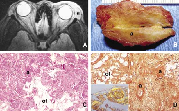
| Fig. 36 Orbital amyloidosis. Axial T2-weighted MRI depicting superior temporal amyloid deposits in anterior orbit (A). Frame B shows an irregular fragment of orbital fibroadipose tissue infiltrated with yellow---orange amyloid deposits. The histopathological appearance of amorphous, acellular deposits of amyloid (a) are shown in between orbital fat (of) in PAS (C) and Congo red (D) stains. (inset). Apple green by the birefringence of perivascular amyloid deposits under polarizing light. (Frames A and B are courtesy of Barrett G. Haik, MD of Memphis, TN) |