|
|
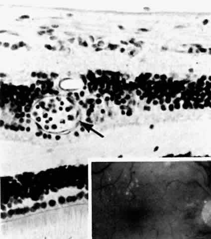
| Fig. 5. Retinal capillary microaneurysm (arrow) is characterized by its thin wall and location in the capillary area of the retina (middle retinal layers) rather than the major vessel area (inner retinal layers). Inset. Fundus appearance of microaneurysms and hard or waxy exudates. (Main figure, H&E, × 176) |