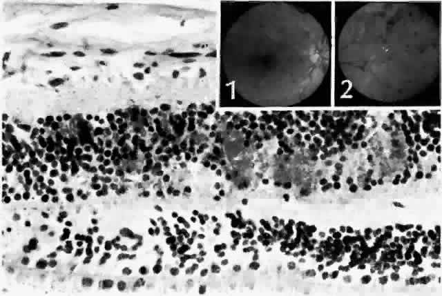
Fig. 9.
Dot and blot hemorrhages consist of small collections of blood in the inner nuclear and outer plexiform layers of the retina.
Insets.
Fundus appearance of dot (
1
) and blot (
2
) hemorrhages, respectively. (
Main figure,
H&E, × 260)