|
|
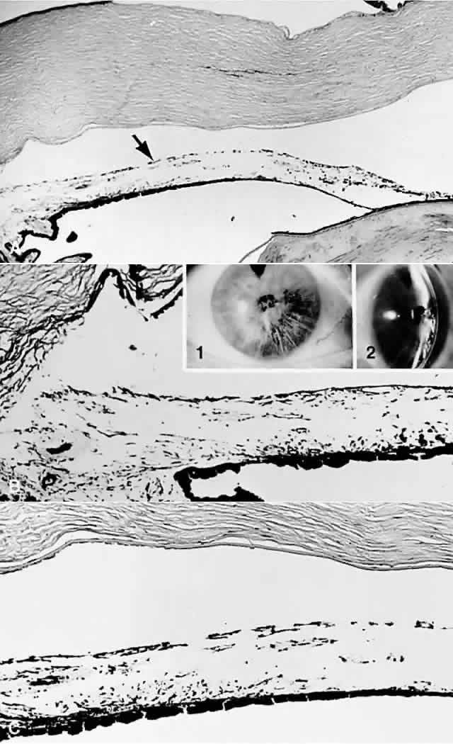
| Fig. 9. Iridoschisis. (A) Anterior iris stromal layers (arrow) are separated widely from the deeper layers. Note the bullous keratopathy. (B and C) Anterior stromal layers are separated from the deeper layers extending from (B) the iris root toward (C) papillary iris. (B, insets) Clinical appearance in another case as viewed (1) directly and (2) with a slit beam. (A, H&E, × 16; B, PAS × 40; inset 1, clinical; inset 2, slit beam; C, H&E, × 40) |