Chapter 22
Bioavailability
JOEL S. MINDEL
Main Menu   Table Of Contents

Search

DEFINITIONS
CAUSES OF BIOINEQUIVALENCE
CLINICAL APPLICATION
REFERENCES

DEFINITIONS
The bioavailability of a drug product is the percentage of the drug that is absorbed. It is assumed that different pharmaceutical manufacturers start with a chemically identical active ingredient and intend to deliver as much of it as possible to the area of absorption. The factors relevant to drug manufacture and drug storage that influence this goal (i.e., the biopharmaceutical aspects) are considered. For example, an overly acidic preparation of mydriatic drops may produce little pupil dilation because of a marked reflex tearing that washes most of the medication away. This is a bioavailability problem because it is a result of drug manufacture. If the increased tearing were the result of a child crying from fear, however, this would not be a bioavailability problem. The child's fear of eye drops could not be blamed on a manufacturing defect. In both instances, the therapeutic result would be the same (i.e., decreased mydriasis) but the mechanism would be different. The term pharmacokinetics is applied to these patient-related factors that influence therapeutic efficacy.

The preceding definition of bioavailability is somewhat arbitrary in the sense that a universally acceptable definition does not exist.1,2 The US Food and Drug Administration definition is as follows:

Bioavailability means the rate and extent to which the active drug ingredient or therapeutic moiety is absorbed from a drug product and becomes available at the site of drug action.

Different definitions are used by the European Economic Council, textbooks, and workshops addressing the subject.3 There is a strong tendency to define bioavailability as the percentage of drug absorbed into the blood. This is because most data have been collected from studies of oral medications, in which absorption was monitored by assaying blood content. To those who prescribe topical medications, however, such as the ophthalmologist and dermatologist, the blood levels are often important only for monitoring side-effects and toxicities, not therapeutic efficacy.

Bioavailability studies are concerned with not only the amount of drug absorbed but also with how the manufacturing process alters both the rate of drug absorption and the consistency of drug absorption. Three brands of a drug may be absorbed completely (i.e., have 100% bioavailability) and yet the first may be therapeutically effective; the second, therapeutically ineffective; and the third, toxic. This would occur (Fig. 1) if the toxic brand was absorbed too rapidly and if the ineffective brand was absorbed too slowly. In the former instance, the therapeutic level would be exceeded; in the latter instance, the therapeutic level would not be attained. Wide variations in the disintegration rates of three oral tablets could produce this problem. Batch-to-batch consistency of absorption is important, especially in treating chronic disease. The ophthalmologist following the intraocular pressures of a glaucoma patient would prefer using a brand that is always 80% absorbed rather than a brand that has an absorption rate that fluctuates between 60% and 100%. In the latter instance, the physician would be unable to distinguish between fluctuations in the disease process and fluctuations in the effectiveness of the medication.

Fig. 1. Variation in therapeutic efficacy from differences in the absorption rate of three brands of the same drug. The total absorption (area under the curve) is the same for all three brands.

Bioavailability data have obvious therapeutic implications. They are only one determinant of therapeutic efficacy and are not therefore a direct measure of therapeutic efficacy. For example, if a patient with streptococcal blepharitis were given 5 million units of a penicillin G preparation with 50% bioavailability, the response would probably be as adequate as though a brand with 100% bioavailability were given—the reason being that the minimum therapeutic blood level would be exceeded in both instances. Because bioavailability does not correlate strictly with the patient's response, attempts to equate the two may lead to erroneous conclusions. Smolen and coworkers4 and Kuehn and coworkers5 have found that the degree of miosis correlates well with the bioavailability of chlorpromazine. This allows pupillometric bioavailability analysis at doses below the maximum sensitivity of the serum assay. Smolen and coworkers note,4 however, that nonbioavailability factors, such as the rate of drug metabolism to pupillometrically inactive molecules or the degree of enterohepatic recycling of active molecules, influence the degree and duration of the miosis.

Back to Top
CAUSES OF BIOINEQUIVALENCE

ORAL MEDICATIONS

Excipients

Different brands that are generically equivalent must have the same amounts of the same active ingredient. Generic equivalents may have totally different excipients, however. Excipients are the therapeutically inactive components of medications:

  • Diluents to give bulk (e.g., lactose, mannitol, cellulose)
  • Binders to give cohesive strength (e.g., gums, gelatin)
  • Lubricants to reduce friction as the tablet is ejected after compression (e.g., oleate)
  • Glidants and antiadherents to improve the flow of granules across the tablet presses (e.g., talcum)
  • Disintegrants to aid the tablet's disintegration in body fluids (e.g., starches)
  • Colorants
  • Flavorants and sweeteners
  • Adsorbents to absorb water and yet maintain the tablet in an apparently dry state (e.g., silicon dioxide)

Although excipients have not received a great deal of attention in the past, it has become evident that they must be included as important determinants of bioavailability.6–8 Even though therapeutically inactive, they can significantly alter the stability and absorption of the active ingredient.

Lederle's Diamox 250-mg tablet contains 250 mg acetazolamide but weighs 600 mg. Less than half of the tablet is the active ingredient. The same is true of the other carbonic anhydrase inhibitors. Daranide (Merck) contains 50 mg dichlorphenamide but weighs 100 mg. Neptazane (Storz) contains 50 mg methazolamide but weighs 150 mg. Different manufacturers may use different excipients, and the same manufacturer may change the excipients from batch to batch. There are virtually no legal requirements governing their use. The percentage of active ingredient in many commonly used medications can be calculated from the data shown in Table 1.


TABLE 22-1. Amount of Excipient in Commonly Used Medications


Generic NameBrand NameTablet Weight (mg)% Excipient
Chlordiazepoxide, 25 mgLibrium (Hoffman-La Roche)253 ± 490
Chlorothiazide, 500 mgDiuril (Merck Sharp & Dohme)555 ± 310
Dexamethasone, 0.5 mg-(Philips Roxane)101 ± 299
Digoxin, 0.25 mgLanoxin (Burroughs Wellcome)122 ± 199
Erythromycin, 250 mgErythrocin (Abbott)734 ± 1056
Hydrochlorothiazide, 50 mgOretic (Abbott)170 ± 271
Prednisone, 5 mgDeltasone (Upjohn)98 ± 195
Prednisone, 50 mgDeltasone (Upjohn)387 ± 387
Propranolol, 10 mgIndural (Ayerst)121 ± 291
Propranolol, 50 mgIndural (Ayerst)210 ± 281


Variations in Dissolution Rate

An orally ingested medication must break up into numerous smaller particles (disintegration rate) and these in turn must go into solution in the intestinal fluids (dissolution rate). Several variables in drug manufacturing reveal themselves by altering this “common final pathway”—the dissolution rate.

COMPRESSION FORCES. The compression forces of the machines stamping out the tablets determine how rapidly they disintegrate. Levy found that the United States Pharmacopeia (USP) disintegration times for a series of commercial aspirins varied from less than 10 seconds to more than 4 minutes.9

PARTICLE SIZE. Depending on how the tablet is manufactured, the size of the drug particles produced by disintegration varies. The smaller the particles, the greater is their combined surface area and the more rapid is the dissolution time.10 Micronized sulfadiazine and griseofulvin are absorbed more rapidly and produce higher blood levels than the nonmicronized forms.11 In 1968, chloramphenicol brands were shown to be bioinequivalent because of variations in particle size.12

ALTERATION OF WATER SOLUBILITY. Using a drug salt instead of its free base or acid alters its dissolution rate. The blood level of potassium penicillin V is more than 65% greater than that of penicillin V acid.13 Excipients, such as bicarbonate, change the pH of the fluid bathing the dissolving particles. In this way, buffering of aspirin, which may or may not decrease gastric irritation, increases blood levels.

CRYSTAL FORM. Many drugs exist in more than one crystalline form; they are polymorphic. At a given temperature and pressure, one form is the most stable and goes into solution less readily. Different brands may contain different forms of drug crystals.14,15 The result is generic equivalents with varying bioavailability. Acetazolamide exists in two polymorphic forms that differ only slightly in their dissolution rates, crystal structures, and solubilities.16 Therefore, in this instance polymorphism is of little clinical consequence.

HYDRATION STATE. Crystals of a drug in the anhydrous state are usually more unstable and go into solution more rapidly than when the molecules are already hydrated. Ampicillin in the anhydrous state has been shown to enter solution more rapidly than ampicillin trihydrate.17

A list of oral medications with known bioavailability problems, compiled from two review articles, is given in Table 2.18,19 The US Food and Drug Administration has reviewed nonantibiotic medications and has compiled a list of 110 drugs it considers to have actual or potential bioavailability problems.20 Of particular interest to the ophthalmologist is the inclusion of 12 corticosteroids and all four carbonic anhydrase inhibitors (Table 3). This is not to say that different brands of carbonic anhydrase inhibitors are always therapeutically inequivalents but that significant differences in the amounts, and the rates of absorption frequently occur.21–23 Schoenwald and coworkers22 showed that sustained release forms of acetazolamide were 40% to 70% less well absorbed than was a rapid-release brand. Acetazolamide has also been shown to interfere with the absorption of the anticonvulsant primidone.24


TABLE 22-2. Oral Medications With Bioavailability Problems


AcetaminophenOxytetracycline
Acetohexamidep-Aminosalicylic acid
AminophyllinePenicillin G
Amphotericin BPenicillin V
AmpicillinPentaerythritol tetranitrate
AspirinPentobarbital
BishydroxycoumarinPhenacetin
ChloramphenicolPhenylbutazone
ChlordiazepoxidePhenytoin
DextroamphetaminePrednisolone
DiazoxidePrednisone
DiethylstilbestrolQuinidine
DigoxinReserpine
ErythromycinRiboflavin
Ferrous sulfateSalicylamide
GriseofulvinSecobarbital
HydrochlorothiazideSpironolactone
HydrocortisoneSulfadiazine
IndomethacinSulfamethoxazole
IsoniazidSulfisoxazole
LevodopaTetracycline
MeprobamateTheophylline
MethandrostenoloneThyroid
MethaqualoneTolbutamide
MethylprednisoloneTriamterene
Nalidixic acidWarfarin
Nitrofurantoin 
(Poole JW: Drug formulation and biologic availability. Semin Drug Treatment 1:148, 1971; Koch-Weser J: Drug therapy: Bioavailability of drugs [second of two parts]. N Engl J Med 291:503, 1974.)


TABLE 22-3. Oral Medications With Known or Potential Bioavailability Problems


Antiarrhythmics Reserpine Dyphylline Estrone
 Procainamide Spironolactone Oxtriphylline Estrogenic substances
 Quinidine Syrosingopine Theophylline Ethinyl estradiol
Anticoagulants TrichlormethiazideCarbonic anhydrase inhibitorsHemostatics
 BishydroxycoumarinAntiinfective agents Acetazolamide Menadione
 Warfarin Nitrofurantoin Dichlorphenamide Phytonadione
Anticonvulsants Sulfadiazine EthoxzolamideHypoglycemics
 Ethosuximide Sulfadimethoxine Methazolamide Chlorpropamide
 Ethotoin SulfamerazineCardiac glycosides Tolbutamide
 Mephenytoin Sulfamethoxypyridazine AcetyldigitoxinTranquilizers
 Methsuximide Sulfaphenazole Digitoxin Chlordiazepoxide
 Paramethadione Sulfapyridine Digoxin Chlorpromazine
 Phenacemide Sulfasalazine Getalin Fluphenazine
 Phensuximide Sulfisomidine Lanatoside C Prochlorperazine
 Phenytoin SulfisoxazoleCorticosteroids Promazine
 Primidone Trisulfapyrimidine Betamethasone Promethazine
 TrimethadioneAntiinflammatories Cortisone Thioridazine
Antihypertensive/diuretics Oxyphenbutazone Desoxycorticosterone Trifluoperazine
 Alseroxylon Phenylbutazone Dexamethasone Trifluopromazine
 Bendroflumethiazide Sulfinpyrazone Fludrocortisone Trimeprazine
 BenthiazideAntineoplastics FluprednisoloneMiscellaneous
 Chlorthiazide Chlorambucil Hydrocortisone Ethisterone
 Chlorthalidone Methotrexate Methylprednisolone Imipramine
 Deserpidine Triethylene melamine Paramethasone Isoproterenol
 Hydralazine Uracil mustard Prednisolone Liothyronine
 HydrochlorothiazideAntituberculars Prednisone Methaqualone
 Hydroflumethiazide Aminosalicylic acid Triamcinolone Methyltestosterone
 Methychlorthiazide Benzoyl p-aminosalicylic acidEstrogens Probenecid
 Polythiazide Aminosalicylate Dienestrol Propranolol
 Quinethazone Phenylaminosalicylate Diethylstilbestrol Propylthiouracil
 Rauwolfia serpentinaBronchial dilators Estradiol Pyrimethamine
 Rescinnamine Aminophylline  Sulfoxone
(US Food and Drug Adminsitration: Holders of Approved New Drug Applications for Drugs Presenting Actual or Potential Bioequivalence Problems, publication No. [FDA] 76-3006. Rockville, MD: US Department of Health, Education and Welfare, 1976.)


Aging

Bioavailability is not static. A given drug preparation may have altered bioavailability with time due to changes that collectively are called aging.

CHANGING CRYSTAL FORM. Drugs that exhibit polymorphic crystal structure change with time to the crystal form most stable at the temperature and pressure of storage. Generally, this results in a decreased rate of dissolution and a lower blood level of drug.

INTERACTION WITH AIR AND MOISTURE. Different brands containing different excipients and stored under different conditions absorb moisture, atmospheric gases, and light at different rates. Water can alter drug hydration and stability. Atmospheric CO2 and H2O can react with calcium to form calcium bicarbonate and rock-hard tablets. Oxygen oxidizes drugs. Light can decompose molecules directly or indirectly. For example, Baugh and associates8 reported that phenylbutazone's instability in the presence of light depended on the colorant. Different batches of a drug that were chemically and physically identical at the time of manufacture may become dissimilar. Edelman and coworkers25 note that the USP recommendations on packaging and storage of nitroglycerin tablets were limited only to “preserve in tight containers.” Loss in potency of less than 5% was demonstrated when the nitroglycerin was stored 201 days in amber or clear glass vials. The corresponding loss in potency when tablet boxes were used was 72.4%.

TOPICAL MEDICATIONS

Solutions

EXCIPIENTS.

Ophthalmic solutions contain buffers, antimicrobial agents, antioxidants, salts, and detergents. A partial listing of commonly used additives is given in Table 4. Excipients in ocular solutions can influence bioavailability in many ways.


TABLE 22-4. Excipients Frequently Added to Ophthalmic Solutions


BenzalkoniumPhenylmercuric acetate and nitrate
BenzethoniumPolyoxypropylene-polyoxyethelenediol
Boric acidPolysorbate 80
ChlorbutanolPropylparaben
CreatineSodium bisulfite
Ethylenediaminetetraacetic acid (EDTA)Sodium carbonate
GlycerineSodium citrate
Hydrochloric acidSodium nitrate
MannitolSodium phosphate
MethylcelluloseSodium sulfite
MethylparabenTyloxapol
Oxine sulfateZinc sulfate
Phenylethyl alcohol 


PH. The pH of a drug solution determines whether the drug is in the ionized or nonionized form. The nonionized form, being more lipid-soluble, is better able to penetrate the cell membrane of the corneal epithelium. The manufacturer, however, may not choose to use a pH favoring the nonionized form because of drug stability: the ionized form of the drug is more stable and has a longer shelf life.

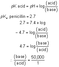
The pH of tears is about 7.4. Penicillin, an acid, exists at this pH primarily in the dissociated form, the relative proportions of charged and uncharged molecules being calculated by the HendersonHasselbach equation:

There is only one molecule of undissociated penicillin for every 50,000 dissociated molecules. This is one important reason why penicillin eye drops do not penetrate the intact corneal epithelium well. Conversely, the pKa of sulfamethazine is 7.4:

Half the molecules of sulfamethazine are undissociated at the pH of the tears, which is a far better fraction than that of penicillin. It is understandable why so many topical eye medications are “alkaloids”—they tend to be nondissociated at the pH of tears.

The pH of the eye-drop preparation and the degree to which the preparation is buffered have an obvious influence on tear pH and drug penetration. Mitra and Mikkelson26 found that as the concentration of citrate buffer increased from 0 to 0.11 mol, the miosis from 1.0% pilocarpine nitrate pH 4.5 eye drops decreased. Presumably, the higher the buffer concentration, the longer the time for the precorneal tear film to return to physiologic pH and the higher the ratio of dissociated to nondissociated molecules of pilocarpine. Hammarlund and coworkers27,28 manipulated this variable in a clever way. They prebuffered the tear film of volunteers using a pH 9.2 solution and found that they could then give one tenth the amount of cyclopentolate, phenylephrine, or homatropine that is usually administered (e.g., 0.1% cyclopentolate instead of 1% cyclopentolate) and still obtain the same pharmacologic response. Smith and coworkers29 and Saari and coworkers30 circumvented the effect of eye-drop pH by applying pilocarpine base in castor oil. The eyes of volunteers given the oil preparation had more marked and longer-lasting miosis-hypotension than did eyes given an equimolar water solution of pilocarpine nitrate.

Riegelman and Vaughan31 reviewed the effect of pH on ocular discomfort. They state that the buffering capacity of the tears is adequate to bring unbuffered solutions of pH 3.5 to 10.5 to within “tolerable” (i.e., not painful) limits almost immediately. The greater the degree to which the solution is buffered, the more narrow is the range of comfort. It is not clear how far the pH of a solution must deviate from that of tears to induce a reflex increase in lacrimation, however. That is, it is unknown whether the solution must be subjectively painful to induce increased lacrimation. It is possible that even though the pH does not cause pain, it may initiate an increase in reflex tearing that removes additional drug.

Not only may a buffer alter the proportion of nondissociated drug and the amount of reflex tearing, but there may be drug-buffer interactions unrelated to pH. Apel and Horsch32 found that at the same pH, pilocarpine 2% solutions were more stable in acetate buffer than in borate buffer.

VISCOSITY. Excipients such as hydroxypropyl methylcellulose and polyvinyl alcohol are frequently added to increase the viscosity of ophthalmic solutions. Presumably, drug-corneal contact time is prolonged by retarding lacrimal drainage. Although simple in concept, this has been a controversial area. Some reports state that viscosity increases drug contact time and bioavailability.33–35 Other reports state that increases in viscosity are of little or no value.36,37 To add to the confusion, some experiments indicate hydroxypropyl methylcellulose is therapeutically superior to polyvinyl alcohol, whereas other experiments give the opposite result.38–40 Swanson and coworkers41 reported an intermediate position: hydroxypropyl methylcellulose was superior to polyvinyl alcohol for the 15- to 30-minute period after drop instillation but polyvinyl alcohol was superior to hydroxypropyl methylcellulose for the 2- to 8-hour period after drop instillation.

The explanations for these discrepancies have been multiple and not entirely satisfactory. Species differences in tear, blink, and corneal physiology have been invoked by those who state that human and rabbit studies are not comparable. Wang and Hammarlund28 added to both the validity and the complexity of this argument by reporting that 0.5% hydroxypropyl methylcellulose was of little value in whites but of significant value in Asians. Another explanation has been that both polyvinyl alcohol and hydroxypropyl methylcellulose exist as polymers of varying size and they tend to coagulate further on heating. Perhaps the solutions of 1.4% polyvinyl alcohol and 0.9% hydroxypropyl methylcellulose used in different laboratories were nonequivalent. A third explanation has been that in those instances in which viscolizers are of value, the effect may have been due not to prolonged contact time but to better initial mixing of the drug with the tears.

Patton and Robinson42 have provided some important insights that may ultimately resolve the preceding controversies. They were careful to work in units of viscosity (cs) rather than percent concentration of viscolizer. They found that when polyvinyl alcohol and hydroxypropyl methylcellulose were compared on a viscosity basis, there was little difference in their influence on drug bioavailability. Furthermore, the effect of increasing the concentration of viscolizer was nonlinear relative to viscosity. For example, increases in polyvinyl alcohol up to 3% resulted in only small increases in viscosity and corneal contact time. Beyond 3%, however, there were rapid increases in the viscosity and contact time of polyvinyl alcohol, presumably because of increased hydrogen bonding between molecules. Further increases beyond 5% and up to 10% were of little additional value. An optimum effect of viscosity was found beginning at 12 cs (Fig. 2). Commercial preparations of 0.5% hydroxypropyl methylcellulose exceeded this minimum, whereas 1.4% polyvinyl alcohol solutions did not. Patton and Robinson42 have also noted that a highly viscous solution may not be especially useful because of the shear created by blinking. This could cause the tear film to thin to the extent that large increases in viscosity may provide only slight increases in contact time.

Fig. 2. Relation of the manner in which increasing the viscosity of a solution increases the corneal contact time. A nearly optimum effect occurs at 12 to 15 cs.

SURFACTANTS. Detergents are added to ocular solutions for several reasons: they increase the solubility of drugs that are relatively hydrophobic; they may act as preservatives because of their antibacterial activity; or they may be added for their ability to partially break down the barrier presented by the corneal epithelium, thereby enhancing drug penetration and bioavailability.

Benzalkonium chloride is the detergent most commonly used as a preservative in ophthalmic solutions. It is a cation and usually used in low concentrations (e.g., 1/10,000). If concentrations of 1/3000 or stronger are used repeatedly, there is significant protein denaturation. This is because the positively charged benzalkonium interacts with the negative charges on the amphoteric corneal proteins. It also interacts with anions such as fluorescein and sulfonamides and is therefore incompatible.43 Smolen and coworkers44 reported that the bioavailability of carbachol was enhanced significantly in the presence of 1/5000 benzalkonium chloride. Rabbits given a 0.1% carbachol solution with 1/2500 benzalkonium chloride obtained as much miosis as if given a 2% carbachol solution without surfactant. Tonjum45 has used the electron microscope to study the effect of benzalkonium chloride on rabbit corneal epithelium. He found that this surfactant broke down the tight junctions of the epithelial cells and allowed penetration of horseradish peroxidase. Similar increases in drug penetration have been reported by others using this and other (e.g., cetyl pyridinium chloride) ionic detergents.46–50

OSMOTICS. Agents may be added to ophthalmic solutions to adjust their tonicity to that of tears, 0.9% sodium chloride.51 If the tonicity deviates too far from this value, the pain produced evokes a reflex tearing that washes the drug from the eye. The range of acceptable tonicities for comfort appears to be wide. Riegelman and coworkers52 found that volunteers did not complain of discomfort when sodium chloride concentrations from 0.5% to 2% were instilled. It is not known whether awareness of pain is needed to evoke increments in reflex tearing, however.

ANTIOXIDANTS. The oxygen in the atmosphere can oxidize many drugs (e.g., phenylephrine and epinephrine) to inactive molecules.53 Bisulfite is an antioxidant that has been added to epinephrine solutions to prevent degradation, yet bisulfite reacts with epinephrine to form 1-(3,4-dihydroxyphenyl)-2-methylaminoethanesulfonic acid.54 The presence or absence of antioxidants can thereby alter the therapeutic efficacy of different epinephrine preparations. Haddad and coworkers55 noted that 10% phenylephrine commercial solutions had a potency equivalent to freshly prepared solutions of 1% to 5% phenylephrine.

AGING. If the same drug were prepared as both an ocular solution and an oral tablet, the former would be most likely to have aging problems. Drugs tend to be less stable in water. One reason is that water can react with several drugs directly. Pilocarpine can be hydrolyzed to an inactive form, as can the local anesthetics and all of the phosphorylating (e.g., echothiophate) and carbamylating (e.g., physostigmine) cholinesterase inhibitors. Oxygen may also react more readily with drugs in solution; for example, catechols such as epinephrine are oxidized to adrenochrome. Impurities in the glass or plastic container may slowly be leached into the solution with time. These impurities (e.g., polymerization residues, plasticizers, pigments, and lubricants) may alter the drug structure or the physical characteristics of the drug solution.

Chloramphenicol eye drops are sold in opaque or heavily tinted—usually amber—containers. This reduces photochemical decomposition from light wavelengths of 285 mm or longer.56 Exposure of an unprotected 0.25% chloramphenicol solution for four minutes to sunlight equivalent to that of a normal summer day decomposes 8% of the drug to p-nitrobenzaldehyde. The presence or absence of a mercuric preservative is not a factor. Such decomposition may affect drug toxicity and efficacy: it has long been proposed that nitroso breakdown products of chloramphenicol are the cause of its associated aplastic anemia. Although not the subject of this chapter, it is interesting to speculate what may be happening to chloramphenicol within the aqueous humor—whether absorbed from topical application or oral ingestion—while the well-medicated patient sits reading in a brightly lit room.

Drop Volume

The size of the eye drop is a bioavailability factor with easily recognizable economic implications. When the eye-drop volume exceeds that which can be retained by the lids and conjunctival sac, the excess fluid runs down the face. The ocular bioavailability of the overflow is zero. Drop size is not only a function of the bottle tip or dropper design but also of the viscosity and temperature of the solution and the angle at which the bottle is held.57 For example, the greater the viscosity, the larger the drop volume. These factors resulted in one timolol preparation being delivered in drops of 28 μl average size and levobunolol in 46-μl drops.58 To address the effect of tip of manufacturing, the bottles were refilled with saline; the respective average volumes of the timolol and levobunolol drops became 48 and 55 μl.

A different situation exists if the drop volume is less than the maximum that can be held by the lids. A single 5-μl drop of 0.5% tropicamide was a less effective mydriatic than a 16-μl drop.59 This deficiency was overcome, however, by increasing the drug concentration in the micro drop (e.g., to 1%).

Suspensions are saturated solutions of a drug in which excess molecules, unable to enter solution, exist as small particles or crystals. These particles represent a drug depot. For this depot to be useful, however, it must enter solution. This can occur in two ways: either by increasing the water volume of the solution or by removing the drug in solution. If the preparation is mixed with a solution not containing drug (e.g., tears), it is no longer saturated and additional drug can diffuse out from the depot. This is important therapeutically only if the depot particles are kept in the conjunctival sac long enough for diffusion to take place there. The particles are of no value if they release their drug after entering the lacrimal duct system. The second way the depot can contribute drug is for the molecules already in solution to be rapidly absorbed by the corneal epithelium. Without changing volume, this produces desaturation of the solution and promotes diffusion from the depot. Examples of suspensions used in ophthalmology are the “forte” preparations of glucocorticosteroids. Little documentation exists to support the belief that significant amounts of depot drug are absorbed. Sieg and Robinson60 found that suspensions of the highly lipophilic corticosteroid fluorometholone gave higher and more prolonged levels of drug in rabbit aqueous humor than did saturated solutions. In those instances in which suspensions are superior to solutions, the cause may be that the depot particles rub the corneal epithelium and alter the epithelial barrier by their irritant properties. It has been observed that although patients do not usually complain of discomfort from suspensions, if one permits a basis of comparison by giving one eye a glucocorticoid solution and the other a glucocorticoid suspension, most subjects state that the latter is more irritating.61 To minimize irritation, manufacturers generally attempt to keep particle size less than 10 μm.

Two factors influencing depot drug bioavailability are particle size and excipient-particle interaction. The smaller the size of the particles, the greater is their total surface area and therefore the more rapid is the diffusion of depot drug molecules into solution. Howard and coworkers62 and Schoenwald and Stewart,63 studying prednisolone acetate and dexamethasone suspensions, found this relation valid. In addition, they found that the 0.2% hydroxypropyl methylcellulose present in some suspensions retarded the dissolution of the depot prednisolone acetate. In a suspension without hydroxypropyl methylcellulose, 20% of the prednisolone acetate dissolved within 100 seconds; in an identical suspension with hydroxypropyl methylcellulose, 270 seconds were required.

Contemporary suspensions largely consist of particles of insoluble drug molecules. Pharmaceutical manufacturers also can create suspensions with nondrug particles, however.64 These particles (less than 10 μm or they cause patient discomfort), have a porous or solid matrix to which the drug is absorbed or trapped. The particles can consist of a natural polymer (e.g., albumin) or a synthetic one (e.g., a polyalkylcyanoacrylate). The drug can be present during the particles' formation or added after they are formed. Alternatively, micro- or nanocapsules can be made. These capsules consist of a central solid or liquid drug reservoir surrounded by a polymer membrane.

Ointments

The drug molecules on the surface of an ointment are immediately accessible to the tear fluid and can easily enter solution. The bulk of the drug is trapped in the ointment, however. Ointments can be either water- or oil-based. A common oil-based ophthalmic ointment consists of petrolatum and mineral oil—the latter being added to promote melting and flow of the ointment at body temperature. The problem of trapped depot drug is greatest in these oil-based ointments. Riegelman65 noted that the solubility of most drugs in commercial petrolatum eye ointments is low. As a result, most of the medication is present as small solid microcrystals within the ointment. To escape, the drug must diffuse through the petrolatum, a slow process. As a result, over a prolonged period (e.g., while the patient sleeps) more drug may get into the tear film from an ointment than from a single drop but the unanswered question is whether the amount of drug released reaches therapeutic levels (Fig. 3). Different brands of neomycin ointment may contain 5 mg/g neomycin; however, their bioavailability can differ, depending on the number and size of drug particles on the surface, the number and size of drug particles within the ointment, and the composition of the ointment.

Fig. 3. Less total drug (area under the curve) is absorbed from a single drop than from a single application of ointment. In this example, however, the drop achieves therapeutic levels, whereas the ointment does not.

Sieg and Robinson60 studied the aqueous humor levels of radiolabeled fluorometholone in rabbits using solutions, suspensions, and ointments. They found that although the appearance of drug in the aqueous humor was delayed, the ointment gave peak levels comparable to those of the solutions and suspensions. This peak level was sustained for a longer period (about 3 hours versus 1.5 hours) than was found for the suspension.

Hendrickson and Hanna66 found little difference in the induction time needed for mydriasis when volunteers were given cyclopentolate, tropicamide, or phenylephrine either in the usual concentrations as solutions or in smaller quantities as ointments. In addition, the duration and degree of mydriasis-cycloplegia did not improve with ointments.67 Nine hours after instillation, a single dose of 4% pilocarpine hydrochloride gel was no more effective in lowering intraocular pressure than was a single drop of 4% pilocarpine hydrochloride solution.68 The gel produced a clinically effective ocular hypotension for a longer time, however.69

It appears that sweeping generalizations that ointments are better or worse than solutions cannot be made. The physical and chemical characteristics of each drug and each ointment must first be examined, and superiority must be specified in terms of rapidity of onset of action, peak tissue levels, and duration of action.

Water-based ophthalmic ointments are called hydrogels. Usually they are made up of polymers that swell in water.70 Adding small amounts of such polymers to a solution increases its viscosity. Adding larger amounts results in a gel. Two types of hydrogels are manufactured for clinical use. One is preformed gels that are in tubes. The other type are formulated as liquids and are applied as eye drops; physiologic conditions within the tears convert the polymers to gels. This latter type of in situ-formed gels have the advantage of being able to be applied in more precise doses than ointments squeezed from tubes. Tube-dispensed preformed gels can be made up of such polymers as carbomer (e.g., pilocarpine in Pilopine HS [Alcon Laboratories]), hyaluronic acid, polyvinyl alcohol, or cellulose. In situ-formed gels can be manufactured using gellan gum (e.g., timolol in Timoptic XE [Merck]), poloxamer, or nanoparticle dispersions of cellulose acetate phthalate. Gellan gum is an anionic polysaccharide that gels when exposed to cations in the tear film; reflex tearing helps retain the drug rather than to wash it away. Poloxamer gelling depends on the higher temperature of the tears. Dispersions of cellulose acetate phthalate gel because of the higher pH of lacrimal fluid, which neutralizes the acid groups on the polymer.

Membrane Systems

Membrane systems have been developed to provide both a steady rate of drug delivery and an improved ratio of therapeutically useful drug. Solutions give a pulse of medication. The therapeutic level is exceeded and then falls with time. Suspensions and ointments, to the degree that their depots release medication, provide a more even and prolonged form of therapy. Drugs contained within a membrane and placed in the conjunctival sac should produce a more even (i.e., approaching zero order) release of drug. The rate of drug release depends on the lipophilic or hydrophilic nature of the membrane, pore size, and membrane thickness. Small amounts of continuously released drug are less likely to be lost in the lacrimal drainage.

The advantages of membrane therapy over solutions and suspensions are somewhat mitigated by the ability of the cornea to act as a depot. Aqueous humor drug levels may be more prolonged after drop therapy than would otherwise be expected because significant amounts diffuse from the corneal depot into the aqueous humor.

If the drug is maintained within the membrane as an anhydrous central-core reservoir, drug stability is enhanced and the possibility of contamination, compared with solutions, is reduced. Excipients, such as preservatives, and their effects can be avoided. For example, Longwell and coworkers71 note that solutions of pilocarpine salts (e.g., pilocarpine hydrochloride or pilocarpine nitrate) are maintained by the manufacturer at acidic pH to promote drug stability. This favors formation of the ionized form of the drug, however, which does not penetrate the corneal epithelium well. In a commercially available membrane system (Ocusert [Alza]), pilocarpine exists in the reservoir as the unionized base. This gives the membrane two advantages: the most lipid-soluble form of the drug is being delivered, and the tear pH is not acutely lowered. The authors suggest that this may partially explain why the small amounts of drug delivered by this device are therapeutically effective. Urbanyi and coworkers72 found that the Ocusert membrane system contained less of the therapeutically inactive stereoisomer isopilocarpine than did many solutions of pilocarpine. Additional advantages and disadvantages of this particular membrane system (Ocusert) unrelated to bioavailability have been mentioned in articles by Pollack and coworkers73 and Hennig.74

Soft contact lenses, dipped in solutions of drugs, have been used as drug reservoirs by Podos and coworkers,75 who studied pilocarpine, and by Kaufman and coworkers,76 who studied fluorescein. Their findings were similar. The former found that a single set of 0.5% pilocarpine eye drops did not reduce ocular hypertension. If the pilocarpine drops were placed on a soft contact lens after the lens had been inserted, little effect was discernible. If the lens had been presoaked in 0.5% pilocarpine for 2 minutes and worn 23 hours, however, a significant reduction in intraocular pressure remained when the lens was removed at the end of this period. In 10 patients so treated, the mean intraocular pressure before contact lens insertion was 29 mmHg in the right eye and 28 mmHg in the left eye. Twenty-three hours later, the mean intraocular pressure in the right eye, which had worn the 0.5% pilocarpine-soaked lens, was 21 mmHg, whereas the mean intraocular pressure in the left eye, which had worn a saline-soaked lens, was 27 mmHg. Maximum contact lens uptake of pilocarpine occurred 30 to 60 minutes after placing the lens in the solution; about 40% of this maximum value was achieved within 2 to 4 minutes of soaking. After 30 minutes of contact lens wear, about 60% of the drug had been lost from the lens. After 2 hours of wear, about 75% of the drug had been lost. If the lens were presoaked for 2 minutes in 0.5% pilocarpine and worn only 1 hour, a mean intraocular pressure of 21 mmHg was found 23 hours later (baseline mean pressure, 26 mmHg). Krohn and Breitfeller77 used a 30% gel of non-cross-linked soft contact lens polymer. In an in vitro rabbit corneal system, the gel gave the same transcorneal flux of pilocarpine as a presoaked soft contact lens for periods of up to 90 minutes. Beyond that, the soft contact lens was much more effective. The transcorneal flux from an 8% pilocarpine drop was equivalent in the gel and soft contact lens for the first 20 minutes after administration. Presumably, the superiority of the lens resided in its physical state rather than in any chemical differences. Differences in the soft contact lenses of various manufacturers may be expected to produce different results. McCarey and coworkers,78 studying gentamicin release in an in vitro model, found that the drug diffusion rate was not simply proportional to the water content of a hydrogel lens, however. The main pathway for drug movement was not through the lens but around its edge.

If a contact lens with 40% water content were soaked overnight in 0.5% gentamicin sulfate and placed on a normal eye, therapeutic drug levels against Pseudomonas species were found in the cul-de-sac tear fluid for at least 72 hours.79 Jain80 was able to measure therapeutically effective levels of gentamicin in the aqueous humor for up to 6 hours after patients had worn a presoaked hydrogel contact lens for a half hour before cataract surgery.

Collagen shields are dehydrated contact lens-shaped preparations, usually of porcine or bovine collagen. The shield can either be manufactured with water-insoluble drugs (e.g., cyclosporine) in them or can be rehydrated with solutions containing water-soluble drugs (e.g., aminoglycosides). Once placed on the patient's cornea, the shield dissolves over a period up to 72 hours, depending on the manufacturing process. The rate of a drug's release from the shield depends on its solubility, its binding to collagen, and the rate that the shield dissolves. Collagen shields immersed in 0.01% sodium fluorescein for 10 minutes provided patients with (1) higher aqueous humor levels (as measured by fluorophotometry) at 2 and 4 hours than 0.01% eye drops given every 30 minutes, and (2) higher levels than a daily wear contact lens soaked in 0.01% fluorescein.81

Liposomes, which are synthetic phospolipid vesicles, absorb to corneal epithelium cell membranes and directly transfer the drug they contain.82 Liposomes can improve bioavailability only to the degree that they release their sequestered drug. Liposomal epinephrine bioavailability is less than that of epinephrine eye drops, whereas liposomal iodoxuridine efficacy is superior to that of drops.83,84 The primary limitations of liposomes are limited binding to corneal epithelium and expense.85 One attempt to improve binding has been the incorporation of antibodies (i.e., the creation of site-specific immunoliposomes).86 Incorporation of monoclonal antibodies to herpes simplex virus has allowed targeted release of acyclovir in infected corneas. Another approach has been the use of temperature-sensitive liposomes.87 Application of microwave irradiation at the corneal limbus after intravenous administration of liposomes presumably raised the ciliary body temperature and caused drug release. The concentration of cystosine arabinoside in the aqueous humors of treated rabbit eyes was four times greater than the concentration in contralateral unheated control eyes.

Surgically placed nonbiodegradable intravitreal membrane systems have been developed for sustained release of ganciclovir during treatment of cytomegalovirus retinitis.88 Ganciclovir can diffuse through a membrane of polyvinyl alcohol but not through a membrane of ethylvinylacetate. The rate of release of ganciclovir into the vitreous is determined by the rate of drug diffusion through the polyvinyl alcohol core and the amount of the surface of that core that is not covered by ethylvinylacetate.

Pro-Drugs

Improved drug delivery has been achieved by chemically transforming active drug molecules into derivatives called pro-drugs that more readily enter the body and are then reconverted to the parent compound. N-Mannich bases of acetazolamide have enhanced aqueous solubility and dissolution rates.89 The dihydro form of 2-PAM is a more lipophilic tertiary amine that once in the eye can be rapidly oxidized to 2-PAM.90 Dipivefrin is a commercially available pro-drug of epinephrine. Two pivalic acid molecules are joined by ester bonds to epinephrine. The resultant compound penetrates the cornea about 20 times as well as the parent structure. Diesters of pilocarpic acid act as pro-drugs and may enhance pilocarpine bioavailability.91

PARENTERAL MEDICATIONS

It is generally assumed that bioavailability is not a problem for parenterally administered drugs. Intramuscular injections of digoxin, phenytoin, chlordiazepoxide (Librium [Roche Products]), and diazepam (Valium [Roche Products]) produce lower blood levels and poorer clinical responses than similar oral doses, however.92,93 The explanation for these surprising findings is unknown.

Back to Top
CLINICAL APPLICATION
It is likely that many bioavailability problems are not identified as such because clinicians have not been trained to question the quality of the drugs they use. When a disease fails to respond to therapy, physicians consider the following possibilities: the disease has progressed, the choice of drug was incorrect, the dose of drug was incorrect, or the patient did not take the drug.

If a glaucoma patient whose intraocular pressure had been controlled for years with pilocarpine 4% is found to have a pressure of 28 mmHg, the medications are usually immediately changed because it is assumed that the disease has progressed. What the ophthalmologist has not asked in the past is, “Was that last bottle of pilocarpine therapeutically effective?” A study by MacDonald and coworkers94 found that 66% of pilocarpine 1% solutions used by their patients were prepared by local pharmacists. Nearly 80% of these were contaminated. The assayed concentrations of drug ranged from less than 0.5% to nearly 3%. Most of the errors were in those bottles prepared in the local pharmacies but manufactured bottles of incorrect strength were also found. Not only are the conditions of manufacture important but subsequent storage by the pharmacist and patient may also pose problems. Mathews and coworkers95 have shown that pilocarpine stability is temperature-dependent. When pilocarpine solutions of 11 manufacturers were submitted by hospital pharmacies across the United States, it was found that although the drug decomposed into isopilocarpine and pilocarpic acid during storage, most had sufficient pilocarpine to keep the preparations within compendial limits.96 Eight of 242 bottles were outside the upper USP limits, and these seemed to be the result of evaporation. The ophthalmologist may be able to detect whether the patient is using an ineffective bottle of pilocarpine by keeping on hand bottles known to be effective. The effective bottles could be obtained from patients whose intraocular pressures were under control. If a drop from a therapeutically effective bottle of pilocarpine produced a response in a patient whose glaucoma seemed to be out of control, the ophthalmologist would suspect that the problem was due to a defective preparation and not to the disease. Clinicians can relate how occasionally pilocarpine has been more effective than a cholinesterase inhibitor in lowering intraocular pressure. Becker and Shaffer97 presented a case of responsiveness to pilocarpine after resistance to phospholine iodide. Clinically, this occurs rarely; pharmacologically, it is difficult to explain. Perhaps the answer is that the bottles of cholinesterase inhibitor were partially inactive.

The problem of inactive ophthalmic solutions may be more widespread than has been realized. Watson and Lawrence98 describe the USP assay for epinephrine solutions as limited in specificity and the USP assay for phenylephrine as lacking precision. Frank and Chafetz99 describe the USP assay for carbachol as erratic.

The drugs used to treat glaucoma can be easily tested for efficacy in the clinical situation. This is because their onset of action is rapid and their hypotensive effect can be quantified using tonometry. The unfortunate realities, however, are that most drug responses are less rapid in onset and not as easily quantifiable (e.g., the response of iridocyclitis to a drop of glucocorticosteroid). Kay and coworkers100 reported a case in which the prophylactic use of systemic prednisone after ocular trauma did not prevent sympathetic ophthalmia. These authors questioned both the prophylactic value of corticosteroids and whether the dose was adequate. The possibility of diminished prednisone bioavailability was not considered, although documented cases exist.101–103 Even if altered bioavailability were considered, these ophthalmologists would have had great difficulty proving it. This inability to detect clinically many bioavailability problems means that the clinician must be dependent on the pharmaceutical manufacturer and governmental regulatory agencies to ensure therapeutically effective drugs. The reasons why these have been less than adequate have been discussed elsewhere.104

Because the physician depends on the drug manufacturer, an argument can be made for brand name prescribing. Inconsistency of bioavailability can be minimized by prescribing the same reliable brand for the same patient. The verb “minimized” is used rather than “prevented” because there can be batch-to-batch variation from the same manufacturer. This variation is usually less than that found between different manufacturers. Consumer pressures to reduce drug costs by generic prescribing have made the defense of brand name prescribing unpopular. It is also fair to say that in the past, physicians have prescribed by brand name not because of an awareness of bioavailability but because of less defensible reasons, such as prescribing habits learned during residency and the effectiveness of pharmaceutical company advertising and salespeople.

Back to Top
REFERENCES

1. Francke DE: Pharmacokinetic and biopharmaceutic terminology (editorial). Drug Intell Clin Pharm 11:304, 1977

2. Zathurecky L: Progress in developing a standard terminology in biopharmaceutics and pharmacokinetics. Drug Intell Clin Pharm 11:281, 1977

3. Consensus Workshop: Is there a need for more precise definitions of bioavailability? Eur J Clin Pharmacol 40: 123, 1991

4. Smolen VF, Murdock HR, Williams EJ: Bioavailability analysis of chlorpromazine in humans from pupillometric data. J Pharmacol Exp Ther 195:404, 1975

5. Kuehn PB, Jhawar AK, Wiegand WA et al: Pharmacokinetics of chlorpromazine-induced miotic response in rabbits. J Pharm Sci 65:1593, 1976

6. Jacob JT, Plein EM: Factors affecting dissolution rate of medicaments from tablets. II. Effect of binder concentration, tablet hardness, and storage conditions on the dissolution rate of phenobarbital from tablets. J Pharm Sci 57: 802, 1968

7. Lovering EG, Mainville CA, Rowe ML: Drug permeation through membranes. V. Interaction of diazepam with common excipients. J Pharm Sci 65:207, 1976

8. Baugh R, Calvert RT, Fell JT: Stability of phenylbutazone in presence of pharmaceutical colors. J Pharm Sci 66:733, 1977

9. Levy G: Comparison of dissolution and absorption rates of different commercial aspirin tablets. J Pharm Sci 50: 388, 1961

10. Fencher JH: Particle size of drugs and its relationship to absorption and activity. J Pharm Sci 57:1825, 1968

11. Reinhold JG, Phillips FJ, Flippin HF: A comparison of the behavior of microcrystalline sulfadiazine with that of ordinary sulfadiazine in man. Am J Med Sci 210:141, 1965

12. Glazko AH, Kinkel AW, Alegnani WC et al: An evaluation of the absorption characteristics of different chloramphenicol preparations in normal human subjects. Clin Pharmacol Ther 9:472, 1968

13. Juncher H, Raaschou F: The solubility of oral preparations of penicillin. V. Antibiot Med Clin Ther 4:497, 1957

14. Mesley J, Clements RL: Infrared identification of barbiturates with particular reference to the occurrence of polymorphism. J Pharm Pharmacol 20:341, 1968

15. Summers VP, Carles JE, Enever RP: The polymorphism of aspirin. J Pharm Pharmacol 22:615, 1970

16. Umeda T, Ohnishi N, Yokoyama T et al: Physicochemical properties and isothermal transition of acetazolamide polymorphs. Chem Pharm Bull 33:3422, 1985

17. Poole J: Physicochemical factors influencing the absorption of the anhydrous and trihydrate forms of ampicillin. Curr Ther Res 10:292, 1968

18. Poole JW: Drug formulation and biologic availability. Semin Drug Treatment 1:148, 1971

19. Koch-Weser J: Drug therapy: Bioavailability of drugs (second of two parts). N Engl J Med 291:503, 1974

20. US Food and Drug Administration: Holders of Approved New Drug Applications for Drugs Presenting Actual or Potential Bioequivalence Problems, publication No. (FDA) 76-3006. Rockville, MD, US Department of Health, Education and Welfare, 1976

21. Ellis PP, Price PK, Kelmenson R et al: Effectiveness: Of generic acetazolamide. Arch Ophthalmol 100:1920, 1982

22. Schoenwald RD, Garabedian ME, Yakatan GJ: Decreased bioavailability of sustained release acetazolamide dosage forms. Drug Dev Indust Pharm 4:599, 1978

23. Straughn AB, Gollamudi R, Meyer MC: Relative bioavailability of acetazolamide tablets. Biopharm Drug Dispos 3:75, 1982

24. Syversen GB, Morgan JP, Weintraub M et al: Acetazolamide-induced interference with primidone absorption. Arch Neurol 34:80, 1977

25. Edelman BA, Contractor AM, Shangraw RF: The stability of hypodermic tablets of nitroglycerin packed in dispensing containers. J Am Pharm Assoc 11:30, 1971

26. Mitra AK, Mikkelson TJ: Ophthalmic solution buffer systems. I. The effects of buffer concentration on the ocular absorption of pilocarpine. Int J Pharm 10:219, 1982

27. Boberg-Ans J, Grove-Rasmussen KV, Hammarlund ER: Buffering technique for obtaining increased physiologic responses from alkaloidal eye drops. Br J Ophthalmol 43:670, 1959

28. Wang ESN, Hammarlund ER: Corneal absorption reinforcement of certain mydriatics. J Pharm Sci 59:1559, 1970

29. Smith SA, Smith SE, Lazare R: An increased effect of pilocarpine on the pupil by application of the drug in oil. Br J Ophthalmol 62:314, 1978

30. Saari M, Koskela P, Masar S: Effect of vehicle on pilocarpine-induced ocular hypotension. Acta Ophthalmol 56: 489, 1978

31. Riegelman S, Vaughan DG: A rational basis for the preparation of ophthalmic solutions. Surv Ophthalmol 3:471, 1958

32. Apel P, Horsch W: Untersuchungen zur Vorbereitung der industriellen Produktion von Pilocarpin: Augentropfen. Pharmazie 31:657, 1976

33. Hass JS, Merrill DL: The effect of methylcellulose on responses to solutions of pilocarpine. Am J Ophthalmol 54: 21, 1962

34. Blaug SM, Canada AT Jr: Relationship of viscosity and prolongation of contact time in methylcellulose containing ophthalmic solutions. Am J Hosp Pharm 22:662, 1965

35. Chrai SS, Robinson JR: Ocular evaluation of methylcellulose vehicle in albino rabbits. J Pharm Sci 63:1218, 1974

36. Adler CA, Maurice DM, Paterson ME: The effect of viscosity of the vehicle on the penetration of fluorescein into the human eye. Exp Eye Res 11:34, 1971

37. Smolen VF, Schoenwald RD: Drug absorption analysis from pharmacological data. III. Influence of polymers and pH on transcorneal biophasic availability and mydriatic response of tropicamide. J Pharm Sci 63:1582, 1974

38. Waltmann SR, Patrowicz TC: Effects of hydroxypropylmethylcellulose and polyvinyl alcohol on ocular penetration of topical fluorescein in man. Invest Ophthalmol 9:966, 1970

39. Bach PC, Riddel G, Miller C et al: The influence of vehicles on neomycin sulfate prevention of experimental ocular infection in rabbits. Am J Ophthalmol 69:659, 1970

40. Krishna N, Brow F: Polyvinyl alcohol as an ophthalmic vehicle: Effect on regeneration of corneal epithelium. Am J Ophthalmol 57:99, 1964

41. Swanson AA, Jeter DJ, Cregor CR: The influence ophthalmic vehicle on 3H thymidine in normal rabbit corneas. Ophthalmologica 156:425, 1968

42. Patton TF, Robinson JR: Ocular evaluation of polyvinyl-alcohol vehicle in rabbits. J Pharm Sci 64:1312, 1975

43. Lawrence CA: Chemical preservatives in ophthalmic solutions. Am J Ophthalmol 39:385, 1955

44. Smolen VF, Clerenger JM, Williams EJ et al: Biophasic ability of ophthalmic carbachol. I. Mechanisms of cationic polymer and surfactant promoted miotic activity. J Pharm Sci 62:958, 1973

45. Tonjum AM: Permeability of rabbit corneal epithelium to horseradish peroxidase after the influence of benzalkonium chloride. Acta Ophthalmol 53:335, 1975

46. O'Brien CS, Swan KC: Carbaminoylcholine chloride in the treatment of glaucoma simplex. Arch Ophthalmol 27:253, 1942

47. Ginsburg M, Robson JM: Further investigations on the action of detergents on the eye. Br J Ophthalmol 33:574, 1949

48. von Sallmann L, Meyer K: Penetration of penicillin in the eye. Arch Ophthalmol 31:1, 1944

49. Green KG, Tonjurn A: Influence of various agents on corneal permeability. Am J Ophthalmol 72:897, 1971

50. Keller N, Moore D, Carper D et al: Increased corneal permeability induced by the dual effects of transient tear film acidification and exposure to benzalkonium chloride. Exp Eye Res 30:203, 1980

51. Krogh A, Lund CG, Pedersen-Bjergaard K: The osmotic concentration of human lacrimal fluid. Acta Physiol Scand 10:88, 1945

52. Riegelman ER, Vaughan DG Jr et al: Compounding ophthalmic solutions. J Am Pharm Assoc 16:742, 1955

53. Sokoloski TD, Higuchi T: Kinetics of degradation in solution of epinephrine by molecular oxygen. J Pharm Sci 51: 172, 1962

54. Higuchi T, Schracter LC: Reactivity of bisulfite with a number of pharmaceuticals. J Am Pharm Assoc 48:535, 1959

55. Haddad MJ, Moyer NJ, Riley FC Jr: Mydriatic effect of phenylephrine hydrochloride. Am J Ophthalmol 70:729, 1970

56. deVries H, Beijersbergen van Henegouwen GMJ, Huf FA: Photochemical decomposition of chloramphenicol in a 0.25% eyedrop and in a therapeutic intraocular concentration. Int J Pharm 20:265, 1984

57. Ball SF, Schneider E: Cost of beta-adrenergic receptor blocking agents for ocular hypertension. Arch Ophthalmol 110:654, 1992

58. Schwartz JS, Christensen RE, Lee DA: Comparison of timolol maleate and levobunolol: Doses and volume per bottle. Arch Ophthalmol 107:17, 1989

59. Gray RH: The influence of drop size on pupil dilation. Eye 5:615, 1991

60. Sieg JW, Robinson JR: Vehicle effects on ocular bioavailability. I. Evaluation of fluorometholone. J Pharm Sci 64: 931, 1975

61. Mindel JS, Goldberg J, Tavitian HO: Similarity of the intraocular pressure response to different corticosteroid esters when compliance is controlled. Trans Am Acad Ophthalmol 86:99, 1979

62. Howard SA, Mauger JW, Phusanti L: Dissolution profiles for multisized prednisolone acetate suspensions. J Pharm Sci 66:557, 1977

63. Schoenwald RD, Stewart P: Effect of particle size on ophthalmic bioavailability of dexamethasone suspensions in rabbits. J Pharm Sci 69:391, 1980

64. Zimmer A, Kreuter J: Microspheres and nanoparticles used in ocular delivery systems. Adv Drug Del Rev 16:61, 1995

65. Riegelman S: Pharmacokinetic factors affecting epidermal penetration and percutaneous absorption. Clin Pharmacol Ther 16:873, 1974

66. Hendrickson RO, Hanna C: Use of drugs in ointment for routine mydriasis. Ann Ophthalmol 9:333, 1977

67. Cable MK, Hendrickson RO, Hanna C: Evaluation of drugs in ointment for mydriasis and cycloplegia. Arch Ophthalmol 96:84, 1978

68. Goldberg I, Ashburn FS Jr, Kass MA et al: Efficacy and patient acceptance of pilocarpine gel. Am J Ophthalmol 88:843, 1979

69. Johnson DH, Epstein DL, Allen RC et al: A one-year multicenter clinical trial of pilocarpine gel. Am J Ophthalmol 97:723, 1984

70. Zignani M, Tabatabay C, Gurny R: Topical semi-solid drug delivery: Kinetics and tolerance of ophthalmic hydrogels. Adv Drug Del Rev 16:51, 1995

71. Longwell A, Birss S, Keller N et al: Effect of topically applied pilocarpine on tear film pH. J Pharm Sci 65:1654, 1976

72. Urbanyi T, Piedmont A, Willis E et al: Simultaneous determination of pilocarpine and isopilocarpine in pharmaceutical preparations by liquid chromatography. J Pharm Sci 65:257, 1976

73. Pollack IP, Quigley HA, Harbin TS: The Ocusert pilocarpine system: Advantages and disadvantages. South Med J 69:1296, 1976

74. Hennig J: Pilocarpintropfen-Ocusert-Pilocarpine: Eine vergleicherde Untersuchung. Klin Monatsbl Augenheilkd 169:112, 1976

75. Podos SM, Becker B, Asseff C et al: Pilocarpine therapy with soft contact lenses. Am J Ophthalmol 73:336, 1972

76. Kaufman HE, Uotila MH, Gasset AR et al: The medical uses of soft contact lenses. Trans Am Acad Ophthalmol Otolaryngol 75:361, 1971

77. Krohn DL, Breitfeller JM: Quantitation of pilocarpine delivery across isolated rabbit cornea by noncross-linked high viscosity polymer gel. Invest Ophthalmol 15:324, 1976

78. McCarey BE, Schmidt FH, Wilkinson KD et al: Gentamicin diffusion across hydrogel bandage lenses and its kinetic distribution on the eye. Curr Eye Res 3:977, 1984

79. Busin M, Spitznas M: Sustained gentamicin release by presoaked medicated bandage contact lenses. Ophthalmology 95:796, 1988

80. Jain MR: Drug delivery through soft contact lenses. Br J Ophthalmol 72:150, 1988

81. Reidy JJ, Limberg M, Kaufman HE: Delivery of fluorescien to the anterior chamber using the corneal collagen shield. Ophthalmololgy 97:1201, 1990

82. Schaeffer HE, Krohn DL: Liposomes in topical drug delivery. Invest Ophthalmol Vis Sci 21:220, 1982

83. Stratford RE, Yang DC, Redell MA et al: Ocular distribution of liposome-encapsulated epinephrine and insulin in the albino rabbit. Curr Eye Res 2:377, 1983

84. Smolin G, Okumoto M, Feiler S et al: Idoxuridine-liposome therapy for herpes simplex keratitis. Am J Ophthalmol 91:220, 1981

85. Lee VHL, Urrea PT, Smith RE et al: Ocular drug bioavailability from topically applied liposomes. Surv Ophthalmol 29:335, 1985

86. Norley SG, Sendele D, Huang L et al: Inhibition of herpes simplex virus replication in the mouse cornea by drug containing immunoliposomes. Invest Ophthalmol Vis Sci 28: 591, 1987

87. Khoobehl B, Peyman GA, McTurnan WG et al: Externally triggered release of dye and drugs from liposomes into the eve. Ophthalmology 95:950, 1988

88. Smith TJ, Pearson A, Blandford DL et al: Intravitreal sustained-release ganciclovir. Arch Ophthalmol 110:255, 1992

89. Bundgaard H, Johansen M: Pro-drugs as drug delivery systems: Bioreversible derivatization of phenytoin, acetazolamide, chlorzoxazone and various other NH- acidic compounds by N-aminomethylation to effect enhanced dissolution rates. Int J Pharm 7:129, 1980

90. Bodor N, Shek E, Higuchi T: Improved delivery through biological membranes. I. Synthesis and properties of 1-methyl-1, 6-dihydropyridine-2-carbaldoxime, a pro-drug of N-methylpyridinium-2-carbaldoxime chloride. J Med Chem 19:102, 1976

91. Bundgaard H, Falch E, Larsen C et al: Pilocarpic acid esters as novel sequentially labile pilocarpine pro-drugs for improved ocular delivery. J Med Chem 28:979, 1985

92. Greenblatt DJ, Shader RI, Koch-Weser J: Slow absorption of intramuscular chlordiazepoxide. N Engl J Med 291: 116, 1974

93. Hillestad L, Hansen T, Melsom H et al: Diazepam metabolism in normal man. Clin Pharmacol Ther 16:479, 1974

94. MacDonald R Jr, Keller K, Blatt MM et al: Sterility and concentration of pilocarpine solutions. Am J Ophthalmol 68:1099, 1969

95. Mathews A, Nunes A, Brochmann-Hanssen E: Hydrolysis and epimerization kinetics of pilocarpine in aqueous solution. J Pharm Sci 63:716, 1974

96. Kreienbaum MA, Page DP: Stability of pilocarpine hydrochloride and pilocarpine nitrate ophthalmic solutions submitted by U.S. hospitals. Am J Hosp Pharm 43:109, 1986

97. Becker B, Shaffer RN: Diagnosis and Therapy of the Glaucomas, p 417. 2nd ed. St. Louis, CV Mosby, 1965

98. Watson JR, Lawrence RC: Selective GLC determination of epinephrine, isoproterenol, and phenylephrine in pharmaceutical dosage form. J Pharm Sci 66:560, 1977

99. Frank J, Chafetz L: IR spectrophotometric assay of carbachol solutions. J Pharm Sci 66:439, 1976

100. Kay ML, Yanoff M, Katowitz JA: Development of sympathetic uveitis in spite of corticosteroid therapy. Am J Ophthalmol 75:90, 1974

101. Campagna FA, Cureton G, Mirigan RA et al: Inactive prednisone tablets U.S.P. XVI. J Pharm Sci 52:605, 1963

102. Levy G, Hall NA, Nelson E: Studies on inactive prednisone tablets U.S.P. XVI. Am J Hosp Pharm 21:402, 1964

103. Healey LA: Generic equivalents. Arthritis Rheum 16:775, 1973

104. Mindel JS: Bioavailability and generic prescribing. Surv Ophthalmol 21:262, 1976

Back to Top