Chapter 23
Pharmacokinetics
JOEL S. MINDEL
Main Menu   Table Of Contents

Search

DEFINITION
TOPICAL OCULAR MEDICATIONS
PERIOCULAR INJECTIONS
SYSTEMIC THERAPY
REFERENCES

DEFINITION
Pharmacokinetics refers to how the patient's tissues affect the therapeutic response. Factors such as drug penetration, drug metabolism, and drug excretion are considered.
Back to Top
TOPICAL OCULAR MEDICATIONS

EYELIDS

The eyelids can be a very effective barrier, as can be seen in uncooperative children who squeeze their lids tightly. One technique for delivering topical medications to these eyes is to place the drops on the medial canthus with the patient lying supine.1 Eventually, the child will open the lids and the drops will run in. One study compared this technique with instilling eyedrops with the lids open, in the usual manner.2 Eighty adult volunteers received two 30-μl drops of cyclopentolate 1% to one eye, once with the eyes open and once with the eyes closed. There was no significant difference between the two methods for the following parameters: time to reach peak mydriasis, peak cycloplegia, and peak cyclopentolate blood level and amount of peak mydriasis, peak cycloplegia and peak cyclopentolate blood level.

LACRIMAL VOLUME AND TURNOVER

The lacrimal volume is 6 to 7 μl.3,4 If the lids are permitted to blink, only an additional 3 μl of fluid can be added without overflow, giving a maximum of 10 μl.5 If the lids are prevented from blinking and the head is held upright, a maximum of 30 μl can be retained. Assume that a 50-μl eye drop is placed on the blinking eye (depending largely on the outside diameter of the eye-dropper tip, commercial eye drops can vary greatly in volume; e.g., glaucoma medications can range from 24 to 56 μl.6–8) The best retention possible, if the tear fluid were entirely replaced by the medication, would be 20% (i.e., 10 μl/50 μl). Eighty percent of the drug given has been lost onto the patient's lid because of overflow. Compounding this poor retention is a tear turnover rate of approximately 16% per minute. Five minutes after the drop has been instilled, only 40% of the medication initially retained by the lids is still present.9 In the example given, this means that, at best, only 8% of the medication in the original 50 μl drop is present in the tear film 5 minutes after it is applied. The additional problem of a variable but usually significant reflex tearing after eye drop administration leads one to conclude that very little of the medication applied has the opportunity to be absorbed. This situation compares very unfavorably to that found with oral medications in which the drug is in contact with the intestine for hours to days. Reflex tearing can be minimized to some extent. Prior administration of a local anesthetic drop will reduce tear turnover, as well as increase drug absorption, by altering the corneal epithelium.10

Zimmerman and coworkers10a have provided evidence that continuous eyelid closure and/or occlusion of the nasolacrimal duct by digital compression of the nasal canthal area produces increased drug absorption. Both techniques reduce the drainage of tear fluid and prolong eye-drug contact. Digital compression obstructs tear flow. Continuous lid closure prevents blink-induced nasolacrimal pump drainage. Total punctal occlusion, with a plug, improves the efficacy of glaucoma medications.11

White and coworkers12 reported a study in which technetium pertechnetate levels in the tear film were measured twice in the same eye—once with the eye allowed to blink normally for 5 minutes and once with the eye closed for 5 minutes. The clearance rate was significantly faster in the blinking eye for 0 to 2 minutes after adding the technetium, but there was no significant difference after 2 minutes. The mean ± standard error technetium half-life for 0 to 2 minutes was 93 ± 10 seconds in the blinking eyes and 1290 ± 578 seconds in the closed eyes; for 2 to 5 minutes, it was 1080 ± 223 seconds for blinking eyes and 1040 ± 151 seconds for closed eyes.12

In another study, when nasolacrimal occlusion was performed for 3 minutes, a single drop of pilocarpine 2% gave as effective a hypotensive response for 12 hours as a single drop of 4% pilocarpine.13 The same was true for carbachol 1.5% with nasolacrimal occlusion versus carbachol 3%. Timolol maleate 0.25%, after nasolacrimal occlusion, was as effective for 24 hours as timolol maleate 0.5%. When aqueous humor samples were obtained without nasolacrimal occlusion or after 5 minutes of nasolacrimal occlusion in patients given a single dose of timolol maleate 0.5%, peak timolol levels occurred in both groups 1 hour after instillation. However, the mean level of aqueous humor timolol was significantly higher, by approximately 70%, for up to 180 minutes in those eyes with occlusion.14 Nasolacrimal occlusion achieved no additional hypotensive benefit from pilocarpine 4%, epinephrine 1%, or dipivefrin 0.1%, which indicated that these doses were already at their peak therapeutic concentrations in amount of intraocular pressure lowering and duration of effect.15

The rate of drainage through the canaliculi may be related to the volume of drug solution instilled.16 The larger the volume, the more rapid the drainage. Therefore, it would seem that smaller eye drops containing drugs in higher concentration would be more effective. However, the situation is more complex.17 In humans, drop sizes ranging from 1 to 10 μl18 and from 5 to 15 μl19 did not alter the drainage rate. When drops approached 20 μl in volume or became larger, their size may have altered lacrimal drainage, but overflow occurred, complicating the analysis. Thus, a report by Brown and coworkers20 that two 8-μl drops of 10% phenylephrine were more effective as a mydriatic than two 32-μl drops of 2.5% phenylephrine (6.7 ± 0.3 mm vs. 5.7 ± 3 mm) is difficult to interpret.

There are a number of arguments that can be made for and against the use of drug combinations. The rabbit studies of Chrai and coworkers21 support their use. These investigators found that if different drugs were given separately 1 minute apart, the second drug washed out a disproportionately greater amount of the first drug. The loss of each drug was proportional to the volumes instilled. If 0.01 M pilocarpine nitrate were given before 0.01 M epinephrine hydrochloride, the aqueous humor concentration of the former was 0.388 μg/ml and that of the latter, 0.024 μg/ml. If the order of administration was reversed, then the concentration of aqueous humor epinephrine fell to 0.016 μg/ml, whereas the pilocarpine concentration rose to 0.573 μg/ml. With regard to applying a second drop of the same medication, rabbit studies found very little benefit.21 If the drops were given less than a minute apart, the gain in tear film concentration of the drug was only 10%. If the drops were given 3 minutes apart, the increase in tear film concentration was only 20%.

The beneficial effect of a second drop in humans might be greater because the rabbit tear turnover rate of 7.1% is less than half that of humans (16%).22 In addition, the nictitating membranes of rabbits may provide prolonged drug contact time. Miller and O'Connor23 removed the nictitating membranes from a number of rabbit eyes and found that tear film fluorescein was present nearly three times as long, 8.0 ± 0.7 minutes versus 3.2 ± 0.3 minutes, in rabbits with nictitating membranes. In another study, drops of tropicamide, 0.125%, or phenylephrine, 2.5%, were instilled in two different groups of 50 volunteers.24 One eye received two drops of the same drug, one drop immediately after the other; the other eye received only one drop of that drug. Pupil measurements were made for 60 minutes after tropicamide and for 90 minutes after phenylephrine. For tropicamide the mean dilation 20 minutes after instillation was 2.2 ± 0.75 mm in eyes receiving two drops and 1.4 ± 0.72 mm in eyes receiving one drop. At maximum dilation, 60 minutes, the corresponding dilations for two drops and one drop were 3.16 ± 0.83 mm and 2.34 ± 0.75 mm. For phenylephrine, the mean dilation 30 minutes after instillation was 2.49 ± 0.75 mm in eyes receiving two drops and 1.56 ± 0.68 mm in eyes receiving one drop. At maximum dilation, 90 minutes after instillation, the corresponding increases in pupil diameter for two drops and one drop were 4.19 ± 0.83 mm and 3.21 ± 0.71 mm. These differences in dilation, greater by approximately one third in eyes receiving two drops, were highly significant.

The lacrimal fluid contains protein, which can bind drugs and diminish their therapeutic efficacy. The tears in humans have an estimated 0.7% protein content, with 0.4% albumin.9 Mikkelson and coworkers found that addition of 3% bovine serum albumin to rabbit tears lowered the miotic response to 50 μl of 0.025 M pilocarpine nitrate to a level equivalent to that of 0.00025 M pilocarpine (i.e., one hundredth as concentrated).25 They conjectured that in certain disease states, such as conjunctivitis, increased lacrimal protein might alter the therapeutic response. They have also provided proof that excipients in pharmaceutical preparations can compete for the same lacrimal and corneal protein binding sites as pilocarpine.26 Cetylpyridinium chloride 0.02% enhanced the therapeutic efficacy of pilocarpine solutions tenfold. This may, in part, have been due to a surfactant effect that promoted penetration through a partially disrupted corneal epithelium barrier. However, addition of cetylpyridinium chloride to fluorometholone solutions did not increase corneal penetration. The neutral fluorometholone molecule does not compete for the same protein binding sites as the cationic cetylpyridinium chloride. Green and Downs27 found that corneal penetration of the anion prednisolone phosphate was not improved by the presence of 0.01% benzalkonium chloride, a cationic surfactant. The explanation may be that they compete for different protein binding sites. Perrin and Nelson28 have shown that benzalkonium chloride binds strongly to albumin.

Tear pH and buffering capacity are important to corneal penetration of drugs. The manufacturers almost always make their preparations at a pH level below 7 (e.g., Propine is pH 2.5 to 3.5). This increases the shelf life of drugs, most of which have pK values above 7. The manufacturer depends on the patients' tear pH and buffering capacity to rapidly overcome the unbuffered pH of the eye drops. Carney and coworkers29 have shown that the mean ± standard deviation (SD) pH of tears is 7.50 ± 0.16 and that the buffering capacity is most effective against an acidic challenge. The buffering system is believed to have bicarbonate and proteins as its two major components.

CORNEA

The cornea presents a physical barrier to intraocular drug penetration. Active transport has not been demonstrated. Rather, drugs appear to penetrate by diffusion. This process is characterized by the following factors: the rate of diffusion parallels drug concentration, the process is not saturable, the process is not retarded by metabolic inhibitors, and penetration will not occur against an electrochemical gradient.

Because of the limited time that drugs in the lacrimal fluid are in contact with the cornea, the first barrier to penetration, the corneal epithelium is the most important. Drugs must enter the corneal epithelium rapidly or they will be washed away.

The corneal epithelium is about five cell layers thick. Cell membrane lipids limit drug penetration. The greater the lipid solubility of a drug, the greater its diffusion into the corneal epithelium.30 Lipid solubility is expressed as the partition coefficient. It is determined by using a phase separation of an oil (e.g., olive oil) floating on water. The drug is added, and, after allowing enough time for an equilibrium to be achieved, the amounts in the oil phase and in the water phase are measured. The partition coefficient is the ratio of the amount of drug in the oil phase as numerator and the amount of drug in the water phase as denominator. The higher the partition coefficient, the greater is the lipid solubility. For example, the β-adrenergic antagonists propranolol, levobunolol, and timolol lower the intraocular pressure when applied topically and have high partition coefficients, respectively, of 1640, 249, and 82. Nadolol and sotalol are β-adrenergic antagonists that are relatively ineffective when given topically; their partition coefficients are low, 8.4 and 0.24, respectively. These latter drugs penetrate the cornea less readily.31

Leibowitz and Kupferman and their coworkers32–38 have written extensively about the effect of lipid solubility on glucocorticoid penetration. The lipid solubilities of the different esters of dexamethasone and prednisolone increase in the following sequence: phosphate << free alcohol that is < acetate. They found that rabbit corneal epithelial levels of corticosteroid esters, administered both as drops and ointments, followed their lipid solubilities (Table 1). Intraocular inflammation produced by intravitreal injections of clove oil enhanced epithelial penetration of the less lipid-soluble esters (i.e., the phosphate and free alcohol). However, there was little or no increase in penetration of the more highly lipid-soluble acetate (Table 2). Presumably, the inflammation disrupted the epithelial barrier and aided penetration of the less lipid-soluble esters. The increase in corneal penetration was insufficient to overcome the advantage that increased lipid solubility gave the acetate ester. As a result, in both the normal and inflamed eye, the acetate ester gave the highest corneal concentrations. These data cannot be directly translated into statements about the relative therapeutic efficacy of these corticosteroids because their intrinsic potencies may differ.


Table 1. Corticosteroid in Rabbit Corneas Following Topical Administration of 0.05 ml


Corticosteroidμg/min/g cornea
0.1% Dexamethasone ointment 
 Phosphate ester0
 Free Alcohol348
 Acetate ester1573
0.1% Dexamethasone 
 Phosphate ester solution0
 Free alcohol suspension452
1.0% Prednisolone 
 Phosphate ester solution968
 Acetate ester suspension2336
(Kupferman A, Liebowitz HM: Topically applied steroids in corneal disease: IV. The role of drug concentration in stromal absorption of prednisolone acetate; V. Dexamethasone alcohol; VI. Kinetics of prednisolone phosphate. Arch Ophthalmol 91:377, 92:329, 331, 1974)


Table 2. Effect of Inflammation on Corticosteroid Penetration in Rabbit Corneas Following Topical Administration of 0.05 ml


CorticosteroidIncrease in Steroid* (μg/min/g cornea)
0.1% Dexamethasone ointment 
 Phosphate ester40
 Free alcohol122
 Acetate ester-323
0.1% Dexamethasone 
 Phosphate ester solution1068
 Free alcohol suspension91
1.0% Prednisolone 
 Phosphate ester solution106
 Acetate ester suspension59

*Compared to uninflamed corneas in Table 1.
(Kupferman A, Liebowitz HM: Topically applied steroids in corneal disease: IV. The role of drug concentration in stromal absorption of prednisolone acetate; V. Dexamethasone alcohol; VI. Kinetics of prednisolone phosphate. Arch Ophthalmol 91:377, 92:329, 331, 1974)


The corneal epithelium contains hydrophilic constituents that retard drug penetration. Cell membrane phospholipids and proteins have anionic and cationic binding sites. Smolen and coworkers39 have made the following observation: The miotic response to anterior chamber injections of carbachol was increased by placing 0.1 ml of 0.06% diethylaminoethyl dextran on rabbit corneas. The molecular weight of the dextran was approximately 2 million, which made it unlikely that it penetrated the corneal epithelium. They attributed the identical effect of 0.03% benzalkonium chloride to a corneal surface interaction: Binding of these two cationic molecules with surface anionic sites might produce a significant displacement of electrons. This would alter protein-carbachol binding in the deeper layers of the cornea. Smolen and coworkers40 showed that the fixed charge density of the corneal surface could be altered by the pH of an applied solution. This occurred in a manner similar to the pH titration curves of amphoteric macromolecules. The epithelial surface was predominantly acidic at physiologic pH 7.4. The exposed negatively charged sites could interact with positively charged molecules such as the quaternary amine carbachol.

Ramselaar and coworkers41 studied the effect of local anesthetics and preservatives on the penetration of topical fluorescein into the human cornea. Five drops of each topical anesthetic, oxybuprocaine or tetracaine, did not significantly increase penetration. The preservatives, chlorhexidine and benzalkonium, and combinations of preservatives and local anesthetics did increase penetration. However, their effectiveness seemed due to punctate breakdown of the corneal epithelium. For example, three drops of a tetracaine-benzalkonium combination failed to increase fluorescein permeability. Four drops resulted in 40% of treated eyes having punctate staining.

Largely of historic interest, iontophoresis was used to aid corneal penetration of ionizable drugs having very limited lipid solubility. One electrode was placed on the cornea in the presence of a drug solution. The other electrode could be placed anywhere else on the body. If the corneal electrode were made the anode, positively charged drugs were driven into the eye by the current. If the corneal electrode were made the cathode, negatively charged drugs were driven into the eye. The epithelial damage produced by the electrode (usually placed in a scleral contact lens), the current, the topical anesthetic, the hypertonic solution, and the nonphysiologic pH aided drug penetration. It is questionable whether the current alone was ever responsible for increased corneal epithelial penetration. Boyd,42 for example, applied hypertonic 5% sodium sulfathiazole, pH 10.5, to rabbit corneas using an iontophoresis contact lens. He observed that currents of more than 3 mA when applied for more than 5 minutes produced gross epithelial damage. Once the corneal epithelium was breached, the current would drive molecules into the stroma. Boyd found the corneal concentration of sulfathiazole three times greater if a 1-mA current were used compared with using no current. The development of topical drugs with increased lipid solubilities made iontophoresis unnecessary. Nonionizing antibiotics, such as tetracycline, chloramphenicol, and bacitracin, would not benefit from iontophoresis.43

The corneal stroma presents a barrier unlike that presented by the epithelium. The stroma is relatively acellular and made up primarily of collagen. Although it makes up approximately 90% of the cornea by weight, it contains only 1% of the lipid. As a result, drugs need not have lipid solubility to traverse the corneal stroma. Water solubility is required instead. Leibowitz and Kupferman studied the stromal absorption of different glucocorticosteroid esters after removal of the corneal epithelium.34,38 As expected, the water-soluble phosphate ester penetrated the stroma most readily. The acetate ester and free alcohol produced lower stromal drug levels (Table 3).


Table 3. Total Corticosteroidin Denuded Rabbit Corneas Following Topical Administration of 0.05 ml


Corticosteroidμg/min/g cornea
0.1% Dexamethasone ointment 
 Phosphate ester3,250
 Free alcohol140
 Acetate ester2,588
0.1% Dexamethasone 
 Phosphate ester solution4,642
 Free alcohol solution1,361
1.0% Prednisolone 
 Phosphate ester solution16,338
 Acetate ester suspension4,574
(Leibowitz HM, Kupferman A: Bioavailability and therapeutic effectiveness of topically administered corticosteroids. Trans AM Acad Ophthalmol Otolaryngol 79:78, 1975)


The third component of the cornea, the endothelium, is one cell layer thick. Presumably, it acts as a lipid barrier, although its thinness makes it much less formidable than the corneal epithelium.

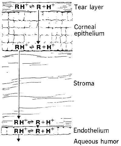
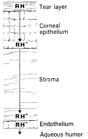
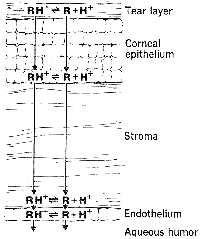
The need for drugs to have both lipid and water solubility if they are to penetrate the cornea is called the differential solubility concept. It has been used to explain the diffusion characteristics of many drugs. A logical application can be made for weak bases such as pilocarpine, tropicamide, epinephrine, cyclopentolate, and atropine.44 These exist in both ionized and nonionized molecular forms at physiologic pH 7.4. The nonionized form, being the more lipid soluble, penetrates the corneal epithelium and endothelium. The ionized form, being the more water soluble, penetrates the corneal stroma (Fig. 1). However, this diagram does not seem valid for all drugs. For example, the quaternary amines, such as carbachol, echothiophate, and demecarium, carry positive charges at all pH levels. Despite the low lipid solubilities of these ions, they penetrate the cornea in sufficient amounts to be therapeutically useful (Fig. 2). Other exceptions are those molecules with significant water solubility in the nonionized form and significant lipid solubility in the ionized form (Fig. 3). Pilocarpine is an important example. Ionized pilocarpine can penetrate all these corneal layers, as can nonionized pilocarpine. Increasing evidence suggests that although transcorneal penetration of the nonionized form of a drug is determined by its lipid solubility, the penetration of the ionized form of a drug into the anterior chamber is much greater than lipid solubility would allow. For example, Maren and Jankowska45,46 found that the highly ionized molecule bromacetazolamide had 20 times the permeability at pH 7.6 than could be explained by penetration of the nonionized form of the molecule. However, penetration of the drug into the posterior chamber more closely reflected the lipid solubility of the drug. The authors interpreted this latter finding as evidence that entry into the posterior chamber is achieved by limbal penetration of the drug followed by direct diffusion through iris and ciliary body. Ahmed and Patton47 also found evidence that topically applied drugs could enter the eye yet bypass the anterior chamber. In their studies, the drug was believed to enter the conjunctiva, diffuse across the sclera, and then penetrate the uveal tissues.

Fig. 1. The diffusion of a weak base across the cornea (e.g., epinephrine).

Fig. 2. The diffusion of a permanently charged molecule across the cornea (e.g., carbachol).

Fig. 3. The transcorneal diffusion of a molecule with significant water solubility in the un-ionized form and significant lipid solubility in the ionized form (e.g., pilocarpine).

In the past, the cornea has been looked on only as a barrier to drug penetration. More recently, the work of Makoid, Sieg, and Robinson48–50 has emphasized a different aspect: the cornea as a drug reservoir. Krohn and Breitfeller51 developed an in vitro rabbit corneal chamber with an epithelial irrigating system mimicking tear flow. When an 8% pilocarpine solution was allowed to remain in contact with the corneal epithelium for only 30 seconds, pilocarpine molecules could be detected diffusing through the cornea 90 minutes later. Van Hoose and Leaders52 hypothesized that the endothelium retarded pilocarpine diffusion into the aqueous humor, creating a stromal drug reservoir. However, Sieg and Robinson50 provided data supporting an alternative view. After applying 10-2 M pilocarpine to intact corneas, they scraped off the epithelium at various time intervals. The amounts of pilocarpine in the stroma and endothelia were assayed. These values were compared with those from corneas with intact epithelia (Table 4). The data indicated it was the epithelium that acted as the reservoir. The levels in stroma and endothelium (in micrograms per gram tissue) and in aqueous humor (in micrograms per milliliter) were nearly identical during the 2-hour period studied (i.e., they were in equilibrium and could be considered a single drug compartment). Mindel and coworkers53 found that although drugs were distributed in the cornea according to their partition coefficients, with a more lipid-soluble drug found in higher concentration (calculated as milligram of drug per milligram of tissue) in the corneal epithelium and a more hydrophilic drug found in higher concentration in the stroma, the ninefold greater mass of the stroma tended to make it the more important reservoir (calculated as milligram of drug per whole tissue).


Table 4. Corneal Concentration Following Topical 10-2 M Pilocarpine


 μg Pilocarpine/g Cornea
Minutes After ApplicationEpithelium IntactEpithelium Removed
24.310.23
55.500.53
104.770.81
302.720.89
600.830.32
(Sieg JW, Robinson JR: Mechanistic studies on transcorneal permeation of pilocarpine. J Pharm Sci 65:1816, 1976)


The existence of a corneal reservoir might explain an observation by Harris and Galin,54 who found that if a single pilocarpine drop were given to patients with open-angle glaucoma, it made little difference whether a 1% or 10% solution were used. For a 2-hour postinstillation period, the decreases in intraocular pressure were indistinguishable. However, chronic administration of 4% pilocarpine drops produced a reduction in intraocular pressure that was far superior to that from chronic administration of 1% pilocarpine. This superiority could result from a greater accumulation of pilocarpine in the corneal epithelium using the 4% concentration.

Metabolizing enzymes in the cornea, especially in the corneal epithelium, can enhance or limit a drug's effect. Dipivefrin is a prodrug of epinephrine. The conversion of dipivefrin to epinephrine is, at least in part, dependent on the activity of tissue esterases, which remove the two pivalic moieties by hydrolysis.55 These esterases do not appear to be cholinesterases because they are not inhibited by diisopropylfluorphosphate.56 Acetylating enzymes in the cornea, and especially in the iris-ciliary body, seem to explain why the topical carbonic anhydrase inhibitor aminozolamide is effective as an ocular hypotensive agent. Retention of high levels of the active metabolite, 6-acetamido-2-benzothiazolesulfonamide, has been found in these tissues.57 Degradation of pilocarpine to inactive pilocarpic acid has been demonstrated in the rabbit eye58; the corneal stroma-endothelium, as well as the corneal epithelium and iris-ciliary body, were sites of metabolism. The presence of atropinase and cocainesterase in rabbit ocular tissues and plasma has also been documented.59

CONJUNCTIVA AND SCLERA

The cornea has long been considered the route of drug entry into the eye. The possible role of a transconjunctival and scleral route has been largely overlooked. However, in rabbits this pathway has been shown to exist.60,61 Both the large area of the conjunctival surface and the prolonged conjunctival contact time of drug trapped under the lids may make this neglected route an important factor. The conjunctival surface of the eyelids and globe provides a surface area approximately 17 times larger than that of the cornea.62

Blood flow in the conjunctiva is usually thought of only as affecting systemic drug absorption. However, vasoconstrictors or vasodilators, by decreasing or increasing conjunctival drug absorption, alter both the amount and duration of drug in the tear film. These in turn will affect corneal and intraocular drug penetrations. This, too, has been demonstrated in rabbits.63 Human sclera, in vitro, has been shown to be permeable to hydrophilic compounds.64 Permeability was inversely proportional to molecular weight.

Chronic use of medications may cause low-grade conjunctival inflammation and secondary fibrosis, altering transconjunctival drug permeability. Histopathologic studies of human conjunctiva obtained after prolonged topical therapy have produced differing results; one study found evidence of inflammation and fibrosis, whereas the other did not.65,66

AQUEOUS HUMOR

Just as the protein content of tears increases with extraocular inflammations, so, too, the protein content of aqueous humor increases with intraocular inflammations. This increased protein results from a breakdown in the blood-aqueous humor barrier. The protein content approaches that of serum. Drugs entering the aqueous humor will be bound by these proteins and become therapeutically inactive. On the other hand, these protein-bound molecules can subsequently act as a reservoir.

Aqueous humor protein not only binds drug molecules but may degrade them as well. Schonberg and Ellis67 found that 0.5 ml of serum or secondary aqueous humor from humans and rabbits partially inactivated 0.5 ml of 0.1% pilocarpine when incubated at 37°C for 1 hour. Rabbit secondary aqueous humor inactivated 70% of the pilocarpine, rabbit serum inactivated 100% of the pilocarpine, human secondary aqueous humor inactivated 10% of the pilocarpine, and human serum inactivated 40% of the pilocarpine. The inactivation may have been enzymatic because a number of enzyme inhibitors such as penicillamine and ethylenediaminetetraacetic acid (EDTA) prevented the inactivation. Serum cholinesterase was not the inactivator because cholinesterase inhibitors were not protective. Mikkelson and coworkers25 demonstrated this phenomenon in vivo. They reported a decreased miotic response in rabbits 1 and 4 hours after paracentesis, for example, the peak miotic response to a 50-μ1 eye drop of 0.01 M pilocarpine nitrate instilled 1 hour after paracentesis fell from 2.5 to 0.5 mm.

IRIS AND CILIARY BODY

The uveal pigment binds drugs. Surprisingly, when the amounts of melanin were assayed in blue and brown donor irides, there was virtually no difference between the two.68 However, the ciliary bodies and choroid and retinal epithelia of brown-eyed patients had statistically more melanin. Cocaine, homatropine, pilocarpine, and ephedrine have been shown to produce less iris mydriasis or constriction in blacks69,70 and Asians71 than in whites. These differences could be due to racial characteristics other than pigmentation. Harris and Galin72 compared the hypotensive response to pilocarpine in lightly pigmented (blue-eyed) whites and heavily pigmented (brown-eyed) whites. The latter responded more poorly, consistent with uveal pigment binding more of the drug in dark-eyed subjects. Emiru73 showed that albino Africans responded to mydriatics in a manner similar to whites with pale irides.

Lyons and Krohn74 placed iris-ciliary body preparations of pigmented and albino rabbits in dilute pilocarpine solutions for 30 minutes. The mean uptake of pilocarpine by pigmented tissues was 8.1 μg per milligram, whereas that of albino tissues was 3.9 μg per milligram (p < 0.05). This appeared to be a passive uptake rather than an active transport because there was no difference if the tissues were incubated below body temperature, 22°C (71.6°F), or at body temperature, 37°C (98.6°F). Tsuchiya and coworkers75 showed that chloroquine and thioridazine bound to bovine deproteinized acid-insoluble melanin isolated from uveal tissue but that pilocarpine and epinephrine did not; these authors suggested that their isolation procedure may have altered the pigment.

Atropine has a much longer duration of action in the eye than in other organs of the body. For example, parenteral atropine blocks salivary secretions for a few hours, whereas its ocular mydriatic and cycloplegic actions lasts for days. Salazar and coworkers76 proposed that the slow onset and long duration of action of atropine in pigmented eyes were due to the uveal pigment. Binding of atropine by the pigmented cells would explain the delayed onset of action; subsequent slow release from this reservoir would explain the prolonged action. When Salazar and coworkers applied tritiated atropine 2% to pigmented rabbit eyes negative for atropinesterase, significant radioactivity was found 4 days later in the iris; nonpigmented irides had little or no activity. The accumulation of tritiated atropine by pigmented human iris and pigment epithelium was similar to that of pigmented rabbit irides. Median iris sphincter recovery time for rabbits given atropine was as follows: albino atropinesterase positive, 3.8 hours; pigmented atropinesterase positive, 12.4 hours; albino atropinesterase negative, 29.7 hours; and pigmented atropinesterase negative, greater than or equal to 96 hours. However, the duration of atropine's mydriatic-cycloplegic action does not seem to be correlated with pigmentation alone. There is prolongation of efficacy with increasing age. A 2-year-old child may recover from the ocular effects of atropine in 1 or 2 days, but an adult will not.

COMPARTMENT INTERACTION

The interaction of those pharmacokinetic factors important in topical therapy has been studied in detail by Makoid, Sieg, and Robinson.48–50 They placed single drops of pilocarpine nitrate on rabbit corneas. On the basis of their data, they proposed a four-compartment model that is represented diagrammatically in Figure 4. Pilocarpine was removed by precorneal tear flow and aqueous humor drainage. The corneal epithelial peak drug level was reached rapidly, in about 4 minutes, not because of the rapid uptake of pilocarpine but because of the rapid elimination of drug from the precorneal area by tear flow. Epithelial absorption was so limited that approximately 1% of a 25-μ1 drop of 0.1 M pilocarpine nitrate was absorbed. The epithelial uptake of pilocarpine was essentially one way. Flushing the corneal surface with saline resulted in very little drug recovery. This allowed the corneal epithelium to act as a depot. Determinations on whole cornea gave a peak value of 9.0 μg pilocarpine per gram of wet cornea. Two hours later, the corneal level was 1.2 μg per gram of wet cornea. Twelve hours later, 0.043 μg pilocarpine per gram of wet corneal tissue was still detectable.

Fig. 4. Four compartment model of topical drug pharmacokinetics.

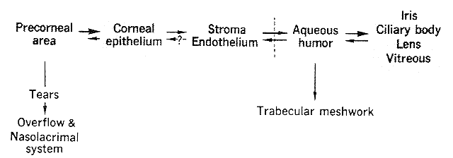
Epithelial pilocarpine molecules entering the stroma diffused rapidly across it and came into equilibrium with the aqueous humor. Stroma, endothelium, and aqueous humor could, therefore, be considered one compartment. Aqueous humor drainage via the trabecular meshwork provided the major route of drug egress. The peak aqueous humor level was 1.2 μg per milliliter of aqueous humor. It was achieved 20 minutes after instillation, at which time the corneal concentration was 6 μg per gram of wet corneal tissue (i.e., five times greater). Three to 12 hours after instillation the cornea to aqueous humor drug ratio was 3:1. Because these assays were performed on whole cornea, rather than epithelium alone, the gradient between the epithelium compartment and the stroma-endothelium-aqueous humor compartments was higher than the figure given. Virtually all the drug absorbed into the corneal epithelium ultimately reached the aqueous humor. In albino rabbits, the iris and ciliary body pilocarpine concentrations were essentially the same as those of the aqueous humor (i.e., stroma, endothelium, aqueous humor, iris, and ciliary body were a single compartment). If uveal pigment had been present, the iris and ciliary body concentrations might have been higher. The role of the lens and vitreous as drug reservoirs was not clear.

The lens and its zonules would be expected to act as a barrier between the aqueous and vitreous cavities. Intracapsular cataract extraction completely removes this barrier; extracapsular cataract extraction leaves it partially intact. Patients with intracapsular cataract extractions and anterior chamber pseudophakes were compared with those with extracapsular extractions (intact posterior capsules) and posterior chamber pseudophakes. The ratios of their aqueous humor-to-anterior vitreous humor fluorescein concentrations were measured.77 Fluorescein had been given orally. The ratios measured during the first 90 minutes after administration were significantly higher in the extracapsular cataract extraction group.

Back to Top
PERIOCULAR INJECTIONS
Local injections offer several advantages: A larger proportion of the medication reaches the target tissues (compared with systemic administration); prolonged drug exposure is achieved (compared with topical therapy), an advantage that can be maximized by using depot forms of medication; and therapeutic drug levels can be attained in the posterior segment (compared with topical therapy). Another advantage of periocular injections is that they bypass the conjunctival drug barrier. The epithelial cells of the conjunctiva would prevent the penetration of drugs with low lipid solubility. If the injection is truly subconjunctivally, only Tenon's capsule and the scleral barriers remain. Both are relatively acellular connective tissues. Therefore, the only barriers to subconjunctivally injected drugs are readily penetrated by hydrophilic molecules. Drugs with low lipid solubility, such as penicillin, penetrate the eye if given subconjunctivally but not if given topically.

Drug in sub-Tenon's injections must penetrate only the sclera. Tenon's capsule is very thin and it would be expected to produce only a slight delay in drug penetration. This difference would be short lived and of no significance from a therapeutic view. The evidence seems to support this logic. Swan and colleagues78 made patients miotic with pilocarpine before strabismus surgery. In the operating room, 50 μl of 1/1000 epinephrine was injected subconjunctivally on one side of the eye and below Tenon's capsule on the other. In 11 of the 12 patients there was a more rapid segmental dilatation of the iris on the side receiving the sub-Tenon's injection. After sub-Tenon's injections, mydriasis occurred within 5 to 6 minutes, whereas after subconjunctival injections, 10 to 16 minutes elapsed before dilatation.

In the older literature, the surprising conclusion that subconjunctival injections produce higher drug levels than sub-Tenon's injections is occasionally encountered. For example, Hardy and Paterson79 injected radiolabeled chloramphenicol in both sites and found higher levels with the subconjunctival technique. However, the levels being measured were in the anterior segment (in this case, in the aqueous humor). It had previously been shown by Wine and coworkers80 that drugs leak out through the puncture sites in the conjunctiva and diffuse across the cornea from the tear film. This leakage would occur more readily after a subconjunctival injection. In rabbits given subconjunctival and sub-Tenon's injections through the skin of the lid, without perforating the conjunctiva, there were no differences in anterior segment drug levels.

Levine and Aronson,81 using rabbits, compared the intraocular penetration of cortisol following retrobulbar (posterior sub-Tenon's) injections with that following systemic administration. They also compared anterior sub-Tenon's injections with systemic administration. In both instances local injections were superior. Forty-one times more intraocular cortisol was present after retrobulbar injection than after systemic administration. This ratio doubled in the presence of intraocular inflammation, not because more retrobulbar drug penetrated but because there was a reduction in intraocular levels that was disproportionately greater following systemic administration than that following retrobulbar injection.

Barza and coworkers82 compared the efficacy of anterior and posterior (retrobulbar) sub-Tenon's gentamicin injections in the treatment of rabbit endophthalmitis (Table 5). Anterior injections proved superior in uninflamed eyes. Higher drug levels were achieved not only in the anterior segment (cornea, iris, and aqueous humor) but, more surprisingly, in the posterior segment (vitreous, choroid, retina, and sclera) as well. This superiority was maintained in the presence of endophthalmitis, with one exception, the vitreous. Vitreous levels were similar after anterior and posterior injections in inflamed eyes. Inflammation lowered drug levels following anterior injections but elevated drug levels following posterior injections.


Table 5. Gentamicin Levels in Six Rabbit Eyes 1 Hour After Local Injections of 40 mg/0.75 ml


 μg Gentamicin per Gram Tissue
 ControlInflamed
 Anterior sub-TenonPosterior sub-TenonAnterior sub-TenonPosterior sub-Tenon
Superior cornea651302
Anterior superior sclera671462
Anterior superior choroid-retina762332
Iris8132
Aqueous humor5143
Vitreous humor0.20.010.50.5
(Barza M, Kane A, Baum JL: Regional differences in ocular concentration of gentamicin after subconjunctival and retrobulbar injection in the rabbit. Am J Ophthalmol 83:407, 1977)


McCartney and coworkers83 found evidence that scleral penetration was greatest in those areas adjacent to the injection. They injected the eyes of rabbits with 6.25 mg of tritium-labeled hydrocortisone subconjunctivally. The eyes were removed at intervals from 30 minutes to 24 hours, divided into quarters, and examined using autoradiography. The sclera and choroid under the injection site showed the greatest amount of radioactivity, and this radioactivity decreased as the distance became farther. There was much more penetration in the presence of inflammation, with the maximum occurring 2 to 4 hours postinjection.

Clinicians assume that placing the injected material over the site of pathology increases the therapeutic response and that ocular penetration is greater in the presence of inflammation. The preceding discussion was intended to demonstrate that the evidence does not entirely support these beliefs. Two variables that usually are not considered are the effects of episcleral and conjunctival vessels in carrying drugs into the general circulation and the effect of transcleral flow of intraocular fluid in retarding drug penetration. Subconjunctival medications are in contact with conjunctival vessels. Sub-Tenon's medications are in contact with episcleral vessels. Schlegel and Swan84 found that the addition of epinephrine to subconjunctival procaine solutions enhanced penetration, presumably by limiting conjunctival vessel absorption. Depot forms of medication minimize systemic absorption by using drugs with lower water solubility. Glucocorticoids have been especially amenable to this form of administration. For example, methylprednisolone sodium succinate (Solu-Medrol) is very soluble in body fluids and will rapidly give high blood and tissue levels of short duration. However, by using a less water-soluble ester, methylprednisolone acetate (Depo-Medrol), prolonged exposure to the drug is achieved. Rodger85 injected 10 mg of methylprednisolone acetate subconjunctivally in a volunteer. Three weeks later, 41% of the drug was recovered from the subconjunctival space. Hyndiuk and Reagan86 found detectable methylprednisolone acetate in the posterior segment of the eyes of monkeys 9 days after retrobulbar injection of 12 mg of the drug (Table 6).


Table 6. Methylprednisolone Levels Following Retrobulbar Injection of 12 mg Methylprednisolone Acetate


 μg Methylprednisolone Acetate per 100 mg Tissue
 Day 2Day 9
Number of monkeys33
Cornea0.6 ± 0.10.6 ± 0.1
Lens1.7 ± 0.80.4 ± 0.1
Anterior uvea0.2 ± 0.00.0
Posterior uvea-retina48 ± 30.5 ± 0.0
Vitreous22 ± 133.7 ± 1.3
Sclera14 ± 15.8 ± 1.3
Optic nerve3.6 ± 1.84.3 ± 1.9
(Hyndiuk RA, Reagan MG: Radioactive depot-corticosteroid penetration into monkey ocular tissue: I. Retrobulbar and systemic administration. Arch Ophthalmol 80:499, 1968)


Bill87 studied transcleral movement of intraocular fluid by injecting Evans blue dye and radioactive iodine (131I)-labeled albumin in the suprachoroidal space of rabbit eyes. More than 85% of the materials left the eyes by bulk flow through the sclera. By lowering the intraocular pressure, the rate of flow through the sclera diminished. The intraocular pressure may, therefore, alter the rate of drug penetration. The lower the pressure (e.g., hypotony from decreased ciliary secretion during inflammation), the slower is the rate of transcleral fluid movement from inside the eye to outside and the greater is the penetration of extraocular drugs.

Leopold and coworkers88 found that vitreous levels of streptomycin and penicillin, injected retrobulbarly, could be markedly enhanced by iontophoresis. One electrode was placed on the cornea and the other behind the neck of rabbits. A current of 3 to 5 mA was applied for 5 minutes. No penicillin or streptomycin had been detectable in the vitreous 30 and 120 minutes after retrobulbar injection alone, but following iontophoresis, penicillin, 0.3 units per milliliter of vitreous, and streptomycin, 12 to 15 μg per milliliter of vitreous, were detectable at both these intervals. These vitreous humor levels were comparable with those achieved by aqueous humor injection of these drugs. Barza and coworkers89 and Maurice90 used transscleral iontophoresis to achieve high vitreous levels of cefazolin, ticarcillin, gentamicin, and fluorescein in phakic rabbit eyes.

Gruntzig and coworkers91 injected radiolabeled colloid and albumin unilaterally in the retrobulbar space of 24 rabbits. They found significant radioactivity in the contralateral retrobulbar space. They speculated that there might be interorbital connecting lymphatic drainage channels. Whether a similar contralateral effect occurs in humans is unknown.

Back to Top
SYSTEMIC THERAPY
Systemic therapy will deliver drugs to both the anterior and the posterior segments of the eye. Drug levels may be lower than those achieved by periocular injections, but systemic administration is more readily accepted by patients, especially those in the pediatric age group.

Drugs do not readily enter the eye from the systemic circulation. There are multiple barriers retarding drug passage and, in toto, they are referred to as the blood-eye barrier. In the past, the blood-eye barrier was conceptually subdivided into a blood-aqueous and a blood-vitreous barrier. The reason was that clinical interest was oriented toward determining drug levels in the aqueous and vitreous humors. Physiologically and pharmacologically, these terms are not always appropriate. For example, it is difficult to write of a “blood-vitreous” barrier when evidence exists of diffusion between the posterior chamber and the anterior vitreous.92

The tight intercellular junctions form the most important barrier to diffusion of drugs from the systemic circulation. The three sites where they are most important are the retinal blood vessel endothelium, the retinal pigment epithelium, and the ciliary body nonpigmented epithelium.93,94 Drug molecules must have lipid solubility to penetrate these tight junctions. At sites where they are less frequent, for example, the endothelium of the uveal blood vessels, drugs can escape by diffusion. At certain sites, drugs enter the eye by the energy-requiring mechanisms of pinocytosis and active transport.

BLOOD-RETINA BARRIER

The retina has two blood supplies: the choroidal and retinal circulations. Drug penetration from the retinal blood vessels is retarded by the tight intercellular junctions of the capillary endothelium.93 The choroidal blood vessels, with endothelial pores of 70 nm, permit large molecules to penetrate. However, the tight junctions of the pigment epithelium prevent their entering the retina.95 Bill96 proposed that the inability of choroidal serum proteins to traverse the pigment epithelium produced an osmotic gradient of 12 mm Hg or more. This gradient would draw fluid out of the eye and retard drug penetration.

The endothelium of the retinal blood vessels behaves as though a limited number of small pores were present. Larger lipid-insoluble molecules, such as inulin (1.4 nm) and sucrose (0.5 nm), are unable to enter the vitreous. Smaller lipid-insoluble molecules, such as glycerol (0.3 nm) and glucose, can penetrate to some degree.97 Larger molecules penetrate according to their lipid solubility (e.g., penicillin diethylaminoethyl iodide enters the vitreous more readily than the smaller penicillin molecule because its partition coefficient is 60 times greater).98

Cunha-Vaz and Maurice99 obtained evidence that an active ion transport system existed that removed organic anions from the vitreous. Fluorescein was shown to move rapidly from the vitreous into the general circulation against a concentration gradient. However, fluorescein could not enter the vitreous from the circulation. The transport system was saturated at 3 × 10⁻⁴ M fluorescein. Its location was in the endothelium of the retinal vessels and possibly the pigment epithelium as well. This transport system explains, in part, why intravenous fluorescein is useful as an indicator of the integrity of these two tissues. Other anions, such as iodopyracet, penicillin, and probenecid compete for this transport system (Table 7). Ogura and coworkers100 have found indirect evidence in humans, using fluorophotometry, that fluorescein is actively transported out of the vitreous. The large protein molecule, horseradish peroxidase, will not enter the vitreous from the bloodstream because of the blood-retina barrier, but it readily enters the blood of the retinal arterioles, venules, and capillaries after injection into the vitreous of monkeys and rabbits.101 This unidirectional movement is accomplished by the cytoplasmic vesicles of the endothelial cells.


Table 7. Vitreous Concentration Required for Complete Inhibition (I100) of Fluorescein Transport by Rabbit Anion Transport System


InhibitionApproximate I100 (M)
Competitive 
 Iodopyracet1×10-3
 Probenecid3×10-5
Metabolic 
 Dinitrophenol1×10-5
 Sodium fluoride3×10-5
(Cunha-Vaz JG, Maurice DM: The active transport of fluorescein by the retinal vessels and retina. J Physiol 191:467, 1967)


Transport systems from the systemic circulation into the retina occur for certain metabolites. Dollery and coworkers102 found that intravenous injection of labeled D- and L-glucose in rats, rabbits, and guinea pigs resulted in selectively higher retinal levels of the metabolically active D form. The D/L ratios were rats, 24; rabbits, 12; and guinea pigs, 8.

Breakdown of the blood-retina barrier can be produced by a variety of insults, such as chemical, thermal, or traumatic injury to the retinal vessels.103 Ischemia will initially affect the arteriolar component of the blood-retina barrier, but after 24 hours, the venous component has decompensated to a greater degree.104

BLOOD-OPTIC NERVE BARRIER

The optic nerve head can be thought of as an anatomic defect in the retina-pigment epithelium barrier. Several groups have shown that if a blood-optic nerve head barrier exists, it is leaky.105,106 Horseradish peroxidase, molecular weight 42,000 and diameter 3 nm, moved from the blood into the optic nerve head of monkeys. This might explain late fluorescence in the optic nerve head after intravenous fluorescein injection.107,108 Glial cells at the edge of the optic disc form a barrier that prevents the spread of peroxidase into the retina. However, penetration of drugs from the nerve into the vitreous does occur. This process is retarded, but not prevented, by the flow of vitreous fluid into the optic nerve head.109

BLOOD-AQUEOUS HUMOR BARRIER

The ciliary body capillaries are leaky like those of the posterior uvea. However, the tight junctions of the ciliary nonpigmented epithelium limit drug diffusion into the aqueous humor.110 Posteriorly, an area of overlap exists, formed by the pigment epithelium of the blood-retina barrier and the nonpigmented epithelium of the blood-aqueous barrier. Shabo and Maxwell111 have identified this area as the posterior pars plana.

The iris vessels of primates contain endothelial tight junctions, which decrease their permeability. These are important in maintaining a blood-aqueous barrier because the anterior iris surface is not covered by either pigmented or nonpigmented ciliary epithelium. It is assumed that small lipid-insoluble molecules enter the aqueous humor from the anterior iris surface. Davson and Duke-Elder97 found that as miosis increased iris surface area, penetration of sugars into the aqueous humor increased.

Saari112 found anatomic evidence of iris vessel pores. The pores behave as though they are about 4 nm in diameter.113 Ten minutes after intravenous injection of glucose (molecular weight 180), the blood-aqueous humor concentration gradient in dogs had decreased by 25%. It took only 3 minutes for the same gradient reduction of xylose (molecular weight 150). Sucrose (molecular weight 342) required 3 hours, and raffinose (molecular weight 504) did not penetrate at all. It was concluded that substances above molecular weight 500 must have lipid solubility if they are to enter the aqueous humor. Langham114 found that chloramphenicol (molecular weight 335) entered the aqueous humor twice as fast as penicillin G (molecular weight 334). Although their molecular weights were similar, their lipid solubilities were not. Using an interface of ether/0.01 M phosphate buffer, pH 7.4, the partition coefficient for chloramphenicol was 4.25, whereas that of penicillin G was 0.0004. Ross115 also found that aqueous humor penetration of drugs followed their partition coefficients.

Drugs are carried from the ciliary processes into the posterior chamber by active transport. The mechanism of this transport may be pinocytosis. Shabo and Maxwell116 found no histologic evidence of pinocytosis, but Smith and Rudt94 demonstrated horseradish peroxidase transport into the posterior chamber via pinocytosis. Davson and Matchett117 found evidence of active transport of sucrose, thiourea, and sulfonamides. Whatever the reasons—active transport and/or the absence of an anterior iris barrier—drugs enter the aqueous humor more readily than they enter the vitreous humor. When Davson and Matchett118 injected sulfonamides systemically, they found the aqueous humor rates of penetration of sulfapyridine and sulfadiazine to be 2.5 and 4.5 times greater, respectively, than their rates of entry into the vitreous humor.

Raviola and Butler119 found evidence in monkeys of a unidirectional vesicular transport system in iris blood vessel endothelial cells that carried anions from the aqueous humor into the blood. Neither anionic ferritin nor cationic ferritin entered the iris stroma when injected intravenously because of the blood-aqueous barrier. However, when these molecules were placed in the aqueous humor, anionic ferritin, but not cationic ferritin, entered the iris blood vessel lumen.

The blood-aqueous barrier can be broken down or bypassed. Raviola120 found that when intravenous hypertonic urea, 3 g/kg, produced an 80% reduction in monkey intraocular pressure, horseradish peroxidase entered the aqueous humor. The route of entry was by back-diffusion from episcleral veins into Schlemm's canal and then into the anterior chamber. The endothelium of Schlemm's canal had clefts 10 to 20 nm wide. Electron microscopy showed that the effect was not due to a breakdown of the tight intercellular junctions that formed the blood-aqueous barrier. Okisaka and coworkers121 found that 2 M urea injected directly into the carotid artery did break down in the blood-aqueous barrier. Shabo and coworkers122 confirmed that the high urea concentration achieved by intracarotid injection was destructive, but the lower intravenous urea level was not.

Intraocular inflammation and paracentesis will also disrupt the blood-aqueous barrier. Langham114 found that the secondary aqueous humor (i.e., that present after aspiration) contained the same concentration of antibiotic as plasma and the same relative amounts of protein bound and unbound drug as plasma. Salminen123 reported enhanced anterior chamber levels of ampicillin, cloxacillin, doxycycline, and tetracycline following paracentesis. The increased antibiotic, if adherent to protein, may act as a reservoir, but, until it dissociates, it will be of no therapeutic value.

Cataract surgery, especially when an anterior chamber pseudophake is implanted, may increase the permeability of the blood-aqueous barrier. Miyake and coworkers124 found an increase in aqueous humor fluorescein when they performed aqueous humor fluorophotometry on patients who had had phacoemulsification cataract surgery 12 to 15 months previously. The operated eye was compared with the unoperated eye 30 minutes after intravenous injection of the fluorescein. The ratios were

  Without pseudophake implant: 1.26 plus or minus 0.55
  With intracapsular pseudophake: 1.73 plus or minus 0.73
  With ciliary sulcus pseudophake: 1.56 plus or minus 0.52
  With anterior chamber open loop pseudophake: 2.35 ± 1.56

However, Sanders and coworkers125 and Liesegang and colleagues126 found no difference in the concentration of fluorescein in the anterior chamber between the aphakic or pseudophakic eye and the control eye.

OTHER FACTORS

Serum Protein Binding

Sulfonamides are very lipid soluble. However, they penetrate the eye relatively poorly because of a high degree of protein binding (more than 90%). Langham114 found that 50% of penicillin G but only 27% of procaine penicillin G was plasma protein bound. As a result, even though the partition coefficient of penicillin G was somewhat higher, procaine penicillin G had a slightly greater rate of aqueous humor penetration. Protein binding is not correlated with any particular structure of a drug. It occurs with drugs that are aromatic or aliphatic, anionic or cationic, and lipophilic or hydrophilic. Generally, as the structure of a drug is altered to make it more lipophilic, it becomes more protein bound.

The ocular penetration of a drug is proportional to the unbound level. To best ensure that a therapeutic intraocular level is achieved, systemically administered drugs should be given in pulses. For example, 250 mg methicillin injected intravenously within a 5-minute period every 4 hours would be expected to give a higher intraocular level than a slow steady drip. The general rule is that the peak eye level is determined by the peak blood level.

Protein binding of anionic drugs is principally to the albumin fraction. Cationic drugs bind to a1-acidic glycoprotein.127 Both may be diminished in uremia, hypoalbuminemia, hyperbilirubinemia, and hepatic failure. Endogenous substances such as bilirubin128 and fatty acids129 compete for binding sites. The decreased drug-protein binding that occurs in renal disease is not due solely to hypoproteinemia (e.g., sulfamethoxazole and cefazolin, but not trimethoprim, are less protein bound in uremia).130,131 Neonates have less protein binding because of a relative hypoproteinemia. Cephalothin, diazoxide, phenytoin, and imipramine are 4% to 12% less bound in the neonate than in the adult.132 The percentage of antibiotic bound at normal serum protein levels is shown in Table 8.133,134 Different binding values were reported for the aminoglycoside antibiotics when different buffers were used.135,136 Therefore, binding studies performed in vitro may not mirror the in vivo situation.


Table 8. Protein Binding of Antibiotics


AntibioticSerum Concentration (μg/ml)% Bound
Ampicillin518
Carbenicillin8050
Cefazolin3086
Cephalexin3015
Cephalothin3065
Cephaloridine3020
Cephradine8712
Chlortetracycline2087
Clindamycin594
Cloxacillin5094
Demeclocycline1038
Doxycycline1052
Erythromycin573
Erythromycin estolate496
Erythromycin propionate593
Lincomycin572
Methicillin1049
Nafcillin2087
Oxacillin2085
Oxytetracycline1031
Penicillin V3053
Tetracycline2070
(Robinson GN, Sutherland R: The binding of antibiotics to serum proteins. Br J Pharmacol 25:638, 1965; Vallner JJ: Binding of drugs by albumin and plasma protein. J Pharm Sci 66:447, 1977)


Binding may occur at more than one site. For example, Powis137 found that tetracycline was bound at two different sites, a high affinity site with limited capacity and a low affinity site with large capacity.

The possibility has been investigated of determining plasma levels of non-protein-bound drug by measuring the drug level secreted in tears. The development of techniques, such as immunoassay, gas chromatography, and high-performance liquid chromatography, that can determine minute amounts of drugs have made such investigations feasible. The drug concentrations in tears relative to those unbound in plasma have been determined.138 Nonionized drugs and acidic drugs with high pKa values seem to show the best correlation (e.g., carbamazepine, sulfanilamide, and phenobarbital), but the general lack of predictability makes tear assay unreliable.

Cellular Binding of Drugs

Drugs are bound not only to serum proteins but also by erythrocytes, leukocytes, and platelets. Acetazolamide was shown to be bound to erythrocytes by two processes, a nonlinear saturable process and a linear diffusion process.139 Much of the binding appeared to be intracellular, possibly resulting from intracellular carbonic anhydrase. The erythrocyte uptake was concentration dependent. Blood with a hematocrit of 50% gave an erythrocyte-plasma acetazolamide ratio of 5.2:1 when acetazolamide 5 μg per milliliter of blood was added; at a concentration of 30 μg per milliliter of blood, the ratio was 1.6:1.

DRUG METABOLISM

Drugs administered systemically are subject to metabolic alteration. This may lower or increase drug activity. The effect of systemically administered dexamethasone phosphate is due in part to its conversion to the free alcohol. Hare and coworkers140 found, in vivo, that peak dexamethasone alcohol levels were achieved as soon as 10 minutes after intravenous injection of dexamethasone phosphate. Hydrolysis occurred in vivo at a rate 25 times greater than in blood in vitro. They concluded that most hydrolysis occurred in the organs. Rapid conversions of the phosphate esters of cortisol and prednisolone have also been demonstrated.141

A cytochrome P-450 enzyme, CYP2D6, inactivates a number of drugs, for example, timolol, quinine, propranolol, nortriptyline, and fluphenazine.142 Approximately 10% of Caucasian northern Europeans are defective in this enzyme; the percent varies in other populations. When a 30-μl drop of timolol 0.5% was given bilaterally to supine subjects, the individual variability in conjunctival/mucous membrane absorption of timolol, as much as fivefold, was more important in determining blood levels of active drug than the presence or absence of a defective CYP2D6.143

INDUCED DRUG RESISTANCE

P-glycoprotein is a plasma membrane protein that is associated with multidrug resistance. The protein is expressed at sites where an adenosine triphosphate (ATP)-utilizing pump prevents drug entry into the cell. Cytotoxic drug use can result in selective survival of those tumor cells with increased P-glycoprotein activity. After an initial good response, tumor retreatment may fail. Resistance to one drug in a class of natural products, for example, vinca alkaloids, results not only in cross resistance to other drugs in that class but also to agents that have the features of being lipophilic, having a molecular weight to 300 to 900 daltons, and entering the cell by passive diffusion. Retinoblastoma drug resistance and drug sensitivity has been shown to correlate with P-glycoprotein levels.144

DRUG EXCRETION

The rates at which drugs are removed from the circulation depend largely on the kidneys and liver. At therapeutic levels, about 70% of lidocaine and 90% of propranolol are removed from the circulation by the liver. Hepatic excretion occurs via the bile. Substances may be excreted unchanged but usually have to be conjugated in the liver to a strongly polar group such as a glucuronide.

Renal excretion of drugs depends on several factors. The greater the lipid solubility, the more readily the drug is reabsorbed from the kidney tubules. If the drug molecule is very large, or protein bound, it will not enter the glomerular filtrate. Separate transport systems exist for basic and acidic ionized drugs.

A number of less important factors that affect drug absorption and excretion have been documented. Vukovich and coworkers145 reported that blood levels of cephradine were lower in females than in males following intramuscular injection. Shimizu and Smith146 reported that urinary clearance of lithium occurred more rapidly in the recumbent than in the upright position.

Back to Top
REFERENCES

1. Smith SE: Eyedrop instillation for reluctant children Br J Ophthalmol 75:480, 1991

2. Lahdes KK, Huupponen RK, Kaila TJ: Ocular effects and systemic absorption of cyclopentolate eyedrops after canthal and conventional application. Acta Ophthalmol 72:698, 1994

3. Mishima A: Some physiologic aspects of the precorneal tear film. Arch Ophthalmol 73:233, 1965

4. Mishima S, Gasset A, Klyce SD et al: Determination of tear volume and tear flow. Invest Ophthalmol 5:264, 1966

5. Maurice DM: The dynamics and drainage of tears. Int Ophthalmol Clin 13:103, 1973

6. Brown RH, Lynch MG: Design of eye-dropper tips for topical beta-blocking agents. Am J Ophthalmol 102:123, 1986

7. Lederer CM Jr, Harold RE: Drop size of commercial glaucoma medications. Am J Ophthalmol 101:691, 1986

8. Brown RH, Lynch MG: Drop size of commercial glaucoma medications. Am J Ophthalmol 102:673, 1986

9. Adler FH: Physiology of the Eye. St Louis: CV Mosby, 1965:38

10. Patton TF, Robinson JR: Influence of topical anaesthesia on tear dynamics and ocular drug bioavailability in albino rabbits. J Pharm Sci 64:267, 1975

10a. Zimmerman TZ, Kooner KS Kandarakis AS et al: Improving the therapeutic index of topically applied ocular drugs. Arch Ophthalmol 102:551, 1984

11. Huang TC, Lee DA: Punctal occlusion and topical medications for glaucoma. Am J Ophthalmol 107:151, 1989

12. White WL, Glover AT, Buckner AB: Effect of blinking on tear elimination as evaluated by dacryoscintigraphy. Ophthalmology 98:367, 1991

13. Zimmerman TJ, Sharir M, Nardin GF et al: Therapeutic index of pilocarpine, carbachol, and timolol with nasolacrimal occlusion. Am J Ophthalmology 114:1, 1992

14. Elllis PE, Wu P-Y, Pfoff DS et al: Effect of nasolacrimal occlusion on timolol concentrations in the aqueous humor of the human eye. Am Pharm Assoc 81:219, 1992

15. Zimmerman TJ, Sharir M, Nardin GF et al: Therapeutic index of epinephrine and dipivefrin with nasolacrimal drainage. Am J Ophthalmol 114:8, 1992

16. Chrai SS, Patton TF, Mehta A et al: Lacrimal and instilled fluid dynamics in rabbit eyes. J Pharm Sci 62:1112, 1973

17. Lee VHL, Takemoto KA, Jimoto DS: Precorneal factors influencing the ocular distribution of topically applied liposomal inulin. Curr Eye Res 3:585, 1984

18. Ludwig A, Van Ooteghem M: The influence of the drop size on the elimination of an ophthalmic solution from the pre-corneal area of human eyes. Pharm Acta Helv 62:56, 1987

19. Sorensen T, Jensen FT: Methodological aspects of tear flow determination by means of a radioactive tracer. Acta Ophthalmol 55:726, 1977

20. Brown RH, Wood TS, Lynch MG et al: Improving the therapeutic index of topical phenylephrine by reducing drop volume. Ophthalmology 94:847, 1987

21. Chrai SS, Makoid MC, Eriksen SP et al: Drop size and initial dosing frequency problems of topically applied ophthalmic drugs. J Pharm Sci 63:333, 1974

22. Chrai SS, Patton TF, Mehta A et al: Lacrimal and instilled fluid dynamics in rabbit eyes. J Pharm Sci 62:1112, 1973

23. Miller D, O'Connor P: The influence of the nictitating membrane on steroid inhibition of limbal wound healing. Acta Ophthalmol 55:586, 1977

24. Loewenstein A, Geyer O, Blanc A et al: Dosing problems in the use of topical ophthalmic drops. Graefes Arch Clin Exp Ophthalmol 230:378, 1992

25. Mikkelson TJ, Chrai SS, Robinson JR: Altered bioavailability of drugs in the eye due to drug-protein interaction. J Pharm Sci 62:1648, 1973

26. Mikkelson TJ, Chrai SS, Robinson JR: Competitive inhibition of drug-protein interaction in eye fluids and tissues. J Pharm Sci 62:1942, 1973

27. Green K, Downs SJ: Prednisolone phosphate penetration into and through the cornea. Invest Ophthalmol 13:316, 1974

28. Perrin JH, Nelson DA: Displacement of sulfaethidole from bovine serum albumin by some alkyldimethylbenzylammonium chlorides. Biochem Pharm 23:3139, 1974

29. Carney LG, Mauger TF, Hill RM: Buffering in human tears: pH responses to acid and base challenge. Invest Ophthalmol Vis Sci 30:747, 1989

30. Swan KC, White NG: Corneal permeability: I. Factors affecting penetration of drugs into the cornea. Am J Ophthalmol 25:1043, 1942

31. Schoenwald RD, Huang H-S: Corneal penetration behavior of β-blocking agents. I: Physicochemical factors. J Pharm Sci 72:1266, 1983

32. Cox WV, Kupferman A, Leibowitz HM: Topically applied steroids in corneal disease: I. The role of inflammation in stromal absorption of dexamethasone. Arch Ophthalmol 88:308, 1972

33. Cox WV, Kupferman A, Leibowitz HM: Topically applied steroids in corneal disease: II. The role of drug vehicle in stromal absorption of dexamethasone. Arch Ophthalmol 88:549, 1972

34. Kupferman A, Pratt MV, Suckewer K et al: Topically applied steroids in corneal disease: III. The role of drug derivative in stromal absorption of dexamethasone. Arch Ophthalmol 91:373, 1974

35. Kupferman A, Leibowitz HM: Topically applied steroids in corneal disease: IV. The role of drug concentration in stromal absorption of prednisolone acetate. Arch Ophthalmol 91:377, 1974

36. Kupferman A, Leibowitz HM: Topically applied steroids in corneal disease: V. Dexamethasone alcohol. Arch Ophthalmol 92:329, 1974

37. Kupferman A, Leibowitz HM: Topically applied steroids in corneal disease: VI. Kinetics of prednisolone phosphate. Arch Ophthalmol 92:331, 1974

38. Leibowitz HM, Kupferman A: Bioavailability and therapeutic effectiveness of topically administered corticosteroids. Trans Am Acad Ophthalmol Otolaryngol 79:78, 1975

39. Smolen VF, Clevenger JM, Williams EJ et al: Biophasic availability of ophthalmic carbachol: I. Mechanisms of cationic polymer- and surfactant-promoted miotic activity. J Pharm Sci 62:959, 1973

40. Smolen VF, Park CS, Williams EJ: Drug-biomolecule interactions: Bioelectrometric study of the mechanism of carbachol interactions with the cornea and its relation to miotic activity. J Pharm Sci 64:520, 1975

41. Ramselaar JAM, Boot JP, van Haeringen NJ et al: Corneal epithelial permeability after instillation of ophthalmic solutions containing local anesthetics and preservatives. Curr Eye Res 7:947, 1988

42. Boyd JL: Sodium sulfathiazole iontophoresis. Arch Ophthalmol 28:205, 1942

43. Witzel SH, Fielding IZ, Ormsby HL: Ocular penetration of antibiotics by iontophoresis. Am J Ophthalmol 42, 1956

44. Cogan DG, Hirsch EO: The cornea: VII. Permeability of weak electrolytes. Arch Ophthalmol 32:276, 1944

45. Maren TH, Jankowska L: Ocular pharmacology of sulfonamides: The cornea as barrier and depot. Curr Eye Res 4:399, 1985

46. Jankowska LM, Bar-Ilan A, Maren TH: The relations between ionic and nonionic diffusion of sulfonamides across the rabbit cornea. Invest Ophthalmol Vis Sci 27:29, 1986

47. Ahmed I, Patton TF: Disposition of timolol and inulin in the rabbit eye following corneal versus non-corneal absorption. Int J Pharm 38:9, 1987

48. Makoid MC, Sieg JW, Robinson JR: Corneal drug absorption: An illustration of parallel first-order absorption and rapid loss of drug from absorption depot. J Pharm Sci 65:150, 1976

49. Makoid MC: Pharmacokinetics of Topically Applied Pilocarpine in the Rabbit Eye. [Thesis]. University of Wisconsin, Madison, 1977

50. Sieg JW, Robinson JR: Mechanistic studies on transcorneal permeation of pilocarpine. J Pharm Sci 65:1816, 1976

51. Krohn DL, Breitfeller JM: Transport of pilocarpine by isolated rabbit cornea. Invest Ophthalmol 13:312, 1974

52. Van Hoose MC, Leaders FE: The role of the cornea in the biologic response to pilocarpine. Invest Ophthalmol 13:377, 1974

53. Mindel JS, Smith H, Jacobs M et al: Drug reservoirs in topical therapy. Invest Ophthalmol Vis Sci 25:346, 1984

54. Harris L, Galin M: Dose response analysis of pilocarpine induced ocular hypotension. Arch Ophthalmol 84:605, 1970

55. Mindel JS, Cohen G, Barker LA et al: Enzymatic and nonenzymatic hydrolysis of dipivefrin. Arch Ophthalmol 102:457, 1984

56. Anderson JA, Richman JB, Mindel JS: Effects of echothiophate iodide on the enzymatic hydrolysis of dipivefrin. Arch Ophthalmol 102:913, 1984

57. Putnam ML, Schoenwald RD, Duffel MW et al: Ocular disposition of aminozolamide in the rabbit eye. Invest Ophthalmol Vis Sci 28:1373, 1987

58. Wood RW, Robinson JR: Ocular disposition of pilocarpine in the pigmented rabbit: Site of metabolism. Int J Pharm 29:127, 1986

59. Cauthen SE, Ellis RD, Larrison SB et al: Resolution, purification and characterization of rabbit serum atropinesterase and cocainesterase. Biochem Pharmacol 25:181, 1976

60. Lee VHL, Carson W, Takemoto KA: Macromolecular drug absorption in the albino rabbit eye. Int J Pharmacol 29:43, 1986

61. Hamalainen KM, Kananen K, Auriola S et al: Characterization of paracellular and aqueous penetration routes in cornea, conjunctiva and sclera. Invest Ophthalmol Vis Sci 38:627, 1997

62. Watsky MA, Jablonski M, Edelhauser HF: Comparison of conjunctival and corneal surface areas in rabbit and human. Curr Eye Res 7:483, 1988.

63. Finne U, Vaisanen V, Urtti A: Modification of ocular and systemic absorption of timolol from ocular inserts by a buffering agent and a vasoconstrictor. Int J Pharm 65:19, 1990

64. Olsen TW, Edelhauser JF, Lim JI et al: Human scleral permeability. Invest Ophthalmol Vis Sci 36:1893, 1995

65. Sherwood MB, Grierson I, Miller L et al: Long-term morphologic effects of antiglaucoma drugs on the conjunctiva and Tenon's capsule in glaucomatous patients. Ophthalmology 96:327, 1989

66. Baun O, Heegaard S, Kessing SV et al: The morphology of conjunctiva after long-term topical anti-glaucoma treatment. Acta Ophthalmol 73:242, 1995

67. Schonberg SS, Ellis PP: Pilocarpine inactivation. Arch Ophthalmol 82:351, 1969

68. Menon IA, Wakeham DC, Persad SD: Quantitative determination of the melanin contents in ocular tissues from human blue and brown eyes. J Ocular Pharmacol 8:35, 1992

69. Cameron AJ: Ophthalmic work in a British general hospital in North Africa. Br J Ophthalmol 29:12, 1945

70. Melikian HE, Lieberman TW, Leopold IH: Ocular pigmentation and pressure and outflow response to pilocarpine and epinephrine. Am J Ophthalmol 72:70, 1971

71. Chen KK, Poth EJ: Racial differences as illustrated by the mydriatic action of cocaine, euphthalmine and ephedrine. J Pharmacol Exp Ther 36:429, 1929

72. Harris LS, Galin MA: Effect of ocular pigmentation on hypotensive response to pilocarpine. Am J Ophthalmol 72:923, 1971

73. Emiru VP: Response to mydriatics in the African. Br J Ophthalmol 55:538, 1971

74. Lyons JS, Krohn DL: Pilocarpine uptake by pigmented uveal tissue. Am J Ophthalmol 75:885, 1973

75. Tsuchiya M, Hayasaka S, Mizuno K: Affinity of ocular acid-insoluble melanin for drugs in vitro. Invest Ophthalmol Vis Sci 28:822, 1987

76. Salazar M, Shimada K, Patil PN: Iris pigmentation and atropine mydriasis. J Pharmacol Exp Ther 197:79, 1976

77. Smith RT, Campbell CJ, Koestler CJ et al: The barrier function in extracapsular cataract surgery. Ophthalmology 97:90, 1990

78. Swan KC, Crisman HR, Bailey PF: Subepithelial versus subcapsular injections of drugs. Arch Ophthalmol 56:26, 1956

79. Hardy RG, Jr, Paterson CA: Ocular penetration of 14C-labeled chloramphenicol following subconjunctival or sub-Tenon's injection. Am J Ophthalmol 71:1307, 1971

80. Wine NA, Gornall AG, Basu PK: The ocular uptake of subconjunctivally injected 14C hydrocortisone. Am J Ophthalmol 58:362, 1964

81. Levine ND, Aronson SB: Orbital infusion of steroids in the rabbit. Arch Ophthalmol 83:599, 1970

82. Barza M, Kane A, Baum JL: Regional differences in ocular concentration of gentamicin after subconjunctival and retrobulbar injection in the rabbit. Am J Ophthalmol 83:407, 1977

83. McCartney HJ, Drysdale IO, Gornall AG et al: An autoradiographic study of the penetration of subconjunctivally injected hydrocortisone into the normal and inflamed rabbit eye. Invest Ophthalmol 4:297, 1965

84. Schlegel HE, Swan KC: Ocular penetration of procaine following subconjunctival injection. Arch Ophthalmol 52:774, 1954

85. Rodger FC: Repository corticotherapy in ophthalmic theory and practice. Br J Ophthalmol 49:298, 1965

86. Hyndiuk RA, Reagan MG: Radioactive depot-corticosteroid penetration into monkey ocular tissue: I. Retrobulbar and systemic administration. Arch Ophthalmol 80:499, 1968

87. Bill A: Movement of albumin and dextran through the sclera. Arch Ophthalmol 74:248, 1965

88. Leopold IH, Wiley M, Dennis R: Vitreous infections and streptomycin. Am J Ophthalmol 30:1345, 1947

89. Barza M, Peckman C, Baum J: Transscleral iontophoresis of cefazolin, ticarcillin and gentamicin in the rabbit. Ophthalmology 93:133, 1986

90. Maurice DM: Iontophoresis of fluorescein into the posterior segment of the rabbit eye. Ophthalmology 93:128, 1986

91. Gruntzig J, Schicha H, Krem J et al: Studien zur Lymphdrainage des Auges: I. Quantitative Erfassung des Lymphtransportes aus der Orbita des Kaninchens mit radioaktiven Tracem. Klin Monatsbl Augenheilkd 170:713, 1977

92. Kinsey VE: Ion movement in the eye. Circulation 21:968, 1960

93. Shakib M, Cunha-Vaz JG: Studies on the permeability of the blood-retinal barrier. Exp Eye Res 5:229, 1966

94. Smith RS, Rudt LA: Ultrastructural studies of the blood-aqueous barrier: II. The barrier to horseradish peroxidase in primates. Am J Ophthalmol 76:937, 1973

95. Cunha-Vaz JG: The blood-retinal barriers. Doc Ophthalmol 41:287, 1976

96. Bill A: Blood circulation and fluid dynamics in the eye. Physiol Rev 55:383, 1975

97. Davson H, Duke-Elder WS: The distribution of reducing substances between the intraocular fluids and blood plasma and the kinetics of penetration of various sugars into these fluids. J Physiol 107:141, 1948

98. Beeker GM, Maas EH: Penetration of penethamate, a penicillin ester, into the tissues of the eye. Arch Ophthalmol 60:1013, 1958

99. Cunha-Vaz JG, Maurice DM: The active transport of fluorescein by the retinal vessels and retina. J Physiol 191:467, 1967

100. Ogura Y, Tsukahara Y, Saito I et al: Estimation of the permeability of the blood-retinal barrier in normal individuals. Invest Ophthalmol Vis Sci 26:969, 1985

101. Raviola G, Butler JM: Unidirectional vesicular transport mechanism in retinal vessels. Invest Ophthalmol Vis Sci 24:1465, 1983

102. Dollery CT, Henkind P, Orme M: Assimilation of D and L 1-C-14 glucose into the retina, brain and other tissues. Diabetes 20:519, 1971

103. Cunha-Vaz JG: Studies on the permeability of the blood-retinal barrier: II. Breakdown of the blood-retinal barrier by injury. Br J Ophthalmol 50:454, 1966

104. Cunha-Vaz JG: Studies on the permeability of the bloodretinal barrier: III. Breakdown of the blood-retinal barrier by circulatory disturbances. Br J Ophthalmol 50:505, 1966

105. T'so MOM, Shih C, McLean IW: Is there a blood-brain barrier at the optic nerve head? Arch Ophthalmol 93:815, 1975

106. Hoffman P, Hoyng P, vander Werf F et al: Lack of blood-brain barrier properties in microvessels of the prelaminar optic nerve head. Inv Ophthalmol Vis Sci 42:895, 2001

107. Grayson MC, Laties AM: Ocular localization of sodium fluorescein. Arch Ophthalmol 85:600, 1971

108. Ben-Sira I, Riva CE: Fluorescein diffusion in the human optic disc. Invest Ophthalmol 14:205, 1975

109. Peyman GA, Apple D: Peroxidase diffusion processes in the optic nerve. Arch Ophthalmol 88:650, 1972

110. Tonjum AM, Pedersen OD: The permeability of the human ciliary and iridial epithelium to horseradish peroxidase. Acta Ophthalmol 55:781, 1977

111. Shabo AL, Maxwell DS: Structural organization of the pars plana-ora serrata transition in the human and monkey eye with emphasis on protein barriers. Lab Invest 29:511, 1973

112. Saari M: Morphological basis of diffusion through the iris vessels in the dynamics of the aqueous humor. Acta Ophthalmol 123(Suppl):26, 1974

113. Weld CB, Feindel WH, Davson H: The penetration of sugars into the aqueous humor. Am J Physiol 137:421, 1942

114. Langham M: Factors affecting the penetration of antibiotics into the aqueous humor. Br J Ophthalmol 35:614, 1951

115. Ross EJ: The transfer of non-electrolytes across the blood-aqueous barrier. J Physiol 112:229, 1951

116. Shabo AL, Maxwell DS: The blood aqueous barrier to tracer protein: A light and electron microscopic study of the primate ciliary process. Microvasc Res 4:142, 1972

117. Davson M, Matchett PA: The kinetics of penetration of the blood-aqueous barrier. J Physiol 122:11, 1953

118. Davson H, Matchett PA: The penetration of some sulphonamides into the intraocular fluids of the cat and rabbit. J Physiol 110:416, 1950

119. Raviola G, Butler JM: Asymmetric distribution of charged domains on the two fronts of the endothelium of iris blood vessels. Invest Ophthalmol Vis Sci 26:597, 1985

120. Raviola G: Blood-aqueous barrier can be circumvented by lowering intraocular pressure. Proc Natl Acad Sci 73:638, 1976

121. Okisaka S, Kuwabara T, Rapoport SI: Selective destruction of the pigmented epithelium in the ciliary body of the eye. Science 184:1298, 1974

122. Shabo AL, Maxwell DS, Kreiger AE: Structural alterations in the ciliary process and the blood-aqueous barrier of the monkey after systemic urea injections. Am J Ophthalmol 81:162, 1976

123. Salminen L: Effect of paracentesis on ocular antibiotic concentration. Acta Ophthalmol Suppl 123:75, 1974

124. Miyake K, Asakura M, Kobayashi H: Effect of intraocular lens fixation on the blood-aqueous barrier. Am J Ophthalmol 98:451, 1984

125. Sanders DR, Spigelman A, Kraff C et al: Quantitative assessment of post-surgical breakdown of the blood-aqueous barrier. Arch Ophthalmol 101:131, 1983

126. Liesegang TJ, Bourne WM, Brubaker RF: The effect of cataract surgery on the blood-aqueous barrier. Ophthalmology 91:399, 1984

127. Piafsky KM, Borga O: Plasma protein binding of basic drugs: II. Importance of a1-acid glycoprotein for interindividual variation. Clin Pharm Ther 22:545, 1977

128. Odell GB: Influence of binding on the toxicity of bilirubin. Ann N Y Acad Sci 226:225, 1973

129. Prandota J, Pruitt AW: Furosemide binding to human albumin and plasma of nephrotic children. Clin Pharmacol Ther 17:159, 1975

130. Welling PG, Craig WA, Amidon GL et al: Pharmacokinetics of trimethoprim and sulfamethoxazole in normal subjects and in patients with renal failure. J Infect Dis 128(Suppl):556, 1973

131. Welling PG, Craig WA, Amidon GL, Kunin CM: Pharmacokinetics of cefazolin in normal and uremic subjects. Clin Pharmacol Ther 15:344, 1974

132. Pruitt AW, Dayton PG: A comparison of the binding of drugs to adult and cord plasma. Eur J Clin Pharmacol 4:59, 1971

133. Robinson GN, Sutherland R: The binding of antibiotics to serum proteins. Br J Pharmacol 25:638, 1965

134. Vallner JJ: Binding of drugs by albumin and plasma protein. J Pharm Sci 66:447, 1977

135. Gordon RC, Regamey C, Kirby WMM: Serum protein binding of the aminoglycoside antibiotics. Antimicrob Agents Chemother 2:214, 1972

136. Ramirez-Ronda CH, Holmes RK, Sanford JP: Binding of gentamicin and related aminoglycosides to human serum. J Clin Invest 53:63, 1974(abst)

137. Powis G: A study of the interaction of tetracycline with human serum lipoproteins and albumin. J Pharm Pharmacol 26:113, 1974

138. Van Haeringen NJ: Secretion of drugs in tears. Curr Eye Res 4:485, 1985

139. Wallace SM, Riegelman S: Uptake of acetazolamide by human erythrocytes in vitro. J Pharm Sci 66:729, 1977

140. Hare LE, Yeh KC, Ditzler CA et al: Bioavailability of dexamethasone: II. Dexamethasone phosphate. Clin Pharmacol Ther 18:330, 1975

141. Melby JC, St. Cyr M: Comparative studies on absorption and metabolic disposal of water-soluble corticosteroid esters. Metabolism 10:75, 1961

142. Caporaso NE, Shaw CL: Clinical implications of the competitive inhibition of the debrisoquin-metabolizing isozyme by quinidine. Arch Int Med 151:1985, 1991

143. Huupponen R, Kaila T, Lahdes K et al: Systemic absorption of ocular timolol in poor and extensive metabolizers of debrisoquine. J Ocular Pharmacol 7:183, 1991

144. Chan HSL, Thorner PS, Haddad G et al: Multidrug-resistant phenotype in retinoblastoma correlated with P-glycoprotein expression. Ophthalmology 98:1425, 1991

145. Vukovich RA, Brannick LJ, Sugerman AA et al: Sex differences in the intramuscular absorption rate and bioavailability of cephradine. Clin Pharmacol Ther 17:246, 1975

146. Shimizu M, Smith DF: Salivary and urinary lithium clearance while recumbent and upright. Clin Pharmacol Ther 21:212, 1977

Back to Top