Chapter 51
Basics of Inheritance
ARLENE V. DRACK
Main Menu   Table Of Contents

Search

OVERVIEW OF GENETIC TRANSMISSION
TAKING AN OPHTHALMIC FAMILY HISTORY
PATTERNS OF INHERITANCE
CURRENT MOLECULAR GENETIC TECHNIQUES
CURRENT KNOWLEDGE OF INHERITED EYE DISEASES
SUMMARY
REFERENCES
GLOSSARY

Most physicians think of inherited eye diseases as “zebras”—rare, one of a kind disorders seldom encountered in a typical practice. In fact, most patients seeking ophthalmologic care in industrialized nations have an inherited eye disorder. A typical patient's axial length, corneal curvature, lens shape, and age at loss of lens clarity, as well as their predisposition to develop lattice degeneration, glaucoma, or age-related macular degeneration, are largely directed by their genes, as surely as is the more dramatic and devastating photoreceptor cell death in retinitis pigmentosa. From refractive errors and strabismus to cataracts and retinal detachments, ophthalmologists are in the business of treating genetic eye disease, often without realizing how current molecular genetic knowledge might aid in their diagnosis and treatment, or how further evaluation of certain patients and their families might contribute to our understanding of these disorders.

For centuries, ophthalmologic diagnosis has been a clinical and descriptive science. “Lumpers” argue with “splitters” over the significance of flecks and patterns in the retina and small differences in various parameters between groups of patients with similar disorders. The basic assumption has been that an observant, astute clinician can make accurate diagnoses on clinical grounds alone.

With the advent of molecular medicine, however, a paradigm shift is in order. For the first time in history we can offer some patients a blood test for gene mutation analysis that will tell us without a doubt what the diagnosis is. In the case of autosomal-dominant retinitis pigmentosa (ADRP), approximately 33% of patients can now receive a molecular diagnosis of either a rhodopsin or peripherin gene mutation.

For some mutations, a prognosis for progression can be given. Family members can have a blood test to determine whether they carry the gene. The same is possible for many patients with retinoblastoma, allowing children at risk to be identified early and treated at a stage when their vision and eye, as well as their life, often can be salvaged. Perhaps most illustrative of the power of this technique is the story of “butterfly” or “pattern” macular dystrophy. This entity was first described by Deutmann and associates in 19701 and has since become a “waste-basket” term used to describe different entities. In 1993, several groups of investigators2,3,4 discovered that novel mutations in the peripherin or RDS gene, a gene already known to cause ADRP, caused butterfly macular dystrophy in several large pedigrees. Once all family members had been genotyped, it was apparent that there was great phenotypic variation among persons with the same gene even in the same family. Some older members appeared to have age-related macular degeneration or Best's disease-type lesions, whereas their siblings had typical butterfly lesions. One man with 20/200 vision had a lesion very similar to that of his sister, who had 20/25 vision and was almost the same age.

Studies such as these made it clear that the same genetic mutation can cause a wide range of clinical pictures even in the same family, whereas mutations of different genes, or different mutations of the same gene, can cause identical clinical pictures as in ADRP, in which many different mutations in the rhodopsin gene give the same clinical picture (i.e., bone spicules, disc pallor and flat ERG). Thus, we are at the dawn of a new era in which clinical appearance and tentative clinical diagnosis will indicate which genes should be tested, but the final diagnosis will be made on a molecular basis. A more refined and accurate nomenclature will be developed, and clinical trials will have the advantage of certainty that all patients in the study group actually have the same disorder. To offer their patients state-of-the-art care and current information, practitioners must acquaint themselves with this new form of diagnosis. In doing so, all physicians become part of the knowledge-gathering phase of this exciting and hopeful new era in ophthalmology.

Back to Top
OVERVIEW OF GENETIC TRANSMISSION
Most organisms, including humans, store and transmit genetic information via deoxyribonucleic acid (DNA). DNA is a long, linear polymer composed of many functional units called genes. The DNA of each gene makes a specific ribonucleic acid (RNA) by a process called transcription, which then directs production of a protein by a process called translation: DNA makes RNA makes protein. The protein may serve to maintain the cell in which it is made, or may be transported out of the cell to build or sustain body structures. The genes, together with certain proteins, are organized into rod shaped structures called chromosomes. When a cell divides, the DNA becomes more compact and the chromosomes can be seen under a light microscope. Normal human cells contain 46 chromosomes. This “compacting” during cell division is not for gene function, but rather to assist in the orderly and equal distribution of DNA between daughter cells.

DNA

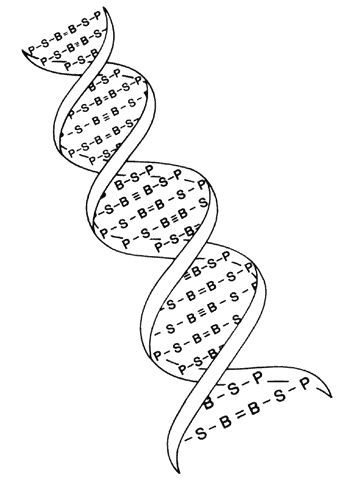
The structure of DNA is a double helix with four bases: adenine, guanine, cytosine, and thymine. These are paired by hydrogen bonds between the two helices like rungs in a ladder (Fig. 1). Each base is associated with a sugar and phosphate forming a nucleotide. The nucleotides are numbered in order, giving the DNA sequence. Nucleotides in the linear polymer chains that make up the two helices are connected by phosphodiester bonds. The sequence of bases (in nucleotide form), in groups of three called codons, makes up the genetic code. Each triplet codes for a specific amino acid. Since 64 different triplets are possible from the four bases, and there are only 20 amino acids, more than one triplet can code for a given amino acid.5 For example, the codon AAA will always and only produce the amino acid phenylalanine in the protein, but other codons may also produce phenylalanine in the protein. Amino acids are ultimately arranged into proteins, the building blocks of living structures.

Fig. 1. Drawing of the DNA double helix, which is held together by hydrogen bonds between the bases (B). The other two units in the DNA molecule are deoxyribose sugar (S) and phosphate (P).

In 1911, Johannsen coined the term “gene” as the basic unit of hereditary characteristics.6 In 1901, the monk Gregor Mendel described the dominant and recessive patterns by which some traits are inherited, leading to the term Mendelian inheritance.7 Garrod developed the concept of human biochemical disease (i.e., inborn errors of metabolism) and its inheritance in a recessive Mendelian fashion in 1908 and8,9

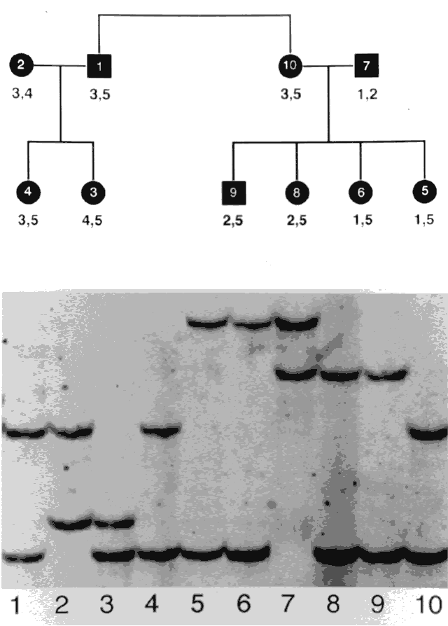
Genes are arranged into coding sequences, called exons, and intervening sequences that do not code, called introns. In addition, there may be separate promoter regions. Only 3% to 5% of human DNA codes for proteins; the rest has no known function.10 Although variation (mutation) in exons is likely to cause disease and is therefore rare, variation in introns does not cause a problem, yet is inherited in a Mendelian fashion. Since these variations are benign, they are also plentiful and vary from family to family. These variations are therefore used as genetic markers (e.g., in linkage analysis).

DNA sequences are written in a 5' to 3' direction (Fig. 2), and the sequence given is for the sense strand. The other strand is complementary and is called the template strand or antisense strand and runs in the opposite direction.10 DNA is transcribed in the following manner:

Fig. 2. DNA sequences for the sense strand (5' and 3' directions). In the double helix, the two strands run in opposite directions. Synthesis proceeds from 5' to 3'.

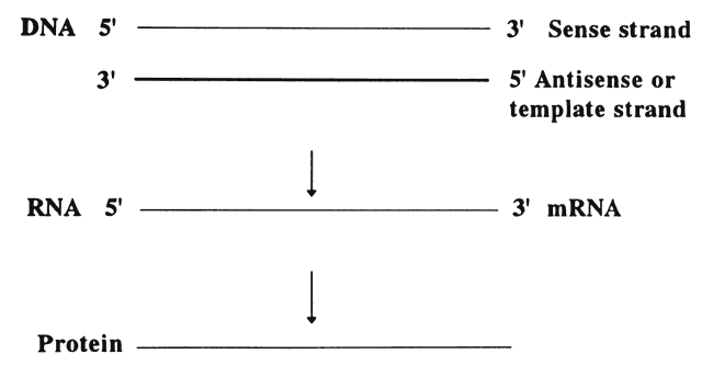
  1. Near the 5' end of genes, there are promotor sequences that instruct messenger RNA (mRNA) to start transcription using the DNA antisense strand as a template. That is, a new RNA strand is made that is complementary in its base composition to the DNA antisense strand and thus is the same as the sense strand. Each DNA codon is thereby turned into a corresponding RNA codon.
  2. The double helix uncoils, and the appropriate bases are added along the strand. RNA processing then occurs, cutting out (splicing) the introns, which do not code for anything, and rejoining the edges.
  3. Translation then occurs. This refers to the new mRNA strands, which include only coding sequences that move out of the nucleus and into the cytoplasm where they bind to the ribosomes. The ribosomes assemble a polypeptide according to the recipe specified by the sequence of codons.
  4. Various modifications may then occur before the chain is transported to its functional location within or outside of the cell.

DNA replicates itself in an analogous fashion. The two helical strands separate, and each is copied by a series of enzymes that position a complementary base opposite each base in the strand of DNA. One double helix thereby gives rise to two identical double helices.

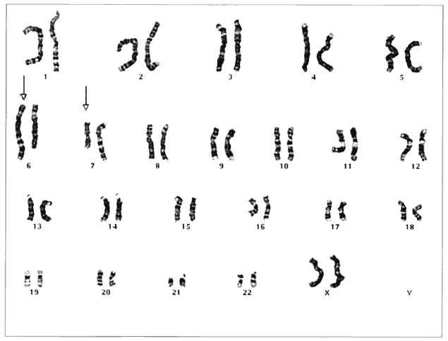
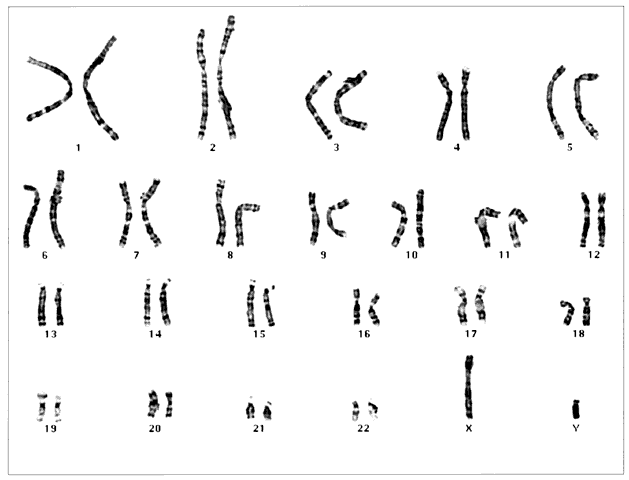
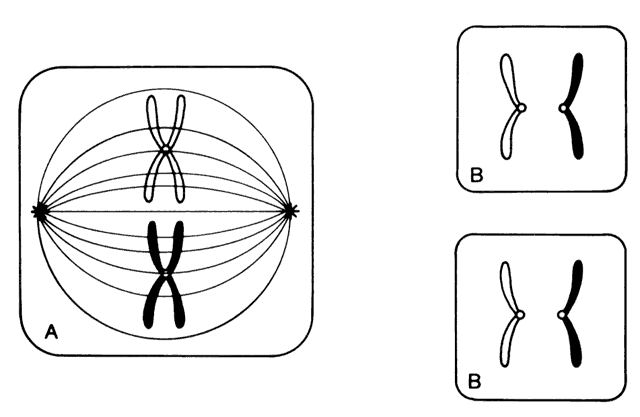
Humans have approximately 2 m of DNA arranged into 46 chromosomes composed of 23 homologous pairs. This includes 44 autosomes, which are the same in males and females, plus either an XY in males (the Y chromosome determines testis development) or an XX in females (Fig. 3). Each member of a homologous pair carries corresponding, although not necessarily identical, genes in the same sequence. One member of each chromosome pair is inherited from the father, the other from the mother. For most of the cell cycle, each chromosome exists as a single chromatid. When a cell is committed to division, however, each chromatid is replicated. This is the form in which we usually see pictures of chromosomes: two identical sister chromatids joined at a centromere, making an “X” shape.

Fig. 3. Normal human male XY karyotype showing 46 chromosomes. Sample was obtained from amniotic fluid. (Courtesy of Paul Fernhoff, MD, Emory University Genetics Laboratory)

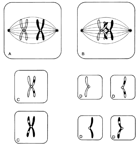
Each somatic cell has 46 chromosomes and divides by mitosis, which results in daughter cells with 46 chromosomes (Fig. 4). Mitosis gives rise to the body cells needed for growth and development. The normal sperm or ova, however, are the result of division by meiosis (Fig. 5) and have 23 chromosomes each, only one from each chromosome pair. In this manner, each parent passes on half of his or her genetic information to each child. Interestingly, the drive toward genetic diversity is so great that it can be seen even within a single sperm or egg. Each parent has two copies of every chromosome and passes on only one. However, the version of each chromosome that is passed on is not identical to either of the copies that the parent possesses. During meiosis, before the two copies of each chromosome separate to go to different eggs or sperm, the homologous chromosomes exchange genetic material in a process called crossing over (see Fig. 5B). At this stage, each homologue is made of two identical sister chromatids joined at the centromere. A crossover involves an exchange of genetic material between two chromatids on two different homologues.

Fig. 4. Mitosis. Only one pair of homologous chromosomes is shown. A. Each chromosome has replicated to form two identical chromatids joined at the centromere. The color difference indicates that one chromosome is paternally derived and one maternally derived. Chromosomes line up in tandem at the equatorial plate before cell division. B. Daughter-cell chromosomes, shown as chromatids, are identical to those of the parent cell.

Fig. 5. Meiosis. In the first meiotic division, homologous chromosomes line up parallel to each other at the equatorial plane (A). Crossing over occurs between two of the sister chromatids (B), permitting the exchange of corresponding chromosomal segments. Reduction division results in daughter cells (C) that contain only one chromosome of each homologous pair. The second meiotic division produces daughter cells (D) with just one chromosome from each homologous pair, the haploid number. Here they are shown as chromatids, each one representing a different “arm” of the 4-armed homologous chromosome pair formed at the beginning of meiosis.

For example, a person has one copy of chromosome 1 from her mother and another copy from her father. Assume that she has brown eyes and is esotropic. Assume hypothetically that an esotropia gene is present on the maternal chromosome 1 as well as a gene for brown eyes. On the paternal chromosome 1 are genes for straight eyes, color blue. We might expect that in meiosis, when one copy of each chromosome goes to each egg, that the resultant eggs would all have either the maternally derived chromosome 1 with brown eyes and esotropia or the paternal straight blue. In fact, however, crossing over exchanges material between this person's two copies of chromosome 1, making unique versions to be passed on: very possibly esotropia with blue eyes or straight brown eyes. The chance that traits will be separated by crossovers increases as their genetic distance from each other increases.

Many crossovers may occur involving one, two, three, or four arms. In human male meiosis, an average of 55 crossovers occurs, with at least 1 crossover per homologous pair. Crossovers in females are even more frequent.10 Thus, the version of chromosome 1 passed on by a parent is actually an amalgam of different pieces from each of his or her two different chromosome 1 copies. This is important to understand because in some families, diseases caused by mutations in genes that are physically close together on the chromosome may occur together in some family members, whereas others may have only one trait or the other. It is also vital to understand crossing over because it plays an important role in understanding linkage analysis.

At conception, a sperm and ovum unite to form a zygote, which reestablishes the 46-chromosome complement. These chromosomes represent a random assortment of the genetic information present in each parent, in a unique combination that allows for advantageous new gene interactions in the offspring. Conversely, it may bring together genes that add to each other in a negative way, producing what we know as human malformation or disease. In addition, with such a complex process it is understandable that “mistakes” sometimes occur: chromosome pairs may not completely separate during meiosis, leading to extra genetic material in the offspring called trisomies or rearrangements called translocations. Trisomies occur with increased frequency as maternal age increases; mutations occur with increased frequency as paternal age increases. Pieces of DNA may be lost (deleted), or one or more bases may not pair up normally, changing a codon (mutations). These occurrences are not rare, and often result in early miscarriages. Although they are genetic in nature, they may or may not be inherited or heritable; it may be a chance occurrence without increased risk of recurrence.

A mutation is a stable, heritable alteration in DNA transmitted from parent to progeny.11 From an evolutionary standpoint, mutations are necessary to generate genetic diversity, which makes adaptation to differing environments possible. While some mutations cause disease, others contribute to success and survival of the fittest. Some mutations do both: when one of a person's two homologous hemoglobin genes carries a sickle mutation, the person is resistant to malaria. Thus, this particular mutation protects persons who live in areas in which malaria is endemic, and it has been passed on to large numbers of progeny in some populations. If a person carries two copies of the sickle mutation, however, sickle cell anemia results, which is associated with high morbidity and mortality.

Back to Top
TAKING AN OPHTHALMIC FAMILY HISTORY
It is important to take a complete history, including pregnancy and birth, past medical history, medication use, and a detailed family history. For most patients, a few directed questions will yield the necessary information; for others, painstaking detail is required. Always remember that patients do not know which aspects of their family history will be important for the ophthalmologist to know, so if the right questions are not asked, valuable information will be lost.

Even a brief family history should be put in pedigree form using standard symbols for clarity (Fig. 6). Questioning should begin with names, ages, and genders of parents, siblings, and children of the proband (the person presenting for care). Initial questions should be general, and the type of questions to ask are common sense:

Fig. 6. The family history, or pedigree, is often of immeasurable value in reaching the correct diagnosis and ensuring appropriate genetic counseling. Above are the standard symbols used in drawing a pedigree.

  Does anyone else in your family have the same problem?
  Does anyone else have eye problems (e.g., glaucoma, retinal detachment, cataract) at an early age?
  Is anyone in the family blind in one or both eyes, and if so, what was the cause?

Such general questions should be part of every eye examination. A routine cataract patient with multiple family members who have suffered retinal detachments is no longer routine. A young patient with borderline high intraocular pressure and cup-to-disc ratio of 0.60 needs to be followed far more closely if several relatives are blind as a result of glaucoma. A child who is found to be myopic will usually have relatives who wear glasses for driving or distance; if instead there is a history of night blindness, or family members who could not get a driver's license because of poor best-corrected vision, the diagnosis and prognosis change markedly.

For patients who clearly have a syndrome or severe genetic disorder, the family history becomes even more crucial. Diagnosis and recurrence risk, of paramount importance to most families, often are determined by the pedigree. Directed history taking is vital, with attention to symptoms or systemic problems that commonly occur as part of the suspected syndrome. Table 1 provides a list of questions that should be asked when taking a directed history. It is important to describe clinical signs in lay terms: patients may not know if anyone in the family had “nystagmus,” but will quickly remember an uncle with “jumping or jiggling eyes.” It is often useful to take an additional history on subsequent visits, since patients may talk to relatives and remember more details after the initial interview. It is imperative to ask specifically about associated systemic findings. The polydactyly of Bardet-Biedl syndrome often is ligated at birth. The infantile skin vesicles of incontinentia pigmenti resolve spontaneously, making the history—not the physical examination—the key to the diagnosis. Aortic aneurysms in relatives of patients with ectopia lentis, or recurrent miscarriages in a family with a child who has multiple congenital anomalies, may not seem relevant to the patient giving a history, but these may provide clues to the diagnosis. A history of consanguinity should be sought if a recessive disorder is suspected.


TABLE ONE. Directed History Taking

  Is anyone in the family blind?
  Has anyone in the family lost an eye?
  Has anyone had jumping or jiggling eyes (nystagmus)?
  Has anyone been unable to get a driver's license because of poor vision?
  Do you or other family members have trouble seeing the stars at night or finding a seat in a dark theater?
  Has anyone had glaucoma or cataracts at a young age?
  Does anyone wear very thick glasses?
  Has anyone had cancer at an early age?
  *Is there a history of mental retardation?
  *Is there a history of sudden death at an early age? Double-jointedness? Sunken chest?
  *Is there a history of early arthritis, deafness, or cleft lip/palate?
  *Is the patient much taller or shorter than others in the family?
  *Is the patient much more lightly pigmented than others in the family?
  *Is there a history of miscarriage? Was the gender of the fetus or reason for the miscarriage known?
  *Are spouses distantly or closely related? What are your family names and where did your ancestors come from?
  *Has anyone had extra fingers or toes present at birth?


*These questions are aimed at uncovering related systemic disorders. Positive responses are most suggestive of the following, respectively: retinoblastoma; homocystinuria or chromosomal defects; Marfan's syndrome or other connective tissue disorders, such as Ehlers-Danlos syndrome; Stickler's syndrome; homocystinuria or Marfan's syndrome (tall), Weill-Marchesani (short); homocystinuria or albinism; chromosomal anomalies; consanguinity increasing the risk of recessive disorders; Bardet-Biedl syndrome.


If the ocular disorder is associated with a systemic syndrome, consultation with a geneticist, internist, or pediatrician is essential. In isolated ocular conditions, however, it falls to the ophthalmologist to discuss recurrence risk. The first step in any such discussion is to relieve parents of guilt. When told there is a genetic component to a disorder, some parents may feel responsible for being carriers, but they often do not articulate this to the clinician. The problem may be even more pronounced in dominant or X-linked disorders, where only one parent is the carrier. This may engender guilt on the part of one parent and blame on the part of the other. Although grandparents and other members of the extended family often lend valuable support, in some cases they experience feelings of guilt, fear, or anger as well, which can have an impact on the reaction of the patient or parents to the discussion of recurrence risk.

It is beneficial to state at the outset that no one is to blame for the inherited disorder. It often helps to point out that affected persons inherited many good qualities as well as the detrimental one, and that if there were a choice, we would all pass on only beneficial genes. Many parents feel relieved to hear it stated that nothing they ingested or did during a child's in utero development caused the disorder, even if this seems obvious. It may help to have parents or other family members articulate their feelings. Sometimes it is beneficial to tell patients that all humans carry abnormal genes and that it is simply a matter of chance whether a combination is present that causes disease. In families where it becomes clear that excessive blame or guilt is not being dealt with in a healthy manner, professional counseling should be recommended. Various support groups, such as the National Retinitis Pigmentosa Foundation, the von Hippel-Lindau Family Forum, and others should also be recommended as appropriate.

If genetic studies are contemplated, several considerations are important. Confidentiality must be maintained because jobs and insurance coverage may be affected by labeling someone a carrier of a disease. Before testing, especially in linkage studies that offer little or no immediate benefit to patients, informed consent must be obtained, with careful consideration being paid to current guidelines. Informed consent is especially complicated in the case of children. Current recommendations are that children older than 7 years should have not only parental consent, but voluntarily assent to participation.12 Appropriate consent/assent forms should be signed. It is also important to obtain permanent addresses where participants may be reached in future years, since results of some genetic testing can be delayed for years.

Back to Top
PATTERNS OF INHERITANCE
In 1901, the Austrian monk Gregor Mendel described autosomal-dominant and autosomal-recessive inheritance of traits in pea plants.7 Since then, these patterns—as well as X-linked dominant and recessive, mitochondrial, and polygenic inheritance—have been described in human disease transmission. A careful pedigree should elucidate the mode of inheritance in a given family.

AUTOSOMAL DOMINANT DISORDERS

In dominant disorders, the disease trait is manifested when only one copy of the gene is abnormal. These disorders often are passed directly from parent to child through several generations, with examples of transmission to children of both sexes from parents of either sex, including male-to-male transmission (Fig. 7). The risk of having affected offspring is 50% for each offspring. Many parents interpret this to mean that one half of an affected person's children will be affected and half unaffected, and thus believe they are “safe” after having one or two affected children. It is important to clarify to these parents at the outset of the discussion that there is a 50/50 chance for each offspring to be affected. For example, each flip of a coin has a 50% chance of being “heads” and a 50% chance of being “tails,” but the order is random: only the final result after an infinite number of tosses results in one half of the tosses being heads and the other half tails. If neither parent is affected, the risk of each future child's being affected is usually very small, often not greater than the population risk. It should be noted, however, that some dominant disorders, such as coloboma or neurofibromatosis, have such variable expressivity that affected persons may not know they are affected.

Fig. 7. Autosomal-dominant inheritance. There is vertical transmission through at least three generations in this pedigree. Only affected persons transmit the condition, and there is a 50% chance that each offspring of an affected person will be affected. Both sexes are affected in approximately equal numbers, and there is male-to-male transmission.

Dominant disorders often are caused by genes that code for structural proteins such that specific tissues are affected: for example, Stickler's syndrome in which mutations of COL2A1 make structurally abnormal collagen leading to abnormalities of the joint, palate, and face as well as abnormal vitreous, which predisposes the affected patient to myopia and retinal detachment.13,14 Often there is no family history. Remember: someone has to be the first to have a new mutation producing a dominant disorder and subsequently a pedigree. This person's father may have been advanced in age, which increases the risk of occurrence of a new dominant mutation.

Dominant disorders often show great variability in expressivity of the trait. For example, coloboma may be autosomal dominant in 22% of cases15, but some members of the family may have only a small chorioretinal “scar” as their manifestation, whereas others may have coloboma of the iris, lens, retina, and choroid, causing microphthalmia and blindness. Some dominant disorders appear to skip generations; this is often dependent on the means used to diagnose the disorder. For example, Best's macular dystrophy may appear to be absent in the young children of a large pedigree with many affected members when diagnosed by ophthalmoscopy. But if an electro-oculogram is used as the diagnostic test, more affected patients, often young children, will be found who do not yet have visible retinal changes.

In pedigrees with retinoblastoma, an abnormal copy of the Rb gene is transmitted as a dominant; however, for a person to be affected, a second somatic mutation must occur in that person. This second mutation occurs in approximately 90% of patients, but in 10% it does not, and no retinoblastoma develops. For example, a seemingly normal person who has an affected parent and an affected child is deemed “unaffected” by ophthalmoscopy, but DNA testing reveals the presence of the Rb mutation. What appeared to be a “skipped generation” was actually a fortunate lack of the second mutation needed for manifestation of the disease, although the inheritance pattern of the gene is dominant.

In some autosomal-dominant and X-linked recessive disorders, there is a phenomenon called anticipation. This means the manifestations of the disease seem to worsen in succeeding generations. An example of this is olivopontocerebellar atrophy, in which a parent may have late-onset, mild cerebellar symptoms and slowly progressive macular degeneration, while his or her child develops complete blindness and becomes wheelchair-bound at an early age. Some cases of apparent anticipation may be due to ascertainment. That is, once it is known that an inherited disorder runs in the family, everyone is more aware of symptoms, children are diagnosed at an earlier age, and they are diagnosed correctly. In some cases, however, anticipation is due to expansion of abnormal sequences of DNA; once these sequences are already longer than normal, they are likely to become more mutated with succeeding generations. This phenomenon has been well described in the Fragile X Syndrome, an important cause of inherited mental retardation in males.

X-linked dominant disorders are very rare, but do exist. In these disorders the genetic abnormality in a gene on the X chromosome is so severe that even the presence of a normal X in females cannot protect from its effects and females are severely affected. Males, who have no normal activity of these presumably vital genes, cannot survive and usually die in utero (Fig. 10). Examples which may be seen in ophthalmologic practice include incontinentia pigmenti (IP) and perhaps Aicardi's syndrome. Patients with Aicardi syndrome usually represent new mutations, while IP is not infrequently present in the family, often with variable expressivity.

Fig. 10. X-linked dominant inheritance. Male offspring of an affected female die in utero. Female offspring have a 50% chance of being affected.

AUTOSOMAL RECESSIVE DISORDERS

Autosomal-recessive inheritance means that two copies of an abnormal gene are required to produce a trait or manifest a disease. Both parents can be normal but can have one or more children of either sex who are affected. Once parents have a child with a recessive disorder, the risk of their having another affected child is one in four (25%) for each new offspring (Fig. 8). Recessive disorders usually result from gene mutations that affect the structure of an enzyme. There is a dose effect, heterozygotes having some normal enzyme function (usually enough to be asymptomatic) but homozygotes having little or no normal enzyme function. Biochemical testing is available to determine carrier versus affected status for many such disorders, including galactosemia, homocystinuria, gyrate atrophy, and Tay-Sachs disease. Recessive disorders are, on average, more severe than dominant ones. This may be partly because dominant disorders persist in the population only if affected persons are healthy enough to reproduce and pass them on, whereas recessive genes are “hidden” in healthy heterozygotes, and therefore persist for generations, finally causing disease so severe that the affected person cannot reproduce.

Fig. 8. Autosomal-recessive inheritance. Normal siblings of an affected (homozygous) person have a 2/3 chance of being carriers (heterozygotes). The double line indicates a consanguineous union, which increases the risk of recessive mutations coming together in an offspring.

The likelihood of manifestation of a recessive disorder increases dramatically with consanguinity, inbreeding, or geographically isolated populations. When persons who have an unusually large number of genes in common marry, recessive disorders increase in frequency. Populations in which this is known to occur include Ashkenazi Jews, especially those with ancestors who lived in the Pale; Bedouins; the Amish; and other groups that frequently marry within a small group. If autosomal-recessive inheritance is suspected, information on ethnicity of both parents and even review of family names on both sides often suggests shared ancestry.

X-LINKED DISORDERS

The key to diagnosing an X-linked recessive disorder is recognizing a pedigree in which only males are affected and all carrier females have an affected father (Fig. 9). There is never male-to-male transmission. Rarely, a woman may manifest some or all signs of the disorder, in which case she would be diagnosed as a manifesting carrier. Because men have only one X chromosome, defects in genes on X that do not have a counterpart on Y cannot be mitigated by the normal homologous gene as it can in women, who have two X chromosomes.

Fig. 9. X-linked recessive inheritance. There is never male-to-male transmission. All of an affected male's daughters will be carriers. A carrier female has a 50% chance of each son's being affected and of each daughter's being a carrier.

NON-MENDELIAN INHERITANCE

Mitochondrial Disorders

Some disorders clearly run in families, but just as clearly do not follow simple Mendelian patterns. An example of this is mitochondrial inheritance. Mitochondria are organelles within human cells. Mitochondria have their own circular DNA, which replicates with and is passed on like the chromosomes already described for nuclear DNA, but is physically separate, residing in the cytoplasm rather than in the nucleus of cells. These mitochondrial genes encode 13 proteins. A fascinating fact about mitochondria is that although in both men and women these genes encode protein subunits in the respiratory chain enzyme complexes and transfer and ribosomal RNAs, all of a person's mitochondria are inherited from his or her mother only. Because mitochondrial DNA is contained in the cytoplasm of cells, including that of egg and sperm, and because only the nuclear DNA of the sperm unites with the nuclear DNA of the egg, the zygote receives only the mitochondrial DNA present in the cytoplasm of the egg (i.e., from the mother).

Thus, only the mother transmits mitochondrial DNA to offspring, and only her daughters pass it on. The inheritance pattern therefore is not Mendelian. Disorders run in families through the maternal lineage, but affected women or women carriers never have affected fathers. Men are affected more frequently than women (for unknown reasons), but more females are affected than in a typical X-linked disorder (Fig. 11). The two most common mitochondrial disorders seen in ophthalmologic practice are Leber's hereditary optic neuropathy and chronic progressive external ophthalmoplegia.

Fig. 11. Mitochondrial inheritance. Women with the mutation pass it on to 100% of their offspring, but not all are affected. Males never pass it on, whether affected or unaffected. Males with the mutation are much more likely to be affected than females with the mutation.

Polygenic Disorders

Mitochondrial disorders are quite rare; more common causes of non-Mendelian inheritance are polygenic or multifactorial disorders. Polygenic disorders obviously have a heritable component, but when rates of affected status are calculated and pedigrees examined, there is not a perfect fit with any of the known modes of inheritance; this may be due to an environmental component. A good example is refractive error. In myopia, for example, children of myopic parents are more likely to be myopic than are children of emmetropic or hyperopic parents,16 but many exceptions occur and frequencies do not strictly fit a dominant, recessive, or X-linked model. This may be because refraction depends on the interplay of axial length, lens shape, and corneal shape, each of which may be inherited separately, as well as a proposed emmetropization factor. Thus, refractive error also may be considered a multifactorial disorder because the conditions that combine to form the disorder are partially determined by one or more genetic factors (i.e., are inherited) but are also modified by environment. Certainly a predisposition for myopia is inherited; however, many studies have shown an effect of the amount of time spent reading on the development of myopia17 as well as the effect of environmental factors on myopia development in animal models.18 The combination of genetic and environmental factors makes evaluation of the genetic component especially complex.

Chromosomal Abnormalities

Chromosomal abnormalities are obviously genetic, but they may or may not be inherited or heritable. Down syndrome, which often has significant ocular morbidity, is a common chromosomal abnormality present in all populations. It is most commonly caused by trisomy 21, a disorder in which the affected person has an extra chromosome 21 that did not separate properly during meiosis. In some cases, however, a translocation occurs, causing some extra chromosome 21 material to adhere to another chromosome. This type of Down syndrome may be inherited: a parent may be carrying a balanced translocation, in which the total amount of DNA is correct but packaged asymmetrically, leading to a chromosomal imbalance in the offspring. Trisomy 13, Turner's syndrome, and many other congenital syndromes have a genetic basis, but are usually sporadic and non-Mendelian.

Back to Top
CURRENT MOLECULAR GENETIC TECHNIQUES
Although a sophisticated understanding of current laboratory techniques in molecular genetics is beyond the needs of most practicing ophthalmologists, a basic knowledge of how to proceed in a genetic workup is within every ophthalmologist's grasp. A stepwise approach simplifies this admittedly complex and “high-tech” field.

STEP 1. Develop a working diagnosis and as detailed a pedigree as possible. Like amniocentesis, which screens only for disorders for which an individual patient is at risk, a molecular genetic workup also must be directed toward “hot-spots” with the use of current techniques. Autosomal-dominant, X-linked recessive, and mitochondrial disorders are far easier to study than autosomal-recessive ones. Families with many affected members statistically are more powerful than singleton patients. Once a diagnosis and family survey have been done, ask yourself this question: Does this patient have an isolated disorder (e.g., autosomal-dominant coloboma), with effects only in the eye, or a syndrome (i.e., a multiorgan abnormality)? In a patient with significant congenital anomalies in two or more organ systems, a high-resolution banding karyotype should be considered (Fig. 12). In patients with sporadic (vs autosomal-dominant) aniridia, the chance of finding a detectable abnormality (usually a deletion in the short arm of chromosome 11), is also significant, especially if Wilms' tumor, growth retardation, or developmental delay are present. Children with retinoblastoma plus other systemic abnormalities may demonstrate deletions in the q arm of chromosome 13. In cases of isolated retinoblastoma, abnormalities are unlikely to be manifested on a high-resolution banding karyotype. Only large deletions, transpositions, and trisomies can be detected with this test; point mutations cannot. Thus, this test is indicated only if there are ocular abnormalities known to be associated with gross chromosomal structural changes, or if there are other congenital anomalies suggesting multiple abnormal genes. If a child has an abnormal karyotype, both parents' karyotypes should be examined even if the parents appear to be normal. Any patient suspected of having Down syndrome, Turner syndrome, or trisomy 13 should have a karyotype analysis.

Fig. 12. High-resolution banding karyotype performed on peripheral blood. This is written as 46 XX t(6;7)(q23.1; q11.23). This means that a break has occurred in the long or “q” arms of one chromosome 6 and one chromosome 7 (arrows). Chromosomal material from each chromosome was switched, that is, translocated to the other chromosome. Since genetic material is not lost, this would be a balanced reciprocal rearrangement and would not usually cause phenotypic effects. An individual who carries this balanced rearrangement would be at increased risk for having an offspring with an unbalanced karyotype which would result in a miscarriage, a stillbirth, or a child with birth defects. (Courtesy of Paul Fernhoff, MD, Emory University Genetics Laboratory)

STEP 2. If the abnormality is isolated/subtle or the karyotype is normal, determine whether there is a candidate gene for the disorder. If the disorder obviously runs in the family, it is genetic. If there is only a singleton patient, a literature search may be done to determine whether inherited or genetic forms of the disorder have been described. Always remember that even in a dominant disorder there may be no family history: someone has to be the first new mutation to start the pedigree. If the gene is known (e.g., in ADRP, in which approximately 33% of patients are known to have mutations of the rhodopsin or peripherin genes; in Leber's hereditary optic neuropathy, in which most patients have one of three major mutations of mitochondrial DNA), a blood sample from one affected person may be sent to a laboratory that screens for that disorder using gene mutation analysis. Currently, most of these tests are being done on a research basis, each laboratory looking at a few specific disorders. If the index patient's DNA is negative for a mutation, the presence of a genetic disorder cannot be ruled out, since none of these tests is positive in all cases. By the same token, the presence of a mutation does not always prove causality.

Many changes in DNA do not cause disease. These changes are called polymorphisms. To determine whether a mutation found in a patient with a given disease is a polymorphism or a disease-causing mutation, other family members—both affected and unaffected—must be tested. If the mutation does not change an amino acid in the final protein, it is not disease-causing. If the DNA change is found in all affected persons and never in unaffected persons, the likelihood that it is responsible for the disease is high. Statistical analysis can be done to determine whether the concordance between mutation and disease state is sufficient to prove causality. If only a singleton patient is available for study, and a mutation known to segregate with the disease is found in other families, causality can be assumed. In the case of a new mutation, however, if it changes both a conserved amino acid and the final protein, it is likely to cause disease, but in the absence of a known segregation of the mutation with the disease in a family, this cannot be proved. Once a mutation is found, gene sequencing is done to detect the exact abnormality.

Figure 13 demonstrates the denaturing gradient gel method of screening for mutations in butterfly macular dystrophy, as well as the mutation segregating with the disease.

Fig. 13. Denaturing gradient gel electrophoresis of polymerase chain reaction products containing exon 1 of the peripherin gene from members of a family with butterfly macular dystrophy. This method may be used to screen single patients or entire families with disorders for which there is a candidate gene. Each gel lane contains a sample from the person whose pedigree symbol is directly above the lane. Clinically affected persons are indicated by closed symbols, clinically unaffected by open symbols. Spouses are indicated by unnumbered symbols, which are connected to an affected patient's symbol by a horizontal line. Affected persons have multiple bands on the gel (representing homoduplexes and heteroduplexes caused by the presence of a mutation), whereas normal persons have only one band, corresponding to normal homoduplex molecules. (Nichols BE, Sheffield VC, Vandenburg K et al: Butterfly-shaped pigment dystrophy of the fovea caused by a point mutation in codon 167 of the RDS gene. Nat Genet 3:202, 1993)

STEP 3. If gene mutation analysis is negative, or a gene has not been identified for a specific patient's disorder, linkage analysis may be performed (Fig. 14). For disorders in which no gene has been implicated, linkage analysis uses genetic markers to determine the location of the gene. Genetic markers are patterns of DNA sequences with a known location on a chromosome, which are inherited in a Mendelian fashion.

Fig. 14. Linkage analysis. In this figure, all symbols are arbitrarily filled in; the affection status is not shown. Each person in the pedigree is numbered within the symbols to correspond with the gel lane containing his or her DNA sample. Each person has two bands with this marker: one band inherited from the father, the other from the mother. Bands are numbered from 1 to 5 starting with the band nearest the top of the gel. Each person's banding pattern is printed under his or her symbol. Now the paternal origin of each band can be determined for all of the children in generation II. For example, person 3 received band No. 5 from her father and band No. 4 from her mother. Person 4 received band No. 5 from her father and band No. 3 from her mother. We know this even though both parents carry band No. 3 because band No. 5 can come only from the father. Each parent contributes one band, therefore band No. 3 is maternal. (Courtesy of Edwin Stone, MD, PhD, University of Iowa)

Linkage analysis works in the following way: first the entire family is typed at each marker as in Figure 14; then the inheritance of each marker is correlated with whether each individual is affected or unaffected.

For purposes of discussion (please refer to Figure 14), assume persons 1 and 10 are both affected with an autosomal-dominant ocular disorder. Assume further that all of the children in the second generation are affected. In this scenario, we can see that all affected persons carry the No. 5 band, but neither of the unaffected spouses do. Thus, the disease may be close to this marker and may travel with it during DNA replication and division in meiosis. A statistical analysis can be done to determine the likelihood of this, called the LOD score. If the LOD score is -2 or more, the disease is very unlikely to be in the immediate vicinity of the marker. If it is + 3 or greater, it is very likely to be in the vicinity of the marker and therefore considered linked. Thus, in the above case, if we knew this marker mapped to chromosome 1q11 (the long arm of chromosome 1), we could begin to look for candidate genes in this area.

Let us now change our assumptions. Assume now that persons 1 and 10 are affected, but only offspring No. 3 and No. 9 are affected. The other children in generation II are normal. Now, although all affected persons carry the No. 5 band, many unaffected persons do also. The marker no longer perfectly segregates with the disease. This can occur (1) as a result of recombination, in which pieces of DNA that are adjacent to each other in a parent are no longer close in the offspring because of crossing over during meiosis; or (2) because the marker is not linked with the disease. The LOD score can be calculated to determine the probabilities of each of these two options, with different rates of recombination.

Linkage analysis has pointed the way to several important disease-causing genes. In one study,19 linkage analysis in ADRP families showed linkage to chromosome 3. This was known to be the location of rhodopsin, an important retinal substance. Patients with ADRP were thus screened for mutations in the rhodopsin gene, which were in fact present in many patients and families. In another study,13 linkage analysis led the way to identifying the COL2A1 gene as a cause of Stickler's syndrome. The advantage of linkage analysis is that it points to a specific region of one chromosome. Researchers can then investigate known genes in a specific chromosomal area that are likely to cause the disease in question (candidate genes) or search for new genes in that area. It generally requires at least 10 family members with definitely known affection status to achieve a significant linkage result.

Back to Top
CURRENT KNOWLEDGE OF INHERITED EYE DISEASES
Currently, disorders such as ADRP, pattern macular dystrophy, retinoblastoma, coloboma, aniridia, albinism, Stickler's syndrome, Leber's hereditary optic neuropathy, and gyrate atrophy have known genetic mutations, and therefore molecular genetic workup and counseling can be offered to patients. Since the list grows and changes all the time, frequent searches of the literature and consultation with inherited eye disease specialists are vital for providing patients with state-of-the-art care. Although most molecular genetic diagnoses currently are significant only for family planning and prognosis, some disorders such as gyrate atrophy have treatments that can halt progression of the retinal degeneration. More treatments will become available in the years ahead.

For some other disorders, the genetic locus is known through linkage analysis, but no gene has yet been found. Thus, single patients cannot be tested, but family linkage studies can be done. This is true for diseases such as Best's vitelliform macular dystrophy, primary open-angle glaucoma, Rieger's syndrome, and congenital cataracts.

Genetic testing for retinoblastoma is readily available and should be offered to all families with this disease. If there are two or more affected persons in the family, DNA markers within the Rb gene can type the entire family; in this case, a modified form of linkage analysis (see Fig. 14) can be used to determine which persons are at risk. Even singleton patients may benefit: because the Rb gene is known, the entire gene also can be sequenced directly to look for mutations. This process takes a long time to complete, but it is useful for parents who want to know if either of them is a carrier before having other children. The Rb gene product is involved in the regulation of cell growth, so persons with an Rb mutation are also at an increased lifelong risk for development of other cancers. Gene therapy is being attempted in animal models, but very early detection, treatment, and close follow-up are currently the mainstays of treatment.

Although the potential for gene therapy exists for all genetic conditions once the defective gene function is known, simpler yet equally effective therapies may be implemented in some cases. Gyrate atrophy is a progressive autosomal-recessive retinal degeneration caused by mutations in the ornithine amino transferase (OAT) gene. Patients with certain mutations are responsive to dietary change and vitamin B supplementation, which can halt the progression of retinopathy. Genetic testing for these patients may be sight-saving. Patients with ADRP often can be given a more precise prognosis based on which rhodopsin or peripherin mutation is present. Patients with Leber's hereditary optic neuropathy are more likely to regain vision if their disease is caused by the more benign versus the more severe mitochondrial mutation. Family members who carry the mitochondrial mutation can be cautioned to avoid drugs and alcohol because there is some evidence that they may precipitate vision loss. Children with sporadic aniridia and a known deletion must be followed carefully for Wilms' tumor; children with coloboma and a chromosomal abnormality must be carefully screened for cardiac and other defects. A patient with a maculopathy and a peripherin mutation can be reassured that good vision will probably be retained, unlike the case in age-related macular degeneration or other entities that may have a similar fundus appearance. A child with myopia and a COL2A1 mutation may need examinations under anesthesia until he or she is able to cooperate with scleral depression, given the very high risk of retinal detachment in Stickler's syndrome; serial hearing tests can also be scheduled to be sure that the often associated decreased hearing does not interfere with life and learning. A child with nystagmus may be spared an electroretinogram and magnetic resonance imaging, and the parents spared much anguish, if a mutation associated with albinism can be detected with blood screening. This is especially important in children from darkly pigmented ethnic groups, who may have enough pigmentation to make the clinical diagnosis of albinism difficult. Thus, although we are far from specific treatment for most inherited eye diseases, molecular genetic diagnosis can often aid in management of patients.

Back to Top
SUMMARY
For the practicing ophthalmologist, the important message is this: every ocular disorder has some genetic component. Inherited eye diseases are not zebras—we see them everyday. If the patient has a strong family history or other systemic abnormalities, or if a known genetic factor has been reported in the literature, a genetic workup should be considered. In disorders associated with other major congenital malformations, a high-resolution banding karyotype should be performed. In isolated cases or if the karyotype is negative, an index patient's blood should be sent for gene mutation analysis if there is a known or candidate gene for that disorder. If the analysis is positive, it must be shown to segregate with the disease in the family or to be a known causative mutation in other large families before definitive genetic counseling can be given. If the analysis is negative or if no candidate gene exists, the entire family may be recruited for linkage analysis. Generally, 10 or more family members with definite diagnoses must participate in order for linkage analysis to have enough statistical power to find a genetic locus. For some singleton patients or small families with certain disorders, it is still not possible to offer definitive molecular genetic testing. All patients benefit, however, from the knowledge that there is intense research being done in this field and that in the not-too-distant future, much more will be learned about the diagnosis and treatment of all inherited eye diseases.
The authors and editors wish to express their appreciation to G. Frank Judisch, MD, author of the original Basis of Inheritance chapter. Some of his material is included in this revision.
Resources for families: National Retinitis Pigmentosa Foundation, Inc., d/b/a RP Foundation Fighting Blindness, 1401 Mt. Royal Avenue, 4th Floor, Baltimore, MD 21217; (800) 683-5555, Fax (410) 225-3936, TDD (410) 225-9409.
VHL Family Forum, 171 Clinton Rd, Brookline, MA 02146.
For retinoblastoma testing, contact B. Galli, MD, Visible Genetics, 700 Bay St., Suite 100, Toronto, Ontario M5G1Z26; (416) 813-3260.
Back to Top
REFERENCES

1. Deutman AF, van Blommestein JDA, Henkes HE et al: Butterfly-shaped pigment dystrophy of the fovea. Arch Ophthalmol 83:558, 1970

2. Nichols BE, Sheffield VC, Vandenburgh K et al: Butterfly-shaped pigment dystrophy of the fovea caused by a point mutation in codon 167 of the RDS gene. Nature Genet 3:202, 1993

3. Nichols BE, Drack AV, Vandenburgh K et al: A 2 base pair deletion in the RDS gene associated with butterfly-shaped pigment dystrophy of the fovea. Hum Molec Genet 2:601, 1993

4. Wells J, Wroblewski J, Keen JW et al: Mutations in the human retinal degeneration slow (RDS) gene can cause either retinitis pigmentosa or macular dystrophy. Nature Genet 3:213, 1993

5. Watson JD, Hopkins NH, Roberts JW et al: Molecular Biology of the Gene. Menlo Park, Benjamin/Cummings, 1987

6. Johannsen W: The genotype concept of heredity. Am Nat 45:129, 1911

7. Mendel G: Versuche über pfanzenhybriden. Leipzig, Engelmann, 1901

8. Garrod AE: Inborn errors of metabolism (Croonian lectures). Lancet 2:1,73,142,214,1908

9. Garrod AE: Inborn Errors of Metabolism, 2nd ed. London, Oxford University Press, 1923

10. Read A: Medical Genetics: An Illustrated Outline. Philadelphia, JB Lippincott, 1989

11. Beaudet AL, Scriver CR, Sly WS et al: Human Biochemical and Molecular Genetics. New York, McGraw-Hill, 1990

12. Wertz DC, Fanos JH, Reilly PR: Genetic testing for children and adolescents: who decides? JAMA 272:875, 1994

13. Francomano CA, Liberfarb RM, Hirose T et al: The Stickler syndrome: evidence for close linkage to the structural gene for type II collagen. Genomics 1:293, 1987

14. Ahmad NN, Al-Kokko L, Knowlton RG et al: Stop codon in the procollagen II gene (COL2A1) in a family with Stickler syndrome (arthroophthalmopathy). Proc Natl Acad Sci USA 88:6624, 1991

15. Fujiki K et al: Genetic analysis of microphthalmus. Ophthalmic Paed Genet 1:139, 1982

16. Zadnik K, Satariano WA, Mutti DO et al: The effect of parental history of myopia on children's eye size. JAMA 271:1323, 1994

17. Rosner M, Belkin M: Intelligence, education and myopia in males. Arch Ophthalmol 105:1508, 1987

18. Raviola E, Weisel TN: An animal model of myopia. N Engl J Med 312:1609, 1985

19. Dryja TP, McGee TL, Reichel E et al: A point mutation of the rhodopsin gene in one form of retinitis pigmentosa. Nature 343:364, 1990

Back to Top