Chapter 54A
Inborn Errors of Metabolism Affecting the Eye
SCOTT E. OLITSKY and LEONARD B. NELSON
Main Menu   Table Of Contents

Search

DISORDERS OF AMINO ACID METABOLISM
DISORDERS OF CARBOHYDRATE METABOLISM
REFERENCES

An inborn error of metabolism is a clinical disorder caused by a genetic inability to produce the full complement of a given functioning protein in its normal configuration. Many of these disorders are rare events with often devastating ocular manifestations. Because of the high incidence of these ocular manifestations, the ophthalmologist is often involved in the care of these patients. Also, there is an increasing diagnostic role for the ophthalmologist in detecting subtle, early, and occasionally pathognomonic lesions to aid in the diagnosis of these rare disorders.
Back to Top
DISORDERS OF AMINO ACID METABOLISM

HOMOCYSTINURIA

Homocystinuria is an inborn error of metabolism of the sulfur-containing amino acids. Carson and Neill first detected homocystinuria using urine chromatography while systematically searching for metabolic abnormalities in mentally retarded institutionalized persons in Northern Ireland.1 Simultaneously and independently, Gerritsen and associates in Madison, Wisconsin, found the same metabolic defect in an infant with mental retardation and failure to thrive.2

Homocystinuria is an autosomal recessive disorder caused by a deficiency of the enzyme cystathionine beta-synthase, an intermediate in the degradation of homocysteine to cystine.3 The resulting blockage in this biochemical pathway causes accumulation of homocysteine precursors, homocystine and methionine, with increased concentrations of these amino acids in blood and urine. The genetic mutation has been mapped to chromosome 21 (21q22.3).

Four organ systems show major involvement in patients with homocystinuria: the skeletal, central nervous, and vascular systems and the eye . Excessive height and long thin extremities are common skeletal findings in the homocystinuric patient. These findings are similar to those of the Marfan syndrome. The most consistent skeletal abnormality is osteoporosis, which may lead to vertebral collapse.4 Mental retardation occurs in approximately 50% of cases and may be progressive. Thrombotic vascular occlusions constitute the main threat to survival in patients with homocystinuria. Large and small arteries and veins may be affected. Therefore, premature deaths from myocardial infarctions, pulmonary emboli, and cerebrovascular accidents are not uncommon.

Anesthesia may present significant risks to the homocystinuric patient.5–7 Mudd and colleagues documented 25 postoperative thromboembolic events in 586 homocystinuric patients undergoing surgical procedures, 6 of them fatal.8 Medications that may predispose these patients to a hypercoagulable state, such as oral contraceptives, should also be avoided.9

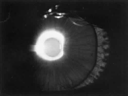
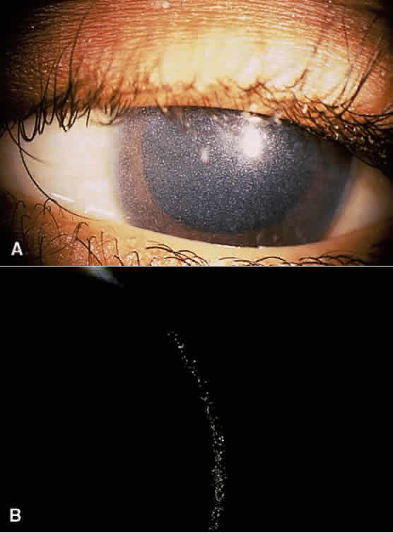
Ectopia lentis is the ocular hallmark of homocystinuria and can be detected in approximately 90% of patients.10,11 Ectopia lentis is an acquired and progressive abnormality in this disorder. The dislocation is bilateral, with the lens usually migrating either inferiorly or inferonasally. The causes of ectopia lentis are summarized in Table 2.


TABLE TWO. Differential Diagnosis of Ectopia Lentis

  Genetic

  Without systemic manifestation

  Simple ectopia lentis

  Congenital (recessive and dominant varieties reported)
  Delayed onset (recessive and dominant varieties reported)


  Ectopia lentis et pupillae (recessive)
  Megalocornea (most commonly X-linked; dislocation of the lens a rare complication)


  With systemic manifestation

  Marfan syndrome
  Homocystinuria
  Weill-Marchesani syndrome
  Hyperlysinemia
  Sulfite oxidase deficiency


  Nongenetic

  Trauma
  Leutic


(Modified from Maumenee IH: The eye in the Marfan syndrome. Trans Am Ophthalmol Soc 79:684, 1981)


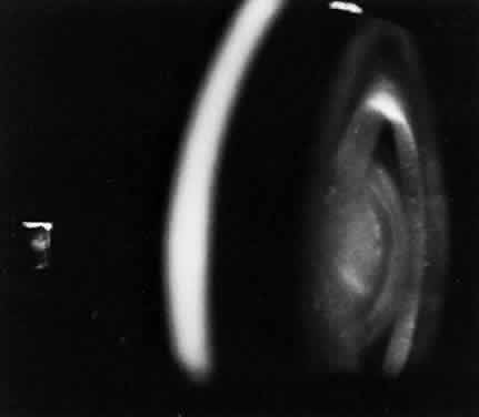
The lens in homocystinuria is much more mobile than in the Marfan syndrome. This may be related to the clinical observation of progressive irregularity of the zonular fibers and the appearance of a fringe of white zonular remnants at the equator of the lens and on the surface of the ciliary body (Fig. 1).12,13 Henkind and Ashton first reported histopathologically the ocular findings in four eyes of three homocystinuric patients. They found the zonular fibers to be deficient adjacent to the lens. These zonules had recoiled to the surface of the ciliary body and were matted and retracted into a feltwork that fused with a greatly thickened basement membrane of the nonpigmented epithelium. The greatly thickened basement membrane overlying the ciliary body in homocystinuria has subsequently been shown by electron microscopy to be composed of degenerate zonular material.14 In addition, Ramsey and coworkers noted that the degree of zonular abnormality was related to age: the younger the patient, the more normal-appearing zonular fragments composed of oriented filaments that could be identified.14 The zonular fibers are composed of glycoproteins with a high concentration of cysteine, which may explain their susceptibility to abnormal function in homocystinuria.15,16

Fig. 1. Inferior dislocation of the lens in a homocystinuric patient. Note the absence of most zonular fibers. (Nelson LB, Maumenee IH: Ectopia lentis. Surv Ophthalmol 27:143, 1982)

Myopia is another common ocular finding and may precede the detection of ectopia lentis by several years.17 The development of progressive lenticular myopia is often the first sign of a lens dislocation. Retinal detachment is usually a complication of lens surgery, although it may occur spontaneously.11 Other ocular findings associated with homocystinuria are listed in Table 3.


TABLE THREE. Ocular Findings Associated With Homocystinuria and the Frequency of Their Occurrence


FindingFrequency
Dislocation of lensesAlmost all cases
MyopiaAlmost all cases
Fringe of zonules on dislocated lensMost cases
Peripheral cystoid retinal degenerationUsual
Optic atrophyFrequent
Secondary glaucomaFrequent
White deposits on the ciliary processes and zonular fibersUncertain
Congenital cataractRare
Central retinal artery occlusionRare
AniridiaRare
Coloboma of choroid and optic discRare
Coloboma of irisRare
MicrocorneaRare
MicrophthalmosRare
(Modified from Spaeth GL: Other inborn errors of metabolism affecting the eye. In Harley RD [ed]: Pediatric Ophthalmology. Philadelphia, WB Saunders, 1983).


One treatment of homocystinuria to correct the metabolic defect has involved a low-methionine and high-cystine diet, which has not been uniformly successful.18–22 This diet has not only failed to normalize completely the abnormal biochemical findings, but it is difficult to prescribe (all eggs, meat, and cow's milk are forbidden). When this diet is prescribed to infants, failure to gain weight may be a serious complication.23

Another approach to therapy is supplementation with coenzymes. Pyridoxine (vitamin B6) is the coenzyme necessary to activate cystathionine beta-synthetase.24 Barber and Spaeth were the first to demonstrate that certain homocystinuric patients respond biochemically to oral pyridoxine with lowered plasma concentrations of homocystine and methionine.25 A different mutation in the cystathionine beta-synthase gene is responsible for the observed difference in the individual response to pyridoxine.26,27 Homocystinurics who are B6-responsive have a less devastating clinical course, with a higher average IQ and less frequent major clinical abnormalities. In addition, B6-responsive individuals may show a decrease in the incidence of clinical abnormalities when treated with pyridoxine.8,28

Early identification of patients with homocystinuria is essential so that early and effective treatment can be provided. Spaeth and Barber first described a sodium-nitroprusside test to screen for homocystine in the urine.29 Because the test results are positive in many conditions in which sulfur-containing metabolites are excreted (e.g., cystinuria), urine chromatography or high-voltage electrophoresis is necessary for a definitive diagnosis. Some B6-responsive cases can be missed with current screening protocols.8,30

ALBINISM

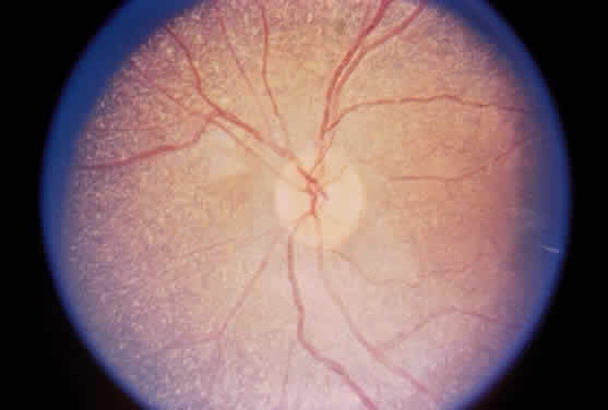
Albinism comprises a heterogeneous group of clinical syndromes exhibiting hypomelanosis based on heritable metabolic defects of the melanin pigment system. All types of albinism are characterized by foveal hypoplasia, nystagmus, photophobia, and decreased visual acuity in addition to absent or decreased melanotic pigment in skin, hair, and eyes (oculocutaneous albinism [OCA]) or in the eye alone (ocular albinism).31 The term albinoidism refers to hypomelanotic disorders in which the patients do not have nystagmus, decreased visual acuity, or foveal hypoplasia. Eleven disorders have been identified with clinical features of oculocutaneous albinism and four with features of ocular albinism (Table 4). The ocular findings in oculocutaneous albinism are listed in Table 5 (Figs. 2 and 3).


TABLE FOUR. Classification of Inherited Hypopigmented Conditions With Ocular Involvement

  Oculocutaneous albinism

  Hair bulb tyrosine test negative

  Tyrosinase-negative (AR)
  Yellow-mutant (AR)


  Hair bulb tyrosine test positive

  Tyrosinase-positive (AR)
  Hermansky-Pudlak syndrome (AR)
   Chédiak-Higashi syndrome (AR)
  Cross syndrome (AR)
  Rufous (AR)
  Brown (AR)
  Autosomal dominant
  Black locks: congenital sensorineural deafness (AD)


  Hair bulb tyrosine test questionable


  Ocular albinism

  Nettleship-Falls X-linked
  Forsius-Eriksson X-linked
  Autosomal recessive
  Lentigines-deafness (AD)


AR, autosomal recessive; AD, autosomal dominant


TABLE FIVE. Ocular Signs in Oculocutaneous Albinism

  Subnormal visual acuity (20/80 to 20/400)
  Nystagmus
  Extremes in refractive error
  Strabismus
  Iris transillumination
  Macular hypoplasia
  Visible choroidal vasculature


Fig. 2. Diffuse transillumination in a diaphanous hypopigmented iris of a child with oculocutaneous albinism. Note the lens edge.

Fig. 3. Pigmentary dilution of the retina in oculocutaneous albinism. Note the absence of the macular reflex. Large and tortuous choroidal vessels are easily seen; these vessels are not visible when the retinal pigment epithelium is normal.

Three forms of OCA have been identified, each mapped to a different genetic locus. OCA type I represents tyrosinase-negative albinism and has been mapped to chromosome 11 (11q14-q21). There is a deficiency in the catalytic activity of tyrosinase, which is involved in at least three steps in the melanin biosynthetic pathway.32,33 In OCA type IA, the affected homozygote is “dead white” at birth, with no discernible pigment. In the yellow mutant form of OCA (type IB), some pigmentation of the skin forms and the hair develops a yellow coloration.

Tyrosinase-positive OCA (type II) has been mapped to chromosome 15 (15q11.2-q12) and is the most prevalent type of albinism in the world.34 The mutated protein is a transmembrane polypeptide that may play a role in transporting small molecules such as tyrosine, a precursor of melanin.35,36 Affected individuals show some degree of pigmentation and have less profound clinical manifestations than those with OCA type I.

In OCA type III (brown oculocutaneous albinism), a mutation in the tyrosinase-related protein-1 affects its interaction with tyrosinase, resulting in dysregulation of tyrosinase activity. This promotes the synthesis of brown melanin instead of black melanin.37 The mutation has been localized to chromosome 9 (9p23). Affected individuals have brown hair and skin and are capable of developing a faint tan with sun exposure.38

Individuals who are homozygous for the genetic mutations responsible for OCA have an abnormal proportion of fibers from the ganglion cells of the temporal retina that decussate to the contralateral cerebral hemisphere; this can be demonstrated by monocular visual evoked potential asymmetry.39,40 This may play a role in the high incidence of subnormal binocular vision and strabismus in albinos. This abnormality can be detected early in life and may be helpful in making the diagnosis of OCA during the neonatal period.41

All patients with OCA should be questioned about a history of bleeding because of the association of albinism with a hemorrhagic diathesis, the Hermansky-Pudlak syndrome.42 The hemostatic defect in the Hermansky-Pudlak syndrome is typically mild; the most common manifestations are gingival bleeding, epistaxis, and easy bruisability.43 More serious bleeding episodes have followed surgical procedures, especially when aspirin was used to control postoperative discomfort.44 The Hermansky-Pudlak syndrome is the most common form of albinism seen in Puerto Rico. A deficiency of platelet dense bodies, as observed by electron microscopy, is the most reliable method of diagnosing this syndrome.45

In affected persons with ocular albinism, the cutaneous pigmentary dilution is much less noticeable than is the ocular involvement. The skin pigmentation in these patients typically falls within the normal range. However, when these affected persons are compared with their nonaffected siblings, they typically have a lighter complexion.46 The ocular findings in ocular albinism are similar to those in OCA. However, in patients with ocular albinism, the pigmentary dilution of the eye may be subtle. In whites, there is usually iris transillumination. In blacks, the iris often does not transilluminate and the retina is usually moderately pigmented.47 Regardless of the pigmentation of affected patients, all have foveal hypoplasia.

In the Nettleship-Falls form of X-linked ocular albinism, there is an inborn error of metabolism affecting the pigment cells. This defect has been localized to the Xp22.3 region. Histopathologic examination of the clinically normal skin in affected males and carrier females has revealed the presence of macromelanosomes.48 Female carriers often show a mud-spattered appearance of the fundus with hy-perpigmented streaks as well as iris transillumination defects. These clinical signs, in addition to skin biopsy, can be used to help in genetic counseling for families at risk.49,50

TYROSINEMIA

There are five clinical syndromes in which elevated serum and urinary tyrosine levels and their metabolites can be detected: (1) tyrosinemia secondary to systemic disease (liver damage, pernicious anemia, vitamin C deficiency); (2) neonatal tyrosinemia, a transient benign process resulting from immature parahydroxyphenylpyruvate acid oxidase producing high serum tyrosine levels in premature infants; (3) tyrosinemia associated with hepatorenal disease (hereditary tyrosinemia, tyrosinemia type I, which is a rapidly fatal disease of infancy), methionemia, aminoaciduria, and glycosuria; (4) tyrosinemia associated with ocular but not hepatorenal disease (essential tyrosinemia, tyrosinemia type II); and (5) tyrosinemia without hepatorenal or ocular disease.

Type I tyrosinemia is caused by a defect in fumarylacetoacetate hydrolase (chromosome 15q23-q25), the last enzyme in the tyrosine catabolism pathway.51 An accumulation of succinylacetone occurs that reacts with other amino acids and proteins. Patients who survive beyond infancy are at high risk for the development of hepatocellular carcinoma; this has been prevented in some children who have undergone neonatal liver transplantation.52

Tyrosinemia type II (Richner-Hanhart syndrome) is a rare congenital error of metabolism characterized by a triad of dendriform keratitis, hyperkeratotic lesions of the palms and soles, and mental retardation (Figs. 4 and 5).53–59 Many of the early reported cases of this disease were in patients who were the product of consanguineous marriages, suggesting a possible autosomal recessive inheritance.55,58–61

Fig. 4. Subepithelial corneal opacification in tyrosinemia type II. (Courtesy Marian Macsai, MD)

Fig. 5. Hyperkeratotic lesion of the soles in a patient with tyrosinemia. (Courtesy Marian Macsai, MD)

The diagnosis of tyrosinemia type II may be confirmed by amino acid analysis of the blood and urine, which shows an increase of tyrosine and its metabolites only. The enzymatic basis of the syndrome is a defect in soluble hepatic cytosol tyrosine aminotransferase, whose gene has been mapped to chromosome 16 (16q22.1-q22.3).55,62,63

Ocular symptoms of photophobia and lacrimation in both eyes usually appear during the first few months of life. The affected patient has bilateral subepithelial central corneal ulcers with herpetiform dendritic branching, which may develop with time into round whitish opacities with superficial new vessel formation.59 The corneal ulceration probably results from accumulation of intracellular crystals, presumably tyrosine. These crystals have been shown to enlarge, lacerate and rupture the cell, attract lysosomal enzymes, and trigger the inflammatory cycles.54,64

A low-tyrosine, low-phenylalanine diet has been used successfully in several patients with tyrosinemia type II, with rapid resolution of the clinical symptoms and signs.55,58,65–68 The keratitis found in tyrosinemia type II can be distinguished from herpes simplex keratitis by its morphologic appearance, bilateral presentation, lack of response to antiviral therapy, and associated systemic findings.53 Therefore, an infant or young child with bilateral dendritic keratitis should undergo serum and urinary evaluation for elevated levels of tyrosine and its metabolic byproducts.

Tyrosinemia type III is due to a defect in the enzyme 4-hydroxyphenylpyruvate dioxygenase, which has been mapped to chromosome 12 (12q24). Liver biopsy in affected individuals has been histologically normal, and mental retardation may be present.69

CYSTINOSIS

Cystinosis is an autosomal recessively inherited disorder of amino acid metabolism characterized by the deposition of cystine crystals in various tissues such as the eye, bone marrow, lymph nodes, and internal organs.70 Cystinosis is a lysosomal storage disorder that results from impaired transport of the disulfide amino acid cystine from cellular lysosomes. It differs from other lysosomal diseases because the main enzyme function of lysosomes, acid hydrolysis, does not play a role in cystine deposition. Plasma levels of cystine are below saturation level, whereas intracellular concentrations are elevated in polymorphonuclear leukocytes, macrophages, and fibroblasts, further indicating the cellular nature of the defect responsible for this disease.

Three clinical types of cystinosis have been described, all of which have ocular involvement (Table 6).71 The infantile, or nephropathic, variety is the most severe form of cystinosis.72 The gene has been mapped to the short arm of chromosome 17 (17p13).73 During early childhood, affected patients develop polyuria and polydipsia as a result of impaired renal tubular water reabsorption. This is followed by growth retardation, renal rickets, metabolic acidosis, progressive renal failure, and, ultimately, death from uremia before puberty.


TABLE SIX. Some Distinguishing Characterisitics of the Three Major Types of Cystinosis


 Nephropathic (Infantile)AdolescentBenign
General Symptoms   
Onset of symptoms6–18 months18 months—17 yearsNo symptoms
GrowthImpairedVariableNormal
Skin pigmentationUsually fairVariableNormal
RicketsPresentVariableAbsent
Bone marrow cystine crystalsPresentPresentPresent
Ocular   
RetinopathyPresentVariableAbsent
Crystalline deposits in cornea and conjunctivaPresentPresentPresent
PhotophobiaUsuallypresentVariableAbsent
Renal   
Tubular dysfunction (Fanconi syndrome)PresentOften incompleteAbsent
Glomerular failurePresentPresent at a later age than in infantile typeAbsent
(Modified from Schneider JA, Schulman JD: Cystinosis. In Stanbury JB,Wyngaarden JB, Fredrickson DS et al [eds]: The Metabolic Basis of Inherited Disease, 5th ed. New York, McGraw-Hill, 1983)


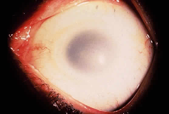
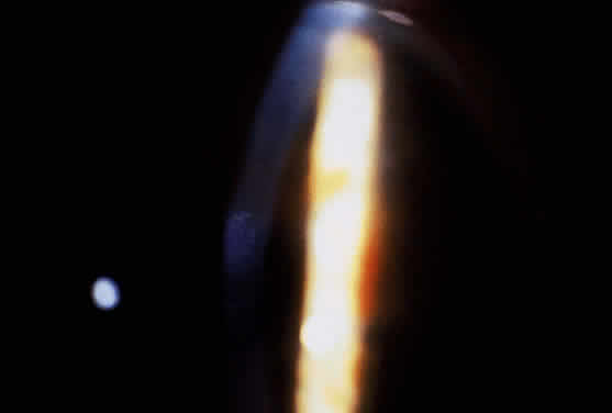
Children with nephropathic cystinosis usually develop severe photophobia within the first few years of life. Slit-lamp examination discloses diffuse scintillating, tinsel-like crystals of the cornea, conjunctiva, and iris (Fig. 6).74–76 Initially, the iridescent crystals occupy the full stromal thickness only in the periphery, while centrally the anterior half to two thirds of the stroma is involved. In patients with longstanding disease, thinning and focal breaks in Bowman's membrane may be present and contribute to the development of severe photophobia.77 Corneal sensitivity may be reduced.78

Fig. 6. Slit-lamp view of crystal deposition in cystinosis. (Courtesy James J. Reidy, MD)

The essential fundus abnormality is a patchy depigmentation of the periphery and a finer “salt-and-pepper” disturbance at the posterior pole.72,79 Crystal deposition may also be seen (Fig. 7). Despite the extensive tissue infiltration of cystine crystals, there is no significant visual disturbance.

Fig. 7. Crystal deposition of the fundus in nephropathic cystinosis. (Courtesy James J. Reidy, MD)

Cysteamine is a cystine-depleting agent used in the treatment of infantile nephropathic cystinosis. When given systemically, it has proven to be successful in retarding glomerular deterioration and enhancing growth, but it does not prevent the development of corneal crystal formation.70,73,80–82 Topical cysteamine drops have been shown not only to provide primary prevention of corneal crystal deposition but may also reverse the corneal complications in older patients.83,84

Adolescent cystinosis is a milder form of the disease, appearing in the first or second decade of life.85,86 This type of cystinosis may or may not include rickets, renal failure, photophobia, or retinopathy. All patients have shown the crystalline material in the cornea, conjunctiva, and reticuloendothelial system.

A benign adult form of cystinosis was first described by Cogan and coworkers in 1957.87 The primary clinical distinction between the benign and the nephropathic variants is failure of the former patients to show either retinopathy or renal dysfunction (see Table 6).88 Although affected patients may have mild photophobia, they are frequently diagnosed by routine ophthalmologic examination when the typical corneal crystals are noted.89,90

GYRATE ATROPHY

Gyrate atrophy of the choroid and retina is a rare autosomal recessively inherited, progressive, metabolic chorioretinal dystrophy beginning in childhood.91 It is caused by a deficiency in the mitochondrial matrix enzyme ornithine aminotransferase (OATase), resulting in hyperornithinemia.92–100 The genetic mutation has been mapped to chromosome 10 (10q26).101

Aside from visual impairment, patients with gyrate atrophy are for the most part asymptomatic. Mild to moderate diffuse slowing on electroencephalography has been reported in fewer than one third of affected patients.102–104 However, seizures have not been documented with increased frequency, and the majority of patients have normal intelligence.103,104 Liver biopsies in patients with gyrate atrophy have demonstrated nonspecific morphologic abnormalities of the mitochondria, with elongation, branching, and segmentation.105 The functional significance of these mitochondrial abnormalities is not known. However, these abnormalities are believed to be a direct result of hyperornithinemia because similar mitochondrial changes have been produced in rats maintained on high-ornithine diets.106

Tubular aggregates have been identified in type 2 skeletal muscle fibers in patients with gyrate atrophy.103–109 These aggregates are not specific for gyrate atrophy and have been identified in disorders such as periodic paralysis, hyperthyroidism, porphyria cutanea tarda, myasthenia gravis, myotonic dystrophy, postviral infections, and alcoholism.91,110 The disease progresses to almost complete loss of type 2 fibers, but the progression of muscle changes is slower than the progression of the ocular disease.

The major clinical problem in patients with gyrate atrophy is a slowly progressive loss of vision leading to blindness, usually by the fourth decade of life (Table 7).102,111–113 Myopia or decreased night vision is the earliest symptom, usually noted before the end of the first decade of life. Constriction of the visual field is obvious by the second decade. By age 40, most patients show visual fields smaller than 10°.112 Posterior subcapsular lens changes develop in late adolescence. After cataract surgery, there is generally a marked improvement in visual acuity, especially in younger patients.


TABLE SEVEN. Ocular Manifestations of Gyrate Atrophy

  Progressive loss of vision
  High myopia and marked astigmatism
  Posterior subcapsular lens changes
  Vitreous may be syneretic and contain clusters of cloudy fibrils Confluent arcuate equatorial areas of chorioretinal degeneration


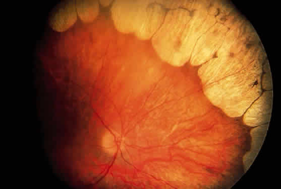
During late childhood, sharply demarcated, circular areas of chorioretinal degeneration in the midperiphery can be detected. There may be increased pigmentation around the margins of these lesions. During the second decade, the lesions enlarge, coalesce, and extend toward the posterior pole of the retina (Fig. 8). By the third decade, much of the retina is involved, although foveal lesions are rarely present until very late in the course of the disease. Histologic examination of an affected retina has shown focal areas of photoreceptor atrophy with adjacent retinal pigment epithelial hyperplasia. Electron microscopy has revealed mitochondrial abnormalities of the photoreceptors.114

Fig. 8. Fundus in gryrate atrophy. (Courtesy Richard Lewis, MD)

The electroretinogram (ERG) eventually diminishes in amplitude and is usually extinguished well before the entire retina is involved clinically.91 The electro-oculogram becomes severely diminished, parallel to the reduction in the ERG.91

The slow progression of the degenerative changes in gyrate atrophy and the difficulty in measuring small changes in ocular function objectively make evaluation of any therapy difficult.110,115–119 Two therapeutic approaches have been attempted in patients with gyrate atrophy: reducing the accumulation of ornithine and stimulation of residual ornithine aminotransferase activity. Arginine is the precursor of ornithine. A chronic diet restricted in arginine has been shown to lower ornithine levels and slow the progression of the ocular disease.120 Some patients respond to pharmacologic doses of pyridoxine (vitamin B6) to increase the level of pyridoxal phosphate, a cofactor of OATase. A different single nonsense mutation within the OATase gene leads to a difference in the affinity of the enzyme for pyridoxal phosphate, accounting for the positive response to pyridoxine in this subset of patients.121,122

OCULOCEREBRORENAL SYNDROME (LOWE SYNDROME)

Renal tubular dysfunction, mental retardation, and congenital ocular defects form the triad of a rare disorder originally described by Lowe and coworkers and known as the oculocerebrorenal syndrome.123 The condition is transmitted in a sex-linked recessive pattern.124 The aminoaciduria is renal in origin; the plasma amino acids are normal. The enzyme responsible for this syndrome is an inositol polyphosphate 5-phosphatase within the Golgi apparatus whose gene has been mapped to the X chromosome.125–127

Systemic manifestations of this disorder usually are present at birth; they consist of hypotonia, failure to thrive, anorexia, and vomiting.123 Later signs are mental retardation, intention tremors, and a peculiar high-pitched cry. The renal tubular defect results in aminoaciduria, glycosuria, proteinuria, acidosis, phosphaturia, and hypophosphatemia with or without rickets. Many affected males also display a characteristic maladaptive behavior pattern that includes tantrums and stubbornness; this is not simply reflective of the developmental impairment or multiple disabilities present in these patients.128,129

The ophthalmologic findings are among the earliest, most prominent and constant clinical features of this disorder.130,131 In a large review series, Abbassi and colleagues found that dense bilateral nuclear or posterior cortical cataracts were present at birth in nearly 100% (68/70) of the cases.124 Glaucoma was not as constant a finding but was present in 66% of cases.124

The cataract in Lowe syndrome is characterized by reduced anterior-posterior and equatorial measurements and malformations ranging from a discoid configuration to a giant posterior lentiglobus.130 Anteriorly, focal areas of lens capsule thickening and localized areas of lens epithelium hyperplasia are present. Punctate cortical opacities can be seen in the lens of female carriers of the disease and increase with age.132,133

Glaucoma, with or without buphthalmos, is usually ascribed to an anomalous development of the anterior chamber angle.131 Corneal opacities, present in at least 50% of cases, are probably related mainly to congenital and chronic glaucoma.131

There is no known effective treatment for the basic disorder; therapy consists of the correction of acidosis and rickets, if present.

Back to Top
DISORDERS OF CARBOHYDRATE METABOLISM

GALACTOSEMIA

Galactosemia is an inborn error of metabolism in which the infant cannot metabolize galactose into glycogen as the result of a deficiency in the activity of the enzyme galactose-l-phosphate uridyl transferase.134,135 This condition is inherited in an autosomal recessive manner. The genetic mutation has been mapped to chromosome 9 (9p13).136,137

An infant with galactosemia may appear normal at birth, but vomiting or diarrhea usually begins within a few days of ingesting galactose in milk.138 Signs of liver dysfunction, including jaundice, hepatomegaly, and even ascites, develop after the first week of life. As the untreated infant grows older, mental retardation becomes evident.

The chemical findings, in addition to those of liver dysfunction, include elevated blood galactose level, galactosuria, hyperchloremic acidosis, albuminuria, and aminoaciduria.139,140 Although an elevated level of red cell galactose-l-phosphate has been used as a diagnostic criterion, the direct assay of red cell transferase activity provides the definitive diagnosis.141

The essential and perhaps only ocular manifestation of galactosemia is cataract formation.142 Galactosemic cataracts usually appear within the first few days of life and may be the first sign of the disease (Fig. 9). The first visible change in the lens is the appearance of an “oil droplet.” Subsequently there is a progressive clouding of the lens. The pathophysiology of the lens change probably involves the excessive accumulation within the lens of dulcitol, which is formed by the enzymatic reduction of galactose and is not metabolized further. Dulcitol is a sugar alcohol in which the plasma membranes are relatively impermeable. This results in a change in the osmotic balance, with a consequent increase in the water drawn into the lens and its subsequent opacification.143–145

Fig. 9. Concentric lamellar opacities of galactosemia.

Another type of galactosemia is caused by galactokinase deficiency.146,147 In this disorder, cataract formation is the sole clinical manifestation. The diagnosis can be made by the finding of normal amounts of galactose-l-phosphate uridyl transferase and an absence of galactokinase in the red blood cells.146 The galactokinase gene is located on chromosome 17 (17q24).148

The management of patients with galactosemia rests on the elimination of galactose from the diet. Early diagnosis and prompt institution of therapy can result in regression of cataract formation and an improvement of the other signs of the disease. Galactosemia can be diagnosed in utero through transabdominal amniocentesis and tissue culture. Restriction of dietary galactose during pregnancy of women who have had children with galactosemia may also be beneficial in preventing damage to the fetus.149

THE MUCOPOLYSACCHARIDOSES

The conditions genetically referred to as the mucopolysaccharidoses result from the deficiency of specific lysosomal enzymes involved in the metabolic degradation of dermatan sulfate, heparan sulfate, or keratan sulfate, either singly or in combination. The incompletely degraded mucopolysaccharides accumulate in various tissues and organs throughout the body and are excreted in the urine.

As a group, the mucopolysaccharidoses are characterized by a rather distinctive spectrum of clinical manifestations. Skeletal deformity, resulting from changes in both the bones and the joints, is a prominent feature. There is also a characteristic facies with coarse, often somewhat grotesque features. Visceromegaly, cardiac disease, respiratory problems, deafness, and mental deficiency occur in some of the syndromes. The principal ocular manifestations of the various mucopolysaccharidoses are progressive corneal clouding, pigmentary retinal degeneration, optic atrophy, and in some cases glaucoma (Table 8).


TABLE EIGHT. Mucopolysaccharidoses


DesignationEnzyme DefectUrinary MucopolysaccharideHeredityGeneral Clinical ManifestationsOphthalmologic Manifestations
MPS IH (Hurler syndrome)Alpha-L-iduronidaseDermatan sulfate and heparan sulfateAutosomal recessiveCoarse facial features, short neck, short stature, kyphoscoliosis, gibbus, crouched habitus, clawhand deformity. Radiologic changes of dysostosis multiplex, severe. Visceromegaly, protuberant abdomen, hernias. Pulmonary and cardiovascular disease. Mental deficiency. Hearing loss. Early death, usually before age 10 years.Prominent wide-set eyes (shallow orbits hypertelorism), prominent supraorbital ridges, heavy brows, puffy lids. Corneal clouding is early, progressive, and severe. Retinal degeneration includes arteriolar attenuation, decreased foveal reflex, pigmentary changes, abnormal ERG. Optic atrophy. Vision impairment. In some cases, megalocornea and/or glaucoma.
MPS IS (formerly MPS V) (Scheie syndrome)Alpha-L-iduronidaseDermatan sulfate and heparan sulfateAutosomal recessiveMinimal to moderate somatic and visceral signs of mucopolysaccharidos is resembling, but less severe than, those of Hurler prototype. Somewhat coarse facial features, joint stiffness, clawhand deformity, carpal tunnel syndrome, and aortic disease, but stature and habitus relatively normal. Intellect normal or nearly normal. Hearing impairment common. Life span relatively normal.Corneal clouding is early and progressive, often more dense peripherally. Retinal degeneration is “RP-like” with retinal pigmentary changes, progressive night blindness, visual field changes. In some cases, glaucoma.
MPS I H/S (Hurler-Scheie compound)Alpha-L-iduronidaseDermatan sulfate and heparan sulfateAutosomal recessivePhenotype intermediate between that of Hurler and Scheie syndromes. Dwarfing, progressive joint stiffness, clawhand deformity, hypertelorism, progressive coarsening of facial features, micrognathia. Hepatosplenomegaly, cardiovascular disease. Hearing impairment. Intellectual impairment. Survivival into teens or twenties.Progressive corneal haze and vision impairment. Possibly papilledema.
MPS II (Hunter syndrome)Iduronate-2-sulfataseDermatan sulfate and heparan sulfateX-linked recessiveCoarse facial features. Dwarfing skeletal deformities resembling but less severe than those of Hurler proto-type. Hepatosplenomegaly, cardiac and respiratory disease, hernias. Hydrocephalus, motor paralysis in some. Hearing impairment. Nodular skin lesions. Rapid psychomotor and physical deterioration and early death (often by age 15 years) in severe form. Fairly normal mental development and longer survival (even into twenties and fifties) in mild form.Clinically clear corneas, but minimal microscopic changes reported. Progressive retinal degeneration is “RP-like,” usually severe, with retinal pigmentary changes, arteriolar attenuation, disc pallor, night vision problems and field defects, and abnormal ERG. Swelling of the nerve heads.
MPS III (Sanfilippo syndrome)Sulfamidase in type AAlpha-N-acetyl-glucosaminidase in type BAcetyl-coenzyme A: alpha-glucosaminide-N-acetyltransferase in type CN-acetyl-glucosamine-6-sulfatase in type DHeparan sulfateAutosomal recessiveFour biochemically different but clinically similar forms, with range of phenotypic variation. Early and severe progressive mental deterioration. Less severe somatic changes. Coarse facial features, megalocephaly,. hypertelorism, moderate skeletal changes Hepatosplenomegaly.Clinically clear corneas, but some microscopic changes reported. Attenuation of retinal arterioles, some pigmentary changes, and subnormal ERG documented. Optic atrophy may occur.
MPS IV-A (Morquio syndrome, classic form)N-acetylgalacto-samine-6-sulfataseKeratan sulfateAutosomal recessiveMarked skeletal changes. Dwarfism with platyspondyly, odontoid hypoplasia. Atlantoaxial instability; may lead to long tract signs and respiratory paralysis. Prominent joints, knock-knees, pigeon breast deformity, kyphosis, semicrouching stance, and waddling gait. Somewhat coarse features. Hypoplastic dental enamel. Cardiopulmonary complications. Hearing impairment. Intellect normal or moderately impaired.Corneal clouding, mild or fine haze, in A and in B. Some evidence of retinal involvement: reduced scotopic responses in some cases, arteriolar attenuation in one adult. Possibly optic atrophy or disc blurring.
MPS IV-B (Morquio syndrome, mild form)Beta-galactosidaseKeratan sulfateAutosomal recessiveSimilar to IV A, but less severe dwarfism, less tendency to atlantoaxial instability, and usually normal dental enamel.See ophthalmologic manifestations for MPS IV-A.
MPS V (vacant, formerly Scheie)     
MPS VI (Maroteaux-Lamy syndrome)N-acetylgalacto-samine-4-sulfatase (arylsulfatase B)Dermatan sulfateAutosomal recessiveStriking dwarfism and skeletal deformities. Coarse facial features. Visceromegaly and cardiac lesions. Hydrocephalus and spinal cord compression. Intellect normal. Granular inclusions in circulating leukocytes.Corneal clouding is dense. Optic atrophy. Retinal vascular tortuosity. Typically no signs of retinal degeneration, but pigmentary and ERG-VER changes in one case of mild variant.
MPS VII (Sly syndrome)Beta-glucuronidaseDermatan sulfate and heparan sulfateAutosomal recessiveVariable, often moderate manifestations. Short stature and skeletal deformity. Coarse facial features, hypertelorism. Hepatosplenomegaly, hernias, cardiovascular and respiratory problems. Inclusions in circulating lymphocytes.Corneal clouding present or absent.

ERG, electroretinogram; RP, retinitis pigmentation


To date, deficiency of ten specific lysosomal enzymes has been demonstrated in the various mucopolysaccharidoses. All are recessively inherited; one mucopolysaccharidosis, MPS type II (Hunter syndrome), is X-linked, and the others are autosomal. The diagnosis of the various mucopolysaccharidoses is made on the basis of the distinguishing clinical features, the presence of excessive mucopolysaccharide substances in tissue and urine, and demonstration of the enzyme deficiency, particularly in cultured fibroblasts.

In reviewing the mucopolysaccharidoses, reference should be made to comprehensive discussions of the clinical, pathologic, biochemical, and genetic features of these disorders.150–152

MPS I: Hurler Syndrome, Scheie Syndrome and Hurler/Scheie Syndrome

The disorders grouped together as MPS I represent a spectrum of clinical severity from the very severe (Hurler syndrome, MPS IH) through an intermediate (Hurler/Scheie syndrome, MPS I H/S) to a more mild form (Scheie syndrome, MPS IS). All forms of MPS I are due to a mutation of the gene encoding alpha-L-iduronidase (IUDA), which has been mapped to chromosome 4 (4p16.3).153 Historically, Hurler and Scheie syndromes were categorized as different entities among the mucopolysaccharidoses, with Scheie syndrome having been named MPS V. Some of the early reported cases of Hurler syndrome probably represented the Hurler/Scheie or the Scheie varieties. McKusick suggested that both Hurler and Scheie syndromes be called MPS I, with the individual subclassifications as currently known.154 Different mutations encoding for IUDA account for the variable activity of the enzyme, leading to the spectrum of clinical features represented by the three disorders.155,156 Because the three forms of MPS I have distinct clinical features as well as different prognoses, it remains useful to examine each individually. All three disorders are autosomal recessive.

MPS IH: HURLER SYNDROME. Hurler syndrome is the prototype of the mucopolysaccharidoses. The disorder is severe and progressive. There is accumulation of acid mucopolysaccharide in virtually every system of the body, producing both somatic and visceral abnormalities and leading to early death, usually by age 10 years.

In MPS IH there is a profound deficiency of IUDA, with excessive urinary excretion of both dermatan sulfate and heparan sulfate in a ratio of approximately 70:30. It occurs in many races and is probably the most common of the mucopolysaccharidoses.

Manifestations develop in infancy and early childhood and become more apparent with increasing age. The head tends to be large and misshapen. Scaphocephaly from premature closure of the sagittal suture is common, and there is often a prominent longitudinal ridge along the sagittal suture. The facial features characteristically are coarse and the expression is dull. Hypertelorism is usual, and the orbits are shallow; the eyes appear wide-set and prominent. The lids tend to be puffy, the brows prominent. The nose is broad with wide nostrils and a flat bridge. The ears may be large and low-set. The lips usually are patulous; the tongue is large and protuberant. The teeth generally are small, stubby, and widely spaced, and the gums are hyperplastic.

Characteristically there are marked skeletal changes. Moderate dwarfism, short neck, kyphoscoliosis, and gibbus are typical, and on plain-film examination the vertebral bodies (particularly those of the lower dorsal and upper lumbar region) are wedge-shaped with an anterior hooklike projection referred to as beaking. The extremities are short, the hands and feet are broad, and the phalanges are short and stubby. Radiologically, the tubular bones show expansion of the medullary cavity and thinning of the cortex. The terminal phalangeal bones commonly appear hypoplastic. The joints are stiff, and flexion contractures develop; clawlike deformity of the hands is especially characteristic. The posture is semicrouching, the gait awkward. Thoracic deformity is another regular feature of the syndrome; the chest appears large and wide with flaring of the lower ribs over the abdomen. On plain-film examination, the ribs appear spatulate or saber-shaped. Typically the medial end of the clavicle is widened. The many radiologic findings in this condition are commonly described by the term dysostosis multiplex.

The abdomen is protuberant, owing in part to abnormalities in supporting tissues and to visceromegaly. As a rule there is enlargement of both the liver and spleen. Diastasis recti, umbilical hernia, and inguinal hernias are common. Recurrent hernias may be a presenting sign.157 The skin tends to be thick. There is usually generalized hypertrichosis.

Manifestations of cardiac involvement, including murmur, angina, myocardial infarction, and congestive heart failure, are common. Pathologic changes in the heart from mucopolysaccharide deposition can be extensive. The great vessels and peripheral vessels also are affected.

Respiratory problems develop in virtually every patient. Recurrent upper respiratory tract infection, bronchitis, and chronic nasal congestion are common, and the patients almost always are noisy mouth breathers. A number of factors, including deformity of the facial and nasal bones, narrowing of the passages, abnormalities of the tracheobronchial cartilage, and deposition of mucopolysaccharide in the lungs, contribute. In addition to the abnormalities of the respiratory passages and lungs, cardiac disease and thoracic deformity may contribute to respiratory difficulties.

The principal neurologic manifestation is mental deficiency. There may also be motor signs. Pathologic changes can be found throughout the nervous system. In some cases hydrocephalus develops. A special feature in some cases is the presence of leptomeningeal cysts. Enlargement of the sella is often due to subarachnoid cysts.158 The optic foramina also may be enlarged. Deafness is frequent; it may be of mixed or sensorineural type. Middle ear infections are common.

The course is one of progressive mental and physical deterioration. Death most frequently results from cardiac or respiratory disease.

The principal ophthalmologic manifestations of MPS IH are progressive corneal clouding, retinal degeneration, optic atrophy, and vision loss. It appears that the corneal and retinal changes relate somewhat to the pattern of mucopolysacchariduria—that is, corneal changes are greater in conditions characterized by higher levels of dermatan sulfate in the urine, as in Hurler syndrome. The retinal degeneration appears to correlate with the degree of heparan sulfaturia; the retinal changes are more severe in Hunter and Sanfilippo syndromes and less in Hurler syndrome.159,160

Corneal clouding was recognized early to be an important feature of this disorder, and classic clinical descriptions of the corneal changes are found in the older literature.161–165 Clouding of the cornea is usually evident by age 2 to 3 years, often by age 1 year; in some cases it may be seen at birth.165,166 Photophobia is a common early symptom. With time there is progression from a generalized haziness or steamy appearance to a dense, milky ground-glass opacification. On slit-lamp examination one sees fine granular opacities in the corneal stroma, often increasing in density from the anterior stroma and subepithelial region to the posterior stromal layers.163,165

In 1939 Berliner provided what is probably the first significant histopathologic study of the eye in Hurler syndrome, describing the corneal changes in detail: he found large vacuolated cells under Bowman's layer, fragmentation of Bowman's layer, separation of the corneal lamellae, and deposits of granular material in the stromal spaces.163 Later studies documented these histopathologic changes and provided further evidence for mucopolysaccharide accumulation in the cornea.166–169 The epithelium may be intact or may show edema and cytoplasmic vacuolization, with accumulation of proteoglycans in and around cells. Bowman's layer usually shows thinning, lamellar splitting, or fragmentation. In the stroma there is swelling and vacuolization of keratocytes, intracellular and extracellular deposition of proteoglycans, and lamellar separation.170 Desçemet's membrane and endothelium usually are described as normal, although cytoplasmic vacuolization and metachromatic staining of the endothelium have been noted.

Progressive corneal clouding may prevent visualization of the fundus, but signs of retinal involvement and optic atrophy have been documented in Hurler syndrome. The ERG findings are abnormal, usually markedly reduced, in Hurler syndrome.171,172 Histologic findings include enlargement and vacuolization of cells of the nuclear layer of the retina, vacuolization of the ganglion cells, atrophy of the optic nerve, and thickening and infiltration of the arachnoid with foam cells.166,167,169

In addition to corneal, retinal, and optic nerve changes, there may be histopathologic evidence of mucopolysaccharide accumulation in the epithelium of the ciliary body, in the walls of the iris capillaries, in the sclera, and in the conjunctiva.166,167,169 Ultrastructural changes have been found (in uveal melanocytes and fibrocytes, ciliary epithelium, smooth muscle cells of ciliary body, pericytes, trabecular endothelium, lens epithelium, and sclerocytes.173

Megalocornea has been described in many cases; in most cases intraocular pressure has been normal, although glaucoma has occasionally been documented.162–166

Progressive impairment of vision is usual, secondary to corneal clouding, retinal degeneration, and optic atrophy, singly or in combination; glaucoma, the effects of cerebral mucopolysaccharide accumulation, and the development of hydrocephalus may also contribute. In view of the extensive systemic and neurologic abnormalities in MPS IH, the poor prognosis for life, and the probability of concurrent retinal degeneration and optic atrophy, corneal transplant in an effort to improve vision is not recommended. Cases in which keratoplasty has been successful may not have represented the more severe Hurler syndrome variant of MPS I.174,175

MPS IS: SCHEIE SYNDROME. MPS IS was first described by Scheie and colleagues in their classic study of ten patients reported in 1962.168 The predominant manifestations are corneal clouding, joint stiffness, clawhand deformity, carpal tunnel syndrome, and aortic valve disease, principally aortic stenosis and regurgitation. The facial features are coarse and the mouth is broad. Other somatic and visceral changes characteristic of mucopolysaccharidosis tend to be minimal. Stature is normal, and the patients do not develop the distorted habitus characteristically seen in Hurler syndrome. Intellect is normal or nearly normal, although psychiatric disturbances have been reported. There may be hearing impairment. The life span is relatively normal. Histopathologic changes are similar to those of the prototype MPS IH. In MPS IS, however, the cortical neurons appear normal.

Corneal clouding is a prominent manifestation of the Scheie syndrome (Fig. 10).168 Developing early in life, sometimes present at birth, the corneal clouding tends to worsen with age and may ultimately interfere with vision. The corneal involvement is diffuse but tends to be densest peripherally. Clinically, the hazy cornea may appear enlarged, edematous, and thickened, initially raising suspicion of glaucoma, particularly when telltale somatic signs of mucopolysaccharidosis are minimal. The pathologic corneal changes are similar to those in Hurler syndrome.168,176 Corneal transplants have been tried but with little success. In most cases reported, the ocular pressure has been normal or in the upper range of normal, but in some cases glaucoma has been documented.177,178

Fig. 10. Corneal clouding in MPS IS. (Courtesy James J. Reidy, MD)

Although retinal changes have not been documented in all reported cases, retinal degeneration is a recognized feature of the Scheie syndrome.168,171,172,179 Manifestations include vision impairment, particularly progressive night blindness, field changes such as ring scotoma, retinal pigmentary changes (“RP-like”), and subnormal or extinguished ERG responses.

MPS I H/S: HURLER-SCHEIE SYNDROME. A number of patients with features intermediate between those of the Hurler and the Scheie syndromes have been reported.154,180,181 As in MPS IH and MPS IS, in MPS I H/S there is deficiency of IUDA and urinary excretion of both dermatan sulfate and heparan sulfate. The histopathologic changes are those of mucopolysaccharide accumulation in connective tissue throughout the body, as well as in parenchymal cells of the liver and brain.

The prominent clinical manifestations are skeletal changes (dysostosis multiplex) with dwarfing and progressive joint stiffness, scaphocephaly, hypertelorism, and progressive coarsening of facial features. In addition, a receding chin (micrognathia) appears to be a distinctive feature. Other manifestations include hepatosplenomegaly, pulmonary and cardiovascular involvement, mental retardation, and hearing impairment. Significant manifestations (destruction of the sella, cerebrospinal fluid rhinorrhea, and loss of vision) related to the presence of arachnoid cysts have also been reported in MPS I H/S. Patients with this syndrome may survive into the teens or twenties.

As in both MPS IH and MPS IS, corneal clouding occurs in MPS I H/S.154,180,181 The corneal haze is diffuse (sometimes denser peripherally) and progressive; it may be evident in childhood and ultimately may interfere with vision.

Bone marrow transplantation has been used in the treatment of patients with MPS I with variable success. A beneficial effect on the visceral features, but not the ocular abnormalities, may be seen.182,183

MPS II: Hunter Syndrome

In the Hunter syndrome (MPS II), the metabolic defect is a deficiency of the lysosomal enzyme iduronate-2-sulfatase. There is urinary excretion of both dermatan sulfate and heparan sulfate in a ratio of approximately 1:1. In contrast to the other mucopolysaccharidoses, MPS II is X-linked recessive. The genetic mutation has been mapped to the long arm of the X chromosome.184 Phenotypically the Hunter syndrome closely resembles the Hurler prototype. The manifestations of MPS II, however, are generally less severe than those of MPS IH, and the Hunter syndrome is distinguished clinically by longer survival and by the absence of gross corneal clouding.

In the Hunter syndrome the facial features are coarse, the supraorbital ridges tend to be prominent, the tongue is large, and the teeth are widely spaced. Dwarfing and stiffness of the joints are prominent features. Clawhand deformity is common. Lumbar gibbus may develop but is usually not severe. As a rule there is hepatosplenomegaly. The abdomen is protuberant. Hernias are common. Cardiac involvement is a regular feature of the syndrome; congestive heart failure and coronary artery disease are major causes of death. Respiratory disability also is evident in most patients. Neurologic manifestations vary. Spastic quadriplegia may develop from impingement on the cervical spinal cord. Hydrocephalus may develop. Mental deterioration occurs, but the severity and the rate of regression vary. Progressive deafness occurs in most patients.

A distinctive feature of the Hunter syndrome is the occurrence of nodular or pebbly ivory-colored skin lesions, most frequently on the back extending from the inferior angle of the scapula toward the axillary line, less often in the pectoral area, nape of the neck, and lateral aspect of the upper arms and thighs. Adults with the Hunter syndrome also tend to have a rosy or ruddy complexion.

In contrast to MPS IH, obvious corneal clouding is not a regular feature of MPS II.185,186 However, slight corneal changes may be detected by slitlamp examination in older patients with Hunter syndrome, and histologic evidence of corneal mucopolysaccharide accumulation has been reported.187,188

The principal ophthalmologic manifestation of MPS II is progressive retinal degeneration with attendant impairment of vision.171,189 Night vision problems and visual field defects are common. The disorder may lead to blindness. The fundus signs include retinal pigmentary changes, sometimes spicule formation, retinal arteriolar attenuation, and optic disc pallor. The ERG is usually reduced or extinguished.171 In some cases it may be normal.172 Blurring of the disc margins thought to be chronic papilledema has also been noted.

In their report of the ocular histopathologic findings by light microscopy in MPS II, Goldberg and Duke described few corneal abnormalities.187 The corneal epithelium and Bowman's layer were intact, except peripherally, where Bowman's layer was split and where eosinophilic material was present beneath the epithelium. Desçemet's membrane and endothelium were intact, although eosinophilic granules were present in the endothelial cytoplasm. Fine granular deposits were present in the corneal stroma, chiefly in interlamellar spaces. The nonpigmented epithelium of the ciliary processes appeared foamy. There were significant retinal changes, including pigment migration, paucity of pigment epithelial cells, diminution of rods and cones, reduction in number of ganglion cells, and gliosis of the nerve fiber layer. The sclera was thickened.

Within the Hunter syndrome there is recognized a wide range of severity, related to the severity of the mutation of the affected gene.190,191 Milder involvement is characterized by slower mental deterioration and is compatible with survival to 50 years or beyond. Severely affected patients show relatively more rapid neurologic deterioration and die earlier, often before age 15 years.

MPS III: Sanfilippo Syndrome

The Sanfilippo syndrome (MPS III), sometimes referred to as polydystrophic oligophrenia, is a mucopolysaccharidosis in which there is severe mental retardation and relatively less severe somatic abnormalities.

Four biochemically different, clinically similar forms of the syndrome occur. In type A there is deficiency of sulfamidase (chromosome 17.q25). In type B there is deficiency of alpha-N-acetylglucosaminidase (chromosome 17q21). In type C there is deficiency of acetyl-coenzyme A: alpha-glucosaminide-N-acetyltransferase (chromosome 14). In type D there is deficiency of N-acetylglucosamine-6-sulfatase (chromosome 12q14). Each of these enzymes is involved in a separate step in the sequential degradation of heparan sulfate, and all forms of this disorder demonstrate urinary excretion of this substance. All forms are autosomal recessive.

Mental retardation, the predominant clinical manifestation of MPS III, usually becomes evident in the first few years of life. With increasing age there is progressive deterioration of intellect and behavior. Because the patients usually are strong, management often becomes a problem as they regress; many require institutionalization.

Somatic abnormalities typical of the mucopolysaccharidoses tend to be mild or inconspicuous. There is some coarseness of facial features. Synophrys is usual. Generalized hirsutism may be marked. Dwarfing, joint stiffness, and clawhand deformity are usually evident but are not as severe as in the Hurler prototype. Radiologically, the skeletal changes of dysostosis multiplex are relatively mild. Slight to moderate hepatosplenomegaly develops, and the abdomen tends to be protuberant. Respiratory difficulties are common. Heart involvement may occur but tends to be less severe than in other mucopolysaccharidoses. The clinical findings tend to be most pronounced in type A.192

Ocular manifestations are mild if present at all. Corneal clouding does not occur in MPS III, although microscopic changes were noted in one of Sanfilippo's patients, and histologic corneal and scleral changes were reported in another patient subsequently.193,194 Some retinal involvement may occur in MPS III. Narrowing of the retinal vessels and pigmentary changes have been noted. Subnormal ERG responses have been recorded.171,172 Optic atrophy may develop.172,195

MPS IV: Morquio Syndrome

The syndrome that bears Morquio's name is characterized by severe dwarfism and skeletal deformity, often with neurologic complications, and a number of extraskeletal abnormalities such as corneal clouding and cardiac valvular disease. In MPS IV, there is defective degradation of keratan sulfate. There is excessive urinary excretion of keratan sulfate, although the amount of keratan sulfate in the urine tends to diminish as affected patients grow older.

As with Sanfilippo syndrome, clinically similar but enzymatically different forms of Morquio syndrome occur. The designation MPS IV-A is used to denote classic Morquio syndrome, in which the enzyme defect is a deficiency of N-acetylgalactosamine-6-sulfatase. The gene has been mapped to chromosome 16q24. The designation MPS IV-B is used to denote a milder Morquio-like syndrome in which the enzyme defect is deficiency of beta-galactosidase. In addition, a third type (MPS IV-C) has been suggested to exist, representing mildly affected Morquio-like persons in which there is normal activity of both N-acetylgalactosamine-6-sulfatase and beta-galactosidase. The enzyme defect in currently unknown. Patients with MPS IV-A may demonstrate significant variability in the severity of their clinical features; this may be due to a difference in the residual activity of the affected enzyme. Therefore, patients with MPS IV-A may be further subdivided into the severe classic, intermediate, and mild types.196 All forms of MPS IV are autosomal recessive.

Patients with Morquio syndrome appear normal in the first months of life, although radiographic signs may be present early. With growth during the first years of life, abnormalities such as retarded growth, knock-knees, flat feet, prominent joints, dorsal kyphosis, sternal bulging, flaring of the rib cage, and awkward gait become evident. The deformities worsen with age. Affected persons characteristically are markedly dwarfed and develop a semicrouching stance. Joint stiffness is not a feature, however; rather, joints may be excessively loose, leading to instability. Barrel chest and pigeon breast deformity are common. The neck typically is short. The face is abnormal, with somewhat coarse features, a broad-mouthed appearance, prominent jaw, and widely spaced teeth. The dental enamel is often thin, giving the teeth a grayish appearance and leading to flaking and fracturing of the enamel and multiple cavities. Cardiac involvement is common and may involve the aortic or mitral valves. Because a murmur is not always detected on physical examination, echocardiography has been suggested to be part of the assessment of these patients.197 Progressive hearing loss occurs in almost all patients. Invariably there is absence or severe hypoplasia of the odontoid process, and there is usually ligamentous laxity of the spinal column. Atlantoaxial subluxation and spinal cord and medullary compression are frequent complications; manifestations may be acute, subacute, or chronic, subtle or severe, ranging from minimal long tract signs to spastic paraplegia, respiratory paralysis, and death. A major treatment issue in this disorder is the prevention of cervical myelopathy. Occipitocervical fusion is often required, and prophylactic surgery has been suggested.198 In general, the course is one of progressive incapacitation. Intelligence usually is normal or mildly impaired.

Corneal clouding is a feature of Morquio syndrome, although some of the earlier literature would suggest otherwise.199,200 The corneal clouding in Morquio syndrome is relatively mild, having the appearance of a fine haze rather than the dense ground-glass opacification common to Hurler syndrome. The changes may not become clinically evident to the unaided eye for several years, often not before age 10 years. In the early stages, the corneal involvement may be overlooked unless careful slit-lamp examination is performed. The biomicroscopic appearance is that of diffuse involvement of the stroma with punctate or granular opacities but usually sparing of the epithelium, Bowman's layer, and endothelium. Depending on the density of the corneal haze, there may be some impairment of vision but usually not of severe degree. Corneal clouding is seen in MPS IV-B as well as in MPS IV-A.201,202

Fundus abnormalities have been reported infrequently in Morquio syndrome. In most cases the fundi and light-adapted ERG are normal in MPS IV.171,172 Optic atrophy has been noted.203

MPS VI: Maroteaux-Lamy Syndrome

The Maroteaux-Lamy syndrome (MPS VI) is characterized by severe dwarfism, visceromegaly, cardiac lesions, and progressive corneal clouding. In some cases hydrocephalus and spinal cord compression develop. Resembling the prototype mucopolysaccharidosis in many ways, the Maroteaux-Lamy syndrome is distinguished by retention of normal intellect, the pattern of mucopolysacchariduria, and the enzyme defect. In MPS VI there is deficiency of N-acetylgalactosamine-4-sulfatase (arylsulfatase B), with urinary excretion of predominantly dermatan sulfate. The genetic mutation has been mapped to chromosome 5 (5q11-q13). Azurophilic cytoplasmic inclusions in the polymorphonuclear leukocytes (Alder granules) are a characteristic finding in MPS VI. The disorder is autosomal recessive. In addition to the classic form of Maroteaux-Lamy, milder variants associated with the same enzyme deficiency are described.

In the severe or classic form of MPS VI, growth retardation affecting both the trunk and limbs is usually evident by age 2 or 3 years. Genu valgum, lumbar kyphosis, and anterior sternal protrusion develop. The lower ribs are flared. Joint movement is restricted. Clawhand deformity develops; carpal tunnel syndrome is common. The head appears relatively large. The facial features tend to be coarse. There is often mild hypertrichosis. As a rule, hepatomegaly develops in patients older than 6 years of age, splenomegaly develops in about half the patients, and the abdomen usually is protuberant. Cardiac involvement, particularly valve lesions, similar to that of Hurler syndrome may develop. Deafness occurs in some patients.

The principal neurologic complications are hydrocephalus and spinal cord compression secondary to atlantoaxial subluxation consequent to hypoplasia of the odontoid process. Survival is variable; the longest survival of a patient with the severe form of MPS VI is said to be into the late twenties.

The principal ophthalmologic manifestation of MPS VI is progressive corneal clouding, usually evident within the first few years of life. The appearance is that of a ground-glass haze distributed diffusely throughout the stroma, sometimes more dense peripherally and usually of sufficient degree to be seen grossly.204,205 In addition to the stromal opacities, some epithelial and endothelial changes may be seen on slit-lamp examination.205

In what appears to be the first reported histopathologic study of the eye in Maroteaux-Lamy syndrome, Kenyon and coworkers in 1972 described changes typical of mucopolysaccharidosis.205 On light microscopy they found cytoplasmic vacuolization of corneal epithelium, interruption of Bowman's layer with accumulation of foamy histiocytes, swelling of keratocytes with foamy cytoplasm and separation of stromal lamellae, some cytoplasmic vacuolization of corneal endothelium, but essentially no alteration of Desçemet's membrane. Other findings included thickening of sclera with vacuolated cells between the fibers, vacuolated cells in the trabecular meshwork, ballooned histiocytes, vacuolated fibrocytes in connective tissue stroma of the ciliary body, involvement of the basal portion of nonpigmented ciliary epithelium, and some changes in the choroid. By histochemical techniques, they documented accumulation of acid mucopolysaccharide in the affected cells and tissues. The retina appeared normal except for the macular area, where reduction of the ganglion cell population and thinning of the nerve fiber layer were noted. The optic nerve showed atrophy and secondary gliosis. Similar corneal changes have been documented in the mild phenotype of MPS VI.206

The fundi in MPS VI generally are normal. As a rule, patients with Maroteaux-Lamy syndrome do not develop ophthalmoscopic signs of pigmentary retinal degeneration, and the ERG usually is normal, although areas of hypopigmentation and a reduced A-wave response have been reported.205–207

MPS VII: Sly Syndrome

In the Sly syndrome (MPS VII), there is deficiency of beta-glucuronidase, leading to a block in the degradation of dermatan sulfate and heparan sulfate, with urinary excretion of both dermatan sulfate and heparan sulfate. MPS VII was the first autosomal mucopolysaccharidosis for which a chromosomal assignment was achieved. The gene is located on the long arm of chromosome 7 (7q21). The disorder is autosomal recessive.

Clinical manifestations within the syndrome vary. The spectrum includes many of the characteristic features of mucopolysaccharidosis, including short stature, progressive skeletal deformity and radiologic signs of dysostosis multiplex, coarse facial features, hypertelorism, hepatosplenomegaly, diastasis recti, protuberant abdomen, hernias, intellectual impairment, cardiovascular involvement, and respiratory problems. Reported patients have shown inclusions in circulating lymphocytes.

In some patients with Sly syndrome, the corneas are clear.208–210 Within the phenotypic variation of this disorder, however, corneal clouding may occur; this may be evident grossly or only on slit-lamp examination.211

Back to Top
REFERENCES

1. Carson NAJ, Neill DW: Metabolic abnormalities detected in a survey of mentally backward individuals in Northern Ireland. Arch Dis Child 37:505, 1962

2. Gerritsen T, Vaughn JG, Waisman HA: The identification of homocystine in the urine. Biochem Biophys Res Commun 9:493, 1962

3. Mudd SH, Finkelstein JD, Irreverre F et al: Homocystinuria: An enzyme defect. Science 143:1443, 1964

4. Schimke RN, McKusick VA, Huang T et al: Homocystinuria. JAMA 193:711, 1965

5. Brown RB, Watson PD, Taussig LM: Congenital metabolic diseases of pediatric patients: Anesthesiologic implications. Anesthesiology 43:197, 1975

6. Crooke JW, Towers JF, Taylor WH: Management of patients with homocystinuria requiring surgery under general anesthesia. Br J Anaesthesiol 43:96, 1971

7. Regenbogen L, Ilie S, Elian I: Homocystinuria: A surgical and anesthetic risk. Metab Pediatr Ophthalmol 4:209, 1980

8. Mudd SH, Skovby F, Levy HL et al. The natural history of homocystinuria due to cystathionine beta-synthase deficiency. Am J Hum Genet 37:1, 1985

9. Ritchie JWK, Carson NAJ: Pregnancy and homocystinuria. J Obstet Gynecol Br Commonw 10:664, 1973

10. Spaeth GL, Barber GW: Homocystinuria: Its ocular manifestations. J Pediatr Ophthalmol 3:42, 1966

11. Cross HE, Jensen AD: Ocular manifestations in the Marfan syndrome and homocystinuria. Am J Ophthalmol 75:405, 1973

12. Ramsey MS, Dartz LD, Beaton JW: Lens fringe in homocystinuria. Arch Ophthalmol 93:318, 1975

13. Ramsey MS, Dickson DH: Lens fringe in homocystinuria. Br J Ophthalmol 59:338, 1975

14. Ramsey MS, Yanoff MN, Fine BS: The ocular histopathology of homocystinuria. Am J Ophthalmol 74:377, 1972

15. Swann DA, Streeten BW: Amino acid and peptide composition of zonular fibers. Invest Ophthalmol Vis Sci (suppl) 17:209, 1978

16. Streeten BW: The nature of the ocular zonule. Trans Am Ophthal Soc 80:823, 1982

17. Nelson LB, Maumenee IH: Ectopia lentis. Surv Ophthalmol 27:143, 1982

18. Brenton DP, Cusworth DC, Dent CE et al: Homocystinuria, clinical and dietary studies. Q J Med 35:325, 1966

19. Carson NAJ: Homocystinuria. Trial treatment of a 5-year-old severely retarded child with a natural diet low in methionine. Am J Dis Child 113:95, 1967

20. Carson NAJ, Dent CE, Field CMB et al: Homocystinuria: Clinical and pathological review of ten cases. J Pediatr 66:5, 1965

21. Dunn NG, Perry TL, Dolman CL: Homocystinuria: A recently discovered cause of mental defect and cerebrovascular thrombosis. Neurology 16:407, 1966

22. Perry TL, Hansen S, Love DL et al: Treatment of homocystinuria with a low-methionine diet, supplemental cystine and methyl donor. Lancet 2:474, 1968

23. Perry TL, Hansen S, MacDougall L: Sulfur-containing amino acids in the plasma and urine of homocystinurics. Clin Chem Acta 15:409, 1967

24. Mudd SH, Edwards WA, Loeb PM et al: Homocystinuria due to cystathionine synthetase deficiency: The effect of pyridoxine. J Clin Invest 49:1762, 1970

25. Barber GW, Spaeth GL: Pyridoxine therapy in homocystinuria. Lancet 1:337, 1967

26. Krauss JP: Biochemistry and molecular genetics of cystathionine beta-synthase deficiency. Eur J Pediatr 1557(Suppl 2):S50, 1998

27. Hu Fl, Gu Z, Kozich V: Molecular basis of cystathionine beta-synthase deficiency in pyridoxine responsive and nonresponsive homocystinuria. Hum Mol Genet 2:1857, 1993

28. Wilcken DE, Dudman NP, Tyrrell PA: Homocystinuria due to cystathionine beta-synthase deficiency—the effects of betaine treatment in pyridoxine-responsive patients. Metab Clin Exp 34:1115, 1985

29. Spaeth GL, Barber GW: Prevalence of homocystinuria among the mentally retarded: Evaluation of a specific screening test. Pediatrics 40:586, 1967

30. Naughten ER, Yap S, Mayne PD: Newborn screening for homocystinuria: Irish and world experience. Eur J Pediatr 157(Suppl 2):S84, 1998

31. Witkop CJ, Quevedo WC, Fitzpatrick TB: Albinism and other disorders of pigment metabolism. In Stanbury JB, Wyngaarden JB, Fredrickson DS et al (eds): The Metabolic Basis of Inherited Disease, 5th ed. New York, McGraw-Hill, 1983

32. Spritz RA: Molecular genetics of oculocutaneous albinism. Hum Mol Genet 3(Spec No):1469, 1994

33. Giebel LB, Tripathi RK, Strunk KM et al: Tyrosinase gene mutations associated with type IB (“yellow”) oculocutaneous albinism. Am J Hum Genet 48:1159, 1991

34. Lee ST, Nicholls RD, Schnur RE et al: Diverse mutations of the P gene among African-Americans with type II (tyrosinase-positive) oculocutaneous albinism (OCA2). Hum Molec Genet 3:2047, 1994

35. Lee ST, Nicholls RD, Bundey S et al: Mutations of the P gene in oculocutaneous albinism, ocular albinism, and Prader-Willi syndrome plus albinism. N Engl J Med 330:529, 1994

36. Lee ST, Nicholls RD, Jong MT et al: Organization and sequence of the human P gene and identification of a new family of transport proteins. Genomics 26:354, 1995

37. Boissy RE, Zhao H, Oetting WS et al: Mutation in and lack of expression of tyrosinase-related protein-1 (TRP-1) in melanocytes from an individual with brown oculocutaneous albinism: a new subtype of albinism classified as “OCA3.” Am J Hum Genet 58:1145, 1996

38. King RA, Cervenka J, Okoro AN et al: The brown albino: A new type of tyrosinase-positive oculocutaneous albinism. Am J Hum Genet 30:56A, 1978

39. Apkarian P, Reits D, Spekreijse H et al: A decisive electrophysiologic test for human albinism. Electroenceph Clin Neurophysiol 55:513, 1983

40. Guo SQ, Reinecke RD, Fendick M et al: Visual pathway abnormalities in albinism and infantile nystagmus: VECPs and stereoacuity measurements. J Pediatr Ophthalmol Strabismus 26:97, 1989

41. Apkarian P, Tijssen R: Detection and maturation of VEP albino asymmetry: An overview and a longitudinal study from birth to 54 weeks. Behav Brain Res 49:57, 1992

42. Hermansky F, Pudlak P: Albinism associated with hemorrhagic diathesis and unusual pigmented reticular cells in the bone marrow: Report of two cases with histochemical studies. Blood 14:162, 1959

43. Simon JW, Adams RJ, Calhoun JH et al: Ophthalmic manifestations of the Hermansky-Pudlak syndrome (oculocutaneous albinism and hemorrhagic diathesis). Am J Ophthalmol 93:71, 1982

44. Witkop CJ, Hill CW, Desnick S et al: Ophthalmologic, biochemical, platelet, and ultrastructural defects in the various types of oculocutaneous albinism. J Invest Dermatol 60:443, 1973

45. Witkop CJ, Nunez Babcock M, Rao GH et al: Albinism and Hermansky-Pudlak syndrome in Puerto Rico. Bol Asoc Med PR 82:333, 1990

46. Waardenburg PJ: Remarkable Facts in Human Albinism and Leukism. Assen, Van Gorcum, 1970

47. O'Donnell FE, Green WR, Fleischman JA et al: X-linked ocular albinism in blacks: Ocular albinism cum pigmento. Arch Ophthalmol 96:1189, 1978

48. O'Donnell FE, Hambrick GW, Green WR et al: X-linked ocular albinism: An oculocutaneous macromelanosomal disorder. Arch Ophthalmol 94:1883, 1976

49. Charles SJ, Moore AT, Grant JW et al: Genetic counseling in X-linked ocular albinism: Clinical features of the carrier state. Eye 6:75, 1992

50. Bergen AA, Schuurman EJ, van den Born LI et al: Carrier detection in X-linked ocular albinism of the Nettleship-Falls type by DNA analysis. Clin Genet 41:135, 1992

51. Berube D, Phaneuf D, Tanguay RM et al: Assignment of the fumarylacetoacetate hydrolase gene to chromosome 15q23-15q25. Cytogenet Cell Genet 51:962, 1989

52. Sokal EM, Bustos R, Van Hoof et al: Liver transplantation for hereditary tyrosinemia—early transplantation following the patient's stabilization. Transplantation 54:937, 1992

53. Charlton KH, Pinder PS, Wozniak L et al: Pseudodendritic keratitis and systemic tyrosinemia. Ophthalmology 88: 355, 1981

54. Burns RP: Soluble tyrosine aminotransferase deficiency: An unusual cause of corneal ulcers. Am J Ophthalmol 73:400, 1972

55. Goldsmith LA, Kang E, Bienfang DC et al: Tyrosinemia with plantar and palmar keratosis and keratitis. J Pediatr 83:798, 1973

56. Holston JL Jr, Levy HL, Tomlin GA et al: Tyrosinosis: A patient without liver or renal disease. Pediatrics 48:393, 1971

57. Bienfang DC, Kuwabara T, Pueschel SM: The Richner-Hanhart syndrome: Report of a case with associated tyrosinemia. Arch Ophthalmol 94:1133, 1976

58. Goldsmith LA, Reed J: Tyrosine-induced eye and skin lesions: A treatable genetic disease. JAMA 236:382, 1976

59. Sammartino A, de Grecchio G, Lembo BG et al: Familial Richner-Hanhart syndrome: Genetic, clinical, and metabolic studies. Ann Ophthalmol 16:1069, 1984

60. Garibaldi LR, Siliato F, DeMartini I et al: Oculocutaneous tyrosinosis: Report of two cases in the same family. Helv Paediatr Acta 32:173, 1977

61. Hanhart E: Neue Sonderformen von Keratosis palmo-plantaris, u.a. eine regelmaessig-dominante mit systematisierten Lipomen, ferner 2 einfach-rezessive mit Schwachsinn und z.T. mit Hornhautveraenderungen des Auges (Ektodermatosyndrom). Dermatologica 94:286, 1947

62. Fellman JH, Vanbellinghen PJ, Jones RT et al: Soluble and mitochondrial forms of tyrosine aminotransferase: Relationship to human tyrosinemia. Biochemistry 8:615, 1969

63. Barton DE, Yang-Feng TL, Francke U: The human tyrosine aminotransferase gene mapped to the long arm of chromosome 16 (region 16q22-q24) by somatic cell hybrid analysis and in situ hybridization. Hum Genet 72:221, 1986

64. Burns RP, Gibson IK, Murray MJ: Keratopathy in tyrosinemia. Birth Defects 12:169, 1976

65. Hill A, Nordin PM, Zaleski WA: Dietary treatment of tyrosinosis. J Am Diet Assoc 56:308, 1970

66. Hill A, Zaleski WA: Tyrosinosis: Biochemical studies of an unusual case. Clin Biochem 4:263, 1971

67. Sandberg HO: Bilateral keratopathy and tyrosinosis. Acta Ophthalmologica 53:760, 1975

68. Hunziker N: Richner-Hanhart syndrome and tyrosinemia type II. Dermatologica 160:180, 1980

69. Giardini O, Cantani A, Kennaway et al: Chronic tyrosinemia associated with 4-hydroxyphenylpyruvate dioxygenase deficiency with acute intermittent ataxia and without visceral and bone involvement. Pediat Res 17:25, 1983

70. Thoene JG: Cystinosis. J Inherit Metab Dis 18:380, 1995

71. Schneider JA, Schulman JD: Cystinosis. In Stanbury JB, Wyngaarden JB, Fredrickson DS et al (eds): The Metabolic Basis of Inherited Disease, 5th ed. New York, McGraw-Hill, 1983

72. Read J, Goldberg MF, Fishman G et al: Nephropathic cystinosis. Am J Ophthalmol 76:791, 1973

73. McDowell GA, Town MM, van't Hoff W et al: Clinical and molecular aspects of nephropathic cystinosis. J Molecular Med 76:295, 1998

74. Wong VG, Kuwabara T, Brubaker R et al: Intralysosomal cystine crystals in cystinosis. Invest Ophthalmol 9:83, 1970

75. Kenyon KR, Sensenbrenner JA: Electron microscopy of cornea and conjunctiva in childhood cystinosis. Am J Ophthalmol 78:68, 1974

76. Sanderson PO, Kuwabara T, Stark WJ et al: Cystinosis: A clinical histopathologic and ultrastructural study. Arch Ophthalmol 91:270, 1974

77. Kaiser-Kupfer MI, Chan CC, Rodrigues M et al: Nephropathic cystinosis: Immunohistochemical and histopathologic studies of cornea, conjunctiva and iris. Curr Eye Res 6:617, 1987

78. Katz B, Melles RB, Schneider JA: Corneal sensitivity in nephropathic cystinosis. Am J Ophthalmol 104:413, 1987

79. Wong VG, Leitman PS, Seegmille JE: Alterations of pigment epithelium in cystinosis. Arch Ophthalmol 77:361, 1967

80. Van't Hoff WG, Gretz N: The treatment of cystinosis with cysteamine and phosphocysteamine in the United Kingdom and Eire. Pediatr Nephrol 9:685, 1995

81. Schneider JA, Clark KF, Greene AA et al: Recent advances in the treatment of cystinosis. J Inherit Metab Dis 18: 387, 1995

82. Markello TC, Bernardini IM, Gahl WA: Improved renal function in children with cystinosis treated with cysteamine. N Engl J Med 328:1157, 1993

83. Kaiser-Kupfer MI, Fujikawa L, Kuwabara T et al: Removal of corneal crystals by topical cysteamine in nephropathic cystinosis. N Engl J Med 316:775, 1987

84. Kaiser-Kupfer MI, Gazzo MA, Datiles MB et al: A randomized placebo-controlled trial of cysteamine eye drops in nephropathic cystinosis. Arch Ophthalmol 108:689, 1990

85. Goldman H, Scriver CR, Aaron K et al: Adolescent cystinosis: Comparisons with infantile and adult forms. Pediatrics 47:979, 1971

86. Zimmerman TJ, Hood I, Gasset AR: “Adolescent” cystinosis: A case presentation and review of the recent literature. Arch Ophthalmol 92:265, 1974

87. Cogan DG, Kuwabara T, Kinoshita J et al: Cystinosis in an adult. JAMA 164:394, 1957

88. Lietman PS, Frazier PD, Wong VG et al: Adult cystinosis: A benign disorder. Am J Med 40:511, 1966

89. Kraus E, Lutz P: Ocular cystine deposits in an adult. Arch Ophthalmol 85:690, 1971

90. Dodd MJ, Pusin SM, Green WR: Adult cystinosis: A case report. Arch Ophthalmol 96:1054, 1978

91. McCulloch C, Arshinoff S: Choroideremia and gyrate atrophy. In Duane TD, Jaeger EA (eds): Clinical Ophthalmology. Philadelphia, JB Lippincott, 1984

92. Simell O, Takki K: Raised plasma ornithine and gyrate atrophy of the choroid and retina. Lancet 1:1031, 1973

93. Takki K, Simell O: Genetic aspects in gyrate atrophy of the choroid and retina with hyperornithinemia. Br J Ophthalmol 58:907, 1974

94. Sengers RCA, Trijbels JMF, Brussart JH et al: Gyrate atrophy of the choroid and retina and ornithine ketoacid aminotransferase deficiency. Pediatr Res 10:894a, 1976

95. Trijbels JMF, Sengers RCA, Bakkeren JAJM et al: Lornithine ketoacid transaminase deficiency in cultured fibroblasts of a patient with hyperornithinemia and gyrate atrophy of the choroid and retina. Clin Chim Acta 79:371, 1977

96. Kennaway NG, Weleber RG, Buist NR: Gyrate atrophy of the choroid and retina: Deficient activity of ornithine ketoacid aminotransferase in cultured fibroblasts. N Engl J Med 297:1180, 1977

97. Shih VE, Berson EL, Mandell R et al: Ornithine ketoacid transaminase deficiency in gyrate atrophy of the choroid and retina. Am J Hum Genet 30:174, 1978

98. O'Donnell JJ, Sandman RP, Martin SR: Gyrate atrophy of the retina: Inborn error of 1-ornithine-2-oxoacid aminotransferase. Science 200:200, 1978

99. Kaiser-Kupfer MI, Valle D, Del Valle LA: A specific enzyme defect in gyrate atrophy. Am J Ophthalmol 85:200, 1978

100. Vannas-Sulonen K, O'Donnell JJ, Sipila I: Gyrate atrophy of the retina: Kinetic mutation of live ornithine aminotransferase. Invest Ophthalmol Vis Sci (suppl) 20:210, 1981

101. Ramesh V, Gusella JF, Shih VE: Molecular pathology of gyrate atrophy of the choroid and retina due to ornithine aminotransferase deficiency. Mol Biol Med 8:81, 1991

102. Takki K: Gyrate atrophy of the choroid and retina associated with hyperornithinemia. Br J Ophthalmol 58:3, 1974

103. McCulloch C, Marliss EB: Gyrate atrophy of the choroid and retina with hyperornithinemia. Am J Ophthalmol 80:1047, 1975

104. Kaiser-Kupfer MI, Kuwabara T, Askansas V et al: Systemic manifestations of gyrate atrophy of the choroid and retina. Ophthalmology 88:302, 1981

105. McCulloch JC, Arshinoff SA, Marliss EB et al: Hyperornithinemia and gyrate atrophy of the choroid and retina. Ophthalmology 85:918, 1978

106. Arshinoff SA, McCulloch JC, Matuk Y et al: Amino acid metabolism and liver ultrastructure in hyperornithinemia with gyrate atrophy of the choroid and retina. Metabolism 28:979, 1979

107. Sipila I, Simell O, Rapola J et al: Gyrate atrophy of the choroid and retina with hyperornithinemia: Tubular aggregates and type-2 fiber atrophy in muscle. Neurology 29:996, 1979

108. Kennaway N, Weleber RG, Buist NRM: Gyrate atrophy of the choroid and retina with hyperornithinemia: Biochemical and histologic studies and response to vitamin B6. Am J Hum Genet 32:529, 1980

109. Shapira Y, Yatziv S, Merin S et al: Myopathy in hyperornithinemic gyrate atrophy of choroid and retina. Isr J Med Sci 17:271, 1981

110. Valle D, Simell O: The hyperornithinemias. In Stanbury JB, Wyngaarden JB, Fredrickson DS et al (eds): The Metabolic Basis of Inherited Disease, 5th ed. New York, McGraw-Hill, 1983

111. Takki K, Simell O: Gyrate atrophy of the choroid and retina with hyperornithinemia. Birth Defects 12:373, 1976

112. Takki K, Milton RC: The natural history of gyrate atrophy of the choroid and retina. Ophthalmology 88:292, 1981

113. Francois J: Gyrate atrophy of the choroid and retina. Ophthalmologica 178:311, 1979

114. Wilson DJ, Weleber RG, Green WR: Ocular clinicopathologic study of gyrate atrophy. Am J Ophthalmol 111:24, 1991

115. Berson EL, Shih VE, Sullivan PL: Ocular findings in patients with gyrate atrophy on pyridoxine and low-protein, low-arginine diets. Ophthalmology 88:311, 1981

116. Weleber RG, Kennaway NG: Clinical trial of vitamin B6 for gyrate atrophy of the choroid and retina. Ophthalmology 88:316, 1981

117. Kaiser-Kupfer, deMontasterio F, Valle D et al: Visual results of a long-term trial of a low-arginine diet in gyrate atrophy of choroid and retina. Ophthalmology 88:307, 1981

118. Valle D, Walser M, Brusilow S et al: Gyrate atrophy of the choroid and retina: Biochemical considerations and experience with an arginine-restricted diet. Ophthalmology 88:325, 1981

119. Sipila I, Rapola J, Simell O et al: Supplementary creatine and treatment for gyrate atrophy of the choroid and retina. N Engl J Med 304:867, 1981

120. Kaiser-Kupfer MI, Caruso RC, Valle D: Gyrate atrophy of the choroid and retina. Long-term reduction of ornithine slows retinal degeneration. Arch Ophthalmol 109:1539, 1991

121. Ramesh V, McClatchey AI, Rsmesh N et al: Molecular basis of ornithine aminotransferase deficiency in B6-responsive and -nonresponsive forms of gyrate atrophy. Proc Natl Acad Sci USA 85:3777, 1988

122. Kennaway NG, Stankova L, Wirtz MK et al: Gyrate atrophy of the choroid and retina: Characterization of mutant ornithine aminotransferase and mechanism of response to vitamin B6. Am J Hum Genet 44:344, 1989

123. Lowe CV, Terry M, MacLachlan EA: Organic aciduria, decreased renal ammonia production, hydrophthalmos and mental retardation. Am J Dis Child 83:164, 1952

124. Abbassi V, Lowe CV, Calcagno PL: Oculocerebrorenal syndrome: A review. Am J Dis Child 115:145, 1968

125. Zhang X, Hartz PA, Philip E et al: Cell lines from kidney proximal tubules of a patient with Lowe syndrome lack OCRL inositol polyphosphate 5-phosphatase and accumulate phosphatidylinositol 4,5-biphosphate. J Biol Chem 273, 1998

126. Suchy SF, Olivos-Glander IM, Nussbaum RL: Lowe syndrome, a deficiency of phosphatidylinositol 4,5-biphosphate 5-phosphatase in the Golgi apparatus. Hum Mol Genet 4:2245, 1995

127. Olivos-Glander IM, Janne PA, Nussbaum RL: The oculocerebrorenal syndrome gene product is a 105-kD protein localized to the Golgi complex. Am J Hum Genet 57:817, 1995

128. Kenworthy L, Charnas L: Evidence for a discrete behavioral phenotype in the oculocerebrorenal syndrome of Lowe. Am J Med Genet 59:283, 1995

129. Kenworthy L, Park T, Charnas L: Cognitive and behavioral profile of the oculocerebrorenal syndrome of Lowe. Am J Med Genet 46:297, 1993

130. Johnson BL, Hiles DA: Ocular pathology of Lowe's syndrome in a female infant. J Pediatr Ophthalmol 13:204, 1976

131. Ginsberg J, Bove KE, Fogelson MH: Pathological features of the eye in the oculocerebrorenal (Lowe) syndrome. J Pediatr Ophthalmol Strabismus 18:16, 1981

132. Fagerholm P, Anneren G, Wadelius C: Lowe's oculocerebrorenal syndrome—variation in lens changes in the carrier state. Acta Ophthalmol 69:102, 1991

133. Cibis GW, Waeltermann JM, Whitcraft CT et al: Lenticular opacities in carriers of Lowe's syndrome. Ophthalmology 93:1041, 1986

134. Isselbacher KJ, Anderson EP, Kurahashi K et al: Congenital galactosemia—a single enzymatic block in galactose metabolism. Science 123:635, 1956

135. Tedesco TA, Wu JW, Boches FS et al: The genetic defect in galactosemia. N Engl J Med 292:737, 1975

136. Shih LY, Suslak L, Rosin I et al: Localization of the structural gene for galactose-1-phosphate uridyl transferase to band p13 of chromosome 9 by gene dosage studies. Am J Hum Genet 34:62A, 1982

137. Kondo I, Nakamura N: Regional mapping of GALT in the short arm of chromosome 9. Cytogenet Cell Genet 37:514, 1984

138. Nadler NL, Inouye T, Hsia DYY: Clinical galactosemia: A study of fifty-five cases. In Hsia DYY (ed): Galactosemia. Springfield, IL, Charles C. Thomas, 1969

139. Komrower GM, Schwartz V, Holzel A et al: A clinical and biochemical study of galactosemia. Arch Dis Child 31:254, 1956

140. Holzel A, Komrower GM, Schwarz V: Galactosemia. Am J Med 22:703, 1957

141. Anderson EP, Kalckar HM, Kurahashi K et al: A specific enzymatic assay for the diagnosis of congenital galactosemia. J Lab Clin Med 50:569, 1957

142. Cordes FC: Galactosemia cataract: A review. Am J Ophthalmol 50:1151, 1960

143. Kinoshita JH, Merola LO, Dikmak E: The accumulation of dulcitol and water in rabbit lens incubated with galactose. Biochem Biophys Acta 62:176, 1962

144. Kinoshita JH, Merola LO, Satoh K et al: Osmotic changes caused by the accumulation of dulcitol in the lenses of rats fed with galactose. Nature 194:1085, 1962

145. Kinoshita J, Merola L: Hydralion of the lens during the development of galactose cataract. Invest Ophthalmol 3:577, 1964

146. Gitzelmann R: Hereditary galactokinase deficiency, a newly recognized cause of juvenile cataracts. Pediatr Res 1:14, 1967

147. Gitzelmann R: Deficiency of erythrocyte galactokinase in a patient with galactose diabetes. Lancet 2:670, 1965

148. Stambolian D, Ai Y, Sidjanin D et al: Cloning of the galactokinase cDNA and identification of mutations in two families with cataracts. Nature Genet 10:307, 1995

149. Holton JB: Effects of galactosemia in utero. Eur J Pediatr 154:S77, 1995

150. McKusick VA: Genetic nosology: Three approaches. Am J Hum Genet 30:105, 1978

151. McKusick VA, Neufeld EF: The mucopolysaccharide storage diseases. In Stanbury JB, Wyngaarden JB, Fredrickson DS et al (eds): The Metabolic Basis of Inherited Disease, 5th ed, p 751. New York, McGraw-Hill, 1983

152. Roden L: Structure and metabolism of connective tissue proteoglycans. In Lennarz WJ (ed): The Biochemistry of Glycoproteins and Proteoglycans, p 267. New York, Plenum Press, 1980

153. Scott HS, Ashton LJ, Eyre HJ et al: Chromosomal localization of the human alpha-L-iduronidase gene (IUDA) to 4p16.3. Am J Hum Genet 47:802, 1990

154. McKusick VA, Howell RR, Hussels IE et al: Allelism, nonallelism and genetic compounds among the mucopolysaccharidoses. Lancet I:993, 1972

155. Tieu PT, Bach G, Matynia A et al: Four novel mutations underlying mild or intermediate forms of alpha-L-iduronidase deficiency (MPS IS and MPS IH/S). Hum Mutat 6:55, 1995

156. Scott HS, Litjens T, Nelson PV et al: Identification of mutations in the alpha-L-iduronidase gene (IUDA) that cause Hurler and Scheie syndromes. Am J Hum Genet 53:973, 1993

157. Cleary MA, Wraith JE: The presenting features of mucopolysaccharidosis type IH (Hurler syndrome). Acta Paediatr 84:337, 1995

158. Neuhauser EBD, Griscom NT, Gilles FH: Arachnoid cysts in the Hurler-Hunter syndrome. Ann Radiol 11:453, 1968

159. Collier E: Corneal clouding and retinitis pigmentosa in the mucopolysaccharidoses. N Engl J Med 292:812, 1975

160. Kaplan D: Classification of the mucopolysaccharidoses based on the pattern of mucopolysacchariduria. Am J Med 47:721, 1969

161. Helmholtz JF, Harrington ER: A syndrome characterized by congenital clouding of the cornea and by other anomalies. Am J Dis Child II:793, 1931

162. Ellis RWB, Sheldon W, Capon NB: Gargoylism (chondro-osteodystrophy, corneal opacities, hepatosplenomegaly, and mental deficiency). Q J Med 29:119, 1936

163. Berliner ML: Lipin keratitis of Hurler's syndrome (gargoylism or dysostosis multiplex): Clinical and pathologic report. Arch Ophthalmol 22:97, 1939

164. Meyer SJ, Okner HB: Dysostosis multiplex with special reference to ocular findings. Am J Ophthalmol 22:713, 1939

165. Cordes FC, Hogan MJ: Dysostosis multiplex (Hurler's disease; lipochondrodysplasia; gargoylism): Report of the ocular findings in five cases, with a review of the literature. Arch Ophthalmol 27:637, 1942

166. Newell FW, Koistinen A: Lipochondrodystrophy (gargoylism): Pathologic findings in five eyes of three patients. Arch Ophthalmol 53:45, 1955

167. Wexler D: Ocular histology in Hurler's disease (gargoylism). Arch Ophthalmol 46:14, 1951

168. Scheie HG, Hambrick GW, Barness LA: A newly recognized form fruste of Hurler's disease (gargoylism). Am J Ophthalmol 53:753, 1962

169. Mailer C: Gargoylism associated with optic atrophy. Can J Ophthalmol 4:266, 1969

170. Huang Y, Bron AJ, Meek KM et al: Ultrastructural study of the cornea in a bone marrow-transplanted Hurler syndrome patient. Exp Eye Res 62:377, 1996

171. Gills JP, Hobson R, Hanley B et al: Electroretinography and fundus oculi findings in Hurler's disease and allied mucopolysaccharidoses. Arch Ophthalmol 74:596, 1965

172. Leung L-S E, Weinstein GW, Hobson RR: Further electroretinographic studies of patients with mucopolysaccharidoses. Birth Defects 7:32, 1971

173. Chan CC, Green WR, Maumenee IH et al: Ocular ultrastructural studies of two cases of the Hurler syndrome (systemic mucopolysaccharidosis I-H). Ophthal Pediatr Genet 2:3, 1983

174. Rosen DA, Haust MD, Yamashita T et al: Keratoplasty and electron microscopy of the cornea in systemic mucopolysaccharidosis (Hurler's disease). Can J Ophthalmol 3:218, 1968

175. Gollance RB, D'Amico RA: Atypical mucopolysaccharidosis and successful keratoplasty. Am J Ophthalmol 64:707, 1967

176. Rummelt V, Meyer HJ, Naumann GO: Light and electron microscopy of the cornea in systemic mucopolysaccharidosis type I-S (Scheie's syndrome). Cornea 11:86, 1992

177. Koskenoja M, Suvanto E: Gargoylism: Report of adult form with glaucoma in two sisters. Acta Ophthalmol 37:234, 1959

178. Quigley HA, Maumenee AE, Stark WJ: Acute glaucoma in systemic mucopolysaccharidosis I-S. Am J Ophthalmol 80:70, 1975

179. Constantopoulos G, Dekaban AS, Scheie H: Heterogeneity of disorders in patients with corneal clouding, normal intellect, and mucopolysaccharidosis. Am J Ophthalmol 72:1106, 1971

180. Kajii T, Matsuda K, Ohsawa T et al: Hurler/Scheie genetic compound (mucopolysaccharidosis I H/IS) in Japanese brothers. Clin Genet 6:394, 1974

181. Stevenson RE, Howell RR, McKusick VA et al: The iduronidase-deficient mucopolysaccharidosis: Clinical and roentgenographic studies. Pediatrics 57:III, 1976

182. Guffon N, Souillet G, Maire I et al: Follow-up of nine patients with Hurler syndrome after bone marrow transplantation. J Pediatr 133:119, 1998

183. Gullingsrud EO, Krivit W, Summers CG: Ocular abnormalities in the mucopolysaccharidoses after bone marrow transplantation. Longer follow-up. Ophthalmology 105:1099, 1998

184. Upadhyaya M, Sarfarazi M, Bamforth JS: Localisation of the gene for Hunter syndrome on the long arm of X chromosome. Hum Genet 74:391, 1986

185. Nja A: A sex-linked type of gargoylism. Acta Pediatr 33:267, 1946

186. Beebe RT, Formel PF: Gargoylism: Sex-linked transmission in nine males. Am Clin Climatol Assoc 66:199, 1954

187. Goldberg MF, Duke J: Ocular histopathology in Hunter's syndrome: Systemic mucopolysaccharidosis type II. Arch Ophthalmol 77:503, 1967

188. Topping TM, Kenyon KP, Goldberg MF et al: Ultrastructural ocular pathology of Hunter's syndrome: Systemic mucopolysaccharidosis type II. Arch Ophthalmol 88:164, 1971

189. Hooper JMD: An unusual case of gargoylism. Guy's Hosp Rep 101:222, 1952

190. Lichtenstein JR, Bilbrey GL, McKusick VA: Clinical and probable genetic heterogeneity within mucopolysaccharidosis II: Report of a family with a mild form. Johns Hopkins Med J 131:425, 1972

191. Hopwood JJ, Bunge S, Morris CP et al: Molecular basis of mucopolysaccharidosis type II: Mutations in the iduronate-2-sulphatase gene. Hum Mutat 2:435, 1993

192. Van de Kamp JJP, Niermeijer MF, von Figura K et al: Genetic heterogeneity in the Sanfilippo syndrome (types A, B and C). Clin Genet 20:152, 1981

193. Sanfilippo SJ, Podosin R, Langer L et al: Mental retardation associated with acid mucopolysacchariduria (heparitin sulfate type). J Pediatr 63:837, 1963

194. Jensen OA: Mucopolysaccharidosis type III (Sanfilippo's syndrome): Histochemical examination of the eyes and brain with a survey of the literature. Acta Pathol Microbiol Scand 79:257, 1971

195. Wallace BJ, Kaplan D, Adachi M et al: Mucopolysaccharidosis type III: Morphologic and biochemical studies of two siblings with Sanfilippo syndrome. Arch Pathol 82:462, 1966

196. Nelson J, Broadhead D, Mossman J: Clinical findings in 12 patients with MPS IV A (Morquio's disease): Further evidence for heterogeneity. Part I: clinical and biochemical findings. Clin Genet 33:111, 1988

197. John RM, Hunter D, Swanton RH: Echocardiographic abnormalities in type IV mucopolysaccharidosis. Arch Dis Child 65:746, 1990

198. Northover H, Cowie RA, Wraith JE: Mucopolysaccharidosis type IV-A (Morquio syndrome): A clinical review. J Inher Metab Dis 19:357, 1996

199. von Noorden GK, Zellweger H, Ponseti IV: Ocular findings in Morquio-Ullrich's disease: With report of two cases. Arch Ophthalmol 64:585, 1960

200. Veasey CA Jr: Ocular findings associated with dysostosis multiplex and Morquio's disease: Report of a case of the former. Arch Ophthalmol 25:557, 1941

201. Trojak JE, Ho C-K, Roesel RA et al: Morquio-like syndrome (MPS IV B) associated with deficiency of a β-galactosidase. Johns Hopkins Med J 146:75, 1980

202. Arbisser AI, Donnelly KA, Scott CI et al: Morquio-like syndrome with beta-galactosidase deficiency and normal hexosamine sulfatase activity: Mucopolysaccharidosis IV B. Am J Med Genet 1:195, 1977

203. Davis DB, Currier FP: Morquio's disease: Report of two cases. JAMA 102:2173, 1934

204. Goldberg MF, Scott CI, McKusick VA: Hydrocephalus and papilledema in the Maroteaux-Lamy syndrome (mucopolysaccharidosis type VI). Am J Ophthalmol 69:969, 1970

205. Kenyon KP, Topping TM, Green WR et al: Ocular pathology of the Maroteaux-Lamy syndrome (systemic mucopolysaccharidosis type VI): Histologic and ultrastructural report of two cases. Am J Ophthalmol 73:718, 1972

206. Quigley HA, Kenyon KR: Ultrastructural and histochemical studies of a newly recognized form of systemic mucopolysaccharidosis (Maroteaux-Lamy syndrome, mild phenotype). Am J Ophthalmol 77:809, 1974

207. DiFerrante N, Hyman BH, Klish W et al: Mucopolysaccharidosis VI (Maroteaux-Lamy disease): Clinical and biochemical study of a mild variant case. Johns Hopkins Med J 135:42, 1974

208. Sly WS, Quinton BA, McAlister WH et al: Beta-glucuronidase deficiency: Report of clinical, radiologic, and biochemical features of a new mucopolysaccharidosis. J Pediatr 82:249, 1973

209. Danes BS, Degnan M: Different clinical and biochemical phenotypes associated with β-glucuronidase deficiency. Birth Defects 10:251, 1974

210. Gehler J, Cantz M, Tolksdorf M et al: Mucopolysaccharidosis VII: β-glucuronidase deficiency. Humangenetik 23:149, 1974

211. Beaudet AL, DiFerrante NM, Ferry GD et al: Variation in the phenotypic expression of β-glucuronidase deficiency. J Pediatr 86:388, 1975[/REF]

Back to Top