Chapter 60
Hereditary Macular Dystrophies
KENNETH G. NOBLE
Main Menu   Table Of Contents

Search

DIFFERENTIAL DIAGNOSIS
CLASSIFICATION
STARGARDT'S DISEASE (JUVENILE MACULAR DEGENERATION) AND FUNDUS FLAVIMACULATUS
BEST'S VITELLIFORM MACULAR DYSTROPHY
DOMINANT DRUSEN OF BRUCH'S MEMBRANE
PIGMENT PATTERN DYSTROPHIES
CENTRAL AREOLAR CHOROIDAL DYSTROPHY
CENTRAL AREOLAR PIGMENT EPITHELIAL DYSTROPHY
FOVEOMACULAR DYSTROPHY ADULT TYPE
HEREDITARY HEMORRHAGIC MACULAR DYSTROPHY (PSEUDOINFLAMMATORY MACULAR DYSTROPHY)
DOMINANTLY INHERITED CYSTOID MACULAR EDEMA
FAMILIAL FOVEAL RETINOSCHISIS
DOMINANT SLOWLY PROGRESSIVE MACULAR DYSTROPHY
FENESTRATED SHEEN MACULAR DYSTROPHY
BENIGN CONCENTRIC ANNULAR MACULAR DYSTROPHY
CENTRAL PIGMENTARY SHEEN DYSTROPHY
CENTRAL PIGMENTARY DYSTROPHY
MOLECULAR GENETICS
MOLECULAR GENETICS AND MACULAR DYSTROPHIES
MACULAR DYSTROPHIES AND MACULAR DEGENERATIONS
REFERENCES

The hereditary macular dystrophies are a group of diseases that share certain similar features. In-herited in a monogenic fashion, all the typicalmendelian modes (autosomal dominant, autosomal recessive, and X-linked recessive) have been documented.

The most common presentation is a history of slowly progressive central vision loss occurring in the first 3 decades of life associated with a bilateral, often symmetric maculopathy. A careful family history should be taken, and appropriate family members should be examined. The retinal abnormalities are confined to the macula or posterior pole. Visual function tests will indicate the retina outside the macula to be normal.

Back to Top
DIFFERENTIAL DIAGNOSIS
Hereditary macular dystrophies must be distinguished from acquired macular degenerations because of the familial considerations and from generalized chorioretinal dystrophies because of the more dire prognosis.

Acquired macular degenerations usually occur later in life (beginning in the fifth decade) and are more likely to be unilateral or, if bilateral, asymmetric. Generalized chorioretinal dystrophies can mimic a macular dystrophy with regard to the history and fundus appearance, but tests of general retina function (e.g., the electroretinogram [ERG]) will indicate more widespread dysfunction. Generalized chorioretinal dystrophies that may mimic macular dystrophies include rod-cone dystrophy (retinitis pigmentosa), cone-rod dystrophy (inverse retinitis pigmentosa), progressive cone dystrophy, Leber's congenital amaurosis, idiopathic juvenileX-linked retinoschisis, and Goldmann-Favre syndrome. These diseases are not discussed in this chapter.

Back to Top
CLASSIFICATION
Various methods have been used to classify the hereditary macular dystrophies, but none are entirely satisfactory. The limitations of categorization simply according to mode of inheritance or age at onset are evident. Currently, the preferred classification is based on the presumed anatomic location of the primary pathologic process. This is determined by the clinical examination and histopathologic correlation.

From a practical standpoint, it is important for the clinician to place these disorders in perspective based on the frequency of occurrence. The disorder of Stargardt's disease and fundus flavimaculatus is easily the most common hereditary macular dystrophy and affects approximately 70% of patients with hereditary macular dystrophies whom I examine. The second most frequent dystrophy is Best's vitelliform macular dystrophy. These two disorders account for nearly 90% of hereditary macular dystrophies. Dominant drusen of Bruch's membrane, pattern dystrophies of the retinal pigment epithelium (RPE), and central areolar choroidal dystrophy are decidedly less common, and the remaining dystrophies are limited to a few reports in the literature.

Back to Top
STARGARDT'S DISEASE (JUVENILE MACULAR DEGENERATION) AND FUNDUS FLAVIMACULATUS
There is no consensus as to whether Stargardt's disease and fundus flavimaculatus are two distinct diseases, perhaps with some overlap, or whether they represent different phenotypic manifestations of the same genetic disease. The typical appearance of Stargardt's disease, described and depicted in Stargardt's initial publications,1–5 is a pigmentary maculopathy surrounded by yellowish white spots. The typical appearance in fundus flavimaculatus6,7 is these yellowish spots or flecks occupying the entire posterior pole out to the midperiphery, with or without a pigmentary maculopathy. Studies of large groups and families8–10 and long-term evaluations suggest a continuum between the typical appearances of each disease.

Stargardt's disease-fundus flavimaculatus, as the typical and most common macular dystrophy, serves as the prototype of this group in much the same fashion as retinitis pigmentosa is the paradigm for the generalized chorioretinal dystrophies. The common presentation occurs in a youngster who is referred because of failing a vision test or who complains of diminishing central visual acuity. At this time, the fundus examination shows a bilateral symmetric maculopathy. Tests of general retinal function (e.g., the ERG) are normal, and the course is gradual visual deterioration to levels of 20/200 to20/400. The mode of inheritance is autosomal recessive, and there is no sex, racial, or ethnic predilection.

Although the preceding description pertains to most patients, there are variations. The autosomal recessive mode of inheritance has been firmly established. There are, however, at least two well-documented dominant pedigrees that express the fundus manifestations of this disease.11,12 The age at onset of symptoms may actually occur at any decade of life depending on the severity of vision loss and perceptiveness of the patient. Visual acuity may be good and remain so despite the increasing disabling symptoms of paracentral central scotoma. I have seen six symptomatic persons in the fourth to sixth decades of life with 20/20 vision, severe and disabling paracentral scotoma, and a typical fundus appearance of this disease.

The fundus morphology at the time of presentation will be bilateral and symmetric, yet it can be varied. There may be a mild granularity of the macula without flecks, diffuse flecks without a maculopathy, circumscribed choroidal atrophy, a bull's-eye appearance, bone spicule pigment clumping, or rarely, subretinal choroidal neovascularization. The flecks can vary in size, shape, and color; they are continually disappearing and reappearing elsewhere, sometimes leaving no trace, and at other times resulting in pigmentary or choroidal atrophy (Fig. 1). The ERG is the single most important test to differentiate a general retinal dystrophy from a local macular dystrophy. The ERG is usually normal in a local disease, but a decrease in the photopic b-wave response with a normal implicit time can be seen.9,10 A more advanced stage has been described in older persons in whom vision is worse than 20/400, there is evidence of diffuse chorioretinal abnormalities, and the cone and rod ERG responses are moderately or profoundly abnormal.10,13

Fig. 1. Stargardt's disease-fundus flavimaculatus. A. The typical characteristic fundus appearance is a mild macular pigment granularity surrounded by yellowish white flecks. B. On the fluorescein angiogram, these flecks show hyperfluorescence or hypofluorescence, and the background choroidal fluorescence is normal. C and D. A 13-year-old boy had a mild pigment granularity without flecks in both eyes associated with 20/200 vision. Although the granularity appears mild, the fluorescein angiogram shows a large central area of transmitted hyperfluorescence. The silent, dark choroid is evident here. The background choroidal fluorescence is markedly diminished and permits visualization of the retinal capillaries. E and F. A 36-year-old man was asymptomatic with 20/20 vision in each eye. The entire posterior pole and area nasal to the disc show irregularly shaped yellowish white flecks. A fluorescein angiogram shows areas of transmitted hyperfluorescence, many of which do not correspond to the flecks. Despite the widespread flecks, the background choroidal fluorescence is normal.

The three histopathologic reports of this disease agree there is an accumulation of an abnormal material in the RPE but disagree as to its nature. The earliest study mentioned hyaluronidase-sensitive acid mucopolysaccharide, which is positive for periodic acid-Schiff.14 The two more recent studies cite a lipofuscinlike material and tubulovesicular lipid membranes.16

In view of the histology of an abnormal material in the RPE, the observations that more than 80% of patients who have had fluorescein angiography will show a decrease in the underlying choroidal fluorescence and an increase in the visibility of the retinal capillaries (a silent18 or dark19 choroid) warrants special attention. It has been postulated that the abnormal accumulation of lipofuscinlike material in the RPE may be responsible for the silent dark choroid because this pigment will absorb the underlying transmitted choroidal fluorescence.15 I have seen two brothers who initially showed a normal choroidal fluorescence. Eight years later, the eyes of one brother remained unchanged, but the other brother developed a silent dark choroid. The change may be the result of increasing amounts of lipofuscin accumulation.

The differential diagnosis depends on the fundus appearance. With regard to the maculopathy, an atypical morphology suggests diseases such as central areolar choroidal dystrophy, progressive cone dystrophy, vitelliform macular dystrophy, the later atrophic stages in X-linked retinoschisis, and various acquired macular degenerations. The yellowish white flecks may be confused with drusen of Bruch's membrane, fundus albipunctatus, retinitis punctata albescens, multiple vitelliform cysts, and pattern dystrophies of the RPE. The mode of inheritance and visual function tests is helpful in distinguishing among these various disorders.

Back to Top
BEST'S VITELLIFORM MACULAR DYSTROPHY
Best's vitelliform macular dystrophy is the second most common hereditary macular dystrophy but, for many reasons, is the most atypical and enigmatic. It has a variable age at onset, having been noted shortly after birth20 or newly arising in the sixth decade of life.21 Vision varies widely and may actually improve considerably with age. The fundus morphology has many appearances. It may be markedly asymmetric or even uniocular22–29, and in some affected patients with the mildest manifestations, may appear completely normal.30

The solid yellow egg yolk macular lesion is one of the most striking in all of ophthalmology. Unfortunately, this finding is not common in vitelliform macular dystrophy nor is it pathognomonic for this disorder. When it does occur, the patient is usually asymptomatic with 20/20 vision. When this egg yolk “ruptures,” the vision will then diminish. The appearance of the “scrambled egg” or “pseudohypopyon” maculopathy are now well recognized. However, the presenting fundus morphology of drusen, subretinal hemorrhage, choroidal neovascularization, choroidal atrophy, subretinal and intraretinal fibrosis, and nonspecific pigment atrophy and clumping will not suggest the correct diagnosis. When one also considers that the lesions may be multifocal, extramacular, asymmetric, and uniocular, establishing the diagnosis of vitelliform macular dystrophy can be most difficult (Fig. 2). In this regard, it is important to recall that in this autosomal dominant disorder, one of the parents is affected. Actually, all family members should be examined to establish the mode of inheritance since asymptomatic affected persons may show macular and extramacular manifestations.

Fig. 2. Best's vitelliform macular dystrophy. A. The solid egg yolk lesion is the classic appearance and is associated with normal vision. B. When the yellowish material begins to resorb, vision will decrease, as occurred in this 40-year-old woman. The outline of the cyst can be seen. Above is the appearance of the “scrambled egg,” and below is the layering of material in a “pseudohypopyon” appearance.

The electrooculogram (EOG) is the one crucial diagnostic test for this disorder because there is a dichotomy between a normal ERG and an abnormal EOG.31,32 Studies of a large pedigree33 and many affected patients34 have indicated that the EOG is invariably abnormal in all affected members; the EOG is abnormal in both eyes of uniocular cases; and the EOG is abnormal in affected patients with a normal fundus. An abnormal EOG is therefore a sensitive indicator for the dystrophy and may be the only evidence of the inheritance of the abnormal gene.

The three most recent histopathologic studies all found an abnormal accumulation of lipofuscin within the RPE throughout the fundus.35–37 These studies, however, disagree as to the primary site of pathology, that is, the RPE36,37 or the sensory retina.35

Pseudovitelliform lesions are a heterogeneous group of disorders in which a yellowish macular lesion resembles the appearance seen in the hereditary macular dystrophy. Unlike true vitelliform dystrophy, however, these patients are older, have no evidence of a dominantly inherited disorder, and, most importantly, always have a normal EOG.33–41 These mimicking lesions have been associated with perifoveal retinal capillary leakage,39 RPE detachments,40 and nonspecific pigment changes.40,41 Therefore, these yellowish lesions probably represent an unusual morphology of an acquired macular degeneration.

Back to Top
DOMINANT DRUSEN OF BRUCH'S MEMBRANE
Dominant drusen of Bruch's membrane is usually asymptomatic, and therefore, its true frequency is certainly higher than the frequency with which it is diagnosed. The relationship between drusen and age-related macular degeneration is well established clinically42,43 and histopathologically.43–45 However, on clinical and histopathologic grounds, there is reason to believe that these degenerative drusen are different from the dominantly inherited dystrophic drusen.

Clinically, the inherited drusen are small, round, yellow to white, and usually discrete, but occasionally they occur in clumps or are confluent. Whatever the particular appearance or geographic distribution, they are virtually always bilateral, symmetric, and similar within affected family members. Patients become symptomatic from an associated exudative or nonexudative detachment (Fig. 3). Histopathologically, inherited drusen appear to be a nodular thickening of the RPE basement membrane.46 This finding is different from the histopathology of the typical acquired degenerative drusen.43–45

Fig. 3. Dominant drusen of Bruch's membrane. A 25-year-old man was asymptomatic with 20/20 vision in each eye. The tiny white dots in the macula of each eye (A) are more dramatically and emphatically displayed as hyperfluorescent dots throughout the posterior pole (B).

Back to Top
PIGMENT PATTERN DYSTROPHIES
Pigment pattern dystrophies is a generic term that encompasses different inherited retinal dystrophies that share certain features. These include bilateral symmetrical pigmentary maculopathy, variable expressivity, normal or near-normal vision, a normal ERG with an EOG that may be normal or abnormal, and a benign prognosis with little progression.

The configuration and distribution of the pigment alterations has been varied, and the terms reticular,47 macroreticular,43 and butterfly49 were attempts to categorize these dystrophies descriptively. Because members of dominant pedigrees show many of the different morphologies in a single family,50,52 classification according to fundus appearance is not useful (Fig. 4). In some patients, the color of the pigment alterations is not the black or gray of melanin, but rather the yellow or orange of lipofuscin.53,54 This has prompted speculation on the relationship of pigment pattern dystrophy to Best's vitelliform dystrophy,55,56 fundus flavimaculatus,57,60 adult-onset macular dystrophy,61 and pseudovitelliform macular degeneration. Although there is no pathology of the pattern dystrophies, it is not unreasonable to assume that, like Stargardt's disease-fundus flavimaculatus and Best's vitelliform macular dystrophy, an abnormal accumulation of a lipofuscinlike pigment would account for the clinical appearance.63

Fig. 4. Pigment pattern dystrophies. Each eye of this 27-year-old man shows that he had irregularly shaped pigment accumulation (A), which is made more vivid on fluorescein angiography (B). The vision was 20/20 in the right eye and 20/50 in the left eye. The electroretinogram and electrooculogram were normal, and other family members were asymptomatic but unavailable for examination.

Back to Top
CENTRAL AREOLAR CHOROIDAL DYSTROPHY
Central areolar choroidal dystrophy is characterized by a well-circumscribed atrophy of the RPE and choroidal vessels confined to the macula and unassociated with any other causes for chorioretinal atrophy.64,65 A fluorescein angiogram will clearly show choriocapillaris atrophy with diminished background choroidal fluorescence and persistentvisualization of the larger choroidal vessels, even when the atrophy is not apparent funduscopically (Fig. 5).66

Fig. 5. Central areolar choroidal dystrophy. A. There is a well-circumscribed macular sheen surrounded by a hyperpigmented border but no evidence of choroidal atrophy in this 23-year-old man. B. The angiogram indicates some degree of choriocapillaris atrophy since there is persistent visualization of the choroid vessel. C. As the lesion progresses, as is seen in this 62-year-old woman, the well-circumscribed area of chorioretinal atrophy overlying the bare sclera is easily seen.

With progression of the disease, the chorioretinal atrophy is obvious, and in the late stages, a few large choroidal vessels may be seen coursing over the white sclera. The yellowish appearance of these vessels prompted the conjecture that choroidal vas-cular “sclerosis” was the primary cause. However, histopathology has clearly shown no evidence of vessel wall sclerosis, but rather a well-delineated macular zone of atrophy of the choriocapillaris.67–70

In the early stages of central areolar choroidal dystrophy, the finding of nonspecific macular pigmentary abnormalities can mimic a variety of macular diseases (e.g., Stargardt's disease, vitelliform dystrophy, progressive cone dystrophy, and acquired macular degenerations). The later appearance of macular chorioretinal atrophy can also be mistaken for a healed chorioretinitis, atrophic nonexudative acquired macular degeneration, and the late stages of other hereditary dystrophies. Therefore, the diagnosis of central areolar choroidal dystrophy should be suspected when there is dominant inheritance with family members showing the various morphologic stages, bilateral symmetric macular lesions in all affected members without drusen or flecks, choriocapillaris atrophy on angiography of all affected members, and a normal ERG.

Back to Top
CENTRAL AREOLAR PIGMENT EPITHELIAL DYSTROPHY
It is now apparent that a number of disorders, previously referred to as hereditary macular degeneration and aminoaciduria71 (dominant progressive foveal dystrophy),72 central areolar pigment epithelial dystrophy,73 central pigment epithelial and choroidal degeneration,4 or North Carolina dystrophy and macular staphyloma (coloboma)75 share a number of similar findings. The characteristics these disorders share are autosomal dominant inheritance, variable expressivity, early onset of fundus findings, variable visual acuity, no evidence of a generalized visual dysfunction, and a stable or extremely slow progression.

The typical fundus appearance includes focal and confluent drusenlike lesions in the macula and periphery, nonspecific mild pigmentary abnormalities, macular hemorrhage and disciformlike degeneration suggesting choroidal neovascularization, and a marked circumscribed area of chorioretinal atrophy suggesting a coloboma. Although one would assume that the milder changes that occur in the younger persons would deteriorate with time, any lesion may be seen in any age group. In addition, a stable fundus appearance has been documented in a 20-year follow-up of the large kindred seen in North Carolina with progression of the macular lesion seen in only 1 of 24 members.76

Back to Top
FOVEOMACULAR DYSTROPHY ADULT TYPE
The appearance of bilateral symmetric yellowish macular lesions (often with central hyperpigmentation) occurring in mildly asymptomatic adults and associated with a normal EOG prompted the terms adult-onset foveomacular vitelliform dystrophy75,77 and adult-onset foveomacular pigment epithelial dystrophy.61

The presence in some patients of small paracentral drusen and associated exudative macular changes in adults who frequently do not have a positive family history strongly suggests an acquired macular degeneration. Other persons will have a markedly abnormal EOG,61 and this strongly suggests the diagnosis of Best's vitelliform macular dystrophy despite the late age at symptoms. Furthermore, some persons from families with dominant pigment pattern dystrophies show the typical appearance of foveomacular dystrophy, adult type, and this disorder may well be part of the morphologic spectrum.75

The three histopathologic reports77–79 show, to a variable degree, eosinophilic periodic acid-Schiff-positive material between the RPE and Bruch's membrane that may be a lipofuscinlike material; focal loss of photoreceptors; focal zones of RPE atrophy, RPE hypertrophy, cells with increased melanin, and extracellular melanin; and drusen.

The fundus picture that clearly defines this disorder may well include many causative entities, including dominant drusen of Bruch's membrane, acquired macular degeneration, Best's vitelliform macular dystrophy in an older person, or a morphologic variation of pigment pattern dystrophy. Studies of large family pedigrees will probably be necessary to further clarify and classify this disorder.

Back to Top
HEREDITARY HEMORRHAGIC MACULAR DYSTROPHY (PSEUDOINFLAMMATORY MACULAR DYSTROPHY)
The report by Sorsby and coworkers80 of five families with the “late onset and dominant inheritance of a central retinal lesion showing oedema, haemorrhage and exudates developing into generalised choroidal atrophy with massive pigment proliferation” summarizes the basic features of this uncommon dystrophy. Although the late stages mimicking an inflammatory condition prompted the term pseudoinflammatory macular dystrophy,31 the presence of subretinal choroidal neovascularization in the early stages suggests the more accurate descriptive name of hereditary hemorrhagic macular dystrophy.32

It is important to distinguish this disorder from other inherited disorders that may be associated with a hemorrhagic maculopathy. These include dominant drusen of Bruch's membrane, dominant drusen of the optic nerve, Best's vitelliform macular dystrophy, pigment pattern dystrophies, fundus flavimaculatus, angioid streaks, and pathologic myopia. When there is an association with any of the above (e.g., in families with angioid streaks and drusen of Bruch's membrane33,84), the diagnosis should be suspect.

Histopathologic findings have been reported from two elderly siblings from the initial publication by Sorsby and coworkers.35 The findings closely resembled those in age-related macular degeneration with colloid bodies, subretinal neovascular tissue, and gliosis related to ruptures in Bruch's membrane, and choriocapillaris atrophy.

Back to Top
DOMINANTLY INHERITED CYSTOID MACULAR EDEMA
The number of dominant pedigrees with cystoid macular edema caused by perifoveal retinal capillary leakage suggests that it may be a distinct entity.86–89 Additional findings include a late stage of an atrophic maculopathy, optic disc capillary leakage, diffuse posterior pole leakage, punctate vitreous opacities and vitreous veils, various degrees of peripheral pigment disturbances, a normal ERG, and a subnormal EOG in some patients.
Back to Top
FAMILIAL FOVEAL RETINOSCHISIS
Three young sisters, including monozygotic twins, had mild vision loss and a foveal retinoschisis that funduscopically resembled the X-linked foveal schisis.90 Unlike the X-linked disease, the ERG in familial foveal retinoschisis was normal and the periphery was normal.
Back to Top
DOMINANT SLOWLY PROGRESSIVE MACULAR DYSTROPHY
Eighteen members in two successive generations showed “perifoveal pigment epithelial atrophy, posterior polar pole flecks, and … an atrophic form of senile macular degeneration.”91 The fundus and angiogram resembled, to a variable degree, pigment pattern dystrophy, adult foveomacular dystrophy, Stargardt's disease-fundus flavimaculatus, and drusen.
Back to Top
FENESTRATED SHEEN MACULAR DYSTROPHY
Five patients from three successive generations had an unusual maculopathy.92 In the earliest stages there was a “yellowish refractile sheen with red fenestrations within the sensory retina.” In older persons, there were RPE abnormalities, including a bull's-eye maculopathy.
Back to Top
BENIGN CONCENTRIC ANNULAR MACULAR DYSTROPHY
Benign concentric annular macular dystrophy was first described in four members from two successive generations93 and subsequently in four members from two successive generations, including monozygotic twins.94 Visual acuity was good despite the typical macular appearance of an annular zone of hypopigmentation surrounding a normal or hyperpigmented fovea. Although tests of general visual function were thought to be normal, 10-year follow-up on the initial pedigree indicates symptomatically, funduscopically, and functionally a progression to a generalized tapetoretinal dystrophy.95
Back to Top
CENTRAL PIGMENTARY SHEEN DYSTROPHY
A mother and her two daughters had a diffuse yellowish sheen of the posterior pole. The vision in the daughters was good, but the disorder progressed to vision of 20/800 in the mother. An ERG in one daughter was normal.96
Back to Top
CENTRAL PIGMENTARY DYSTROPHY
Patients in the third to sixth decade may present with a bilateral symmetrical pigment abnormality unassociated with drusen or choroidal atrophy. The vision loss is usually symmetric and can vary from 20/20 to 20/400. Most cases are sporadic with occasional evidence of recessive97,98 or dominant inheritance. Tests of general retinal function are normal, thus distinguishing it from a generalized tapetoretinal dystrophy, and a slow progression usually occurs.
Back to Top
MOLECULAR GENETICS
The dramatic and rapid advances in molecular genetics constitute a revolution in medicine. An excellent introduction and general overview of this subject is provided in chapter 55 of the third volume of Foundations of Clinical Ophthalmology, entitled “Molecular Genetic Basics of Eye Diseases” by Ming X. Wang, Leonard B. Nelson, and Larry A. Donoso.

The eye in general and the hereditary chorioretinal dystrophies in particular are uniquely well suited to benefit from this revolution for a number of reasons. First, the eye remains the only organ of the body that is easily accessible for complete direct visualization. This allows for accurate and well-documented morphology of the chorioretinal pathology. Second, there are an entire array of noninvasive diagnostic tests (see chapters on Ancillary Diagnostic Techniques) that provide finer discrimination in characterizing these diseases. In addition, historically, there have been a number of well-documented, large family pedigrees who have been carefully worked up and are now available for study. Linkage analysis with multiple highly polymorphic marker loci has been successful in chromosomal assignment. Finally, an understanding of the normal visual process, specifically phototransduction, and of the retinal anatomic architecture has identified proteins of potential importance. For a number of these proteins, the gene has been identified and sequenced. This permits screening of families or individuals to identify whether or not a mutation has occurred in one of these candidate genes.

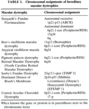
The chromosomal loci of a number of the hereditary macular dystrophies have been determined by the methods of linkage analysis and by the candidate gene approach. The chromosomal assignments and, in some cases, the gene are known for many of these hereditary macular dystrophies and are listed in Table 1.


The importance of the identification of the gene in these diseases cannot be overemphasized. In purely clinical terms, it absolutely identifies those individuals who have the disorder, confirms the diagnosis, and avoids a mistaken diagnosis. The impact on the family is likewise affected because the mode of inheritance is established; affected and unaffected family members are identified; and screening can be performed on carriers and neonates. This information provides a firm foundation for family counseling.

Back to Top
MOLECULAR GENETICS AND MACULAR DYSTROPHIES
It is interesting and perhaps ironic that the first hereditary macular dystrophy to have the gene identified was a rare disorder confined initially to a few English pedigrees, described by Arnold Sorsby, and named Sorsby's fundus dystrophy. Families from different countries, of different origins, with different clinical findings all have the mutation in the TIMP 3 gene first reported by Weber et al.99

Matrix metalloproteinases are proteolytic enzymes that regulate synthesis and degradation of the extracellular matrix throughout the body. Their activity is modulated by the tissue inhibitors ofmetalloproteinases (TIMP), of which there are four kinds. It is postulated that the loss of the inhibiting properties due to a mutation in TIMP 3 in Sorsby's fundus dystrophy could result in abnormalities of the extracellular matrix of Bruch's membrane and hence explain the clinical findings.

Stargardt's disease and fundus flavimaculatus are now apparently different phenotypic expressions of the same genotypic disorder with the identification of the ABCR gene.100 This gene is part of the ABC (ATP-binding cassette) superfamily which are transmembrane proteins modulating energy-dependent transport involving a variety of different substrates. The ABCR gene product is a protein (RMP) that is localized to the rim of rod photoreceptor outer segment disks. The function of this protein is not known, although it may play a role in phototransduction, phagocytosis of photoreceptor outer segments, or vitamin E transport.

Best's vitelliform macular dystrophy, also known as vitelliform macular degeneration type 2, is caused by a mutation in the gene encoding the protein known as bestrophin.101 The function of this protein is not known, but various speculations include a role in transport or metabolism of polyunsaturated fatty acids with resulting lipofuscin deposition.

The disorder referred to in this chapter as central areolar pigment epithelial dystrophy has been called by various names, including North Carolina macular dystrophy. Genetic linkage has established that the disorder that is now referred to as retinal macular dystrophy (MCDR 1) is closely linked to the long arm of chromosome 16 (6q16). Linkage analysis of dominant pedigrees throughout the world have shown that this disease is not restricted to any race, ethnicity, or geography.

The controversy persists as to the relationship, if any, between dominantly inherited drusen of Bruchs's membrane and degenerative drusen in the elderly. Doyne's honeycomb retinal dystrophy and Mallatia Leventinese are two dominant drusen that look somewhat dissimilar in their classic appearance and were first documented in different geographic areas in Europe. Yet both were mapped to chromosome 2p 16–21. A combination of positional and candidate gene methods led to the identification to a single mutation in the gene EFEMP 1 (EGF-containing fibrillinlike extracellular matrix protein 1) in all studied families102. Although this mutation was not present in the control patients or in patients with age-related macular degeneration, the study of this mutated gene might provide insight into the pathogenesis of macular degeneration.

Back to Top
MACULAR DYSTROPHIES AND MACULAR DEGENERATIONS
Why is the macula preferentially affected in a number of retinal disorders? Is there a relationship between the causative factors in hereditary macular dystrophies and in the age-related macular degenerations? These questions have gained a new resurgence with the advances in the genetics of the hereditary dystrophies and the increasing frequency of age-related degenerations with the concomitant visual handicaps.

The pathogenesis of age-related macular degeneration is multifactorial and the genetic component is polygenic. However, twin studies and population-based familial aggregation studies show a genetic component or susceptibility for developing this disease.

Excitement in this field continues to grow when a new gene for a hereditary dystrophy is identified. Many of these dystrophies have phenotypic similarity to their degenerative counterpart (e.g., lipofuscin and choroidal neovascularization in Best's disease, choroidal neovascularization in Sorsby's fundus dystrophy, pigmentary maculopathy with late atrophy in Stargardt's disease, and drusen in hereditary drusen). To date, studies of patients with age-related macular degeneration testing for the abnormal gene in Sorsby's fundus dystrophy and Best's disease have been unrewarding and the association in Stargardt's disease is controversial. However, the knowledge of the abnormal gene may lead to an understanding into molecular pathogenetic mechanisms that result in or predispose to macular degeneration.

Back to Top
REFERENCES

1. Stargardt K: Uber familiare, progressive Degeneration in der Makulagegend des Auges. Graefes Arch Ophthalmol 71:534, 1909.

2. Stargardt K: llber familiare progressive Degeneration in der Makulagegend des Auges. Z Augenheilkd 30:95, 1913.

3. Stargardt K: Zur Kasuistik der Familairen, progressive Degeneration der Makulagegend des Auges. Z Augenheilkd 35:249, 1916.

4. Stargardt K: Uber familiare Degeneration in der Makulagegend des Auges, Nit und ohne psychische Storungen. Arch Psychiatr Nervenkr 58:52, 1917.

5. Stargardt K: Fin Fall von familiarer progressives Makuladegeneration. Klin Monatsbl Augenheilkd 75:246, 1925.

6. Franceschetti A: Uber tapeto-retinal Degeneration im Kindesalter. In: Sautter H, ed. Entwicklung und Rortschritt in der Augenheilkunde. Stuttgart, Enke, 1963:107.

7. Franceschetti A, Francois J: Fundus flavimaculatus. Arch Ophthalmol 25:505, 1965.

8. Hadden OB Gass JDM: Fundus flavimaculatus and Star-gardt's disease. Am J Ophthalmol 82:527, 1976.

9. Noble KG, Carr RE: Stargardt's disease and fundus flavimaculatus. Arch Ophthalmol 97:1281, 1979.

10. Aaberg TM: Stargardt's disease and fundus flavimaculatus: evaluation of morphologic progression and intrafamilial co-existence. Trans Am Ophthalmol Soc 84:435, 1986.

11. Cibis CG, Morey M, Harris DJ: Dominantly inherited macular dystrophy with flecks (Stargardt's). Arch Ophthalmol 98:1785, 1980.

12. Wu G, Welter JJ, Hirose T: Autosomal dominant Star-gardt's disease. Ophthalmology 94(suppl):116, 1987.

13. Fishman GA: Fundus flavimaculatus: a clinical classification. Arch Ophthalmol 94:2061, 1976.

14. Klein BA, Krill AE: Fundus flavimaculatus: clinical, functional and histopathologic observations. Am J Ophthalmol 64:3, 1967.

15. Eagle RC Jr, Lucier AC, Bernardino VB Jr et al: Retinal pigment epithelial abnormalities in fundus flavimaculatus. Ophthalmology 87:189, 1980.

16. McDonnell PJ, Kivlin JD, Maumenee IH et al: Fundus flavimaculatus with maculopathy: a clinicopathologic study. Ophthalmology 93:116, 1986.

17. Fishman GA, Farber F, Patel BS, Derlacki DJ: Visual acuity in patients with Stargardt's macular dystrophy. Ophthalmology 94:809, 1987.

18. Bonin P, Passot M, Triolaire MT. Le signe du silence choroidien dans les degenerescences tapeto-retiennes posterieures. In: De Lacy JJ, ed. International Symposium on Fluorescein Angiography. The Hague, The Netherlands, Dr. W. Junk, 1976:461.

19. Fish G, Grey R, Sehmi KS et al: The dark choroid in posterior retinal dystrophies. Br J Ophthalmol 65:359, 1981.

20. Barkman Y: A clinical study of a central tapetoretinal degeneration. Acta Ophthalmol 39:663, 1961.

21. Wiznia RA, Perina B, Noble KG: Vitelliform macular dystrophy of late onset. Br J Ophthalmol 65:866, 1981.

22. Braley AE: Dystrophy of the macula. Am J Ophthalmol 61:1, 1966.

23. Falls HF: The polymorphous manifestations of Best's disease (vitelliform eruptive disease of the retina). Trans Am Ophthalmol Soc 67:265, 1969.

24. Benson WE, Kolker AE, Enoch JM et al: Best's vitelliform macular dystrophy. Am J Ophthalmol 79:59, 1975.

25. Miller SA, Bresnick GH, Chandra SR: Choroidal neovascular membrane in Best's vitelliform dystrophy. Arch Ophthalmol 82:252, 1976.

26. Miller SA: Multifocal Best's vitelliform dystrophy. Arch Ophthalmol 95:984, 1977.

27. Maloney WF, Robertson DM, Duboff SM: Hereditary vitelliform macular degeneration: variable fundus findings within a single pedigree. Arch Ophthalmol 95:979, 1977.

28. Fletcher RC, Jampol LM, Rimm W: An unusual presentation of Best's disease. Br J Ophthalmol 61:719, 1977.

29. Noble KG, Scher BM, Carr RE: Polymorphous presentations in vitelliform macular dystrophy: subretinal neovascularization and central choroidal atrophy. Br J Ophthalmol 62:561, 1978.

30. Deutman AF: Electro-oculography in families with itelliform dystrophy of the fovea: detection of the carrier state. Arch Ophthalmol 81:305, 1969.

31. Krill AE, Morse PA, Potts AM et al: Hereditary vitelliruptive macular degeneration of the macula. Am J Ophthalmol 61:1405, 1966.

32. Francois J DeRouch A, Fernandez-Sasso D: Electro-oculography in vitelliform degeneration of the macula. Arch Ophthalmol 77:726, 1967.

33. Thorburn W, Nordstrom S: EOG in a large family with hereditary macular degeneration. Acta Ophthalmol 56:455, 1978.

34. Cross HE, Bard L: Electro-oculography in Best's macular dystrophy. Am J Ophthalmol 77:46, 1974.

35. Frangieh GT, Green WR, Fine SL: A histopathologic study of Best's macular dystrophy. Arch Ophthalmol 100:1115, 1982.

36. Weingeist TA, Kobrin JL, Watzke RC: Histopathology of Best's macular dystrophy. Arch Ophthalmol 100:1108, 1982.

37. O'Gorman S. Flaherty WA, Fishtnan GA, Bersori EL: Histopathologic findings in Best's vitelliform macular dystrophy. Arch Ophthalmol 106:1261, 1988.

38. Birndorf LA, Dawson NW: A normal electro-oculogram in a patient with a typical vitelliform macular lesion. Invest Ophthalmol 12:830, 1973.

39. Fishman GA, Trimble S, Raab MF, Fishman M: Pseudovitelliform macular degeneration. Arch Ophthalmol 95:73, 1977.

40. Kingham JD, Lochen GP: Vitelliform macular degeneration. Am J Ophthalmol 84:526, 1977.

41. Snyder DA, Fishman GA, Witteman G, Fishman M: Vitelliform lesions associated with retinal pigment epithelial detachments. Ann Ophthalmol 10:1711, 1978.

42. Gass JDM: Pathogenesis of disciform detachment of the neuroepithelium: I. General concepts and classification. Am J Ophthalmol 63:573, 1967.

43. Gass JDM: Pathogenesis of disciform detachment of the neuroepithelium: III. Senile disciform macular degeneration. Am J Ophthalmol 63:617, 1967.

44. Sarks SH: Ageing and degeneration in the macular region: a clinicopathological study. Br J Ophthalmol 60:324, 1976.

45. Sarks SH: Drusen and their rlationship to senile macular degeneration. Aust J Ophthalmol 8:117, 1980.

46. Gass JDM, Jallow S, Davis B: Adult vitelliform macular detachment occurring in patients with basal laminar drusen. Am J Ophthalmol 99:445, 1985.

47. Sjogren H: Dystrophia reticularis lantinae pigmentosue retinae. Acta Ophthalmol 28:279, 1950.

48. Mesker RP, Oosterhuis JA, Delleman JW: A retinal lesion resembling Sjogren's dystrophy reticulates laminae pigmentosae retina. In: Winkelman JE, Crone RA, eds. Perspectives in ophthalmology, vol 2. Amsterdam, Excerpta Medica, 1970:45

49. Deutman AF, van Blomniestein IDA, Hehkes HE et al: Butterfly-shaped pigment dystrophy of the fovea. Arch Ophthalmol 83:558, 1970.

50. Hsieh RC, Fine BS, Lyons JS: Patterned dystrophies of the retinal pigment epithelium. Arch Ophthalmol 95:429, 1977.

51. Marmor MF, Byers B: Pattern dystrophy of the pigment epithelium. Am J Ophthalmol 84:32, 1977.

52. deJong PTV, Dellman W: Pigment epithelial pattern dystrophy. Arch Ophthalmol 100:1416, 1982.

53. Cortin P, Archer D, Maumenee IH et al: A patterned macular dystrophy with yellow plaques and atrophic changes. Br J Ophthalmol 64:127, 1980.

54. Burgess D: Subretinal neovascularization in pattern dystrophy of the retinal pigment epithelium. Retina 1:151, 1981.

55. Gutman I, Walsh JB, Henkind P: Vitelliform macular dystrophy and butterfly-shaped epithelial dystrophy: a continuum? Br J Ophthalmol 66:170, 1982.

56. Giuffre G, Lodato G: Vitelliform dystrophy and pattern dystrophy of the retinal pigment epithelium: concomitant presence in a family. Br J Ophthalmol 70:526, 1986.

57. Klein R, Lewis RA, Meyers SM et al: Subretinal neovascularization associated with fundus flavimaculatus. Arch Ophthalmol 96:2054, 1978.

58. Leveille AS, Morse PH, Burch JV: Fundus flavimaculatus and subretinal neovascularization. Ann Ophthalmol 14:331, 1982.

59. Bastiacnsen LAK, Hoefnages KLJ: Patterned anomalies of the retinal pigment eithelium: dystrophy or syndrome? Doe Ophthalmol 55:17, 1983.

60. Noble KG: Pathology of the hereditary macular dystrophies. Semin Ophthalmol 2:110, 1987.

61. Vine AK, Schatz H: Adult-onset foveomacular pigment epithelial dystrophy. Am J Ophthalmol 89:680, 1980.

62. Sabates R, Pruett RC, Hirose T: Pseudovitelliform macular degeneration. Retina 2:197, 1982.

63. Miller SA: Fluorescence in Best's vitelliform dystrophy, lipofuscin and fundus flavimaculatus. Br J Ophthalmol 62:256, 1978.

64. Sandvig K: Familial central, areolar, choroidal atrophy of autosomal dominant inheritance. Acta Ophthalmol 33:71, 1955.

65. Noble KG: Central areolar choroidal dystrophy. Am J Ophthalmol 84:310, 1977.

66. Noble KG, Carr RE, Siegel IM: Fluorescein and angiography of the hereditary choroidal dystrophies. Br J Ophthalmol 61:43, 1977.

67. Ashton N: Central areolar choroidal sclerosis: a histopathological study. Br J Ophthalmol 37:140, 1953.

68. Klein BA: Some aspects of classification in differential diagnosis of senile macular degeneration. Am J Ophthalmol 58:927, 1964.

69. Babel MJ: Le role de la choriocapillare dans les afflictions degeneratives du pole posterieur. Bull Mem Soc Fr Ophtalmol 71:389, 1958.

70. Ferry A, Llovera L, Shafer DM: Central areolar choroidal dystrophy. Arch Ophthalmol 88:39, 1972.

71. Lefler WH, Wadsworth JAC, Sidbury JB: Hereditary macular degeneration and aminoaciduria. Am J Ophthalmol 71:224, 1971.

72. Frank HR, Landers MB, Williams RJ, Sidbury JB: A new dominant progressive foveal dystrophy. Am J Ophthalmol 78:903, 1974.

73. Fetkenhour CL, Gurney N, Dobbie JG, Choromokos E: Central areolar pigment epithelial dystrophy. Am J Ophthalmol 81:745, 1976.

74. Leveille AS, Morse PH, Kiernan JP: Autosomal dominant central pigment epithelial and choroidal degeneration. Ophthalmology 89:1407, 1982.

75. Gass JDM: Stereoscopic atlas of macular diseases, 3rd ed. St. Louis, CV Mosby, 1987. <en

76. Small KW: North Carolina macular dystrophy: revisited. Ophthalmology 96:1747, 1989.

77. Gass JDM: A clinicopathologic study of a peculiar foveomacular dystrophy. Trans Am Ophthalmol Soc 72:139, 1974.

78. Patrinely JR, Lewis RA, Font RL: Foveomacular vitelliform dystrophy, adult type: a clinicopathologic study ineluding electron microscopic observations. Ophthalmology 92:1712, 1985.

79. Jaffe GJ, Schatz H: Histopathologic features of adult onset foveomacular pigment epithelial dystrophy. Arch Ophthalmol 106:958, 1988.

80. Sorsby A, Joll-Mason ME, Gardener N: A fundus dystrophy with unusual features. Br J Ophthalmol 33:67, 1949.

81. Duke-Elder S, Perkin ES: Diseases of the uveal tract. In: Duke-Elder S, ed. System of ophthalmology, vol 9. St. Louis, CV Mosby, 1966:715.

82. Carr RE, Noble KG, Nasaduke I: Hereditary hemorrhagic macular dystrophy. Am J Ophthalmol 85:318, 1978.

83. Hoskin A, Sehmi K, Bird AC: Sorsby's pseudoinflammatory macular dystrophy. Br J Ophthalmol 65:859, 1981.

84. Forsius HR, Eriksson AW, Suvanto EA, Alanko HI: Pseudoinflammatory fundus dystrophy with autosomal recessive inheritance. Am J Ophthalmol 94:634, 1982.

85. Ashton N, Sorsby A: Fundus dystrophy with unusual features: a histological study. Br J Ophthalmol 35:751, 1951.

86. Deutman AF, Pinckers AJLG, Aan De Kerk AL: Dominantly inherited cystoid macular edema. Am J Ophthalmol 82:540, 1976.

87. Pinckers AJLG, Deutman AF, Notting JGA: Retinal functions in dominant cystoid macular dystrophy. Acta Ophthalmol 54:579, 1976.

88. Notting JGA, Pinckers AJLG: Dominant cystoid macular dystrophy. Am J Ophthalmol 83:234, 1977.

89. Fishmar GA, Goldberg ME, Trautmann JC: Dominantly inherited cystoid macular edema. Ann Ophthalmol 11:21, 1979.

90. Lewis RA, Lee GB, Martonyi CL et al: Familial foveal retinoschisis. Arch Ophthalmol 95:1190, 1977.

91. Singerman LJ, Berkow JW, Patz A: Dominant slowly progressive macular dystrophy. Am JOphthalmol 83:680, 1977.

92. O'Donnell FE, Welch RB: Fenestrated sheen macular dystrophy. Arch Ophthalmol 97:1292, 1979.

93. Deutman AF: Benign concentric annular macular dystrophy. Am J Ophthalmol 78:384, 1974.

94. Coppetto J, Ayazi S: Annular macular dystrophy. Am J Ophthalmol 93:279, 1982.

95. van Den Biesen PR, Deutman AF, Pinckers AJLG: Evolution of benign concentric annular macular dystrophy. Am J Ophthalmol 100:73, 1985.

96. Noble KG, Sherman J: Central pigmentary sheen dystrophy. Am J Ophthalmol 108:255, 1989.

97. Duke-Elder S: System of ophthalmology, vol X. Diseases of the retina. St. Louis, CV Mosby, 1967.

98. Franceschetti A, Francois J, Babel J: Chorioretina heredodegenerations. Springfield, IL, Charles C. Thomas, 1974.

99. Weber BH, Vogt G, Pruett RC et al: Mutations in the tissue inhibitor of metalloproteinases-3 (TIMP 3) in patients with Sorsby's fundus dystrophy. Nat Genet 8:352–356, 1994.

100. Allikmets R, Sing N, Sun H et al: A photoreceptor cell-specific ATP-binding transporter gene (ABCR) is mutated in recessive Stargardt macular dystrophy. Nat Genet 15:236–246, 1997.

101. Petrukhin K, Kost MJ, Bakail B et al: Identification of the gene responsible for Best macular dystrophy. Nat Genet 19:241–247, 1998.

102. Stone EM, Lotery AJ, Munier FL et al: A single EFEMP 1 mutation associated with both Mallatia Leventinese and Doyne honeycomb retinal dystrophy. Nat Genet 22:199–202, 1999.

Back to Top