Chapter 64
Hereditary Aspects of Glaucoma
JEFFREY D. HENDERER , COURTLAND M. SCHMIDT and RICHARD P. WILSON
Main Menu   Table Of Contents

Search

OVERVIEW OF MOLECULAR GENETICS
GENETICS OF GLAUCOMA
FUTURE APPLICATIONS
CONCLUSION
REFERENCES

Of all the changes in medicine in the past 50 years, few things have been as important as an improved understanding of genetics. This has provided revolutionary insights into how life on this planet functions. The workings of the cell, which 50 years ago were a mysterious collection of proteins and nucleic acids, have become so much better understood. We can now appreciate the organization of thousands of components that must work together for life to exist. Of particular interest are a collection of nucleic acids and proteins that make up DNA. DNA exists in all cells and provides the template from which all of the functional structures of the cell are built. Since its double-helical orientation was deciphered by Watson and Crick in 1956, scientists have been investigating and manipulating its structure and, more recently, its function. The ability to read the blueprint of an organism has enabled medical doctors to understand some diseases to a level of previously incomprehensible detail. This has translated into remedies for certain diseases, and medicine is now at the threshold of an incredible revolution in our approach to diagnosis and treatment of a much wider variety of diseases. For eye disease in general, and glaucoma in particular, this revolution will fundamentally alter the ways in which we diagnose and treat disease. This chapter summarizes our current understanding of the genetics of glaucoma and how it may alter how we care for patients.
Back to Top
OVERVIEW OF MOLECULAR GENETICS
DNA provides the instruction manual for proper protein construction and thereby permits life to exist. DNA is a complicated structure composed of four nucleotide bases with a sugar and phosphate backbone. The four bases, adenine (A), guanine (G), cytosine (C), and thymine (T), protrude from the molecules' backbone to form a stairsteplike arrangement. Two strands of DNA are then joined together by hydrogen bonding between the nucleic acids (adenine always binds thymine and guanine always binds cytosine) creating a wound structure called a double helix with the nucleic acid bases becoming like rungs of a ladder. The helical strand is then further wound around proteins called histones, eventually forming the chromosomes that exist in the nucleus of every cell in our body.

The process of turning the DNA code into a protein product involves several steps. The DNA coil is first transcribed by the enzyme RNA polymerase, which creates a strand of nucleotides called RNA. This is a molecule similar to DNA except that thymine is replaced by uracil (U). This strand then leaves the nucleus, is processed to become messenger RNA (mRNA), and once in the cytoplasm becomes the template for protein formation. Specialized RNA molecules, called transfer RNA (tRNA), then bring amino acids from the cytosol, and with the mRNA as a template, the tRNA joins them together creating the long string of amino acids that will become a protein.

When cells replicate, they undergo a process known as mitosis. The goal of mitosis is to create two daughter cells from the original cell. This requires a duplicate copy of the chromosomes to be created and then distributed appropriately to each daughter cell and results in each daughter cell being identical to the original. The situation is different when reproductive cells are manufactured. As the sperm and egg will eventually unite to form a new complete cell, the sperm and egg can only contain half of a complete set of DNA. The creation of these specialized cells is done by a process known as meiosis. Much like mitosis, meiosis involves a process of DNA replication. However, instead of a complete set of genetic material being distributed to each daughter cell, two successive cell divisions reduce the DNA count so that each of the four daughter cells has half a complete set of chromosomes.

These events have profound implications for human genetics, first discovered by Gregor Mendel in the 19th century. He first formulated the principle of segregation, which suggested genes exist in pairs and only one member of each pair is passed on to the offspring and second described the law of independent assortment, which says that genes at different locations on a chromosome (loci) are inherited independently. The concepts of dominant and recessive traits (one trait can mask the presence of another) were shown by this work.

Although independent assortment is true, genes that are located close to each other on a strand of DNA are often inherited together because of the relative infrequency of recombination events occurring between closely positioned genes. It is an analysis of these two events, recombination and linkage, that form the primary way we perform genetic analysis. A third concept deals with the two sex chromosomes, one of the 23 pairs of human chromosomes. Women have two copies of the same chromosome (X), and men have one X chromosome and one Y chromosome, which is inherited from the father. Conditions that arise from mutations of the DNA on the X or Y chromosome have a pattern of inheritance different from those mutations that affect the other 22 somatic chromosomes.

Back to Top
GENETICS OF GLAUCOMA
For years, a genetic component to glaucoma was known to be present. A positive family history is known to be a significant risk factor for primary open-angle glaucoma1–5 and pseudoexfoliation.6,7 In healthy patients, even traits such as intraocular pressure (IOP) are similar among relatives.8 Associations of glaucoma and HLA type,9 blood type,10 and twin pairs11–14 supported the assumption that genetics is involved. Siblings of affected individuals have the greatest risk of developing glaucoma, followed by other blood relatives and children15 and may be unaware of their disease.16 Furthermore, a strong association of primary open-angle glaucoma or increased IOP and inhaled corticosteroid use has been reported only for persons with a positive family history of glaucoma.17 The molecular explanation for these findings has recently begun to be uncovered.

The glaucomas can be roughly segregated into two major classes, open-angle and closed-angle, with multiple subdivisions for each type. One such subdivision is based on our understanding of the cause of the condition. Primary glaucomas refer to diseases in which we are clinically unable to determine the cause of disease, while secondary glaucomas are those caused by a specific insult, such as inflammation, trauma, or other coexisting eye condition. Of all the forms of glaucoma, the primary open-angle glaucomas are by far the most common among whites and blacks, and primary angle-closure glaucoma is more frequent among the Chinese.18 The primary open-angle glaucomas can be further subdivided into two rough groups based on the age at which the disease is manifest. Juvenile open-angle glaucoma has an onset before age 35 and adult open-angle glaucoma (usually called chronic open-angle glaucoma or even simply primary open-angle glaucoma) has an onset after age 35.

JUVENILE PRIMARY OPEN-ANGLE GLAUCOMA

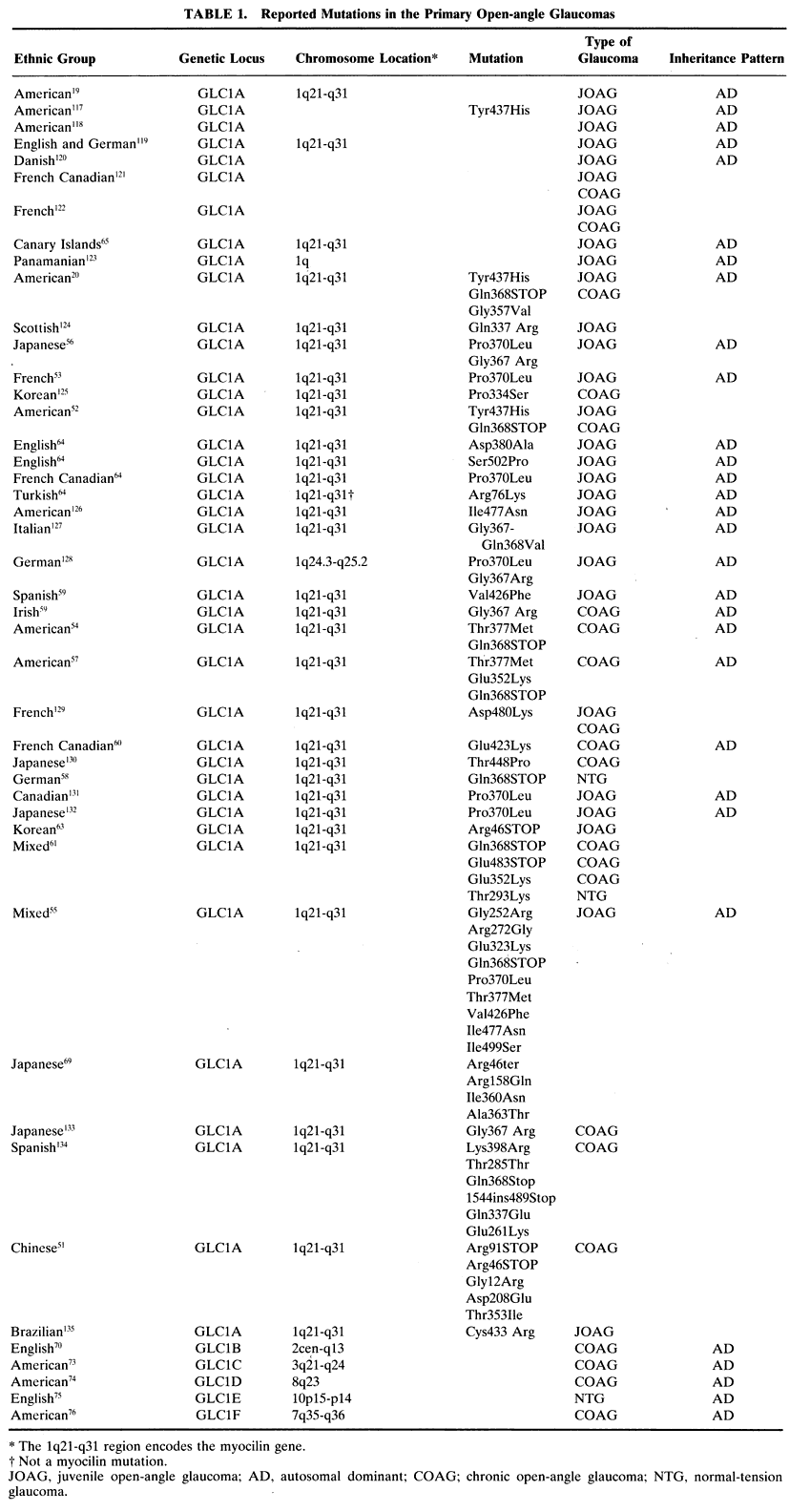
Juvenile open-angle glaucoma (JOAG), although uncommon, has been the most extensively investigated glaucoma. It is a disease that typically affects people between the ages of 3 and 35 in an autosomal dominant fashion and is characterized by elevated IOP with accompanying optic nerve and visual field damage. In 1993 Sheffield and coworkers described an American family with JOAG who appeared to contain an abnormality in the region of the long arm of chromosome 1.19 This locus was given the name GLC1A and several groups subsequently reported pedigrees of JOAG with abnormalities at the GLC1A locus in a variety of ethnic groups (Table 1). In 1997 Stone and coworkers identified the gene associated with the GLC1A locus as the region that codes for the protein myocilin (previously known as the trabecular meshwork-induced glucocorticoid response protein, or TIGR).20 Since then, multiple investigators have reported specific mutations in the myocilin gene and its protein product in families collected from around the world (Table 1).
 

The TIGR gene (now called the myocilin gene, or MYOC) was originally identified by induction of primary cultured trabecular meshwork cells with glucocorticoids. The protein that was induced was named TIGR. This protein has been found in the trabecular meshwork,21,22 ciliary body,23 and iris, but was not found in other ocular tissues24 until recently. Myocilin has now been identified in almost all ocular tissues,25 including the optic nerve,26 in an intracellular and extracellular manner. Its presence is more variable in the endothelial cells of Schlemm's canal.21,27 Systemically, it is also present in skeletal and heart muscle.24 The MYOC gene encodes a 57-kDa protein that is 504 amino acids in length and consists of three exons separated by two introns. One portion of the protein has a myosinlike domain, and one portion has an olfactomedinlike domain. Olfactomedin is the major secretory protein of the mucus layer surrounding olfactory neurons.28 Additional homologous proteins are present in other human tissues.29 Shortly after its description,30 the TIGR gene and its protein product were found to be identical to the myosinlike protein myocilin and its gene, which had been independently identified.31 Since 1998, the gene has been known as myocilin (MYOC), not TIGR. Of the variety of mutations reported in the myocilin gene, the third exon (the olfactomedinlike domain) contains almost all the known mutations of the MYOC gene.32 This may be related to a translocational pause that occurs in the native myocilin protein at the beginning of this domain.33

Myocilin appears to be part of a group of stress proteins and can be induced by subjecting trabecular cells to various insults.34 This class of proteins may bind important intracellular proteins and prevent them from denaturing under stress.32 Mutations in this protein may alter its ability to function and bind other enzymes.35 Although myocilin is present in greater amounts in the trabecular meshwork of glaucomatous eyes,36 it is also present in healthy eyes,25,26,37 arguing that it has a physiologic role. What that role may be is still unknown, but it is well conserved in other mammalian species,38–43 and analysis of the MYOC gene promoter shows regions that are responsive to glucocorticoids.30 How this finding may relate to glaucoma is still under investigation.44,45 Previous work has found that ultrastructural analysis of the trabecular meshwork in steroid-induced glaucoma and JOAG are similar46–50 and that corticosteroid treatment results in the overproduction of myocilin in some cultured trabecular cells.30 Perhaps overproduction of myocilin is common to both glaucomas. Such evidence supports one hypothesis for why myocilin mutations lead to increased IOP. This hypothesis says that myocilin is secreted by trabecular meshwork cells, which then reduces the amount of aqueous that can drain through the meshwork.51 Mutated myocilin might be oversecreted or might have an abnormal extracellular function once secreted that results in reduced aqueous outflow. A second hypothesis however proposes that alterations in myocilin might alter the cytoskeletal structure, thereby affecting cell shape and thereby reducing the size of the pores through which aqueous humor may exit. This second hypothesis suggests that a mutation in the myocilin protein might prevent it from performing its physiologic intracellular function.51 Whether either of these hypotheses is correc is not known. What is clear is that there is a spectrum of clinical findings associated with different mutations (especially the Gln368STOP mutation)32,52 and even within a single mutation.51 Further research into the myocilin protein and its function will be needed to clarify these issues.

Despite the evidence presented above relating open-angle glaucoma to myocilin, mutations in this gene cannot account for most cases of glaucoma, even those that are juvenile onset. Adam and coworkers found five of eight pedigrees to have a mutation at the GLC1A locus;53 Wiggs and coworkers found only 8% of their JOAG pedigrees had mutations in the myocilin gene;54 and Shimizu and coworkers found 33% of their JOAG pedigrees possessed a GLC1A mutation.55 Additionally, some families with mutations of this protein develop adult onset open-angle glaucoma20,52,54,56,57 or normal-tension glaucoma58 instead of JOAG, and some persons with certain mutations in the myocilin gene do not develop glaucoma at all.59–61 A large number of probably non-disease-causing polymorphisms have been found also.62 Recent work among a group of Chinese patients has found stop mutations in the myocilin gene in persons with and without glaucoma, suggesting that a reduction in the amount of myocilin protein may sometimes be, but not necessarily is, a cause of glaucoma.51 Most interesting is the mutation Arg46STOP. This mutation essentially eliminates production of myocilin altogether in homozygous individuals and reduces its production by 50% in heterozygous persons. A 15-year-old Korean girl with severe JOAG was found to be homozygous for this mutation,63 but another larger survey found this mutation present in 3% of normal Asian persons.51 In fact, one unaffected person in this larger study was a 77-year-old woman who was homozygous for this mutation, and only one heterozygous person had glaucoma. Others have found homozygous persons with a different mutation to be unaffected, whereas heterozygous family members manifest glaucoma.60 In the Korean case, the family members who were heterozygous were also unaffected, but two did have increased IOP. These data suggest that nonpenetrance is occurring, that absenceor alteration of every copy of the myocilin protein may not result in glaucoma, or that the Korean girl had other genetic or environmental factors at work. If reductions in or loss of myocilin is not necessary to cause glaucoma, it seems that other mutations that cause simply an alteration in myocilin structure may not cause glaucoma by inacti-vating a normal myocilin function. Rather, the ab-normal protein probably acquires an abnormalfunction.51

Some families have GLC1A-linked disease without mutations in the MYOC gene,64 and one group has reported cases of JOAG that do not even map to the GLC1A locus.65 The myocilin gene hypothesis may not account for the finding that despite the presence of the mutation at birth, JOAG is not congenital. Taken together, such findings indicate that our current understanding of the genetics of JOAG and the GLC1A locus is incomplete and that other genes, such as cyclooxygenase 2 (which is located near MYOC on chromosome 1q)66 and environmental factors may be involved in this disease.32

ADULT-ONSET PRIMARY OPEN-ANGLE GLAUCOMA

Whereas it appears that some of the JOAG cases studied to date have mutations in the GLC1A locus, the situation in adult-onset primary open-angle glaucoma, or chronic open-angle glaucoma (COAG), is much more heterogeneous. There are cases of COAG with mutations of the myocilin gene at locus GLC1A, but also at multiple other locations.

Some investigators have found pedigrees of COAG with mutations of the GLC1A locus (Table 1),20,51,52,54,57,67 and others have excluded this locus in their pedigrees.55,68 In families with mixed JOAG and COAG, 31% of persons have been found to possess a myocilin mutation.55 Overall, it is estimated that only 3% to 5% of patients with COAG have mutations in the myocilin gene,20,52,56,61,62,69 of which the most common is the Gln368STOP mutation.62 This stop mutation, usually associated with increased IOP in elderly patients,52 was reported to be present in one case of normal-tension glaucoma in a young man, indicating there may be other factors involved.58 There appears to be no difference in the frequency of myocilin mutations in COAG cases among racial groups ranging from blacks to whites to Japanese.62

Five additional loci for COAG, termed GLC1B through GLC1F, have been subsequently identified. All are on chromosomes different from GLC1A (Table 1). There are no data that estimate the percentage of patients with COAG who have one of these mutations. It is possible that these mutations may explain only a small number of COAG cases. Such a situation seems to have occurred with locus GLC1B. This was originally described in a pedigree from the United Kingdom,70 but so far, pedigrees from North America do not possess this mutation.71 A recent genome screen of North American patients of various ethnic backgrounds found possible abnormalities in regions of chromosomes 2, 14, 17, and 19. Genomic regions near loci GLC1A, GLC1B, GLC1D, and GLC1E seemed to be more involved than regions near loci GLC1C and GLC1F in this population.72 To date, no gene has been identified with any of these additional loci of COAG.

Clinically, patients with the GLC1B mutation have normal to moderately increased IOP,70 and patients with the GLC1C mutation have markedly increased IOP.73 Patients with a mutation of the GLC1D locus develop open-angle glaucoma in middle age with an associated moderately increased IOP.74 The fourth locus that has been described, GLC1E, was discovered in a British family with low-tension glaucoma,75 and the fifth locus was discovered in a family with increased IOP and has been termed GLC1F (Table 1).76

Other genetic loci have been associated with glaucoma, but not assigned a GLC name. Two groups have found an association of open-angle glaucoma and certain HLA loci among whites77 and Mexicans,78 but not Spaniards.79 It seems likely that adult-onset open-angle glaucoma is a heterogeneous condition and that not only will many more loci and genes be discovered, but there may well be a complex interplay between genes and other factors that eventually result in glaucoma.

SECONDARY OPEN-ANGLE GLAUCOMAS

The secondary open-angle glaucomas have been so named because the anterior segment, including the trabecular meshwork, does not show peripheral anterior synechiae, yet there is an identifiable likely cause of increased IOP and glaucoma. Even with an open angle, this group of glaucomas often has a clinically apparent abnormality of the trabecular meshwork on gonioscopy. There is also typicallyan increased IOP. Examples of this group of glau-comas include uveitic glaucoma, steroid-inducedglaucoma, angle-recession glaucoma, pigment dis-persion syndrome, and the exfoliation syndrome (pseudoexfoliation).

Pseudoexfoliation syndrome is the most common secondary open-angle glaucoma in the world. First described by Lindberg in 1917,80 it is a multisystem condition.81,82 Clinically, the syndrome features deposits of gray-white amyloidlike material throughout the anterior segment. This is most readily apparent on the lens surface and pupillary margin, but the material can be seen on the zonules. The trabecular meshwork is often more heavily pigmented than normal, and there are pupillary margin transillumination defects and atrophy. The condition is particularly prevalent in Scandinavian countries and in other locations where Scandinavians and their descendants live; it is often not apparent until late in life.

For some time there has been evidence of a genetic basis for this disease. Supporting evidence has come from twin12,83 and family84 studies. Analysis of cases in Iceland and Canada found that the disease shows evidence of maternal transmission and suggests an X-linked inheritance, autosomal inheritance with genetic imprinting, or possibly a mitochondrial disease.6,7 To date, there has been no locus or gene identified for this condition.

Pigment dispersion syndrome is a condition that typically affects young myopic persons. In whites, it may affect 2% to 4% of the population between the ages of 20 and 40 years.85 It appears that a posterior bowing of the iris causes iris-zonule touch with subsequent liberation of pigment.86,87 The pigment then is dispersed throughout the anterior chamber, where it accumulates in the trabecular meshwork and causes increased IOP and glaucoma.

Analysis of families with pigment dispersion syndrome shows a pattern of autosomal dominant inheritance.88 Recent work by Andersen and coworkers localized the gene for this condition to chromosome 7q (Table 2).89


CONGENITAL GLAUCOMA

Congenital glaucoma is defined as glaucoma present at birth. In reality, most cases of this type of glaucoma are probably better-termed infantile glaucoma because the disease is often not manifest until several months of age up to 3 years old. From a clinical standpoint, infantile glaucoma is most often an autosomal recessive disease characterized by increased IOP caused by an angle dysgenesis that leads to the classic symptom triad of corneal clouding attributable to edema, epiphora, and photophobia. If left untreated, buphthalmos and amblyopia often develop. This form of glaucoma must be considered separately from secondary congenital glaucomas in which the glaucoma likely arises as a result of other dysmorphic or multisystem abnormal conditions.

Two loci have been identified that appear to segregate with infantile glaucoma, termed GLC3A and GLC3B. These loci have been localized to chromosome 2 and 1, respectively (Table 3). The gene that is mutated at the GLC3A locus is cytochrome P4501B1. Multiple mutations in this gene havebeen identified in populations around the world, in-cluding German-American, Hispanic, French-Canadian, British, Pakistani, Turkish,90 Saudi Arabian,91 Japanese,92 and Slovak Gypsies.93 In Slovak Gypsies affected with congenital glaucoma, the mutation has been reported to be a homozygous glutamic acid substitution for lysine at position 387.94 This location happens to be in a highly conserved portion of the protein and the various mutations most likely affect the normal protein folding or truncate the protein. Position 387 is absolutely conserved among cytochrome P450 proteins and so is likely a critical portion of the protein. A homozygous mutation of glutamic acid for lysine at this position is always associated with congenital glaucoma.94 This matches well with the original descriptions of congenital glaucoma being markedly worse in the Slovak Gypsy population than in other groups.95,96 This is not the case in other families with different mutations. Such families can have incomplete penetrance and a variable clinical picture depending on the mutation and whether it is heterozygous or homozygous.90,97
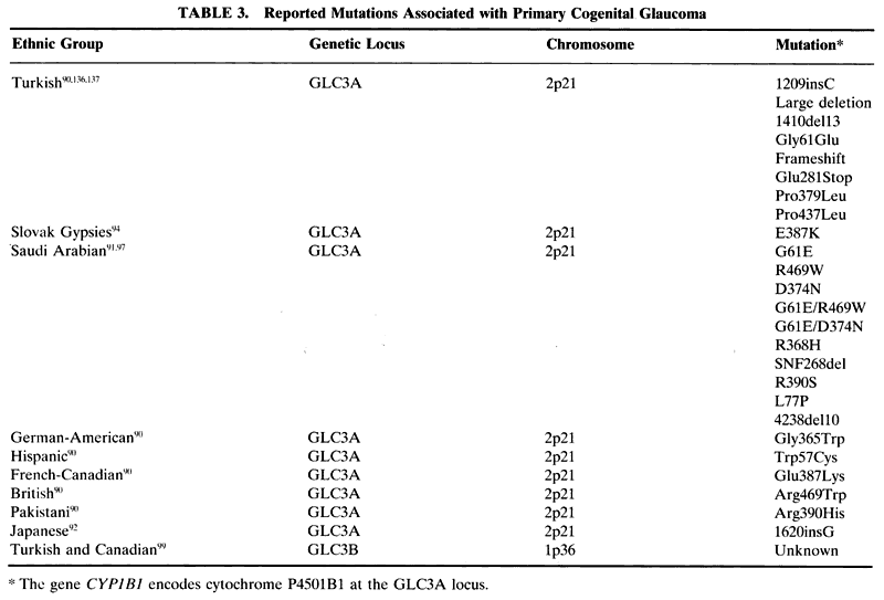


Cytochrome P4501B1 is part of a superfamily of cytochrome P450 proteins. These proteins are involved in metabolism in the cell usually by inserting oxygen into a molecule and seem to assist in keeping the cornea clear.98 In patients with the mutation at the GLC3A locus, loss of this function may be responsible for the corneal edema that is seen in congenital glaucoma.

A second locus for congenital glaucoma was recently identified. Termed GLC3B, it has been localized to chromosome 1p.99 To date, there has been no gene identified with this locus.

CHRONIC ANGLE-CLOSURE GLAUCOMA

Angle-closure glaucoma is a term that describes a group of glaucomas associated with functional obstruction of the trabecular meshwork. Such an obstruction can be from iris peripheral anterior synechiae or from any other scar tissue or cellular layer that covers the trabecular meshwork to impede outflow of aqueous from the eye, such as in iridocorneal endothelial syndrome.

Primary angle-closure glaucoma, in which peripheral anterior synechiae acutely or gradually zipper the angle shut, is thought to be the result of pupillary block that pushes the peripheral iris into the angle and results in scarring. Although there is no clear understanding why this disease occurs, it is found most commonly in the Chinese18 and can be halted by a peripheral iridectomy. The secondary angle-closure glaucomas are a diverse group of conditions in which there is a known cause of angle closure. Conditions such as neovascular glaucoma, malignant glaucoma, and iridocorneal endothelial syndrome represent some of the many diseases that can cause a secondary angle-closure glaucoma.

There have been no reports to date describing any loci for primary angle-closure glaucoma. A single report has placed the gene for dominantly inherited nanophthalmos (NNO1) and angle-closure glaucoma on chromosome 11p.100 No other secondary angle-closure glaucoma has been described to have a genetic basis.

OTHER DEVELOPMENTAL GLAUCOMAS

The term anterior segment dysgenesis describes a variety of ocular conditions in which the anterior segment is malformed at birth. Such conditions are typically bilateral and often associated with glaucoma. Of the various syndromes and anomalies that make up this group of ocular diseases, some have been determined to have a genetic basis (Table 2).

Axenfeld-Rieger syndrome describes a spectrum of anterior segment and systemic findings that are inherited in an autosomal dominant fashion with high penetrance. The condition is associated with an onset of glaucoma in childhood or early adulthood in approximately half of patients. Three genetic loci have been identified on chromosomes 4q25,101 6p25,102 and 13q14.103 The DNA at the 4q25 and 6p25 loci has been found to encode the genes PITX2 (paired-like homeodomain transcription factor 2)104 and FKHL7 (forkhead Drosophila homologue-like transcription factor gene).102 No gene has been identified to date at loci 13q14.

The PITX2 gene is a paired homeobox gene that regulates the expression of other genes during development. Mutations in this gene have also been associated with autosomal dominant iris hypoplasia,105,106 iridogoniodysgenesis syndrome,107 and a single case of Peter anomaly.108 FKHL7 is also a gene that regulates the expression of other genes during development. In the mouse, the homologue gene FOXC1, is expressed in mesenchyme from which the drainage structures of the anterior segment are derived. Although mice with this mutation had anterior segment anomalies, they did not have increased IOP. This suggests an interaction of some sort with another gene is responsible for the glaucoma associated with this condition.109 Mutations at the 6p25 locus have been associated with a variety of anterior segment abnormalities, such as Axenfeld anomaly, iris hypoplasia, Rieger anomaly, posterior embryotoxon,102 iridogoniodysgenesis anomaly,110 and familial glaucoma iridogoniodysplasia.111 Not all the patients with mutations in the 6p25 locus have mutations of the FKHL7 gene. Only those with features of Axenfeld-Rieger anomaly had mutations in this gene.112

The important message from the genetics of Axenfeld-Rieger syndrome is that multiple phenotypic variations can be caused by mutations in the same genes. This supports the notion that there is no useful information to be gained from splitting these conditions into separate diseases and the spectrum of phenotypic findings should be lumped under the name Axenfeld-Rieger syndrome.113

The nail-patella syndrome (NPS1) has been localized to chromosome 9q34114 and its gene LMX1B.115 This gene is a transcription factor important in skeletal development.

Back to Top
FUTURE APPLICATIONS
The excitement of genetics comes from not only a better understanding of the pathophysiology of the disease, but also the possibilities of new approaches to diagnosis and treatment. Genetic testing will help identify those at risk, especially in glaucomas that have an early onset. Early diagnosis will allow earlier treatment and result in less severe disease. Genetic testing will also serve to exclude those without a certain defect, thereby lessening the fear of the patient and possibly reducing the need for frequent monitoring. It is important to realize that because most cases of glaucoma do not seem to have a clear inherited basis and because all the genetic defects that can cause glaucoma have not been identified, the significance of a negative test result would not exclude the possibility of developing glaucoma, but simply means that the risk of developing glaucoma is the same as that in the general population. Nevertheless, for persons in families with a strong history of glaucoma in whom a genetic mutation has been identified, genetic screening might be a useful tool.

The prospect of gene therapy for glaucoma is intriguing. Such therapy might take one of two forms. The first is to attempt to insert a new copy of a defective gene into the cells of the eye by means of a vector and the second is to replace the missing or defective protein product, or somehow block its harmful action, without altering the host DNA. Attempts at these therapies are experimental and likely many years from clinical usefulness, but recent work has focused on developing herpes virus vectors capable of delivering foreign DNA to various ocular structures in animal models.116

Back to Top
CONCLUSION
The study of glaucoma has recently seen an exciting revolution. The identification of genetic defects that can cause this disease offers new hope to understand the pathophysiology of the disease and new avenues for diagnosis and treatment. Intensive work continues to identify other genetic mutations associated with the many forms of glaucoma. Although our understanding of glaucoma has already been deepened, results of this work and its clinical applications will certainly transform the way this group of diseases is viewed and treated in the future.
Back to Top
REFERENCES

1. Shin DH, Becker B, Kolker AE: Family history in primary open-angle glaucoma. Arch Ophthalmol 95:598–600, 1977.

2. Tielsch JM, Katz J, Sommer A, Quigley HA, Javitt JC: Family history and risk of primary open angle glaucoma. The Baltimore Eye Survey. Arch Ophthalmol 112:69–73, 1994

3. Leske MC, Connell AM, Wu SY, Hyman LG, Schachat AP: Risk factors for open-angle glaucoma. The Barbados Eye Study. Arch Ophthalmol 113:918–924, 1995.

4. Wolfs RC, Klaver CC, Ramrattan RS, van Duijn CM, Hofman A, de Jong PT: Genetic risk of primary open-angle glaucoma. Population-based familial aggregation study. Arch Ophthalmol 116:1640–1645, 1998.

5. Budde WM, Jonas JB: Family history of glaucoma in the primary and secondary open-angle glaucomas. Graefes Arch Clin Exp Ophthalmol 237:554–557, 1999.

6. Damji KF, Bains HS, Stefansson E et al: Is pseudoexfoliation syndrome inherited? A review of genetic and nongenetic factors and a new observation. Ophthalmic Genet 19:175–185, 1998.

7. Damji KF, Bains HS, Amjadi K et al: Familial occurrence of pseudoexfoliation in Canada. Can J Ophthalmol 34:257–265, 1999.

8. Levene RZ, Workman PL, Broder SW, Hirschhorn K: Heritability of ocular pressure in normal and suspect ranges. Arch Ophthalmol 84:730–734, 1970.

9. Shin DH, Becker B, Bell CE: HLA in primary open-angle glaucoma. Int Arch Allergy Appl Immunol 55:546–550, 1977.

10. Leske MC, Nemesure BB, He Q, Mendell N, Polednak A: Open-angle glaucoma and blood groups. The Barbados Eye Study. Arch Ophthalmol 114:205–210, 1996.

11. Teikari JM, Airaksinen PJ, Kaprio J, Koskenvuo M: Primary open-angle glaucoma in 2 monozygotic twin pairs. Acta Ophthalmol (Copenh) 65:607–611, 1987.

12. Teikari JM: Genetic factors in open-angle (simple and capsular) glaucoma. A population-based twin study. Acta Ophthalmol (Copenh) 65:715–720, 1987.

13. Teikari JM, Kaprio J, Koskenvo M, Vannas A: Ophthalmic disease in twins: a nationwide record linkage study of hospital discharges and free medications for 16,067 twin pairs. Acta Genet Med Gemellol 36:523–534, 1987.

14. Gottfredsdottir MS, Sverrisson T, Musch DC, Stefansson E: Chronic open-angle glaucoma and associated ophthalmic findings in monozygotic twins and their spouses in Iceland. J Glaucoma 8:134–139, 1999.

15. Nguyen RL, Raja SC, Traboulsi EI: Screening relatives of patients with familial chronic open angle glaucoma. Ophthalmology 107:1294–1297, 2000.

16. McNaught AI, Allen JG, Healey DL et al: Accuracy and implications of a reported family history of glaucoma: experience from the Glaucoma Inheritance Study in Tasmania. Arch Ophthalmol 118:900–904, 2000.

17. Mitchell P, Cumming RG, Mackey DA: Inhaled corticosteroids, family history, and risk of glaucoma. Ophthalmology 106:2301–2306, 1999.

18. Quigley HA: Number of people with glaucoma worldwide. Br J Ophthalmol 80:389–393, 1996.

19. Sheffield VC, Stone EM, Alward WL et al: Genetic linkage of familial open angle glaucoma to chromosome 1q21-q31. Nat Genet 4:47–50, 1993.

20. Stone EM, Fingert JH, Alward WLM et al: Identification of a gene that causes primary open angle glaucoma. Science 275:668–670, 1997.

21. Wang X, Johnson DH: mRNA in situ hybridization of TIGR/MYOC in human trabecular meshwork. Invest Ophthalmol Vis Sci 41:1724–1729, 2000.

22. Ueda J, Wentz-Hunter KK, Cheng EL, Fukuchi T, Abe H, Yue BY: Ultrastructural localization of myocilin in human trabecular meshwork cells and tissues. J Histochem Cytochem 48:1321–1330, 2000.

23. Polansky JR, Fauss DJ, Chen P et al: Cellular pharmacology and molecular biology of the trabecular meshwork inducible glucocorticoid response gene product. Ophthalmologica 211:126–139, 1997

24. Ortego J, Escribano J, Coca-Prados M: Cloning and characterization of subtracted cDNAs from a human ciliary body library encoding TIGR, a protein involed in juvenile open angle glaucoma with homology to myosin and olfactomedin. FEBS Lett 413:349–353, 1997.

25. Karali A, Russell P, Stefani FH, Tamm ER. Localization of myocilin/trabecular meshwork—inducible glucocorticoid response protein in the human eye. Invest Ophthalmol Vis Sci 41:729–740, 2000.

26. Swiderski RE, Ross JL, Fingert JH et al: Localization of MYOC transcripts in human eye and optic nerve by In situ hybridization. Invest Ophthalmol Vis Sci 41:3420–3428, 2000.

27. Stamer WD, Roberts BC, Howell DN, Epstein DL: Isolation, culture, and characterization of endothelial cells from Schlemm's canal. Invest Ophthalmol Vis Sci 39:1804–1812, 1998.

28. Synder A, Rivers A, Yokoe H, Menco B, Anholt R: Olfactomedin: purification, characterization, and localization of a novel olfactory glycoprotein. Biochemistry 30:9143–9153, 1991.

29. Kulkarni NH, Karavanich CA, Atchley WR, Anholt RR: Characterization and differential expression of a human gene family of olfactomedin-related proteins. Genet Res 76:41–50, 2000.

30. Nguyen TD, Chen P, Huang WD, Chen H, Johnson D, Polansky JR: Gene structure and properties of TIGR, an olfactomedin-related glycoprotein cloned from glucocorticoid-induced trabecular meshwork cells. J Biol Chem 273:6341–6350, 1998.

31. Kubota R, Noda S, Wang Y et al: A novel myosin-like protein (myocilin) expressed in the connecting cilium of the photoreceptor: molecular cloning, tissue expression, and chromosomal mapping. Genomics 41:360–369, 1997.

32. Johnson DH: Myocilin and glaucoma: A TIGR by the tail? Arch Ophthalmol 118:974–978, 2000.

33. Zimmerman CC, Lingappa VR, Richards JE, Rozsa FW, Lichter PR, Polansky JR: A trabecular meshwork glucocorticoid response (TIGR) gene mutation affects translocational processing. Mol Vis 5:19, 1999.

34. Tamm ER, Russell P, Epstein DL, Johnson DH, Piatigorsky J: Modulation of myocilin/TIGR expression in human trabecular meshwork. Invest Ophthalmol Vis Sci 40:2577–282, 1999.

35. Rozsa FW, Shimizu S, Lichter PR et al: GLC1A mutations point to regions of potential functional importance on the TIGR/MYOC protein. Mol Vis 4:20, 1998.

36. Lutjen-Drecoll E, May CA, Polansky JR, Johnson DH, Bloemendal H, Nguyen TD: Localization of the stress proteins alpha B-crystallin and trabecular meshwork inducible glucocorticoid response protein in normal and glaucomatous trabecular meshwork. Invest Ophthalmol Vis Sci 39:517–525, 1998.

37. Takahashi H, Noda S, Mashima Y et al: The myocilin (MYOC) gene expression in the human trabecular meshwork. Curr Eye Res 20:81–84, 2000.

38. Abderrahim H, Jaramillo-Babb VL, Zhou Z, Vollrath D: Characterization of the murine TIGR/myocilin gene. Mamm Genome 9:673–675, 1998.

39. Taguchi M, Kanno H, Kubota R, Miwa S, Shishiba Y, Ozawa Y: Molecular cloning and expression profile of rat myocilin. Mol Genet Metab 70:75–80, 2000.

40. Takahashi H, Noda S, Imamura Y et al: Mouse myocilin (Myoc) gene expression in ocular tissues. Biochem Biophys Res Commun 248:104–109, 1998.

41. Tomarev SI, Tamm ER, Chang B: Characterization of the mouse Myoc/Tigr gene. Biochem Biophys Res Commun 245:887–893, 1998.

42. Fingert JH, Ying L, Swiderski RE et al: Characterization and comparison of the human and mouse GLC1A glaucoma genes. Genome Res 8:377–384, 1998.

43. Taniguchi F, Suzuki Y, Kurihara H et al: Molecular cloning of the bovine MYOC and induction of its expression in trabecular meshwork cells. Invest Ophthalmol Vis Sci 41:2070–2075, 2000.

44. Tripathi RC, Parapuram SK, Tripathi BJ, Zhong Y, Chalam KV: Corticosteroids and glaucoma risk. Drugs Aging 15:439–450, 1999.

45. Wordinger RJ, Clark AF: Effects of glucococorticoids on the trabecular meshwork: towards a better understanding of glaucoma. Prog Retin Eye Res 18:629–667, 1999.

46. Rohen JW, Linner E, Witmer R: Electron microscopic studies on the trabecular meshwork in two cases of corticosteroid-glaucoma. Exp Ee Res 17:19–31, 1973.

47. Johnson D, Gottanka J, Flugel C, Hoffmann F, Futa R, Lutjen-Drecoll E: Ultrastructural changes in the trabecular meshwork of human eyes treated with corticosteroids. Arch Ophthalmol 115:375–383, 1997.

48. Tawara A, Inomata H: Developmental immaturity of the trabecular meshwork in juvenile glaucoma. Am J Ophthalmol 98:82–97, 1984.

49. Tawara A, Inomata H: Distribution and characterization of sulfated proteoglycans in the trabecular tissue of goniodysgenetic glaucoma. Am J Ophthalmol 117:741–755, 1994.

50. Furuyoshi N, Furuyoshi M, Futa R, Gottanka J, Lutjen-Drecoll E: Ultrastructural changes in the trabecular meshwork of juvenile glaucoma. Ophthalmologica 211:140–146, 1997.

51. Lam DS, Leung YF, Chua JK et al: Truncations in the TIGR gene in individuals with and without primary open-angle glaucoma. Invest Ophthalmol Vis Sci 41:1386–1391, 2000.

52. Alward WL, Fingert JH, Coote MA et al: Clinical features associated with mutations in the chromosome 1 open- angle glaucoma gene (GLC1A). N Engl J Med 338:1022–1027, 1998.

53. Adam MF, Belmouden A, Binisti P et al: Recurrent mutations in a single exon encoding the evolutionarily conserved olfactomedin-homology domain of TIGR in familial open-angle glaucoma. Hum Mol Genet 6:2091–2097, 1997.

54. Wiggs JL, Allingham RR, Vollrath D et al: Prevalence of mutations in TIGR/Myocilin in patients with adult and juvenile primary open-angle glaucoma. Am J Hum Genet 63:1549–1552.

55. Shimizu S, Lichter PR, Johnson AT, et al: Age-dependent prevalence of mutations at the GLC1A locus in primary open-angle glaucoma. Am J Ophthalmol 130:165–177, 2000.

56. Suzuki Y, Shirato S, Taniguchi F, Ohara K, Nishimaki K, Ohta S: Mutations in the TIGR gene in familial primary open-angle glaucoma in Japan. Am J Hum Genet 61:1202–1204, 1997.

57. Allingham RR, Wiggs JL, De La Paz MA et al: Gln368STOP myocilin mutation in families with late-onset primary open-angle glaucma. Invest Ophthalmol Vis Sci 39:2288–2295, 1998.

58. Mardin CY, Velten I, Ozbey S, Rautenstrauss B, Michels-Rautenstrauss K: A GLC1A gene Gln368Stop mutation in a patient with normal-tension open-angle glaucoma.J Glaucoma 8:154–156, 1999.

59. Mansergh FC, Kenna PF, Ayuso C, Kiang AS, Humphries P, Farrar GJ: Novel mutations in the TIGR gene in early and late onset open angle glaucoma. Hum Mutat 11:244–251, 1998.

60. Morissette J, Clepet C, Moisan S et al: Homozygotes carrying an autosomal dominant TIGR mutation do not manifest glaucoma. Nat Genet 19:319–321, 1998.

61. Williams-Lyn D, Flanagan J, Buys Y et al: The genetic aspects of adult-onset glaucoma: a perspective from the Greater Toronto area. Can J Ophthalmol 35:12–17, 2000.

62. Fingert JH, Heon E, Liebmann JM et al: Analysis of myocilin mutations in 1703 glaucoma patients from five different populations. Hum Mol Genet 8:899–905, 1999.

63. Yoon SJ, Kim HS, Moon JI, Lim JM, Joo CK: Mutations of the TIGR/MYOC gene in primary open-angle glaucoma in Korea. Am J Hum Genet 64:1775–1778, 1999.

64. Stoilova D, Child A, Brice G et al: Novel TIGR/MYOC mutations in families with juvenile onset primary open angle glaucoma. J Med Genet 35:989–992, 1998.

65. WuDunn D, Parrish RK 2nd, Inana G: Genetic heterogeneity in Hispanic families with autosomal dominant juvenile glaucoma. Ophthalmic Genet 17:87–94, 1996.

66. Budde WM: Heredity in primary open-angle glaucoma. Curr Opin Ophthalmol 11:101–106, 2000.

67. Angius A, Spinelli P, Ghilotti G et al: Myocilin Gln368stop mutation and advanced age as risk factors for late-onset primary open-angle glaucoma. Arch Ophthalmol 118:674–679, 2000.

68. Avramopoulos D, Kitsos G, Economou-Petersen E et al: Exclusion of one pedigree affected by adult onset primary open angle glaucoma from linkage to the juvenile glaucoma locus on chromosome 1q21- q31. J Med Genet 33:1043–1044, 1996.

69. Kubota R, Mashima Y, Ohtake Y et al:Novel mutations in the myocilin gene in Japanese glaucoma patients. Hum Mutat 16:270, 2000.

70. Stoilova D, Child A, Trifan OC, Crick RP, Coakes RL, Sarfarazi M: Localization of a locus (GLC1B) for adult-onset primary open angle glaucoma to the 2cen-q13 region. Genomics 36:142–150, 1996.

71. Allingham RR, Wiggs JL, Damji KF et al: Adult-onset primary open angle glaucoma does not localize to chromosome 2cen-q13 in North American families. Hum Hered 48:251–255, 1998.

72. Wiggs JL, Allingham RR, Hossain A et al: Genome-wide scan for adult onset primary open angle glaucoma. Hum Mol Genet 9:1109–1117, 2000.

73. Wirtz MK, Samples JR, Kramer PL et al: Mapping a gene for adult-onset primary open-angle glaucoma to chromosome 3q. Am J Hum Genet 60:296–304, 1997.

74. Trifan OC, Traboulsi EI, Stoilova D et al: A third locus (GLC1D) for adult-onset primary open-angle glaucoma maps to the 8q23 region. Am J Ophthalmol 126:17–28, 1998.

75. Sarfarazi M, Child A, Stoilova D et al: Localization of the fourth locus (GLC1E) for adult-onset primary open- angle glaucoma to the 10p15-p14 region. Am J Hum Genet 62:641–652, 1998.

76. Wirtz MK, Samples JR, Rust K et al: GLC1F, a new primary open-angle glaucoma locus, maps to 7q35-q36. Arch Ophthalmol 117:237–241, 1999.

77. David R, Maier G, Baumgarten I, Abrahams C: HLA antigens in glaucoma and ocular hypertension. Br J Ophthalmol 63:293–296, 1979.

78. Gil-Carrasco F, Vargas-Alarcon G, Zuniga J et al: HLA-DRB and HLA-DQB loci in the genetic susceptibility to develop glaucoma in Mexicans. Am J Ophthalmol 128:297–300, 1999.

79. Abecia E, Martinez-Jarreta B, Casalod Y, Bell B, Pinilla I, Honrubia FM: Genetic markers in primary open-angle glaucoma. Int Ophthalmol 20:79–82, 1996.

80. Lindberg J: Kliniska undersokningar over dipigmentering av pupillarranden odr genomlysbarhet av iris vid fall av alderstarr samt i normala ogon hos gamla personer. Inaug Diss Helsingfors. 1917.

81. Sclotzer-Schrehardt UM, Koca MR, Naumann GO, Volkholz H. Pseudoexfoliation syndrome. Ocular manifestation of a systemic disorder? Arch Ophthalmol 110:1752–1756, 1992.

82. Streeten BW, Li ZY, Wallace RN, Eagle RC Jr: Keshgegian AA. Pseudoexfoliative fibrillopathy in visceral organs of a patient with pseudoexfoliation syndrome. Arch Ophthalmol 110:1757–1762, 1992.

83. Sverrisson T, Gottfredsdottir M, Stafansson E. Chronic open angle glaucoma in monozygotic twins and their spouses. Invest Ophthalmol Vis Sci 35:1471, 1994.

84. Aasved H: Study of relatives of persons with fibrillopathia epitheliocapsularis (pseudoexfoliation of the lens capsule). Acta Ophthalmol (Copenh) 53:879–886, 1975.

85. Ritch R, Steinberger D, Liebmann JM: Prevalence of pigment dispersion syndrome in a population undergoing glaucoma screening. Am J Ophthalmol 115:707–710, 1993.

86. Campbell DG: Pigmentary dispersion and glaucoma. A new theory. Arch Ophthalmol 97:1667–1672, 1979.

87. Liebmann JM, Tello C, Chew SJ, Cohen H, Ritch R: Prevention of blinking alters iris configuration in pigment dispersion syndrome and in normal eyes. Ophthalmology 102:446–455, 1995.

88. Mandelkorn RM, Hoffman ME, Olander KW, Zimmerman T, Harsha D: Inheritance and the pigmentary dispersion syndrome. Ann Ophthalmol 15:577–582, 1983.

89. Andersen JS, Pralea AM, DelBono EA et al: A gene responsible for the pigment dispersion syndrome maps to chromosome 7q35-q36. Arch Ophthalmol 115:384–388, 1997.

90. Stoilov I, Akarsu AN, Alozie I et al: Sequence analysis and homology modeling suggest that primary congenital glaucoma on 2p21 results from mutations disrupting either the hinge region or the conserved core structures of cytochrome P4501B1. Am J Hum Genet 62:573–584, 1998.

91. Bejjani BA, Lewis RA, Tomey KF et al: Mutations in CYP1B1, the gene for cytochrome P4501B1, are the predominant cause of primary congenital glaucoma in Saudi Arabia. Am J Hum Genet 62:325–333, 1998.

92. Kakiuchi T, Isashiki Y, Nakao K, Sonoda S, Kimura K, Ohba N: A novel truncating mutation of cytochrome P4501B1 (CYP1B1) gene in primary infantile glaucoma. Am J Ophthalmol 128:370–372, 1999.

93. Plasilova M, Ferakova E, Kadasi L et al: Linkage of autosomal recessive primary congenital glaucoma to the GLC3A locus in Roms (Gypsies) from Slovakia. Hum Hered 48:30–33, 1998.

94. Plasilova M, Stoilov I, Sarfarazi M, Kadasi L, Ferakova E, Ferak V: Identification of a single ancestral CYP1B1 mutation in Slovak Gypsies (Roms) affected with primary congenital glaucoma. J Med Genet 36:290–294, 1999.

95. Gencik A, Gencikova A, Ferak V: Population genetical aspects of primary congenital glaucoma. I. Incidence, prevalence, gene frequency, and age of onset. Hum Genet 61:193–197, 1982.

96. Gencik A: Epidemiology and genetics of primary congenital glaucoma in Slovakia. Description of a form of primary congenital glaucoma in gypsies with autosomal-recessive inheritance and complete penetrance. Dev Ophthalmol 16:76–115, 1989.

97. Bejjani BA, Stockton DW, Lewis RA et al: Multiple CYP1B1 mutations and incomplete penetrance in an inbred population segregating primary congenital glaucoma suggest frequent de novo events and a dominant modifier locus. Hum Mol Genet 9:367–374, 2000.

98. Schwartzman ML, Balazy M, Masferrer J, Abraham NG, McGiff JC, Murphy RC: 12(R)-hydroxyicosatetraenoic acid: a cytochrome-P450-dependent arachidonate metabolite that inhibits Na+ ,K+ -ATPase in the cornea. Proc Natl Acad Sci USA 84:8125–8129, 1987.

99. Akarsu AN, Turacli ME, Aktan SG et al: A second locus (GLC3B) for primary congenital glaucoma (Buphthalmos) maps to the 1p36 region. Hum Mol Genet 5:1199–1203, 1996.

100. Othman MI, Sullivan SA, Skuta GL et al: Autosomal dominant nanophthalmos (NNO1) with high hyperopia and angle- closure glaucoma maps to chromosome 11. Am J Hum Genet 63:1411–1418, 1998.

101. Murray JC, Bennett SR, Kwitek AE et al: Linkage of Rieger syndrom to the region of the epidermal growth factor gene on chromosome 4. Nat Genet 2:46–49, 1992.

102. Nishimura DY, Swiderski RE, Alward WL et al: The forkhead transcription factor gene FKHL7 is responsible for glaucoma phenotypes which map to 6p25. Nat Genet 19:140–147, 1998.

103. Phillips JC, del Bono EA, Haines JL et al: A second locus for Rieger syndrome maps to chromosome 13q14. Am J Hum Genet 59:613–619, 1996.

104. Semina EV, Reiter R, Leysens NJ et al: Cloning and characterization of a novel bicoid-related homeobox transcription factor gene, RIEG, involved in Rieger syndrome. Nat Genet 14:392–399, 1996.

105. Heon E, Sheth BP, Kalenak JW et al: Linkage of autosomal dominant iris hypoplasia to the region of the Rieger syndrome locus (4q25). Hum Mol Genet 4:1435–1439, 1995.

106. Alward WL, Semina EV, Kalenak JW et al: Autosomal dominant iris hypoplasia is caused by a mutation in the Rieger syndrome (RIEG/PITX2) gene. Am J Ophthalmol 125:98–100, 1998.

107. Kulak SC, Kozlowski K, Semina EV, Pearce WG, Walter MA: Mutation in the RIEG1 gene in patients with iridogoniodysgenesis syndrome. Hum Mol Genet 7:1113–1117, 1998.

108. Doward W, Perveen R, Lloyd IC, Ridgway AE, Wilson L, Black GC: A mutation in the RIEG1 gene associated with Peters' anomaly. J Med Genet 36:152–155, 1999.

109. Smith RS, Zabaleta A, Kume T et al: Haploinsufficiency of the transcription factors FOXC1 and FOXC2 results in aberrant ocular development. Hum Mol Genet 9:1021–1032, 2000.

110. Mears AJ, Mirzayans F, Gould DB, Pearce WG, Walter MA: Autosomal dominant iridogoniodysgenesis anomaly maps to 6p25. Am J Hum Genet 59:1321–1327, 1996.

111. Jordan T, Ebenezer N, Manners R, McGill J, Bhattacharya S: Familial glaucoma iridogoniodysplasia maps to a 6p25 region implicated in primary congenital glaucoma and iridogoniodysgenesis anomaly. Am J Hum Genet 61:882–888, 1997.

112. Mears AJ, Jordan T, Mirzayans F et al: Mutations of the forkhead/winged-helix gene, FKHL7, in patients with Axenfel-Rieger anomaly. Am J Hum Genet 63:1316–1328, 1998.

113. Alward WL: Axenfeld-Rieger syndrome in the age of molecular genetics. Am J Ophthalmol 130:107–115, 2000.

114. Lichter PR, Richards JE, Downs CA, Stringham HM, Boehnke M, Farley FA: Cosegregation of open-angle glaucoma and the nail-patella syndrome. Am J Ophthalmol 124:506–515, 1997.

115. Vollrath D, Jaramillo-Babb VL, Clough MV et al: Loss-of-function mutations in the LIM-homeodomain gene, LMX1B, in nail- patella syndrome. Hum Mol Genet 7:1091–1098, 1998.

116. Kaufman PL, Jia WW, Tan J et al: A perspective of gene therapy in the glaucomas. Surv Ophthalmol 43(Suppl)1:S91–97, 1999.

117. Johnson AT, Drack AV, Kwitek AE, Cannon RL, Stone EM, Alward WL: Clinical features and linkage analysis of a family with autosomal dominant juvenile glaucoma. Ophthalmology 100:524–529, 1993.

118. Richards J, Lichter P, Boehnke M et al: Mapping of a gene for autosomal dominant juvenile-onset open-angle glaucoma to chromosome 1q. Am J Hum Genet 54:62–70, 1994.

119. Wiggs JL, Haines JL, Paglinauan C, Fine A, Sporn C, Lou D: Genetic linkage of autosomal dominant juvenile glaucoma to 1q21-q31 in three affected pedigrees. Genomics 21:299–303, 1994.

120. Graff C, Urbak SF, Jerndal T, Wadelius C: Confirmation of linkage to 1q21-31 in a Danish autosomal dominant juvenile-onset glaucoma family and evidence of genetic heterogeneity. Hum Genet 96:285–289, 1995.

121. Morissette J, Cote G, Anctil J et al: A common gene for juvenile and adult-onset primary open-angle glaucomas confined on chromosome 1q. Am J Hum Genet 56:1431–1442, 1995.

122. Meyer A, Bechetoille A, Valtot F et al: Age-dependent penetrance and mapping of the locus for juvenile and early-onset open-angle glaucoma on chromosome 1q (GLC1A) in a French family. Hum Genet 98:567–571, 1996.

123. Lichter PR, Richards JE, Boehnke M et al: Juvenile glaucoma linked to the GLC1A gene on chromosome 1q in a Panamanian family. Am J Ophthalmol 123:413–416, 1997.

124. Soilova D, Child A, Brice G, Crick RP, Fleck BW, Sarfarazi M: Identification of a new ‘TIGR’ mutation in a family with juvenile-onset primary open angle glaucoma. Ophthalmic Genet 18:109–118, 1997.

125. Kee C, Ahn BH: TIGR gene in primary open-angle glaucoma and steroid-induced glaucoma. Korean J Ophthalmol 11:75–78, 1997.

126. Richards JE, Ritch R, Lichter PR et al: Novel trabecular meshwork inducible glucocorticoid response mutation in an eight-generation juvenile-onset primary open-angle glaucoma pedigree. Ophthalmology 105:1698–1707, 1998.

127. Angius A, De Gioia E, Loi A et al: A novel mutation in the GLC1A gene causes juvenile open-angle glaucoma in 4 families from the Italian region of Puglia. Arch Ophthalmol 116:793–797, 1998.

128. Michels-Rautenstrauss KG, Mardin CY, Budde WM et al: Juvenile open angle glaucoma: fine mapping of the TIGR gene to 1q24.3- q25.2 and mutation analysis. Hum Genet 102:103–106, 1998.

129. Brezin AP, Adam MF, Belmouden A et al: Founder effect in GLC1A-linked familial open-angle glaucoma in Northern France. Am J Med Genet 76:438–445, 1998.

130. Yokoyama A, Nao-i N, Date Y et al: Detection of a new TIGR gene mutation in a Japanese family with primary open angle glaucoma. Jpn J Ophthalmol 43:85–88, 1999.

131. Damji KF, Song X, Gupta SK, Gao J, Rock W, Bulman DE: Childhood-onset primary open angle glaucoma in a Canadian kindred: clinical and molecular genetic features. Ophthalmic Genet 20:211–218, 1999.

132. Taniguchi F, Suzuki Y, Shirato S, Ohta S: Clinical phenotype of a Japanese family with primary open angle glaucoma caused by a Pro370Leu mutation in the MYOC/TIGR gene. Jpn J Ophthalmol 43:80–84, 1999.

133. Taniguchi F, Suzuki Y, Shirato S, Araie M: The Gly367Arg Mutation in the Myocilin Gene Causes Adult-Onset Primary Open-Angle Glaucoma. Jpn J Ophthalmol 44:445–448, 2000.

134. Vazquez CM, Herrero OM, Bastus BM, Perez VD: Mutations in the third exon of the MYOC gene in spanish patients with primary open angle glaucoma. Ohthalmic Genet 21:109–115, 2000.

135. Vasconcellos JP, Melo MB, Costa VP et al: Novel mutation in the MYOC gene in primary open glaucoma patients. J Med Genet 37:301–303, 2000.

136. Sarfarazi M, Akarsu AN, Hossain A et al: Assignment of a locus (GLC3A) for primary congenital glaucoma (Buphthalmos) to 2p21 and evidence for genetic heterogeneity. Genomics 30:171–177, 1995.

137. Stoilov I, Akarsu AN, Sarfarazi M. Identification of three different truncating mutations in cytochrome P4501B1 (CYP1B1) as the principal cause of primary congenital glaucoma (Buphthalmos) in families linked to the GLC3A locus on chromosome 2p21. Hum Mol Genet. 1997;6:641–647.

Back to Top