|
Page 4 of 17
|
Print this Chapter ( K) |
Chapter 15: Ocular Disorders Associated With Systemic Diseases
ENDOCRINE DISEASES
Disturbances of the endocrine glands have a number of important ocular manifestations. By far the most important of these are due to disturbances of the thyroid gland, though parathyroid and pituitary abnormalities also produce significant ocular changes.
THYROID GLAND DISORDERS
1. GRAVES' DISEASE
The general term Graves' disease has been used to denote hyperthyroidism due to an autoimmune process. Patients with the eye signs of Graves' disease but without clinical evidence of hyperthyroidism are referred to as having ophthalmic Graves' disease. Apart from signs of hyperthyroidism, patients may have pretibial myxedema and clubbing of the fingers, and when these signs occur in combination with the ocular signs, the condition is termed thyroid acropachy.
Various laboratory tests are used in the diagnosis of thyroid disease (Table 15-2).
Clinical Findings
Patients may present with nonspecific complaints such as dryness of the eyes, discomfort, or prominence of the eyes. The American Thyroid Association has graded the ocular signs in order of increasing severity from 0 (no signs or symptoms) to 6 (sight loss due to optic nerve involvement).
| UNDEFINED: SIDEBARCONTENT | ||||
Lid retraction is almost pathognomonic of thyroid disease, particularly when associated with exophthalmos. Lid retraction may be unilateral or bilateral and involve the upper and lower lids. It is often accompanied by restrictive myopathy, initially involving the inferior rectus and resulting in impaired elevation of the eyes. The pathogenesis of lid retraction is diverse. Hyperstimulation of the sympathetic nervous system has long been considered a prime element. Direct inflammatory infiltration of the levator muscle is also believed to be a factor. Restrictive myopathy of the inferior rectus muscle can cause lid retraction from the increased stimulation of the levator on attempted upgaze.
A. Exophthalmos:
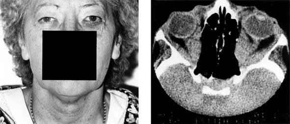
(Figure 15-22.) The degree of exophthalmos may be extremely variable. Measurements using the Hertel or Krahn exophthalmometer range from minimal (20 mm) to excessive (28 mm or more). The condition is usually asymmetric and may be unilateral, and it is important clinically to assess the resistance to manual retropulsion of the globe. The increase in orbital contents that produces the exophthalmos is largely due to an increase in the bulk of the ocular muscles. Visualization of the ocular muscles by CT scan (Figure 15-22) can differentiate exophthalmos from an orbital tumor. In some cases, thickening of the ocular muscles may be restricted to certain muscles only (eg, medial or inferior rectus muscles).

Figure 15-22: Thyroid ophthalmopathy. Left: Proptosis, visual loss, and ophthalmoplegia occurred in this elderly woman with a history of thyroid disease. Right: CT scans showed gross thickening of the ocular muscles, particularly in relation to the orbital apex. The increased intraorbital pressure is producing convexity of the medial orbital wall.
B. Ophthalmoplegia:
This is seen more commonly in ophthalmic Graves' disease, which usually affects older people and may be grossly asymmetric. Limitation of elevation is the most frequent finding, and this is mainly due to adhesions between the inferior rectus and inferior oblique muscles. Confirmation may be gained by measuring the intraocular pressure on elevation, when a substantial increase in the intraocular pressure suggests tethering. Often there is mild limitation of ocular movements in all positions of gaze. Patients complain of diplopia, which may be relieved by corticosteroid treatment, may spontaneously return to normal, or, if it remains static for 6-12 months, can frequently be relieved by operation on one or more extraocular muscles.
C. Retinal and Optic Nerve Changes:
Compression of the globe by the orbital contents may produce elevation of the intraocular pressure and retinal or choroidal striae. The optic disk may become swollen and progress to visual loss from optic atrophy. Optic neuropathy associated with Graves' disease occasionally occurs as a result of compression and ischemia of the optic nerve as it traverses the tense orbit, particularly at the orbital apex.
D. Corneal Changes:
In some patients, a superior limbic keratoconjunctivitis may be seen, though this is not specific for thyroid disease. In severe exophthalmos, corneal exposure and ulceration may occur.
Pathogenesis of the Ocular Signs
The main feature is gross distention of the ocular muscles due to the deposition of mucopolysaccharides. The mucopolysaccharides are strongly hygroscopic, which accounts for the increased water content of the orbits.
The pathogenesis of Graves' disease remains unknown, though an immunologic disorder involving both cellular and humoral elements has been implicated. Long-acting thyroid stimulator (LATS) is unlikely to be of significance in humans, because it is not always found in patients with ocular signs. There has, however, been good correlation between hyperthyroidism and human-specific thyroid stimulator, previously known as LATS protector, although this correlation is not seen in patients with Graves' disease. Thyroid autoantibodies against thyroglobulin and the microsome fraction of thyroid cells are frequently found in Hashimoto's disease and less often in Graves' disease. There are now thought to be two pathogenetic components to Graves' disease: (1) immune complexes of thyroglobulin-antithyroglobulin bind to extraocular muscles and produce a myositis; and (2) exophthalmos-producing substance acts with ophthalmic immunoglobulins to displace thyroid-stimulating hormones from the retro-orbital membranes, which results in the increase of retro-orbital fat.
Treatment
A. Medical Treatment:
Medical treatment includes adequate control of the hyperthyroidism as a primary measure. However, thyroid ophthalmopathy may occur in the euthyroid or hypothyroid states. Severe cases with visual loss, disk edema, or corneal ulceration merit urgent medical treatment with corticosteroids in high doses (eg, prednisolone, 100 mg); low doses are ineffective. Plasmapheresis is occasionally used with good results in the treatment of refractory cases, but full immunosuppression must follow plasmapheresis to prevent rebound increase of immunoglobulins and recurrence of disease. Immunosuppressive agents (eg, azathioprine) may play a supportive role and allow a lower maintenance dose of corticosteroids. Orbital radiotherapy may be useful to avoid operation or as a sequel to surgical decompression.
B. Surgical Treatment:
Decompression of the orbit may be performed by removing the medial and inferior walls via an ethmoidal approach or by endoscopic techniques. Decompression of the orbital apex is essential for a successful outcome.
2. HYPOTHYROIDISM (Myxedema)
Significant ocular signs are not common in myxedema, though the signs of thyroid ophthalmopathy may be seen. Hyperthyroid patients who subsequently become hypothyroid are at greater risk of ophthalmic involvement.
HYPOPARATHYROIDISM
Occasionally at thyroidectomy, the parathyroid glands are removed inadvertently, causing hypo-parathyroidism. Spontaneous cases of hypopara- thyroidism, though rare, should be suspected in young patients with cataracts. The blood calcium decreases, and serum phosphates are increased. Tetany may ensue and can be severe enough to cause generalized convulsions. The ocular manifestations consist of blepharospasm and twitching eyelids. Small, discrete, punctate opacities of the lens cortex develop that may eventually require lens extraction.
Treatment with calcium salts, calciferol, and dihydrotachysterol usually prevents further development of lens opacities, but any that have occurred prior to treatment remain.
Page 4 of 17
10.1036/1535-8860.ch15