

|
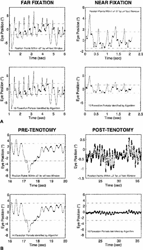
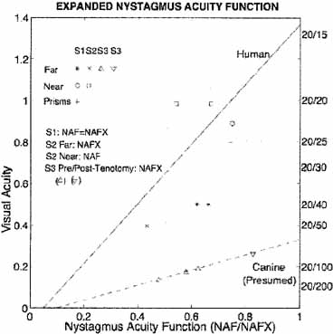
| Fig. 11 (A) The expanded nystagmus acuity function (NAFX) output for a subject with achiasma during far and near fixation, demonstrating improvement at near by the smaller foveation window and lower variation of foveation position. (B) The NAFX for an achiasmatic Belgian sheepdog demonstrating dramatic postoperative improvement by the relative sizes of the nystagmus and the ±3° centralis window. (C) NAFX versus visual acuity for both humans and canines, demonstrating improvement at near fixation (humans S1 and S2), at far with base-out prisms (S1), and post-tenotomy (canine S3). In this and Figures 12, 13, and 18B, the dot-dashed lines indicate the extent of the foveal window (humans) or area centralis (canines). In this and Figure 12, the thickened areas identify foveation (human) or centralis (canine) periods. |