|
|
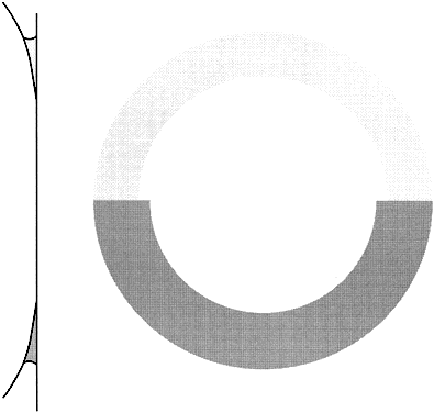
| Fig. 4. Effect of fluorescein concentration on Goldmann applanation tonometry. The circle of applanation is outlined by a meniscus of fluorescein-stained tear film. If the tear film has too low a concentration of dye (upper half of diagram), it is difficult to define the thin inner edge of the meniscus and the examiner may perceive an enlarged circle—thus underestimating IOP. (Moses RA: Fluorescein in applanation tonometry. Am J Ophthalmol 49:1149, 1960.) The optimal fluorescein concentrations have been determined,25 and the most reliable way to obtain accurate readings is through use of a pre-mixed anesthetic/fluorescein drop. (Grant WM: Fluorescein for applanation tonometry. More convenient and uniform application. Am J Ophthalmol 55:1252, 1963; Quickert MH: A fluorescein-anesthetic solution for applanation tonometry. Arch Ophthalmol 77:734, 1967.) Though a “ring of contact” may be seen with anesthetic alone, measurements made without fluorescein underestimate IOP. (Hoffer KJ: Applanation tonometry without fluorescein. Correspondence. Am J Ophthalmol 88:798, 1979; Roper DL: Applanation tonometry with and without fluorescein. Am J Ophthalmol 90:668, 1980.) |