Chapter 10
Retinal Arterial Macroaneurysms
NEAL H. ATEBARA
Main Menu   Table Of Contents

Search

CLINICAL PRESENTATION
FUNDUSCOPIC APPEARANCE
FLUORESCEIN ANGIOGRAPHIC APPEARANCE
PATHOPHYSIOLOGY
DIFFERENTIAL DIAGNOSIS
MANAGEMENT
CONCLUSIONS
REFERENCES

Retinal arterial macroaneurysms, first systematically studied by Robertson in 1973, are fusiform or saccular dilations of the retinal arteries, usually associated with systemic hypertension and atherosclerotic vascular disease. They may follow a benign clinical course, but if they leak fluid or hemorrhage, they can result in significant loss of central visual acuity. Management options depend on the nature of the exudative or hemorrhagic complications of the macroaneurysm. This chapter reviews the clinical presentation, pathophysiology, and management of retinal arteriolar macroaneurysms.
Back to Top
CLINICAL PRESENTATION
The typical patient who presents with a retinal arterial macroaneurysm is older than 60 year of age with a history of systemic hypertension and arteriosclerotic vascular disease. Women are affected three times more often than men.1,2 There may be a sudden loss of central or peripheral vision due to hemorrhage, a gradual blur in central visual acuity due to cystoid macular edema, or the macroaneurysm may be found ophthalmoscopically on a routine examination if there is no exudation or hemorrhage that encroaches upon the fovea. The incidence of retinal arterial macroaneurysms increases with age.
Back to Top
FUNDUSCOPIC APPEARANCE
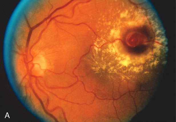
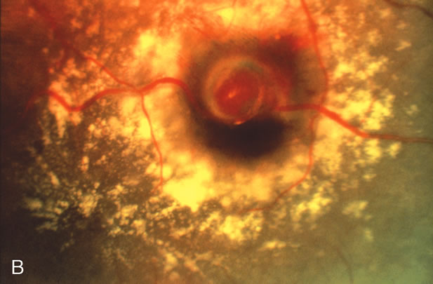
The hallmark for diagnosis of the retinal arterial macroaneurysm is visualization of the macroaneurysm itself (Fig. 1). Because of their relatively large size, macroaneurysms are not usually confused with the microaneurysms of diabetic retinopathy or branch retinal vein occlusion. Microaneurysms typically have a diameter of only 50 to 75 microns and arise from the deeper retinal capillaries; macroaneurysms on the other hand range in size from 100 to more than 500 microns in diameter and arise from the larger retinal arterioles.

Fig. 1. A. Color fundus photograph of a retinal arterial macroaneurysm 750 microns in diameter associated with circinate hard exudates and macular edema involving the fovea. B. High magnification photograph of the same macroaneurysm.

Retinal arterial macroaneurysms usually arise within the first three orders of bifurcation from the optic disc (Fig. 2), often at a point of arteriovenus crossing. Sometimes they occur directly on the optic nerve head3 or on a cilioretinal artery.4 About 20% of affected eyes have multiple aneurysms, and about 10% demonstrate macroaneurysms in both eyes.

Fig. 2. A. Color fundus photograph of a 200-micron macroaneurysm that arises from a retinal artery near the optic disc with thin subretinal hemorrhage that does not threaten the fovea. B. The midphase fluorescein angiogram reveals blockage of choroidal but not retinal vascular hyperfluorescence. C. The late-phase angiogram demonstrates staining of the macroaneurysm.[pa[et[ol0]

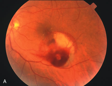
Retinal arterial macroaneurysms often produce retinal edema, circinate hard exudates, and hemorrhage (Fig. 3) into multiple layers of the retina: beneath the retinal pigment epithelium, beneath the retina, within the retina, beneath the internal limiting membrane, between the retina and the posterior hyaloid, and within the vitreous cavity. When the macroaneurysm is obscured by overlying blood, its diagnosis can be challenging.

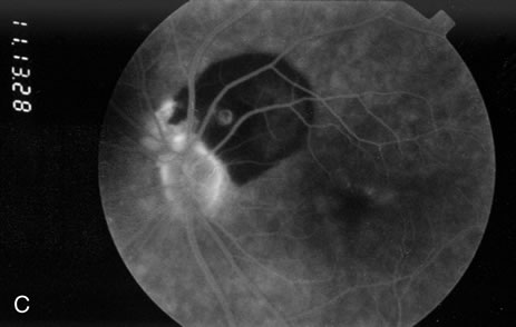
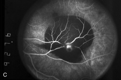
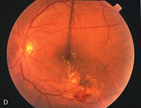
Fig. 3. A. Color fundus photograph of a macroaneurysm along the inferotemporal arcade with surrounding intraretinal and subretinal hemorrhage. There is retinal edema, hard exudates, and thin subretinal hemorrhage extending into the macula. B. Midphase fluorescein angiography reveals hemorrhage that extends just into the foveal avascular zone, threatening foveal vision. C. Late-phase angiogram reveals staining of the macroaneurysm. D. Color fundus photograph of the same macroaneurysm 6 weeks after laser photocoagulation, demonstrating resolution of the edema and hemorrhage.

Back to Top
FLUORESCEIN ANGIOGRAPHIC APPEARANCE
Because of rapid blood flow through retinal arterial macroaneurysms, they typically fill quickly in the early phase of the fluorescein angiogram. In the mid- and late-phases of the angiogram, the macroaneurysm tends to leak to varying degrees, depending on its perfusion and endothelial integrity (Fig. 3C). If there is subretinal fluid surrounding the aneurysm, there may be pooling of dye into the subretinal space. In some cases, a characteristic Z-shaped kink may be identified at the site of the aneurysm.

Perfusion abnormalities caused by the macroaneurysm cause changes in the surrounding retinal vasculature as well, and this is best visualized on fluorescein angiography (Fig. 3B). Ischemia from stagnation of blood flow results in capillary telangiectasis, microaneurysm formation, and capillary nonperfusion.

Some or all of these angiographic features may be obscured by blood or lipid exudation from the aneurysm. If blood collects in front of the retinal vessels (vitreous hemorrhage, preretinal hemorrhage, subinternal limiting membrane hemorrhage, or intraretinal hemorrhage), then blockage of hyperfluorescence may be partial or complete. Subretinal blood blocks hyperfluorescence from the choroid but not the retinal circulation, resulting in dramatically distinct retinal vessels against a dark background (Fig. 3B,C). Lipid exudation and retinal edema may cause partial blockage of hyperfluorescence.

Back to Top
PATHOPHYSIOLOGY
Chronic systemic hypertension is often seen in patients with retinal arterial macroaneurysms, and it is believed this condition has an important role in its pathogenesis. It is hypothesized that chronic vascular wall damage secondary to systemic hypertension and arteriosclerosis leads to focal dilation of the vascular wall, and this leads to macroaneurysm formation.5 There is no adventitia where arteries and veins cross, and this is a point at particular risk for aneurysm formation.

There is also evidence that retinal arterial macroaneurysms may be caused by focal damage to the blood vessel wall from conditions such as embolus,6 toxoplasmosis infection,7 or branch retinal vein occlusion.8

Back to Top
DIFFERENTIAL DIAGNOSIS
The diagnosis of retinal arterial macroaneurysm is based on ophthalmoscopic identification of the macroaneurysm. The macroaneurysm is easily distinguished from microaneurysms—such as those seen in diabetes mellitus, branch retinal vein occlusion, sickle cell disease, and radiation retinopathy—which are smaller in size.

Retinal capillary hemangiomas (von Hippel's angiomatosis) are rare tumors seen in the peripheral retina or on the optic disc. When fully developed, they are usually much larger than macroaneurysms, often larger than 5 mm in diameter. They often result in significant subretinal fluid exudation and cystoid macular edema. They bleed less frequently than macroaneurysms. Peripheral retinal capillary hemangiomas usually arise near the ora serrata, whereas macroaneurysms usually arise within three orders of bifurcation from the optic disc. Retinal capillary hemangiomas of the optic disc grow to sizes much larger than macroaneurysms and almost always result in poor visual acuities.

Hemorrhage can obscure funduscopic visualization and make the differential diagnosis of a macroaneurysm more challenging.

Back to Top
MANAGEMENT
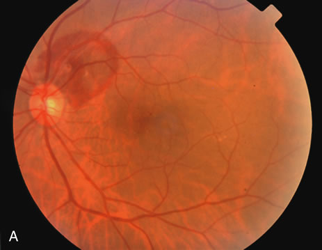
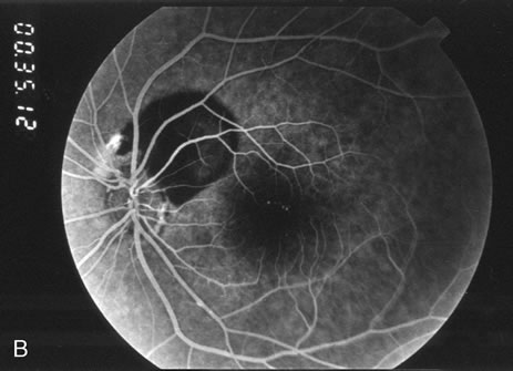
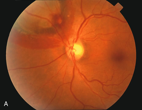
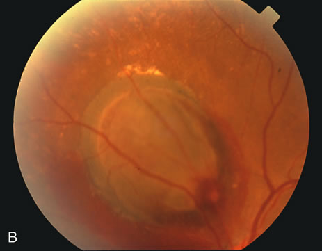
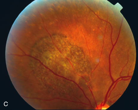
Most retinal arterial macroaneurysms do not require any treatment. If the macroaneurysm produces no exudation or hemorrhage, in most cases, they may be simply observed (Fig. 4). Some macroaneurysms pulsate due to the arterial pulse pressure, and this feature may represent an increased risk of bleeding in the near future.

Fig. 4. A. Color fundus photograph of a left eye with a 300-micron macroaneurysm along the superonasal arcade with a localized area of thick subretinal hemorrhage. B. The macroaneurysm and subretinal hemorrhage were not treated. C. After several months, the subretinal hemorrhage resorbed with visual acuity of 20/20.

If exudation or hemorrhage threaten or affect central vision, several management options exist depending on the clinical situation; laser photocoagulation, Nd-YAG laser photodisruption, pneumatic displacement of hemorrhage, and vitrectomy with surgical evacuation of blood have all been employed in the treatment of retinal arterial macroaneurysms.

Laser photocoagulation (Fig. 3D) directed to the area surrounding the macroaneurysm or to the macroaneurysm itself is indicated when subretinal blood begins to threaten central vision or when exudation threatens or affects central vision.9–11 Photocoagulation causes the macroaneurysm to thrombose and sclerose. This decreases the risk of another hemorrhage and usually reduces the amount of exudation. The main risk of this procedure is that it may itself cause thrombosis of the adjacent branch retinal artery, possibly resulting in a central or peripheral scotoma depending on its location.12 If the macroaneurysm is sufficiently thin walled, photocoagulation may even induce bleeding in rare cases.

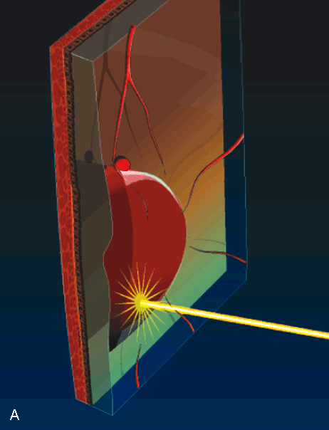
Nd-YAG laser photodisruption may be employed in special cases in which thick, fresh blood is trapped beneath the internal limiting membrane13 or behind the posterior hyaloid.14 Blood in these anatomic locations does not itself cause damage to the retina, but it may limit central vision, and it may prevent the clinician from identifying underlying retinal pathology. Photodisruption results in a localized concussive effect that breaks open the posterior hyaloid or internal limiting membrane. If the trapped blood has not yet solidified, it may then be allowed to “spill out” through the opening into the vitreous cavity (Fig. 5). Risks of this technique include damage to the underlying retina if the laser treatment is applied too close to the retina or if the blood is not thick enough to absorb the concussive effect.

Fig. 5. A. Illustration of a macroaneurysm with thick sub-internal limiting membrane (ILM) hemorrhage Because the hemorrhage obscures the macula, the patient's visual acuity is poor and it is not possible to determine clinically if the hemorrhage extends beneath the macula. Nd-YAG laser photodisruption can break through the ILM. B. If the blood has not yet solidified, it may drain through the laser opening and improve visual acuity and visualization of the macula.

Pneumatic displacement of hemorrhage has been successfully employed in select cases of submacular hemorrhage from a macroaneurysm (Fig. 6). Subretinal blood results in irreversible damage to photoreceptors within 7 days.15–17 Many surgeons attempt to remove or at least displace this blood within 2 weeks and preferably within 7 days in an attempt to preserve central vision. A proposed alternative to vitrectomy with surgical evacuation of the blood is pneumatic displacement, in which a bubble of perfluorocarbon gas is injected into the vitreous cavity. The patient's head is then positioned such that the bubble's buoyant force squeezes the subretinal blood out of the macula and into the inferior fundus.18 Some surgeons precede the gas bubble with an intravitreal injection of tissue plasminogen activator (TPA) in order to lyse the blood clot; whether or not the TPA reaches the blood clot in sufficient quantities to cause an appreciable lysing effect is controversial.19 There is histological evidence that this technique in some cases causes shearing of the photoreceptor outer segments during the pneumatic displacement procedure.20

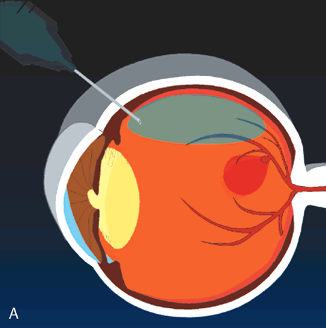
Fig. 6. Fresh submacular blood displaced from the macula with an intravitreal gas bubble injection. A. Air or gas is injected into the vitreous cavity. B. The patient is then placed in the face-down position such that the bubble slowly pushes the blood into the inferior fundus. The blood remains in the subretinal space but is displaced from the macula. Some surgeons precede the gas bubble with an intravitreal injection of tissue plasminogen activator in order to help lyse the blood clot.

Vitrectomy with surgical evacuation of submacular hemorrhage may be indicated when there is a reasonable chance of recovery of some degree of central vision (Fig. 7).21–26 In the eye in which blood that has been under the macula for more than 2 weeks, there is little possibility of visual recovery because of blood toxicity to the photoreceptors. Many authorities consider surgical evacuation of submacular blood when the blood has been present under the macula for 2 weeks or less, preferably less than 7 days. The technique involves a pars plana vitrectomy, followed by a small perforation near the edge of the blood to gain access to the subretinal space. TPA is usually employed to help lyse the blood, thus facilitating its removal with minimal damage to the photoreceptors. The blood is removed using a subretinal cannula; some models feature a double-barreled shaft that allows the injection of fluid into the subretinal space through one barrel and the removal of blood through the other barrel. Some authors note that the use of a bubble of perfluorocarbon liquid on the surface of macula helps express the blood from beneath the macula.

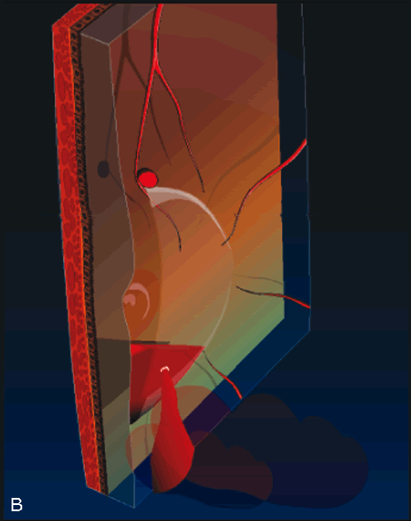
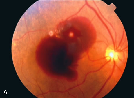
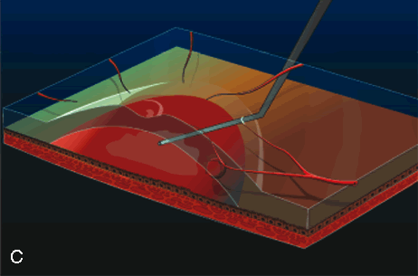
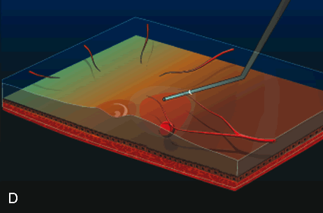
Fig. 7. A. Color fundus photograph of a macroaneurysm causing subretinal and sub-ILM hemorrhage involving the macula. B. Illustration of the spatial relationship between the submacular blood and sub-ILM blood. C. During vitrectomy surgery, a subretinal cannula is used to inject tissue plasminogen activator into the blood clot. D. After the blood is allowed to lyse over the course of 40 minutes, it is removed using a subretinal cannula.

Back to Top
CONCLUSIONS
Retinal arterial macroaneurysms are relatively large outpouchings of a retinal artery associated with systemic hypertension and atherosclerosis. They are often asymptomatic and require no treatment. However, if they produce exudation or hemorrhage that affects or threatens central visual acuity, treatment options include laser photocoagulation, Nd-YAG laser photodisruption, pneumatic displacement of subretinal hemorrhage, and vitrectomy with surgical evacuation of subretinal hemorrhage.
Back to Top
REFERENCES

1. Robertson DM: Macroaneurysms of the retinal arteries. Trans Am Acad Ophthalmol Otolaryngol 77:OP55, 1973.

2. Palestine AG, Robertson DM, Goldstein BG: Macroaneurysms of the retinal arteries. Am J Ophthalmol 93:164, 1982.

3. Brown GC, Weinstock F: Arterial macroaneurysm on the optic disc presenting as a mass lesion. Ann Ophthalmol 17:519, 1985.

4. Giuffre G, Montalto FP, Amodei G: Development of an isolated retinal macroaneurysm of the cilioretinal artery. Br J Ophthalmol 71:445, 1987.

5. Lavin MJ, Marsh RJ, Richardson J: Retinal macroaneurysm: Natural history and guidelines for treatment. Br J Ophthalmol 71:817, 1987.

6. Wiznia RA: Development of a retinal artery macroaneurysm at the site of a previously detected retinal artery embolus. Am J Ophthalmol 114:642, 1982.

7. Ohga H, Egi K, Katayama T, et al: A case of retinal macroaneurysm with congenital ocular toxoplasmosis. Folia Ophthalmol Jpn 41:898, 1990.

8. Panton RW, Goldberg MF, Farber MD: Retinal arterial macroaneurysms: Risk factors and natural history. Br J Ophthalmol 74:595, 1990.

9. Lewis RA, Norton MH, Wise GN: Acquired arterial macroaneurysms of the retina. Br J Ophthalmol 60:21, 1976.

10. Abdel-Khalek MN, Richardson J: Retinal macroaneurysm: Natural history and guidelines for treatment. Br J Ophthalmol 70:2, 1986.

11. Palestine AG, Robertson DM, Goldstein BG: Macroaneurysms of the retinal arteries. Am J Ophthalmol 93:164, 1982.

12. Brown DM, Sobol WM, Folk JC, et al: Retinal arteriolar macroaneurysms: Long term visual outcome. Br J Ophthalmol 78:534, 1994.

13. Tassignon MJ, Stempels N, Van Mulders L: Retrohyaloid premacular hemorrhage treated by q-switched Nd-YAG laser. Graefes Arch Clin Exp Ophthalmol 227:440, 1989.

14. Raymond LA: Neodynium:YAG laser treatment for hemorrhages under the internal limiting membrane and posterior hyaloid face in the macula. Ophthalmology 102:406, 1995.

15. Johnson MW, Olsen KR, Hernandez E: Tissue plasminogen activator treatment of experimental subretinal hemorrhage. Retina 11:250, 1991.

16. Sanders D, Peyman GA, Fishman G, et al: The toxicity of whole blood and hemoglobin. Graefes Arch Clin Exp Ophthalmol 197:255, 1975.

17. Toth CA, Morse LS, Hjelmeland LM, el al: Fibrin directs early retinal damage after experimental subretinal hemorrhage. Arch Ophthalmol 109:723, 1991.

18. Ohji M, Saito Y, Lewis JM, et al: Pneumatic displacement of subretinal hemorrhage without tissue plasminogen activator. Arch Ophthalmol 116:1326, 1998.

19. Hassan AS, Johnson MW, Schneiderman TF, et al: Management of submacular hemorrhage with intravitreal tissue plasminogen activator injection and pneumatic displacement. Ophthalmology 106:1900, 1999.

20. Lewis H: Intraoperative fibrinolysis of submacular hemorrhage with tissue plasminogen activator and surgical drainage. Am J Ophthalmol 118:559, 1994.

21. Hanscom TA, Diddie KR: Early surgical drainage of macular subretinal hemorrhage. Arch Ophthalmol 105:1722, 1987.

22. Peyman GA, Nelson NC, Alturki W, et al: Tissue plasminogen activating factor assisted removal of subretinal hemorrhage. Ophthalmic Surg 22:575, 1991.

23. Moriarty AP, McAllister IL, Constable IJ: Initial clinical experience with tissue plasminogen activator (tPA) assisted removal of submacular hemorrhage. Eye 9:582, 1995.

24. Lim JI, Drews-Botsch C, Sternberg P, et al: Submacular hemorrhage removal. Ophthalmology 102:1393, 1995.

25. Ibanez HE, Williams DF, Thomas MA, et al: Surgical management of subretinal hemorrhage: A series of 47 consecutive cases. Arch Ophthalmol 113:62, 1995.

26. Brent BD, Gonce M, Diamond JG: Pars plana vitrectomy for complicated of retinal arterial macroaneurysms—a case series. Ophthalmic Surg 24:534, 1993.

Back to Top