Chapter 17
Superficial Keratopathy
DOUGLAS J. COSTER
Main Menu   Table Of Contents

Search

BIOLOGIC CONCEPTS RELEVANTTO THE OCULAR SURFACE
CLINICAL EXAMINATION
PATTERNS OF MORPHOLOGIC CHANGE IN THE SUPERFICIAL CORNEA
DENDRITIC KERATOPATHY
PLAQUE KERATOPATHY
LINEAR KERATOPATHY
VORTEX KERATOPATHY
PUNCTATE KERATOPATHY
MORPHOLOGIC CLASSIFICATIONOF SUPERFICIAL SPOTTY LESIONS
ASSOCIATED OCULAR PHENOMENA
SUMMARY
REFERENCES

Corneal surface abnormalities are common. They are important because they can affect vision and are often uncomfortable. The anterior corneal surface, and the tear film it supports, is the most important element in the optical system of the eye. Minor disruptions to epithelium can be painful; it is richly innervated with pain fibers.

The cornea has two distinct functions: first, as an optical element of the eye, the cornea must have a clear, intact ultrastructure for normal vision; second, as a defensive element to protect the internal milieu of the eye. As a protective element, the cornea is equipped to deal with the various challenges facing an exteriorized mucous membrane. It is richly innervated, adjacent to the highly vascularized limbus from which inflammatory cells are recruited, bathed in a constantly moving tear film, and capable of regenerating epithelium rapidly from the limbal stem cells.

The superficial cornea is prone to disease as a result of direct external challenge such as may occur with local infectious agents, from systemic diseases that influence the cornea via the circulation and/or the tear film, such as calcium or immunoglobulin deposition, or from abnormalities of growth and differentiation of the corneal epithelium, such as dysplasia and neoplastic conditions. For some conditions, such as Thygeson's keratopathy, the etiology is unknown.

It is always important when considering patients with diseases of the superficial cornea to be aware that what is seen in the cornea may be the manifestation of a process affecting the other elements of the outer eye, or even the body as a whole. To understand these conditions, it is helpful to keep in mind two concepts: first, that the outer eye is best considered as a coherent ecosystem, and second, that the corneal epithelium is generated from limbal stem cells.

Back to Top
BIOLOGIC CONCEPTS RELEVANTTO THE OCULAR SURFACE

ECOSYSTEM OF THE OUTER EYE

All elements of the outer eye should be considered as part of an interactive ecosystem rather than as isolated elements. Components of this interactive ecosystem are the tear film, corneal epithelium, corneal stroma, limbus, conjunctiva, and lid margins. This environment is also influenced by more remote structures such as the lacrimal drainage system and the skin of the face. No evaluation of the superficial cornea is complete without careful appraisal of all the elements of the ecosystem of the outer eye.

ROLE OF LIMBAL STEM CELLS

Superficial keratopathy, in its various forms, involves the corneal epithelium. The corneal epithelium is crucial to the optical function of the corneal surface and the protective role of the cornea. It is the bulwark of resistance against microbial infection and is necessary for the integrity of the underlying corneal stroma. Unresolved corneal ulceration may result in the loss of the eye.

Under normal circumstances, the corneal epithelium has considerable capacity for replication and repair. This replicative capacity resides in the limbal stem cells. Disturbances of the limbal stem cell population can have a marked effect on the growth and differentiation of the corneal epithelium, with obvious clinical manifestations. In its most severe form, stem cell insufficiency will result in conjunctivalization of the corneal surface. In slightly less severe form, the corneal epithelium will be seen to be abnormal with patchy opacity and irregularity and underlying vascularization. Corneal epithelial cells migrate centripetally from the limbus and tangentially around the central cornea. Cells that are marked in some way, such as those bearing pigment in pigmented races or those opacified by abnormal depositions, as in Fabry's disease, clearly demonstrate this patternof movement. This is the basis of vortex kera-topathy.

Back to Top
CLINICAL EXAMINATION
All ophthalmic examinations must begin with a general inspection of the patient, paying particular at-tention to the globe and surrounding adnexal structures. If a broad general inspection is not routinely performed, potentially important diagnostic clues, such as joint abnormalities, telangiectases of the facial skin, seborrhea, eyelid vesicles, and even the pattern of conjunctival injection, can be overlooked. General examination is particularly important when dealing with corneal epithelial keratopathy, which often is the manifestation of a more widespread disorder.

A general external examination is followed by slit-lamp biomicroscopy, which must be methodical and exhaustive. This should begin with observation of the lid margins and the tarsal and bulbar conjunctiva. A suggested approach to corneal examination includes initial inspection under moderate power using the technique of sclerotic scatter. This can highlight minimal changes that might otherwise be missed. Epithelial abnormalities are frequently best identified with broad-beam tangential illumination. Once an epithelial abnormality has been identified, it can be more carefully characterized under higher magnification, using techniques of direct focal illumination, retroillumination, and specular reflection. Some forms of superficial keratopathy can be seen with the naked eye. These lesions are described as “coarse” lesions to distinguish them from “fine” lesions, which can be seen only with magnification.

Even the most skilled biomicroscopist will find it necessary to augment the examination with the use of special staining techniques. Rose bengal, which has an affinity for devitalized corneal and conjunctival epithelial cells as well as for keratin and mucus, is particularly valuable. It is most useful in the diagnosis of keratoconjunctivitis sicca (KCS), and the pattern of staining can be helpful in differentiating various other forms of epithelial disease. Topical fluorescein is no substitute for rose bengal but can complement it because its affinity is different. Fluorescein does not stain epithelial cells but penetrates epithelial defects and can pool or diffuse through intercellular spaces. One drop of 1% rose bengal placed on the upper bulbar conjunctiva while the patient looks down is generally sufficient. Norn has shown that although a 10% solution identified more subtle defects, it could cause artifactual staining and provoke more discomfort on instillation.1

Once a pattern suggestive of superficial corneal pathology is recognized, the examiner may wish to pursue a more pointed history and external examination, seeking additional clues to support a specific pathologic process.

Back to Top
PATTERNS OF MORPHOLOGIC CHANGE IN THE SUPERFICIAL CORNEA
The diagnostic process involves recognizing a typical clinical pattern that is suggestive of particular diagnosis, and then looking for clinical features to support a specific diagnosis and, if necessary, carrying out laboratory investigations to confirm this. Sometimes more than one visit is required to observe the evolution of the process with time, and perhaps in response to treatment, so that a firm diagnosis can be made. For example, a pattern of superficial corneal spots may suggest a viral cause. The additional presence of an acute follicular conjunctivitis with subconjunctival hemorrhages would encourage a clinical diagnosis of adenoviral keratoconjunctivitis. To confirm this would require identification of the virus.

There is another requirement in formulating a diagnostic process: it is necessary to exclude sight-threatening treatable conditions as a priority rather than accept a diagnosis on the grounds of greatest likelihood. In other words, serious diagnoses demand exclusion rather than simply accepting the most likely causes. For example, superficial keratopathy associated with minimal corneal scarring and corneal anesthesia demands exclusion of a neurologic disorder, such as acoustic neuroma, rather than acceptance of the most likely explanation, which is herpetic keratitis.

As in all forms of clinical medicine, the first step in the diagnostic process is to recognize common patterns of disease expression.

The superficial cornea, consisting of Bowman's layer, the epithelial basement membrane, the epithelium, and the overlying tear film, responds to various disease processes in a limited number of ways. In certain conditions, this response produces a very characteristic pattern of disease, such as the band of rose bengal staining of the cornea and conjunctiva in the palpebral fissure zone caused by KCS. Alternatively, similar patterns of epithelial change can be caused by a wide range of insults, as in the case of diffuse punctate epithelial erosions.

Although corneal ulceration is a commonly encountered pathologic process affecting the superficial cornea, a complete discussion of it is beyond the scope of this chapter; this presentation is confined to the nonulcerative forms of superficial keratopathy. The most important of these are dendriform, plaque, linear, vortex, and superficial punctate keratopathies. Despite the diversity of clinical appearances, these patterns of corneal change share important features. One pattern can be produced by a wide range of pathologic processes, and conversely one etiologic agent can produce more than one pattern of corneal response.

Back to Top
DENDRITIC KERATOPATHY
Corneal epithelial changes characterized by a branching pattern are referred to as dendriform or dendritic keratopathy. The foremost entity manifesting a branching pattern is herpes simplex dendritic keratitis. Few other conditions present with this pattern, but they should be considered in the differential diagnosis of branching epithelial keratopathy. They include healing epithelial abrasions, dendritic plaques in herpes zoster ophthalmicus, and the rare condition of dendriform keratopathy in tyrosinemia (Fig. 1).

Fig. 1. High-magnification slit-lamp photographs of patterns of superficial keratopathy. A. Dendritic pattern of epitheliopathy without ulceration produced by herpes simplex virus infection. B. Mucous plaques arranged in a branching pattern in a patient with herpes zoster ophthalmicus. C. Branching pattern produced by a healing epithelial defect. D. Corneal lesions of tyrosinemia.

Herpes simplex dendritic keratitis is a finely branching epithelial lesion that is sharply marginated and frequently associated with terminal end bulbs (see Fig. 1A). Dendritic keratitis may not be truly ulcerative but consists of swollen, infected cells in a dendritic pattern. If the cytopathic process is overwhelming, epithelial cells are shed and a true ulcer is formed.2 The ulcer base stains strikingly with fluorescein, and the marginated, infected cells stain vividly with rose bengal.3,4

Dendriform keratopathy associated with herpes zoster ophthalmicus consists of raised mucus plaques, which usually appear months after the clinical onset and are rarely encountered early in the disease process. They are not difficult to recognize given the patient's history and clinical appearance. The lesions are coarse (i.e., broad rather than fine), mildly opaque, raised plaques that stain strikingly with rose bengal and poorly with fluorescein5,6 (see Fig. 1B). Fluorescein usually pools about the plaque margin. Débridement results in little damage to the underlying epithelium. It has been assumed that lesions are probably not associated with active viral infection. However, in a recent study in AIDS patients, varicella zoster virus was identified by direct culture of the corneal lesions.7 Although it is exceptionally rare to find herpes simplex and varicella zoster coexisting, they have been encountered together.8

Healing epithelial abrasions can result in a linear or branching pattern along points of epithelial closure (see Fig. 1C). This should not usually be confused with dendritic keratitis because of the history and associated findings. Sliding epithelial cells result in punctate erosions remote from the linear pattern; these stain well with fluorescein or rose bengal, and the staining, distant from the epithelial defect, is helpful in differentiating between the conditions.

Finally, dendritic epithelial figures can be encountered in tyrosinemia type 2, also called the Richner-Hanhart syndrome.9 Primary features are hyperkeratotic skin lesions of the palms, soles, and elbows associated with keratitis in early life. This rare condition is inherited as an autosomal recessive trait with variable phenotypic expression.10 Associated mental retardation is common. Corneal manifestations vary from finely branched intraepithelial opacities resembling small herpetic dendrites9 to severe keratitis with stellate opacification and ulceration.11 The finer intraepithelial lesions do not reach the corneal surface and thus can be distinguished by the absence of staining. The more severe forms appear during the first year of life, with bilateral superficial corneal opacification with radiating opacities forming a pseudoherpetic pattern (see Fig. 1D). Although it is a rare condition, early diagnosis is important, because a striking clinical response can be obtained through dietary therapy.

Back to Top
PLAQUE KERATOPATHY
Adherence of mucus to the corneal epithelium can produce marked symptoms, a striking clinical appearance, and severe sequelae. Two predisposing factors lead to mucus plaque formation: altered corneal epithelium and a relative excess of mucus in the tear film.

Abnormal patches of epithelium function as mucus receptors.6 Foci of abnormal epithelium may result from a number of mechanisms, such as tear film abnormalities, denervation as occurs in herpes zoster ophthalmicus, superficial keratitis due to injury, or inflammation resulting from many causes. A relative excess of mucus can result from aqueous tear deficiency as occurs in KCS,12 altered glycoprotein secretion by goblet cells resulting in increased viscosity of mucus,13 or excess secretion of mucus as occurs in allergic eye disease.

The mucus-epithelium bond can be firm and secure. Filaments result from tenacious adherence between mucus and epithelial cells with subsequent accumulation of epithelial cells, protein, and lipid (Fig. 2A). Filament formation originates from small mucus plaques and may precede ulceration, a serious consequence of plaque formation. Friction between filaments and the upper eyelid can result in small localized areas of epithelial stripping and intense discomfort.

Fig. 2. A. High-magnification photograph of a filament showing a mucous filament based on a patch of abnormal epithelium. B. Slit-lamp photograph of mucous plaques on the corneal surface in a patient with herpes zoster ophthalmicus.

Plaques vary in size and shape. They may be small and round, as occurs in dry eye states,14 or large and complex in shape, as in herpes zoster ophthalmicus (see Fig. 2B). When associated with aqueous tear deficiency, corneal mucus plaques can occur alone or in conjunction with filaments.2 When a more normal tear film is present, as in herpes zoster ophthalmicus with dendriform corneal plaques, associated filamentary keratitis is uncommon. Similarly, large plaques occurring in vernal keratoconjunctivitis are usually not associated with filamentary keratitis.15

The size, shape, and distribution of plaques and filaments are important considerations. As stated earlier, tear film deficiencies tend to produce numerous small round plaques. Large linear or dendriform lesions occur in herpes zoster ophthalmicus, and large solitary oval plaques occur in vernal keratoconjunctivitis.

Slit-lamp examination together with rose bengal and fluorescein staining reveals the extent and nature of these lesions as well as the general condition of the outer eye. Widespread changes invariably occur in the epithelium, and they are more evident when stained with rose bengal.16 Widespread punctate epithelial erosions are usual, indicating a disturbance of the epithelium-tear film complex. Where mucus adheres to the epithelium, intense staining is apparent.

The most common cause of mucus plaque keratopathy and filamentary keratitis is KCS. One fourth of 66 patients reported on by Fraunfelder and colleagues14 demonstrated corneal plaques at some stage during follow-up. Two thirds of these had Sjögren's syndrome. The plaques vary in size and shape, being light gray, slightly elevated, and located in the interpalpebral zone or lower two thirds of the corneal surface. Associated filaments are present in more than half of these cases.17 Abnormalities in the tear film, including mucus, epithelial debris, and decreased marginal tear film, together with interpalpebral rose bengal staining, are important ancillary signs. Associated meibomianitis or blepharitis is commonly present and may have a role in increasing mucus viscosity.14

Superior limbic keratoconjunctivitis (SLK) has mucus plaque changes and filaments as prominent features. This entity was first described in detail by Theodore in 196318 but was commented on earlier by Thygeson.19 The cardinal features include upper tarsal and bulbar conjunctival inflammation, superior limbic proliferation and dysplasia characterized by a thickened limbal epithelium and keratinization, rose bengal staining of the upper bulbar conjunctiva and superior cornea, and filamentary keratitis.20,21 Wright later added mucous discharge, swelling and redness of the upper eyelid, and an increased rate of blinking to these findings.22

The washing of the tear film across the cornea is produced by movement of the upper eyelid. If this process is disturbed by derangement of the posterior eyelid surface or an irregular superior bulbar conjunctiva, there can be a predisposition to epithelial keratinization abnormalities and subsequent plaque formation. This disturbance produces changes in the upper third of the cornea. When there is no obvious associated ocular anomaly, this process is described as SLK. When an obvious cause is present, such as recent glaucoma or cataract surgery,23 the process is simply called filamentary keratitis, even though the findings would fulfill the SLK criteria.

Sometimes SLK is encountered in association with hyperthyroidism,24,25 and many patients with dysthyroid eye disease show features of SLK.26 When the diagnosis of SLK is entertained, a methodical search for disturbances in the topography of the upper globe and lid should be made. If no rectifiable ocular anomalies are present, initial treatment should consist of tear substitutes, with the addition if necessary of a mucolytic agent such as acetylcysteine. Recalcitrant cases can be treated with soft contact lenses27 or conjunctival resection.28,29 Silver nitrate therapy (0.5%) to the upper tarsal conjunctiva may be beneficial in a few patients.

Filamentary keratitis occurs with numerous disorders other than KCS and SLK. Among these are recurrent erosion, postcataract extraction,23 postpenetrating keratoplasty,30 neurotrophic keratitis,31 herpetic keratitis, bullous keratopathy, prolonged patching (padding), and hereditary hemorrhagic telangiectasia.32 Whatever the cause, filament formation results from a derangement in the corneal epithelium and a relative excess of mucus. Here the excess mucus is usually secondary to chronic irritation, and the location of the filaments depends on the type of underlying corneal pathology. In these cases, treatment directed at correcting the underlying disorder involves artificial tear substitutes with or without acetylcysteine, as well as bandage soft contact lenses. Herpes zoster ophthalmicus produces changes in the corneal epithelium, probably as a consequence of denervation as well as alteration in tear secretion. The presentation and treatment of mucous plaque formation in this disease were discussed in the previous section on dendritic keratopathy.

Back to Top
LINEAR KERATOPATHY
Superficial corneal lines may be in the epithelium, basement membrane, or Bowman's layer and can occur in a number of conditions. Two other patters of superficial keratopathy with linear elements, dendritic and vortex keratopathy, are discussed separately in this section. In other instances, epithelial lines may be seen as part of a more generalized corneal disturbance or may occur when the epithelium is not otherwise diseased.

In addition to creating complex dendritic and vortex patterns, healing corneal abrasions may show a simple linear pattern at the forefront of epithelial migration. Such lines are easy to diagnose from the history and their rapid evolution.

Pigmented lines resulting from iron deposition in the basal epithelium are not unusual. Perhaps the most common and well recognized is the Hudson-Stähli line,33,34 a horizontal line at the junction of the middle and lower third of the cornea. These lines have been found in as many as 90% of randomly selected normal eyes in an eye bank.35

A similar line, curving around the front of a pterygium, is known as Stocker's line,36 and Ferry described curvilinear iron deposition at the base of a filtering bleb.37 Fleischer's ring either completely or partially surrounds the base of the cone in keratoconus,38 and, as with all such iron lines, it may be best appreciated with broad cobalt blue illumination. The mechanism of formation of such lines is not clear, although many hypotheses have been put forward. A review of such explanations makes it seem most likely that the iron is derived from the tear film and that it is deposited in corneal epithelium in areas of tear pooling at the base of surface irregularities. In the case of Hudson-Stähli lines, such deposition occurs in conjunction with physiologic pooling in the lower tear meniscus.

Lines at the level of Bowman's layer, the basement membrane, and the basal epithelium are common and are often associated with a tendency for spontaneous epithelial erosion. Although it is possible to categorize lines according to distinct patterns, such as mare's tail lines, fingerprint, and fibrillary lines, in practice it is necessary to divide them only into fine and coarse lines. Fine lines are often associated with microcysts and blebs and indicate a tendency toward spontaneous erosion (Fig. 3). Coarse lines may be seen in association with fine lines and usually indicate the edge of a healed epithelial defect, and as such are evidence of previous corneal ulceration.

Fig. 3. High-magnification clinical photograph of superficial corneal lines in a patient with recurrent corneal erosions.

Back to Top
VORTEX KERATOPATHY
Whorl or vortex patterns are seen in the corneal epithelium in various conditions ranging from epithelial resurfacing to the hereditary enzyme deficiency of Fabry's disease. The strikingly similar morphologic appearance caused by a dissimilar array of underlying conditions suggests that a common mechanism is responsible for generating these patterns. As suggested by Bron,39 the vortex configuration is a manifestation of the growth and repair process of the corneal epithelium.

Such patterns are seen with healing epithelial defects. Epithelial cells adjacent to a defect migrate from the periphery to resurface the wound, followed by an increased mitotic rate of the basal epithelial cells. Epithelialization occurs first from above downward, then from below upward, and finally from the horizontal meridians. The flow of cells from the periphery toward the center of the defect is responsible for the whorllike pattern seen clinically.

Another frequently encountered epithelial keratopathy with a vortex pattern is related to systemic administration of various drugs, most commonlychloroquine40 and amiodarone,41 but also hydroxy-chloroquine, quinacrine, amodiaquine,42 chlorpromaxine, pethidine (meperidine),39 and indomethacin.43 The biomicroscopic appearance is that of yellow-brown corneal epithelial deposits that are symmetrical and bilateral. These appear in a vortex pattern from a line below the pupil and swirl outward, generally sparing the limbus. The pattern observed with chloroquine and amiodarone resembles the keratopathy associated with Fabry's disease. This similarity was noted by Francois, who grouped the three entities and labeled them with the descriptive tern cornea verticillata.44 It has also been noted that once these deposits reach the fully developed, whorled configuration, the keratopathy does not progress but instead gradually disappears after the drug is discontinued. Electron microscopic studies have shown these epithelial deposits to be complex lipids within lysosomelike intracytoplasmic inclusions. They represent the ability of cationic amphiphilic drugs to induce a generalized intralysosomal accumulation of polar lipids.45

The lipid-drug complexes described above create a clinical picture of whorl keratopathy indistinguishable from that seen in Fabry's disease, an X-linked recessive lipidosis characterized by deficiency of α-galactosidase. In this disease, abnormal accumulations of birefringent lipid material are found in the cytoplasm of corneal epithelium; these lipid inclusions are concentrically arranged membranous lamellae with a single limiting membrane.46

Another type of corneal whorl pattern morphologically similar to those so far described was termed striate melanokeratosis by Cowan.47 This phenomenon, usually noted in heavily pigmented patients, is associated with previous corneal inflammation. The whorled striae are continuous with the limbal pigment extensions commonly seen in highly pigmented people and are thought to be due to epithelial pigment slide. This disorder may mimic the clinical picture in systemic toxic corneal epitheliopathies.

Corneal iron lines, especially Hudson-Stähli lines, can produce a vortex pattern at the junction of the middle and lower thirds of the cornea. Careful biomicroscopic examination with broad tangential illumination and cobalt blue light often reveals many minor radial extensions from the major horizontal component of the line. These minor components are most strongly developed toward the periphery.

The morphologic similarity of vortex epithelial patterns observed in various dissimilar conditions suggests that a common mechanism is responsible for their formation. It seems likely that the whorl pattern is due to a centrally directed epithelial slide phenomenon, which originates peripherally and creates a clinically recognizable pattern due to opacification of the epithelial cells.

Back to Top
PUNCTATE KERATOPATHY
Spotty lesions of the superficial cornea are common and can present a difficult diagnostic puzzle. This difficulty is related in part to the wide range of processes that result in an extraordinary diversity of spots. Further, a confusing nomenclature has evolved during the past century. Fortunately, in addition to the often distinctive morphology of the corneal lesions there are numerous clues, particularly in the conjunctiva and lids, that can help make a provisional diagnosis. It is mandatory when evaluating a case of epithelial keratopathy to approach the problem in a way that makes maximum use of all the clinical features. In this regard, a significant contribution came from Jones,48 who classified the corneal lesions, indicating the importance of the accompanying signs in the lids and conjunctiva, and established an approach to punctate epithelial keratopathy that has become widely accepted.

The confused nomenclature arose as a consequence of developments on different continents. Keratitis punctata superficialis was a term used by Fuchs49 in the 1880s to describe an epidemic disease that affected patients in the Third Clinic in Vienna. This condition was an acute keratoconjunctivitis that was usually bilateral, although it commenced in one eye. The conjunctival reaction was follicular, and there were superficial punctate corneal lesions large enough to be seen with the naked eye. Sanders,50 in the United States, was the next to describe the condition. He used the appropriate term epidemic keratoconjunctivitis to describe a quite different clinical entity. It has since emerged that most cases of epidemic keratoconjunctivitis are due to infection with adenovirus and that different serotypes are responsible for the wide range of clinical manifestations. Superficial punctate keratitis has been loosely applied to conditions other than the specific entity identified by Thygeson, and as a result a need has arisen for a descriptive classification based, when possible, on the etiology of the lesions.

Most spotty lesions of the cornea are superficial, but the range of morphology is considerable. Important variations occur in the size, shape, and distribution of lesions as well as in the layer of the cornea involved; these features are important indicators of the etiology. Because it is unusual for corneal disease to occur without accompanying changes in the lids and conjunctiva, it is mandatory to take a broad overview of what is going on in the outer eye and to evaluate any nonocular pathology on the way to a clinical diagnosis. After a clinical diagnosis is made, it can be confirmed with special investigations, observations over a period of time, or in some cases the response of the condition to a trial of treatment.

Back to Top
MORPHOLOGIC CLASSIFICATIONOF SUPERFICIAL SPOTTY LESIONS
Superficial spotty lesions of the cornea can be classified on the basis of their morphology:
  • Epithelial lesions
  • Punctate epithelial erosions
  • Punctate epithelial microcysts
  • Punctate epithelial keratopathy
  • Combined epithelial and subepithelial keratopathy
  • Stromal punctuate keratopathy

Of particular significance are the size and shape of the lesions and the layer of the cornea involved. An additional consideration is the distribution of the lesions.

The depth of the spotty lesions in the cornea and their size are related. Small lesions may be epithelial or subepithelial. Large lesions can be seen with the unaided eye and usually involve the superficial stroma. Small lesions can be seen only with the slit lamp. Epithelial lesions take many forms, and we can subdivide these into three major groups: erosions, microcysts, and keratopathy (Fig. 4).

Fig. 4. Epithelial spotty lesions. A. Punctate epithelial erosions in a patient with keratoconjunctivitis sicca. B. Epithelial microcysts in a recently ulcerated cornea. C. Punctate keratopathy as occurs in Thygeson's superficial punctate keratitis.

EPITHELIAL LESIONS

Punctate Epithelial Erosions

Punctate epithelial erosions are fine lesions, usually slightly depressed below the surrounding normal epithelium, that stain with rose bengal or fluores-cein. They appear to be areas in which there has been a loss of normal surface epithelium, with exposure of underlying immature cells. They represent a nonspecific response to injury and thus occur in a wide range of pathologic conditions. As the characteristic lesions of photokeratopathy and minor chemical injuries of the cornea, punctate erosions can occur without any other changes in the outer eye.

It is common to find these lesions as part of the presentation in almost every form of superficial corneal disturbance, but they are a particularly striking component of staphylococcal blepharokeratoconjunctivitis, KCS, neuroparalytic keratitis, and toxicity reactions. The position of these lesions on the cornea obviously gives some idea of the etiology of the condition. They can be encountered with a corneal injury by a foreign body under the upper lid; in this situation, the distribution of the lesions is determined by the position of the foreign body and its movement across the cornea. In conditions in which the exposed cornea is affected, the lesions are largely confined to the central third of the cornea. Clearly, in seeking the basis of this nonspecific change, not only the nature of the lesions but also the distribution is of importance.

Punctate Epithelial Microcysts

Punctate epithelial microcysts occur in isolated groups or confluently. As localized phenomena, they occur in areas of epithelial healing or sometimes as part of a superficial dystrophic process associated with recurrent erosion. Cystic spaces occur confluently in the epithelium with corneal edema. Whether cysts take up dye depends on the existence of an external opening. Again, microcysts are a nonspecific response of the epithelium and occur in a wide range of conditions.

Punctate Epithelial Keratopathy

Punctate epithelial keratopathy consists of round lesions, which may be very small or at times large enough to be seen with the naked eye. They represent accumulations of abnormal epithelial cells that may be surrounded by an inflammatory cell infiltrate. There has been a tendency to use the specific term superficial keratitis to describe such lesions.51 This term is inappropriate because they are not always inflammatory (i.e., they do not always incite an inflammatory response). Along with the wide variation in size is an assortment of shapes. The lesions tend to stain poorly with fluorescein but well with rose bengal.

Fine punctate keratopathy occurs in staphylococcal blepharokeratoconjunctivitis, viral and chlamydial disease, and molluscum contagiosum, as well as in KCS and exposure keratitis. Viral infections, particularly adenovirus and herpes simplex infection, produce coarse epithelial keratopathy, as can varicella zoster virus and vaccinia.

Keratoconjunctivitis medicamentosa is generally a punctate keratopathy; however, the condition has some other features that enable the physician to distinguish it from other causes of superficial punctate keratopathy. Of course there is a history of useof topical ocular medication. Virtually all topical medication may interfere with epithelial cell membrane physiology,52 and difficulties are encountered when medications are used for prolonged periods. If the ocular surface is disturbed by other factors such as tear film disorders, keratoconjunctivitis can occur with less exposure to a drug than would otherwise be the case.

A coarse punctate keratopathy with widespread punctate epithelial erosions between the larger lesions, with the changes most obvious in the exposed lower third of the cornea, is particularly common.Conjunctival epitheliopathy involving the punctum also may be present. More severe cases may be marked by mucous threads or filaments in the most affected areas. Conjunctival hyperemia, chemosis, and staining tend to be most intense inferiorly.

Severe cases may be affected by corneal ulceration. The epithelial defects tend to be round or oval with grayish rolled margins. They are usually in the inferior or inferonasal region of the cornea, with associated coarse superficial keratopathy.53

This form of lesion is characteristic of Thygeson and Brayley's superficial punctate keratitis, a specific entity characterized by punctate keratopathy in the absence of conjunctival and lid disease and not positively associated with viral infection, despite isolated claims for the identification of an etiologic agent. It is characteristic of the lesions in this particular disease that they remain confined to the epithelium, are evenly distributed to the cornea, tend to regress spontaneously, and show a favorable response to topically delivered corticosteroid preparations.

COMBINED EPITHELIAL AND SUBEPITHELIAL KERATOPATHY

Viral and chlamydial infections for which the cornea is the portal of entry produce a characteristic pattern of superficial pathology. In a predictable sequence, the lesions are initially confined to the epithelium and subsequently involve the superficial layers of the stroma; later, as the epithelial component resolves, the inflammatory lesions in superficial stroma persist (Fig. 5). The size of the lesions and the rate of evolution are characteristic of the etiologic agent.

Fig. 5. Combined epithelial and subepithelial punctate keratopathy. A. The lesions are initially confined to the epithelium. B. They later involve the subepithelial superficial stroma. C. As the epithelial component resolves, they remain as superficial stromal opacities.

This pattern of keratopathy is characteristic of adenoviral keratoconjunctivitis, a common disease with a wide range of clinical expression depending very much on the strain of adenovirus involved. Cases of adenoviral keratoconjunctivitis can generally be considered to be of a mild or a severe form. Serotypes 3 and 7 are associated with a mild form of the disease and serotypes 8, 19, and 29 with a more severe clinical picture.

Pharyngoconjunctival fever, due to serotypes 3 and 7, occurs in epidemics, with patients developing an upper respiratory tract infection with cervical lymphadenopathy before the development of ocular manifestations of a follicular conjunctivitis associated with a watery discharge. Keratopathy is by no means an invariable accompaniment, but when it does occur the lesions are small, undergoing the evolution through the combined epithelial and subepithelial form during a short period usually not exceeding 10 to 12 weeks and often less. Impairment of vision with this form of disease is exceedingly uncommon.

Infection with serotypes 8, 19, or 29 is usually more severe. This form of the disease is spread directly by fingers or instruments, in contrast to infection with serotypes 3 and 7 responsible for pharyngoconjunctival fever, which tends to be spread by droplets. The ocular component of the disease manifests first, with a follicular conjunctivitis and watery discharge. Highly characteristic of the process is the conjunctival edema, and there are occasionally petechial hemorrhages. In severe cases, a membranous layer of exudate forms on the conjunctiva. Keratitis in these cases can be severe. The lesions usually are large and evolve slowly, sometimes lasting as long as 18 months or 2 years. They can be so large that they affect visual acuity.

Not all cases of outer eye infections with adenovirus fall conveniently into these two groups. Intermediate forms are common, and the corneal lesions are very similar to those produced by other infective agents such as chlamydia. Apart from a slight yellow discoloration in the early stages, the corneal lesions produced by chlamydia are indistinguishable from those caused by adenovirus unless the other features in the history and examination are carefully considered. Punctate opacities in the superficial stroma usually result from end-stage scarring in patients with viral, chlamydial, or herpetic keratitis. What a clinician sees in these diseases depends on the point in evolution of the keratopathy at which the observations are made. Punctate opacities of Bowman's layer, in which there is thickening of the layer with gray material, are a nonspecific response to inflammation, particularly in the presence of ulceration, and can result from innumerable disease processes. When this process is more marked, it is recognized as Salzmann's degeneration.54

Back to Top
ASSOCIATED OCULAR PHENOMENA
A wide range of pathologic processes clearly are responsible for the superficial lesions of the cornea, and many of these lesions, particularly the “spotty” varieties, are nonspecific. Careful evaluation of the conjunctiva and lids is essential if we are to reach an accurate provisional diagnosis (Tables 1 and 2; Fig. 6).


Fig. 6. The association of superficial punctate corneal lesions and lid and conjunctival pathology. (Jones BR: The differential diagnosis of superficial keratitis. Trans Ophthalmol Soc UK 80:665, 1961.)

An accompanying follicular conjunctival reaction is of great importance. Adenovirus infection causes an acute follicular reaction associated with a watery discharge. Herpes simplex infection can also produce an acute follicular response, but on the other hand a chronic follicular response associated with punctate keratitis occurs with chlamydial disease, both urban and hyperendemic, as well as with molluscum contagiosum and as part of chronic drug toxicity. The time course of the disease rather than the pattern of conjunctival response is most useful in differentiating these conditions.

The state of the lid margin demands close attention. Lid vesicles occur with primary herpes simplex infections and varicella zoster infection, although the lesions produced by these two agents can usually be differentiated with little difficulty. In the presence of both an acute follicular conjunctivitis and lid vesiculation or ulceration, the diagnosis is likely to be herpes simplex or varicella zoster infection. Although vaccinial infection produces punctate keratopathy and lid vesicles, the conjunctival reaction, which is usually severe, is not usually follicular. Coarse superficial punctate keratopathy in a white eye without lid or conjunctival disease is what Thygeson described as superficial punctate keratitis.55

After making a provisional diagnosis, confirmation can be achieved only by subjecting patients to the appropriate laboratory investigations, watching the evolution of the disease with time, and in some circumstances instituting a treatment trial.

Back to Top
SUMMARY
Although the superficial cornea responds to insult in various ways, it is usually possible to categorize the morphologic response into one of five distinct patterns: dendriform, plaque, linear, vortex, or punctate. Once a pattern has been identified, correlation with the clinical presentation and in some instances laboratory procedures allows clinicians to make an accurate diagnosis in most patients.
Back to Top
REFERENCES

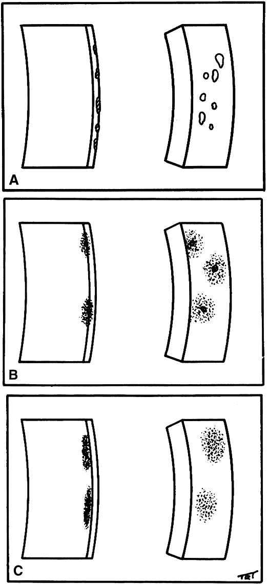
1. Norn MS: Rose bengal vital staining. Acta Ophthalmol 48:546, 1970

2. Coster DJ, Grutzmacher RD: Herpetic eye disease. Aust J Ophthalmol 11:1, 1983

3. Marsh RJ, Fraunfelder FT, McGill JI et al: Herpetic corneal epithelial disease. Arch Ophthalmol 94:1899, 1976

4. Dawson CR, Togni B: Herpes simplex eye infectons: Clinical manifestations, pathogenesis and management. Surv Ophthalmol 21:121, 1976

5. Piebenga LW, Laibson PR: Dendritic lesions in herpes zoster ophthalmicus. Arch Ophthalmol 90:268, 1973

6. Marsh RJ: Herpes zoster keratitis. Trans Ophthalmol Soc UK 93:181, 1973

7. Acers TE, Vaile V: Co-existent herpes zoster and herpes simplex. Am J Ophthalmol 63:992, 1967

8. Chern KC, Conrad D, Holland GN et al: Chronic varicella-zoster virus epithelial keratitis in patients with acquired immunodeficiency syndrome. Arch Ophthalmol 116:1011, 1998

9. Beinfang DC, Kuwabara T, Pueschel SM: The Richner-Hanhart syndrome. Arch Ophthalmol 94:1133, 1976

10. Goldsmith LA, Reed J: Tyrosine-induced eye and skin lesions. A treatable genetic disease. JAMA 236:382, 1976

11. Burns RP: Soluble tyrosine aminotransferase deficiency: An unusual case of corneal ulcers. Am J Ophthalmol 73:400, 1972

12. Abdel-Khalek LMR, Williamson J, Lee WR: Morphological changes in the human conjunctival epithelium, II. In keratoconjunctivitis sicca. Br J Ophthalmol 62:800, 1978

13. Wright P: Sjögren's syndrome. Trans Ophthalmol Soc UK 94:764, 1974

14. Fraunfelder FT, Wright P, Tripathi RD: Corneal mucous plaques. Am J Ophthalmol 83:191, 1977

15. Jones BR: Vernal keratitis. Trans Ophthalmol Soc UK 81:215, 1981

16. Williamson J, Doig WM, Forrester JV et al: Management of the dry eye in Sjögren's syndrome. Br J Ophthalmol 58:798, 1974

17. Wright P: Diagnosis and management of dry eyes. Trans Ophthalmol Soc UK 91:119, 1971

18. Theodore FH: Superior limbic keratoconjunctivitis. Eye Ear Nose Throat Monthly 42:25, 1963

19. Thygeson P: Further observations on superficial punctate keratitis. Arch Ophthalmol 66:34, 1961

20. Theodore FH: Further obsrvations on superior limbic keratoconjunctivitis. Trans Am Acad Ophthalmol Otolaryngol 71:341, 1967

21. Theodore FH, Fury AP: Superior limbic keratoconjunctivitis: clinical and pathological correlations. Arch Ophthalmol 84:481, 1970

22. Wright P: Superior limbic keratoconjunctivitis. Trans Ophthalmol Soc UK 92:555, 1972

23. Dodds HT, Laibson PR: Filamentary keratitis following cataract extraction. Arch Ophthalmol 88:609, 1972

24. Tenzel RR: Comments on superior limbic filamentous keratitis, Part 2. Arch Ophthalmol 79:508, 1968

25. Cher I: Clinical features of superior limbic keratoconjunctivitis in Australia: A probable association with thyrotoxicosis. Arch Ophthalmol 82:580, 1969

26. Fells P: Comments on superior limbic keratoconjunctivitis. Trans Ophthalmol Soc UK 92:50, 1972

27. Bloomfield SE, Gasset AR, Forstot SL et al: Treatment of keratitis with soft contact lens. Am J Ophthalmol 76:978, 1973

28. Tenzel RR: Resistant superior limbic keratoconjunctivitis. Arch Ophthalmol 89:439, 1973

29. Donshik PC, Collin HB, Foster CS et al: Conjunctival resection treatment and ultrastructural histopathology of superior limbic keratoconjunctivitis. Am J Ophthalmol 85:101, 1978

30. Rothis WM, Chandler JW, Forstot SL: Filamentary keratitis following penetrating keratoplasty. Ophthalmology 89:946, 1982

31. Davis WG, Drewsy RD, Wood TO: Filamentary keratitis and stromal neovascularization associated with brain stem injury. Am J Ophthalmol 90:489, 1980

32. Wolper J, Laibson PR: Hereditary hemorrhagic telangiectasis (Rendu-Osler-Weber disease) with filamentary keratitis. Arch Ophthalmol 81:272, 1969

33. Hudson AC: A note on certain peculiar markings in the cornea. R London Ophthalmic Hosp Rep 18:198, 1911

34. Stähli J: Uber den Fleischerschen Ring beim Keratokonus und eine neue typische Epithelpigmentation der mornalen Kornea. Klin Monatsbl Augenheilkd 60:721, 1918

35. Barraguer-Somers E, Chan CC, Green WR: Corneal epithelial iron deposition. Am J Ophthalmol 90:729, 1983

36. Stocker F: Eine pigmentierte Hornhautlinie beim Pterygium. Klin Monatsbl Aukenheilkd 102:384, 1939

37. Ferry AP: A “new” iron line of the superficial cornea: Occurrence in patients with filtering blebs. Arch Ophthalmol 79:142, 1968

38. Fleischer B: Uber Keratokonus und elgenartige Pigmentbildung in der Kornea. MMWR 53:625, 1906

39. Bron AJ: Vortex patterns of the corneal epithelium. Trans Ophthalmol Soc UK 93:455, 1973

40. Hobbs HE, Calnan CD: The ocular complications of chloroquine therapy. Lancet 1:1207, 1958

41. Kaplan LJ, Cappaert WE: Amiodarone keratopathy. Arch Ophthalmol 100:601, 1982

42. Kersley GD, Palin AG: Amodioquine and hydroxychloroquine in rheumatoid arthritis. Lencet 2:886, 1959

43. Burns CA: Indomethacin, reduced retinal sensitivity and corneal deposits. Am J Ophthalmol 66:825, 1968

44. Francois J: Cornea verticillata. Bull Soc Belge Ophthalmol 150:656, 1968

45. Lullmann H, Lullmann-Rauch R, Wassermann O: Lipidosis induced by amphiphilic cationic drugs. Biochem Pharmacol 27:1103, 1978

46. Font RL, Fine BS: Ocular pathology in Fabry's disease. Am J Ophthlmol 73:419, 1972

47. Cowan TH: Striate melanokeratosis in Negroes. Am J Ophthalmol 57:443, 1964

48. Jones BR: The differential diagnosis of punctate keratitis. Trans Ophthalmol Soc UK 80:655, 1961

49. Fuchs E: Keratitis punctate superficial. Wien Klin Wochenschr 2:837, 1889

50. Sanders M: Epidemic keratoconjunctivities (“shipyard conjunctivitis”), Part I. Isolation of a virus. Arch Ophthalmol 28:581, 1942

51. Braley AE, Alexander RC: Superficial punctate keratitis. Arch Ophthalmol 50:147, 1953

52. Burstein NL: Preservative cytotoxic threshold for bensalkonium chloride and chlorhexidine digluconide in cat and rabbit corneas. Invest Ophthalmol Vis Sci 19:308, 1980

53. Schwab IR, Abbott RL: Toxic ulcerative keratopathy. Ophthalmology 96:1187, 1989

54. Abbott RL, Forster RK: Superficial punctate keratitis of Thygeson associated with scarring and Salzmann's nodular degeneration. Am J Ophthalmol 87:296, 1979

55. Thygeson P: Superficial punctate keratitis. JAMA 144:1544, 1950

Back to Top