Chapter 36 The Phakomatoses DAVID SAMI, ANTHONY VIVIAN, DAVID TAYLOR and DAWN SAUNDERS Table Of Contents |
A PERSPECTIVE NEUROFIBROMATOSES TUBEROUS SCLEROSIS COMPLEX VON HIPPEL-LINDAU SYNDROME STURGE-WEBER SYNDROME AND OTHER VASCULAR DISORDERS REFERENCES |
A PERSPECTIVE |
In 1932, Van der Hoeve suggested that the syndromes of Bourneville, Recklinghausen, and
von Hippel-Lindau should be grouped as phakomatoses (“mother-spot”), having in common hereditary multisystem
tumors with risk of malignant transformation.1 The spectrum was extended in 1937 to include Sturge-Weber syndrome.2 How is it that a condition with no clear familial pattern or risk of malignant
transformation was included as a phakomatosis? Its inclusion
was an error based on a single case in which retinoblastoma developed
in a patient with Sturge-Weber syndrome.3 Later on, to accommodate Sturge-Weber syndrome, Hogan and Zimmerman reinterpreted the phakomatoses as conditions of multisystem hamartoses, irrespective of malignancy potential.4 Duke-Elder protested by excluding Sturge-Weber from his discussion of phakomatoses.5,6 Over time it seems that the cutaneous lesions, as a marker of multisystem disease, have gained prominence. This may explain inclusion of the phakomatoses as part of the “neurocutaneous disorders,”7–10 even though cutaneous findings are not part of the diagnostic criteria of von Hippel-Lindau syndrome. Given multiple definitions to choose from, the phakomatoses have become a loose category, expanding as a convenient way to group syndromes.5,7,11 We believe that the term phakomatosis should be restricted to the original three syndromes as molecular genetics support Van der Hoeve's observation: neurofibromatosis, tuberous sclerosis complex, and Von Hippel-Lindau syndromes are disorders of tumor suppressor genes,12–15 a common link explaining the familial pattern (autosomal dominant), variable expressivity, multisystem tumors, and risk of malignant transformation. Thus we will restrict the phakomatoses to the original three syndromes:
Each section is divided into: epidemiology; systemic features (cutaneous, neurologic, skeletal/visceral); ophthalmic features; diagnostic criteria; genetics and screening. Sturge-Weber and related syndromes will be discussed separately. |
NEUROFIBROMATOSES | ||||||||||||||||||||||||||||||||||||
Although up to 10 subtypes have been described,16,17 currently the commonly used clinical subdivision is based on the National
Institutes of Health (NIH) consensus classification.18 This classification recognizes two major forms of neurofibromatosis: types 1 and 2. The
other types were not formally classified. Both major
subtypes are disorders of tumor suppressor genes (Table 1).12,14 Knudsen's two-hit hypothesis states that the statistical odds of developing an inactivating mutation
in both alleles of a tumor suppressor gene, in the absence of an underlying
disorder of DNA repair, is very low unless there is a germ-line
mutation in one of the alleles. The odds of developing “loss of
heterozygosity” through a new mutation is doubled, making the
individual prone to developing tumors in his/her lifetime.19
TABLE 1. Neurofibromatosis Classification/Subtypes
NEUROFIBROMATOSIS TYPE 1 Epidemiology Neurofibromatosis type 1 (NF1) accounts for 90% of the neurofibromatoses and shows autosomal dominant inheritance. Its prevalence is 1/3500 to 1/5000.20,21 (Prevalence is an estimate of the frequency of a condition in the general population; incidence is an estimate of the number of new cases over a given period of time, typically 1 year.) Although the penetrance of NF1 is close to 100% by age 5 years, its expressivity even within the same family may be highly variable. (Penetrance is the proportion of individuals with the defective gene who show clinical evidence of the disorder; expressivity is the degree of variability in clinical findings among individuals with similar mutations.) The NF1 gene has a high mutation rate, with up to half of newly diagnosed cases representing de novo mutations.22 Systemic Features NF1 can involve any organ system. CUTANEOUS FINDINGS.
NEUROLOGIC FINDINGS.
SKELETAL/VISCERAL FINDINGS.
Ophthalmic Features Virtually any part of the visual system may be affected by NF1: the bony orbit, extraocular muscles, orbital nerves, eyelids, conjunctiva, cornea, uvea, retina, optic nerves, chiasm, and optic radiations. OCULAR FEATURES. Conjunctiva. Neurofibromas of the conjunctiva are infrequent, affecting about 2% of patients with NF1, often on the perilimbal conjunctiva.35 Cornea. Enlarged corneal nerves have been reported in association with NF1 but are more commonly seen with multiple endocrine neoplasia syndrome (MEN-IIb).36,37 Uveal Tract. Lisch nodules are hamartomas (a tumor involving only those tissue elements normally found at the involved site) of the iris pigment epithelium. They are dome-shaped discrete lesions, are typically light brown in color, and may also be found in the angle.38 Lisch nodules appear earlier (33% at 2.5 years, 50% at 5 years of age)39 than neurofibromas. They are benign and can help to confirm diagnosis in children who may have café au lait spots as the only other clinical finding. Lisch nodules are present in nearly all adults with NF139,40 but are rare in NF2 (Fig. 3).41,42 Diffuse nodular iris nevi (also known as iris mamillations) should not be confused with Lisch nodules. Its clinical significance is not well established.43,44
Like Lisch nodules of the iris, pigment epithelial hamartomas may affect the choroid. In one series, flat pigmented lesions of the choroid were found in 35% of patients with NF1. Fluorescein angiography suggests these are choroidal nevi.45The entire uveal tract may be thickened by a diffuse neurofibroma, thought to be hamartomatous hyperplasia of Schwann cells. This may result in the development of glaucoma.46,47 Retina. Retinal involvement in NF1 is unusual. Astrocytic hamartomas (similar to those in tuberous sclerosis), retinal capillary hemangiomas, and combined hamartoma of retina and retinal pigment epithelium (RPE) occur.48,49 EYELID AND ORBIT. Orbital neurofibromas may arise from any nerve in the orbit, especially the trigeminal nerve.50 Classically the triad of visual loss, optic atrophy and optociliary shunt vessels have been associated with optic nerve sheath meningiomas,51 but these findings may be nonspecific for optic nerve compression,52,53 particularly in children. If a child with a presumed diagnosis of NF1 is found to have an optic nerve meningioma, NF2 should be considered. The eyelids are frequently involved in NF1. The lateral portion of the upper lid is prone to develop a plexiform neurofibroma, producing a sinusoidal or S-shaped deformity of the lid margin.54–56 Amblyopia develops from ptosis and astigmatism (Fig. 4).
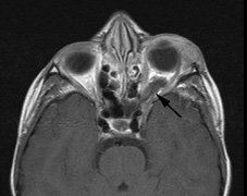
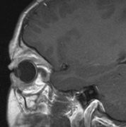
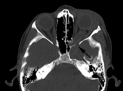
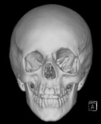
Growth of a plexiform neuroma may be directed posteriorly, infiltrating orbital tissues and involving the bony walls. Congenital plexiform neuromas of the orbit are often associated with absence of the sphenoid wing (Fig. 5). Intracranial pulsation can be transmitted to the orbit causing pulsatile proptosis or enophthalmos.57–59 Plexiform neuromas of the lid and orbit are difficult to treat; complete excision is difficult and potentially disfiguring. The tumors are vascular and may bleed copiously at surgery. They are not radiosensitive and there is also risk of inducing malignant transformation. Trials with antiangiogenic chemotherapeutic agents (thalidomide, interferon alpha) and mitotic-signaling pathway blockers (inhibition of Ras by Farnesyl protein tranferase inhibitor) are in progress.60,61 OPTIC NERVE GLIOMAS Optic pathway gliomas arise from the astrocytes of the optic nerve. Most are pilocytic astrocytomas and typically remain intradural, extending in the subdural space. Malignant transformation is rare. There appear to be two growth patterns62:
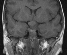
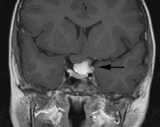
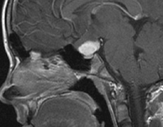
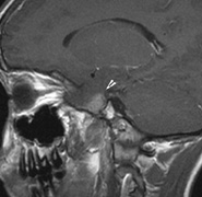
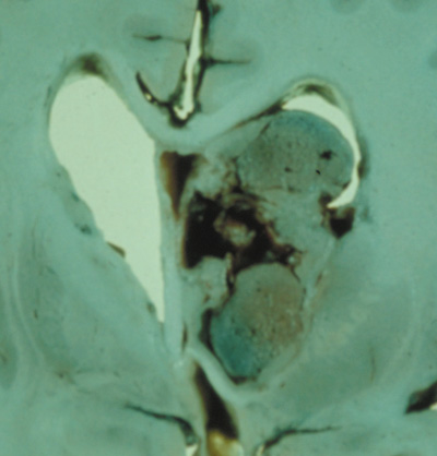
The incidence of optic pathway gliomas in NF1 is difficult to estimate, as the majority are asymptomatic and do not affect vision. In NF1, radiographic evidence of an optic nerve glioma occurs in 15% of patients.64,65 Most tumors are neither suspected historically nor detected by ophthalmic examination.65 Even when optic nerve gliomas become clinically detectable, visual function often remains stable in the absence of any intervention.66,67 Thus routine computed tomography (CT) imaging, even in patients with a known optic nerve glioma, is controversial. The potential risks of repeated radiation exposure in a child with a tumor-suppressor gene defect is a consideration. MRI studies are more revealing than CT but may require sedation or anesthetic and are costly. An MRI is often recommended at the time of diagnosis, but in most cases routine “follow-up” MRIs are unnecessary. Annual ophthalmic clinical examination for relative afferent pupillary defect (RAPD), visual acuity, visual fields, color vision, and funduscopy is usually all that is required, as no therapeutic action is likely to be taken unless there is significant and progressive visual involvement. Optic nerve gliomas may involve the optic chiasm and be associated with endocrine disorders or nystagmus.68–70 Surgical excision of chiasmal gliomas (Fig. 6) carries a high risk of visual loss. Invasion of the hypothalamus or the third ventricle carries a poor prognosis, with greater than 50% 15-year mortality rate.71 One review of radiation treatment for chiasmal gliomas collated data from small case series and found no significant long-term improvement in visual function, progression, or mortality with radiation treatment.71 Other reports suggest that radiation doses over 4500cGy improve symptoms and slow progression of chiasmal gliomas over several years.72 Adequate tumor coverage by radiotherapy results in irradiation of normal brain and nearly all children need hormone replacement.73 Chemotherapy is an alternative.74,75
Diagnostic Criteria The NIH has defined 7 criteria,22 at least two of which must be present:
In young children, nearly half of sporadic cases fail to meet NIH criteria by 1 year of age, but meet them by age 8 to 12 years. The clinical features appear in the following order: café au lait spots, axillary freckling, Lisch nodules, neurofibromas.26 The inclusion of UBOs on MRI also aids the diagnosis of very young children25 but may require a general anesthetic. Genetics and Screening All family members of patients with NF1 should be screened. Lisch nodules are present in 90% or more of patients with the disease, so ophthalmic examination is essential. Annual ophthalmic examination with visual acuity, visual field, color vision, and fundoscopy is recommended for affected individuals. The NF1 gene (17q11.2) was cloned in 1990.76,77 Its protein, neurofibromin, is expressed throughout the body but particularly in the central and peripheral nervous system.12 It is a tumor-suppressor gene with autosomal dominant inheritance, high penetrance, and variable expressivity. NF1 has been called a neurocristopathy, but the disease is not limited to neural crest derived tissues.12,78 Neurofibromin has structural similarity to GTPase, activating proteins that inhibit the function of G proteins such as Ras. Inhibition of Ras by neurofibromin has been shown in vitro and in vivo. NF1-deficient tumors show increased levels of Ras-GTP. Increased Ras function has been shown in a number of human tumors.12,79–81 NEUROFIBROMATOSIS TYPE 2 Neurofibromatosis type 2 (NF2) was described in 1822 by the Scottish surgeon Wishart.82 Initially, NF1 and NF2 were lumped together despite their differences.83 The identification of different genes re-established them as separate entities.84 Epidemiology The NIH consensus conference22 estimated an incidence of 1:50,000 but there are varying estimates internationally.84 NF2 is autosomal dominant with full penetrance by age 60.85 In all, 10% of affected patients are symptomatic by age 10, 50% by 20 years, and 80% by 30 years.86 Some sporadic cases reflect parental mosaicism.87,88 Systemic Features Several studies have established the clinical picture of NF2.85,89–91 Patients may become severely disabled because of the combination of deafness and imbalance from vestibular schwannomas, weakness from multiple spinal tumors, and poor sight from cataracts, optic nerve meningiomas, and retinal hamartomas. CUTANEOUS FINDINGS. Skin findings in NF2 may be subtle but not uncommon. Skin tumors were found in 60% and were the first presenting sign in more than 25% of patients.92 The skin tumors are predominantly schwannomas. A minority are neurofibromas and mixed types. Clinically differentiating the histological types may be difficult.92 They may present as dermal tumors or subcutaneous nodules of peripheral nerves. NF2 plaques are well circumscribed, slightly raised, often pigmented, and hairy. They are a sensitive marker for NF222 and are more common on the limbs and trunk than the neck and face.92 Café au lait spots are not uncommon in NF2, but it is unusual to find more than five and most tend to be restricted to the trunk. Plexiform neurofibromas and axillary/inguinal freckling are uncommon.92,93 NEUROLOGIC FINDINGS.
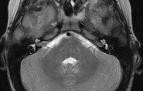
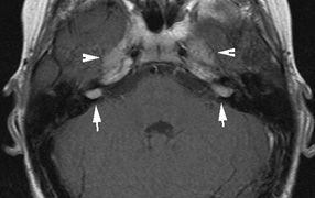
VISCERAL FINDINGS. NF2 does not appear to have significant visceral complications Ophthalmic Features The most common ocular findings in NF2 include premature cataracts and retinal hamartomas. Ocular motor nerve deficits may also be associated with NF2. Cataracts in NF2 were recognized in 1986.97 They are common, reported to be found in 69%,98 81%,91 85%,99 and 87%100of cases. They are typically mild98 and a lens opacity may be the initial manifestation of disease, as found in 10% (5 of 49 patients) in one study.98 Retinal hamartomas affect about 10% to 20% of patients (8% in one study91and 22% in another98) and may be associated with a more severe phenotype of NF2.91,98 Combined pigment epithelial and retinal hamartomas (CPERH) and epiretinal membranes have been reported.101–103 Lisch nodules are rare in NF2. The absence of Lisch nodules, in association with the presence of posterior subcapsular or cortical cataracts (Fig. 8) may aid the differentiation of NF1 and NF2.97,104,105 Optic nerve sheath meningiomas may cause significant visual impairment in the first years of life (Table 2).84,106
TABLE 2. Ocular Findings in Neurofibromatosis Type 298
Diagnostic Criteria The NIH scheme is most commonly used:95,107
A significant proportion of cases (20% to 30%) may present with intracranial meningioma, spinal tumors, or cutaneous tumors. Vestibular schwannomas may account for as little as 15% to 30% of presenting signs in children. Thus, expanded criteria have been proposed to include individuals with multiple schwannomas/meningiomas who have not developed vestibular schwannomas as yet and do not have a clear family history.84 Genetics and Screening Cataracts can affect vision in early life. For children at risk, annual ophthalmic examination is prudent. Central nervous system (CNS) tumors rarely become symptomatic before age 10 years. Vestibular schwannomas tend to grow faster in younger patients; therefore, asymptomatic at-risk individuals may benefit from MRI scanning every 2 years under age 20 and every 3 years over age 20. Epiretinal membranes and combined hamartomas should lead to investigation for NF2. An annual neurologic exam has also been recommended.84 The NF2 tumor-suppressor gene product is known as schwanomin108 or merlin109. The gene is located at 22q11.1–22q13.1 (Online Mendelian Inheritance in Man [OMIM] #607379).110 The NF2 gene shows homology to the highly conserved ezrin-radixin-moesin (ERM) family of membrane-cytoskeleton linking proteins. The NF2 protein is believed to interact with the actin cytoskeleton. Disruption of NF2 function has been linked to loss of cell-to-cell contact inhibition of growth and division.14 |
TUBEROUS SCLEROSIS COMPLEX | ||||||||||||||||||||||||||||||||||
In 1880, Bourneville, a French neurologist, described a girl with the triad
of mental retardation, seizures, and vesiculopapular rash on the
face. At postmortem, tumors were found in the brain (cortical sclerotic
lesions and tuberous extensions into the ventricles) and kidneys. Earlier, Rayer (1835) and Addison and Gull (1850) had
described distinctive facial papules.111 Balzer and Menetrier (1885) described the microscopic appearance
of the facial papules as “adenoma sebaceum,” thought
to be benign tumors of sebaceous glands, although the skin lesions are
actually angiofibromas.3,111,112 Sherlock (1911) coined the acronym epiloia—for epilepsy, low intelligence, and adenoma sebaceum—which
is no longer used.113 Van der Hoeve (1920) described the retinal tumors of tuberous
sclerosis.114 There have been exciting developments in regard to the molecular genetics of tuberous sclerosis complex (TSC). Two tumor suppressor genes have been identified: TSC1 and TSC2. Further insight into the role of these genes promises potential treatments to halt the progression of disease,115 thus emphasizing the need for early diagnosis. EPIDEMIOLOGY Tuberous sclerosis is autosomal dominant with 65% to 75% of new cases representing spontaneous mutations.116 Many “sporadic” cases may actually reflect somatic or germ-line mosaicism of a parent.117,118 Its prevalence is approximately 1:10,000 to 1:15,000.119,120 TSC has high penetrance but variable expressivity, even within the same family.121 SYSTEMIC FEATURES Cutaneous Findings
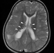
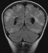
Neurologic Findings INTRACRANIAL LESIONS. Mental retardation and seizures in TSC are often associated with benign CNS astrocytic hamartomas.131 On imaging, the cerebral lesions show three patterns:132
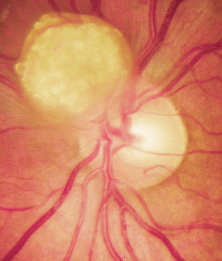
SEIZURES. Typically observed first in infancy, seizures are a common presenting sign of TSC and a frequent source of morbidity. The seizures often begin as “salaam spasms”—repetitive myoclonic spasms that produce head nodding with associated extension/flexion of the trunk and limbs. They often progress to grand mal seizures25,116 and may be difficult to control. Vigabatrin has been advocated for control of infantile spasms but it carries a risk for irreversible visual field loss. Other treatments include corticosteroids, adrenocorticotrophic hormone, and a ketogenic diet.25,137–139 Selective surgical excision of an “epileptogenic tuber” may be an option for intractable epilepsy.140 MENTAL RETARDATION. Mental deficiency, part of Vogt's diagnostic triad (adenoma sebaceum, epilepsy, mental deficiency),3 affects about 50% of patients overall.25,141 TSC2 mutations may carry a higher risk than TSC1.142 AUTISM. This may affect 50% to 60% of TSC patients, possibly correlating with numbers of cerebellar and temporal tubers.25,143,144 Visceral Findings Patients with TSC may have renal, cardiac, pulmonary and other visceral involvement. RENAL. The TSC2 gene was localized by linkage with the polycystic kidney disease gene in patients with TSC and polycystic kidneys. This represents a contiguous gene syndrome in which adjacent genes (PKD1 and TSC2) are deleted together.145–147 Multiple angiomyolipomas of the kidney are benign hamartomas of blood vessels, smooth muscle, and fat. By 10 years of age, the frequency of angiolmyolipomas is 75%, similar to adults.148 The fat signal of angiomyolipomas helps to distinguish them from renal cell carcinoma.25 Renal insufficiency/failure may be the complication of both cystic disease and angiomyolipomas and is one of the leading causes of morbidity and mortality in TSC.138,149 CARDIAC. Rhabdomyoma are usually multiple, typically asymptomatic, and often regress with age.150,151 PULMONARY. Lymphangioleiomyomatosis is a progressive lung disease characterized by bronchiolar smooth muscle infiltration and cystic changes of the lung parenchyma. About 1% of adult females with TSC are affected.116,152,153 OPHTHALMIC FEATURES The principal ophthalmic manifestations of TSC are in the posterior segment. Astrocytic Retinal Hamartomas Although in Van der Hoeve's original description the term phakoma or phakomata was a generic term for the tumors occurring in various organ systems,1 it has become associated with the retinal astrocytic tumors of TSC,3 which occur in about 50% of affected patients and are bilateral in 25% to 30% (Table 3, Fig. 16).154,156
TABLE 3. Astrocytic Retinal Hamartomas in Tuberous Sclerosis
Historically, the presence of mixed (type 3) hamartomas led to the suggestion of evolution from the type 1 to the type 2.157 A long-term photographic study found that although the types remained unchanged, a minority of types 1 and 3 increased in calcification and a new lesion was noted that had not been evident on an earlier photograph.157 All three types of retinal hamartomas may be found in young children. There is no correlation between age and the predominant type of lesion.156 Histologically, phakomata are composed of glial astrocytes—elongated, fibrous astrocytes with small oval nuclei, arising from the retina or the optic disc. The type 2 lesions contain basophilic, calcified drusen-like deposits within a dense vascular network. On fluorescein angiography, the type 2 lesions show autofluorescence, leakage with transit of dye, and marked late hyperflorescence.3,113 Astrocytic retinal hamartomas usually do not affect vision and require no treatment. Vitreous seeding and vitreous hemorrhage have been reported in association with phakomata, but these are rare.158,159 Laser photocoagulation may be considered in rare cases were exudation induces serous retinal detachments.160 The differential diagnosis of “white retinal lesions” includes retinoblastoma, toxoplasmosis and toxocara.3,113 Pigmentary Abnormalities Depigmented “punched-out” chorioretinal lesions in the midperiphery have been reported in up to 40% of patients. Similar lesions may be found in normal patients.156 Nonretinal Features The adnexae may be involved by angiofibromas: eyelid skin and “salmon-colored” subconjunctival nodules. Sector iris depigmentation and iris coloboma have been reported in a minority of patients.113,156,161 DIAGNOSTIC CRITERIA When TSC is classical (“Salaam seizures,” facial angiofibroma, developmental delay), the diagnosis is straightforward. However, children can be mildly affected, and many of the classical signs may become apparent later. Based on the revised clinical diagnostic criteria set forth by the Tuberous Sclerosis Consensus Conference162, a diagnosis of TSC may be made if the patient has two major features, or one major feature plus two minor features. Genetic testing for TSC still has a high false-negative rate (Table 4).25,162,163 TABLE 4. Features of Tuberous Sclerosis Complex
GENETICS AND SCREENING The Tuberous Sclerosis Consensus Conference162 recommendations for screening once a diagnosis of TSC is established are:
Two genes have been identified, TSC1 (9q34, OMIM #605284) and TSC2 (16p13.3, OMIM #191092), which appear to be tumor-suppressor genes. The gene product of TSC1 is hamartin and the gene product of TSC2 is tuberin. Of sporadic cases of TSC, 70% have been linked to TSC2 mutations. Among the familial cases, half are linked to the 9q34 locus and half to the 16p13.3 locus. Hamartin and tuberin appear to interact together in vivo, which may be why mutations of two different genes produce a similar phenotype. Together they form a cytoplasmic protein complex that has an inhibitory effect on cell growth and division. The tuberin-hamartin complex inhibits the activity of TOR (target of rapamycin). This has raised the possibility of reestablishing the regulatory effect of the tuberin-hamartin complex on TOR with drugs such as rapamycin.115,164 |
VON HIPPEL-LINDAU SYNDROME | ||||||||||||||||||||||||||
Von Hippel described the fundoscopic appearance and histology of “angiomatosis
retinae” in 1911165 and Lindau linked the retinal, cerebral, and visceral components of the
syndrome in 1926. The term von Hippel-Lindau syndrome (VHL) was
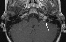
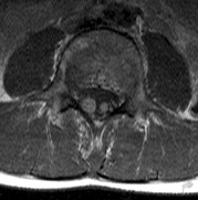
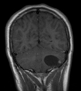
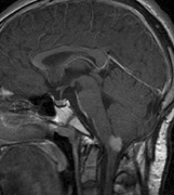
first used in 1936.166 The first major clinical criteria for the syndrome were proposed in 1964.167 The main manifestations of von Hippel-Linau syndrome are: histologically similar hemangiomas of the retina and CNS, renal cysts, renal cell carcinomas, pancreatic cysts, pheochromocytomas, and endolymphatic sac tumors.13 EPIDEMIOLOGY The prevalence of VHL syndrome is about 1 in 35,000 to 40,000.168 It is autosomal dominant with greater than 95% penetrance by age 60 years.169 Its expression, even within the same family, may be quite variable, especially for renal disease and pheochromocytoma.170,171 SYSTEMIC FEATURES Cutaneous Findings Cutaneous findings are not part of the diagnostic criteria for VHL syndrome. Neurologic Findings CNS hemangiomas are the most common tumor of VHL, affecting 60% to 80%, with a predilection for the cerebellum and spinal chord. An enlarging cystic component is a frequent finding in symptomatic tumors. Patients typically present in their early 30s; headaches or neck pain in affected individuals should not be ignored.172,173 On microscopy, CNS hemangiomas resemble retinal capillary hemangiomas. Their malignant potential is low.174 The treatment is surgical (Fig. 17, A and B).13 Endolymphatic sac cystadenomas are a sensitive marker of VHL. Patients may present with hearing loss, tinnitus, disequilibrium or facial nerve palsy.175,176 Visceral Findings KIDNEY. Renal cysts are often asymptomatic and do not require treatment. However, complex cysts may carry a risk for malignant transformation. Renal carcinoma affects about a third of patients. Patients typically present in their late 30s. These tumors may be completely asymptomatic, underscoring the necessity for ultrasound screening.13,177 ADRENALS. Pheochromocytomas are benign neural crest tumors of the adrenal medulla (chromaffin cell derived) and probably affect 10% to 20% of patients. Mean age at diagnosis is at about 30 years of age. Pheochromocytomas may be multiple and bilateral. They are catecholamine-secreting tumors that classically produce severe hypertension and anxiety attacks, but they may also be asymptomaic.178 The National Cancer Institute classification of VHL syndrome is in part based on the absence (type I) or presence (type II) of pheochromocytoma (Table 5).171
TABLE 5. Classification of von Hippel-Lindau Syndrome
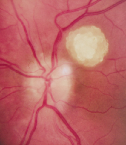
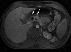
PANCREAS. Pancreatic lesions may be nonsecretory (most commonly cysts or cystadenomas) or secretory (islet cell tumor).179 In one study of 52 patients, 56% (29 patients) were found to have pancreatic lesions. The majority (19 of 29) had cystic changes only (Fig. 17C). Pancreatic lesions were the only abdominal manifestation of disease in 6 of 52 patients.180 In a separate study, pancreatic cysts did not show significant progression on follow-up examinations over an average period of 5 years.181 Like pancreatic cysts, islet cell tumors appear to be frequently asymptomatic.182 EPIDIDYMIS AND BROAD LIGAMENT. Papillary cystadenomas of the epididymis are typically benign and asymptomatic. In one study, about half of patients were affected.183 Papillary cystadenomas of the broad ligament are thought to be the female equivalent of papillary cystadenomas of the epididymis.184 OPHTHALMIC FEATURES VHL syndrome does not appear to have significant anterior segment manifestations. Posterior Segment Retinal capillary hemangiomas are among the most frequent and earliest manifestations of VHL syndrome. Most patients present between 10 and 40 (mean age of 25) years of age and 5% present before age 10. Overall, retinal capillary hemangiomas affect about 60% of individuals with VHL syndrome. Development of new retinal capillary hemangiomas after age 60 years is unusual.169,170 Retinal capillary hemangiomas (Fig. 18) may be the only manifestation of the syndrome,185 and typically involve the midperipheral temporal retina.186 A pair of dilated and tortuous retinal vessels (artery and vein) classically supply the vascular tumor. Without fluorescein angiography, it may be difficult to distinguish artery from vein on fundus examination.170
Retinal capillary hemangiomas may also occur in the absence of VHL syndrome, but the age at presentation is typically later (50 vs. 25 years).187 Small retinal capillary hemangiomas may remain stable for many years, but most tend to enlarge and may become symptomatic secondary to exudation and retinal detachment. On microscopy, retinal capillary hemangiomas are similar in appearance to the CNS hemangiomas of VHL syndrome. Thin vascular channels are lined by endothelial cells and pericytes. The vascular channels are separated by foamy stromal cells. The foamy appearance is due to phagocytosed lipids that escape the fenestrated endothelium of the vessel walls. The lipid is plasma–derived cholesterol stearate.188–190 Hemangioma is more appropriate than hemangioblastoma.170,188,190 The cellular origin of capillary hemangiomas in VHL is controversial,170 but loss of heterozygosity of the VHL gene has been demonstrated in the stromal cells of capillary hemangiomas.191 There are various classification schemes based on morphology (endophytic, exophytic, and sessile) and the presence or absence of exudation. Endophytic tumors grow on the retinal surface, protruding into the vitreous cavity. Exophytic tumors tend to be nodular and orange-colored, growing the in outer layers of the retina. The sessile growth pattern implies a flat tumor, gray or orange in color, and grows within the middle layers of the retina.185 The “natural history” of the retinal capillary hemangioma has been divided into four192 or five193 stages170:
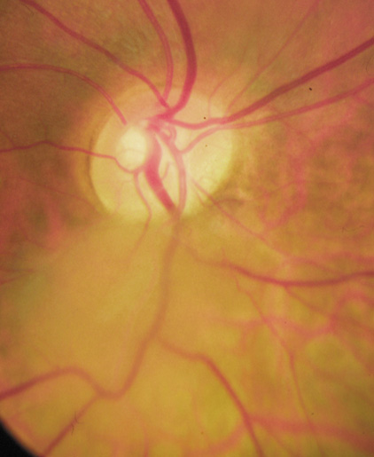
On occasion, an exophytic-type capillary hemangioma adjacent to the optic nerve can masquerade as chronic disc edema.194 The frequency of optic-disc angiomas was 15% in a group of 175 patients.186 Optic-disc hemangiomas may be the only manifestation of disease in VHL syndrome.186 In the absence of effective treatment, exudation from capillary hemangiomas may induce retinal detachment and macular star formation.170,186 Early detection (i.e., when the hemangioma is small) and treatment are associated with better visual outcomes.186 The exception may be small juxtapapillary hemangiomas, in which treatment may produce significant loss of acuity. Small juxtapapillary hemangiomas may remain stable.185 Both direct and feeder vessel photocoagulation with a yellow-dye laser have been shown to be effective for treating small (<1.5mm) capillary hemangiomas. Feeder vessel technique may require a greater number of treatment sessions.195 Direct treatment may be associated with a smaller scotoma than feeder vessel treatment, but direct photocoagulation carries a potential for hemorrhage and retinal detachment.186 Hemoglobin absorbs yellow laser more than green or blue wavelengths. Complete obliteration of the hemangioma may not be necessary to achieve resolution of symptoms.170 When the retinal capillary hemangioma is located in the far anterior periphery, or when it is larger than 3.0 mm in diameter, cryotherapy may be more effective than photocoagulation.170 To treat hemangiomas 4 to 5 mm in diameter, plaque radiotherapy has been recommended.196 The VHL gene product appears to inhibit the production of VEGF (vascular endothelial growth factor). Treatment with SU 5416, a VEGF-receptor inhibitor, has been reported to restore and maintain visual acuity and visual field in a patient with an optic-disc hemangioma.197 Retinal “twin vessels”—a paired retinal arteriole and venule of normal caliber, separated by less than the diameter of the venule—have been described as part of VHL198 but the finding has not been reproduced.186 Twin vessels are not associated with retinal capillary hemangiomas, and may be found in normal patients.199 Differential Diagnosis In the presence of subretinal exudates, the peripheral hemangioma may be overlooked, giving a false impression of Coats disease (diffuse unilateral retinal telangiectasias with exudation in young boys). Dilated feeder vessels may be confused for the arteriovenous malformation of Wyburn Mason disease (racemose hemangioma). Exudation is not a feature of Wyburn Mason disease and there is no angioma. Retinal cavernous hemangiomas lack prominent feeder vessels and do not leak fluid into the subretinal space. Retinal macroaneurysms may leak fluid but do not have dilated feeder vessels. Uveal melanomas usually have only a dilated vein.170 DIAGNOSTIC CRITERIA: If there is a positive family history of VHL syndrome, transmission is likely based on finding any one of the following13:
If family history is negative, a diagnosis is likely based on:
The National Cancer Institute has classified VHL syndrome into two main categories on clinical grounds: type I without pheochromocytoma and type II with pheochromocytoma. Type II is further subdivided based on the presence or absence of renal cell carcinoma and CNS hemangioblastoma, and type IIC implies pheochromocytoma only (Table 5).170 GENETICS AND SCREENING Subclinical lesions are common in VHL syndrome and there is more than one “accepted” protocol.13,169,200 In general, the protocols are similar, differing in the age to start screening for the different manifestations of the syndrome. Annual fundoscopy and urinary catecholamine testing should begin early in childhood (age 1 to 5 years). Spine and brain MRI should be carried out every 1 to 2 years and abdominal ultrasonography annually beginning age 10 years. Autosomal dominant inheritance of multisystem tumors with malignant potential is the “trademark” of tumor-suppressor gene defects and the phakomatoses. New mutations are thought to represent about 10% of affected individuals. The VHL gene product (OMIM #193300) located at 3p25–26 appears to downregulate the transcription factors that turn on the production of VEGF as part of the cellular response to hypoxia. This may account for the predilection to form hemangiomas.201 Molecular analysis suggests that missense mutations of the gene predispose to pheochromocytomas, which is type II (96% of mutations in VHL syndromeand pheochromocytoma were found to be missense mutations in one study).202 No particular type of mutation has been associated with frequency or severity of retinal capillary hemangiomas.186 DNA testing for VHL gene mutations can achieve 99% detection rates.203 This may be valuable for counseling of young parents of an affected child who do not have a clear family history and may still be too young to manifest features of the syndrome themselves. |
STURGE-WEBER SYNDROME AND OTHER VASCULAR DISORDERS | ||||
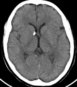
STURGE-WEBER SYNDROME Sturge (1879) reported the case of a child with facial “port-wine stain,” epilepsy, buphthalmos, and a dark choroid.204 Schirmer had noted the association of the facial vascular malformation and glaucoma in 1860.3 Weber (1922) reported brain X-ray findings of parallel streaks (“railtrack” sign), which he interpreted to represent calcific changes (Fig. 19).205
Port-wine stains (PWS) affect 0.3% of live births. The majority of isolated PWS occur in the head and neck.206–208 The possibility of Sturge- Weber syndrome (SWS) is raised when a newborn is noted to have a facial PWS. SWS does not have any clear inheritance pattern. The minimum diagnostic criteria for SWS is a subject of debate.209–211 The complete syndrome includes:
Facial capillary vascular malformation (Fig. 20) is the most obvious feature of SWS. It consists of many dilated capillaries lined by a single layer of endothelial cells.3 The PWS is present at birth and partially blanches with pressure. Historically it has also been referred to as a “naevus flammeus.” The PWS is typically unilateral, and may follow the distribution of the trigeminal (CN V) nerve divisions. The conjunctiva and episclera ipsilateral to the PWS are often involved.
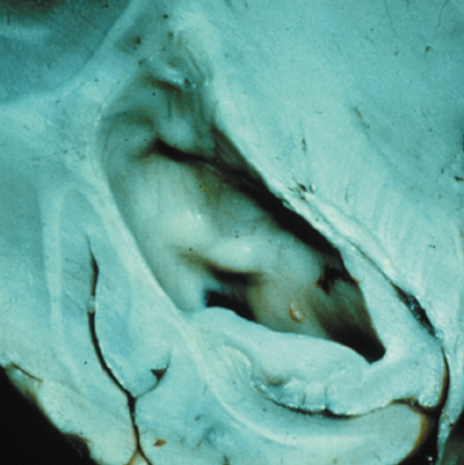
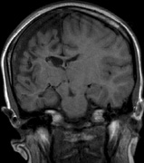
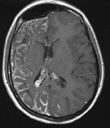
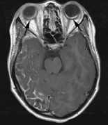
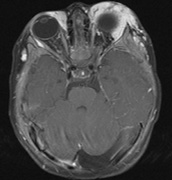
The PWS may not entirely respect the midline, possibly a reflection of transmedian innervation.212 The lesion grows proportionally with the child, often becoming darker in color. 213 The overlying skin may develop nodularity or hypertrophy in two-thirds of patients (by the fifth decade).214 Treatment with a pulsed-dye laser may significantly improve the appearance, but recurrence (i.e., redarkening) is possible.215,216 A deficit in the number of perivascular nerves in port-wine stains suggests that abnormal neural modulation of vascular flow may be involved in their pathogenesis.217 Intracranial leptomeningeal capillary vascular malformation (Figs. 21 and 22) is associated with seizures and contralateral neuromuscular weakness. Central nervous system involvement correlates highly with ipsilateral involvement of the V1 dermatome by a PWS.208,218
It has been hypothesized that the calcific changes reflect cellular anoxic injury secondary to venous stasis/poor drainage.219,220 Abnormal autonomic innervation has also been reported in association with the malformed cortical vessels—innervation only by noradrenergic fibers.221 Studies suggest that the normal increase in brain-blood flow to accommodate increased metabolic demand during seizure activity is blunted in affected brain regions of SWS.222 Thus, seizures in the setting of impaired perfusion may be exacerbating brain injury. Periods of contralateral weakness (days to weeks) can follow a prolonged seizure episode.206,223 When seizures begin early in life, the risk of developmental delay is much higher. In a review of 171 patients with a facial PWS and associated intracranial vascular malformation, 80% were found to have a seizure disorder. Of those, 75% with seizures had an onset during the first year of life, rising to 95% before age 5 years. Nearly all patients had a PWS that included (or was limited to) the V1 distribution. Developmental delay was strongly associated with seizures (71% of children with seizures as compared to 6% without seizures).224 Glaucoma affects 50% to 70% of SWS patients224,225 and it is a therapeutic challenge. From the time of diagnosis, the intraocular pressure, the optic discs, and the corneal diameters need to be measured intermittently, even if an occasional examination under anesthetic is required. The development of glaucoma appears to follow a bimodal age distribution: early-onset/congenital-type and later-onset. The glaucomatous eye is nearly always on the same side as the PWS.224 The highest risk for glaucoma appears to be when the PWS involves both the upper and lower lid: 72% vs. 21% for VI-only (upper eyelid) involvement in one study.224 The bimodal age distribution of glaucoma suggests that more than one mechanism is involved. Angle abnormalities (“trabeculodysgenesis” with or without flat anterior iris insertion) have been reported in association with the early-onset/congential type.225 Other proposed mechanisms include uveal capillary dilation226 and elevated episcleral venous pressure. Reports of blood in Schlemm's canal on gonioscopy support the idea of increased episcleral venous pressure pathomechanics.227,228 Glaucoma in SWS often responds poorly to medical therapy alone. The “trabeculodysgenesis” of early onset Sturge-Webber glaucoma is reported to be similar in gonioscopic appearance to primary congenital glaucoma.229 On this basis, goniotomy was suggested as a treatment of choice in early-onset SWS glaucoma. One group looked at the success of goniotomy, trabeculotomy, trabeculectomy, laser trabeculoplasty and medication in the management of SWS-associated glaucoma.230 In the early onset subgroup receiving goniotomy, median stable interval of intraocular pressure reduction after one procedure was 8 months; with multiple goniotomies and the addition of medical treatment, the median stable interval was extended to 9 years. Other investigators have found that goniotomy alone may be insufficient, requiring filtering surgery at some point. However, trabeculectomy carries a significant risk of choroidal expansion in association with hypotony. Theoretically, the risk may be higher when there is increased choroidal vascularity in association with the facial PWS—a “tomato ketchup” fundus.231 Often, there is associated increased tortuosity of the retinal vessels as well. Placement of a prophylactic posterior sclerotomy prior to filtering surgery to prevent choroidal expansion is controversial.232 Cryo- or thermocoagulation of the ciliary body in combination with medical therapy may be an alternative to surgery.233 KLIPPEL TRENAUNAY—WEBER SYNDROME Klippel-Trenaunay syndrome consists of combined capillary, lymphatic, and venous malformations, in association with varicosities and limb enlargement (Fig. 23). In the majority of cases, the lower limb is affected.234,235
The occurrence of “merged” Klippel-Trenaunay and Sturge-Weber syndromes has recently been questioned. The contention is that the presumed combined cases actually represent Sturge-Weber syndrome with capillary malformations below the head and neck, without the associated lymphatic malformations of Klippel-Trenaunay syndrome.235 Extracranial port-wine stains are relatively common in SWS (45% in a study of 171 patients).224 PHACES SYNDROME PHACES236,237 is an acronym for the association of:
The majority of patients are female, and affected children may have only one extracutaneous manifestation. The facial vascular lesion is typically large, segmental, and plaque-like.238,239 Unlike the vascular malformation of Sturge-Weber Syndrome, the PHACES facial vascular lesion is likely a true hemangioma.240 Visceral and/or CNS hamangiomas may affect up to 50% of patients.241 The most common associated ocular abnormalities appear to be microphthalmos and increased vascularity on examination of the fundus. Glaucoma does not appear to be a common association of the syndrome (Fig. 24).242 |