Chapter 42
The Ophthalmologist's Role in Visual Processing and Learning Disabilities
HAROLD P. KOLLER and AND KENNETH B. GOLDBERG
Main Menu   Table Of Contents

Search

SYSTEMIC CONDITIONS THAT MAY AFFECT READING AND LEARNING
OCULAR CONDITIONS AFFECTING READING
MIGRAINE
LEARNING DISABILITIES
SPEECH AND LANGUAGE DISORDERS
NONVERBAL LEARNING DISORDERS
ATTENTION DEFICIT/HYPERACTIVITY DISORDERS
PERVASIVE DEVELOPMENTAL DISORDERS
CONCLUSION
REFERENCES

SYSTEMIC CONDITIONS THAT MAY AFFECT READING AND LEARNING
Many systemic conditions can be responsible for temporary or chronic visual disturbances in the classroom that can impact performance and reading efficiency. Learning in the long term, however, is not affected. Children with chronic allergies that cause tearing, itching, redness, and burning often have these annoying symptoms during class. Appropriate treatment is readily available with antihistaminic eye drops. Other systemic conditions such as juvenile rheumatoid arthritis, chronic sinusitis, and ocular trauma all, at one time or another, can result in some visual discomfort and make reading a bit stressful (usually leading to frustration or reduction in reading rate or fluency). They never interfere with learning or cerebral processing function and can once again be compensated for with accommodations (i.e., increasing time for reading, books on tape, having a “note-taking” service for class).9

Other disorders affecting learning that an ophthalmologist frequently sees include Tourette syndrome and other developmental neurologic conditions (i.e., cerebral palsy). Idiopathic sleep disorders can also affect learning, as can social and family dysfunction or poor school instruction. Several serious genetic diseases are frequently associated with learning disorders. These include familial dysautonomia (Reilly-Day syndrome10) and mitochondrial cytopathy.11 Both of these conditions involve multiple organ systems. The brain is a high oxygen/energy uptake organ system, and the mitochondria are the oxygen generators of our cells. Any disease of the mitochondria affects most organ systems. The autonomic nervous system is almost nonfunctional in familial dysautonomia, and brain processing is also affected. Much more research regarding mitochondrial (not nuclear) inheritance is required to associate learning disorders with these conditions. Familial dysautonomia may also be a partial mitochondrial disorder, as might certain types of learning disorders.

Back to Top
OCULAR CONDITIONS AFFECTING READING
Koller has described a number of purely ophthalmic conditions that at times may affect reading efficiency and attention (Table 1).12 All of them do not, however, permanently influence learning. These conditions include strabismus, amblyopia, nystagmus, pituitary disorders, infections (such as herpes simplex), trauma, and various congenital anomalies of the anterior and posterior ocular segments. Convergence insufficiency exophoria/tropia may cause reading fatigue but never has been shown to cause a learning disorder. The other conditions may make seeing a bit more difficult and create a need for more testing time and/or visual aids, but they do not directly or indirectly affect comprehension and learning. Intermittent blurring can occur with any of these conditions. IDEA and help with Individual Educational Plans are available to such students, if only for the period their ocular symptoms are active. This of course may be permanent in a number of cases (i.e., albinism, optic atrophy). Orthoptic eye exercises may be of benefit for individuals with true convergence insufficiency to alleviate their reading fatigue by creating diplopia awareness and improving convergence amplitudes.

TABLE 1. Visual Perception and Learning Differences in Pediatric Ophthalmology: What the Ophthalmologist Needs to Know About Learning Disabilities in Clinical Practice

  1. Ophthalmic Causes of Temporary Learning Impairment and Inefficiency
    1. Strabismus, amblyopia, and refractive errors
      1. Decompensated accommodative esotropia with secondary diplopia
      2. Secondary ocular cranial nerve palsies (n. III, IV, VI)
      3. Uncorrected congenital vertical strabismus with abnormal face and head positions
      4. Bilateral very high ametropias, such as bilateral amblyopia of high hyperopia
      5. Bilateral occlusion amblyopia, such as after congenital cataract surgery
      6. Associated untreated “A” and “V” syndromes
      7. Convergence insufficiency exotropia/phoria in certain individuals
    2. Nystagmus of moderate to severe degree causing significant vision loss
    3. Pediatric ocular diseases that can affect learning via visual and emotional effects
      1. Severe juvenile rheumatoid arthritis and related ocular inflammations
      2. Congenital glaucoma with vision loss
      3. Congenital cataracts and major corneal opacities with vision loss and nystagmus
      4. Significant unilateral and/or bilateral ocular trauma with vision loss
      5. Ocular and adnexal neoplasms with secondary disfigurement and vision loss
      6. Severe chronic ocular infections such as HSV-I with vision loss
      7. Congenital and degenerative retinal diseases affecting the macula
  2. Neuro-ophthalmic Causes of Temporary or Intermittent Learning Impairment and Inefficiency
    1. Brain tumors causing a change in personality and behavior in a pediatric patient
    2. Migraine symptoms
      1. Acephalgic pediatric migraine with visual disturbances and variable vision in school
      2. Ophthalmic migraine in childhood
      3. Ophthalmoplegic migraine
      4. Classic migraine in the older student
      Optic nerve disease with significant visual impairment
  3. Systemic Diseases Associated With Vision and Neurologic Dysfunctions Potentially Affecting Learning
    1. Metabolic and endocrinic diseases
    2. Blood dyscrasias affecting the ye and brain
    3. Metastatic neoplastic disease to the eye and/or brain
  4. The Ophthalmologist's Role in Examining a Child and Advising the Family of a Defect in visual processing and/or learning is to rule out the presence of eye disease or related systemic disorder and to refer the pediatric patient to the proper professionals for more definitive diagnoses and subsequent treatment(s). In order to effectively do this, a classification of nonophthalmic learning disorders will now be outlined.
    1. Learning disabilities (differences)
      1. Developmental speech and language based disorders (epidemiology)
        1. Articulation disorders
        2. Expressive language disorders
        3. Receptive
          1. Dyslexia—phonologic processing disorder
            1. genetics
            2. pathophysiology
            3. remediation
            4. early preschool identifying characteristics
          2. Other receptive language disorders
      2. Nonverbal learning disabilities
        1. Definition
        2. Characteristics and affected areas of learning
          1. visual-spatial perception
          2. visual memory
          3. psychomotor coordination
          4. complex tactile-perceptual skills
          5. reasoning
          6. concept formulation
          7. mathematical abilities
          8. psychological behavioral difficulties
          9. good verbal and reading skills
        3. Early identification traits
        4. Differential diagnosis
        5. Methods of treatment. What exactly is optometric vision training?
        6. Rationale of therapy based on the traditional closed head trauma/stroke rehabilitation model
      3. Attention deficit hyperactivity disorder (epidemiology)
        1. Definition
        2. Characteristics (DSM-IV) and diagnostic criteria observable during an eye exam
          1. squirms in seat, fidgets with hand and/or feet
          2. unable to remain seated when required to do so
          3. easily distracted
          4. blurts out answers before a question is finished
          5. difficulty following instructions
          6. undable to sustain attention in work activities
          7. interrupts or intrudes on others
          8. does not appear to listen
          9. loses items for tasks such as toys, pencils, and books
          10. often engages in dangerous activities without considering the consequences
        3. Subclassification
          1. inattention
          2. impulsivity
          3. hyperactivity
        4. Differential diagnosis
          1. Tourette's syndrome
          2. conduct disorder
          3. oppositional defiant disorder
          4. other tic disorders
      4. Pervasive developmental disorders/autistic spectrum disorders (PDD/ASD)
        1. Definition
        2. Characteristics—defects in social relatedness and language/communication skills
        3. Subclassifications
          1. Asperger's syndrome (chief eye symptom is “lack of eye contact”)
          2. Rett syndrome
          3. Classic autism
          4. Unclassified PDD/ASD
        4. Referral and treatment options
        5. Micro and primary dyskinetic strabismus as a presenting sign of PDD
        6. Hyperlexi

Reprinted with permission from: Koller HP: An ophthalmologist's approach to visual processing/learning differences. J Pediatr Ophthalmol Strabismus 39(3);140, 2002.


Back to Top
MIGRAINE
Migraine has been defined as “a familial disorder characterized by recurrent attacks of headache of variable frequency, duration, and intensity, which are usually unilateral and associated with anorexia, nausea, and vomiting, the attacks may be preceded or accompanied by neurological or visual symptoms.”13 The 28th edition of Dorland's Illustrated Medical Dictionary defines migraine as “an often familial symptom complex of periodic attacks of vascular headache, usually temporal and unilateral in onset, commonly associated with irritability, nausea, vomiting, constipation, or diarrhea, and often photophobia. Attacks are preceded by constriction of the cranial arteries, often with resultant prodromal sensory (especially ocular) symptoms and the spreading depression of Leao; the migraines themselves commence with the vasodilation that follows.”14 Migraines can occur without aura in approximately 10% of the population and are responsible for approximately 60% of all types of migraine attacks.15 Classic migraine begins with an aura, whereas common migraine may not. Migraine may also occur without headache. This “acephalgic migraine” variant is more common in children and may consist of any type of aura16 affecting vision alone (Table 2).

TABLE 2. Visual Phenomena of Migraine


Silver streaks
White lights
Light objects appear excessively bright
All objects appear gray or yellow
Photophobia
Distortion of all linear objects
Dancing and moving cobwebs
Moving black veils
Scintillating picket fences
Silver stars
Wavy perpendicular lines
Flashing gold lights
Fourth-of-July sparklers (prevent reading)
Pinwheels
Welder's sparks
Sparking star-like objects
Rotating gears
Visual field interference (multiple)
Diplopia
Zigzag streaks of light
Shimmering spots of light
Small pinpoint circles of light (red, yellow, blue)
Varicolored circles
Green spots (partial loss of vision)
Central bright-red flashing lights
Shimmering gray-brown spots (partial loss of vision)
Corrugated lines of light
Irregular reddish areas
Shimmering reddish circles (enlarge and interfere with vision)
Black spaces surrounded by shimmering triangles
Rotating black or dark gray spots
Halo vision (without glaucoma)
Herringbone pattern
Narrowed peripheral fields (prevents reading)

Reprinted with permission from Miller NR, Newman NJ: Migraine. In Miller NR, Newman NJ, eds: Walsh and Hoyt's Clinical Neuro-Ophthalmology, fifth edition. Philadelphia: Lippincott Williams & Wilkins, 1998:3664.


Although there are many migraine variants, Dorland's14 goes on to define acute confusional migraine, which is a rare variant of classic migraine that occurs in children. It is marked by attacks of confusion and disorientation with agitation that manifest as a mixture of apprehension and combativeness. A headache may occur later. We have observed that the cause of frequent complaints of intermittent blurred vision in school-age children while in the classroom, with an otherwise normal eye examination, is migraine. Photophobia is the most common complaint of children who are found to have migraine. In adults, basal migraine is defined as that which results in a prodrome of visual field constriction with various other visual phenomena. In children, this may manifest in the classroom environment as intermittent blurred vision (fortification spectrum). However, on ophthalmologic examination, visual acuity of 20/20 in each eye, near and far, is frequently obtained, with normal stereopsis and no strabismus. They merely are experiencing the pediatric equivalent of adult, constricting visual fields and the typical “scintillating scotoma.”15,17

Pediatric migraine, paramigraine phenomena, and/or associated symptoms in a school-aged child are very important to diagnose early to understand secondary ocular symptoms that might affect classroom efficiency. Migraine is a genetic disorder, and first-line blood relatives will likely have a history of vascular headache syndromes.18 In addition, the affected child will likely have a history of several of the following symptoms19:

Infantile colic
Lactose intolerance requiring a milk substitute
Frequent infantile febrile seizures
Unexplained abdominal discomfort on a recurrent basis
Photophobia
Phonophobia
Hyper smell sense
Allergic predisposition
Sleep disturbances, including nightmares, night terrors, and sleep walking
Motion sickness, especially when riding in a motor vehicle
Type A personality
Macropsia/micropsia/metamorphopsia, in which things seem larger or closer than they actually are, or smaller or further away than they actually are, with and without associated distortion (the “Alice in Wonderland” Syndrome)
Numerous other visual phenomena such as silver foils, sparkling lights, and seeing various colors at different times

These last two symptoms are classified as ophthalmic or ocular migraine (Table 2).20

DIPLOPIA WITHOUT STRABISMUS

Migraine symptoms often go undetected in childhood and may be the only factor responsible for children with visual difficulties in school without other afflictions. Because these children frequently have type A personalities, the fact that they have recurrent disturbing symptoms without an apparent cause may in and itself cause them to have difficulty concentrating in school, the intermittent blurring notwithstanding. The blurring is believed to be a result of vascular constriction causing decreased blood supply to the retina. There are a significant number of children with defined learning disorders who also have migraine or paramigraine symptoms that may interfere with concentration and attention in the classroom and at home (personal observation).21

A comprehensive medical history and appropriate treatment are always indicated in severe cases that involve functional incapacity. The worst cases of migraine in children can involve ophthalmoplegias in which paralysis of one or more of the extraocular muscles cause diplopia. Double vision may last for hours or months and can be disturbing to the school-aged child. Diplopia without strabismus, however, is more frequent.18

Back to Top
LEARNING DISABILITIES
LD can be defined as “a generic term that refers to a heterogeneous group of disorders manifested by significant difficulties in the acquisition and use of listening, speaking, reading, writing, reasoning, or mathematic abilities. These disorders are presumed to be caused by central nervous system dysfunction and may occur in conjunction with other handicapping conditions (e.g., mental retardation, psychologic problems) and environmental circumstances (e.g., poor instruction).”21 LD can take a variety of presentations based on the symptoms that the child demonstrates (i.e., reading disorder, math disorder, disorder of impulse control, and disorder of social cognition) and can be a result of a variety of pathologic causes (i.e., genetic, environmental base).

The literature has reported that approximately 7% to 15% of all school-aged children have been diagnosed with an LD.22 Other samples have varied depending on definitions of LDs and sampling procedures.23,24 There has been much debate as to how to define and quantify LDs to enhance nosologic systems and diagnostic clarification. These diagnostic systems seem to differ between professional groups (i.e., physicians, psychologists, and educators) and jurisdictions (state by state). Several methods of defining LDs include (a) intellectual functioning versus achievement discrepancy; (b) grade level discrepancy; (c) response to intervention; and (d) cognitive processing models.

MODELS OF LEARNING DISABILITIES

The intellectual functioning versus achievement discrepancy model or aptitude versus achievement model is the most commonly used practice in diagnosing LDs in the school setting.25,26 The premise of this model is that the individual's IQ score should be approximately equivalent with his or her academic achievement. Instances in which the achievement level is below the IQ score, by a certain degree (not defined in strict terms), would lead to the diagnosis of an LD. This model has advantages in that it is the easiest to operationally define and perform in a daily setting. The standards can be held in a rigid fashion by schools and are easy to enforce. However, there are many problems with this model, including the size of the discrepancy that is needed to diagnose LD and psychometric difficulties with testing (i.e., it is difficult to discover LD in younger children in this model because of the test properties and because LD may be more difficult to diagnose in children at the extremes of IQ). Moreover, this approach does not appreciate the aberrant cognitive processes that lead to the diagnosis, which would ultimately aid with the development of remediation strategies.

The second model, less commonly used by schools, is that the child shows a discrepancy from his achievement grade norm. Inherently this model does not account for aptitude and thus would place individuals with low aptitude into LD classifications. This method, rarely used, would change the definitions of mental retardation and would likely not provide adequate programming for individuals with low aptitudes who would not be able to achieve even with specialized instruction in the LD classroom environment with an academic-based curriculum (as opposed to a more functionally based approach training children at low levels in life skills).

A recently proposed model is the response to intervention model.27 This model proposes that children who fail to progress in school, with intensive instruction geared toward individual learning needs, are learning disabled. The problem inherent with this model is the failure-based position of having to watch a child struggle until he or she can receive specialized instruction, possibly taking many years. Shaywitz has reported that early intervention and specialized instruction have led to better functional outcomes.28 This methodology would hamper the child's access to early interventions.

The cognitive processing model is one put forth by the field of neuropsychology.

Neuropsychology is a branch of psychology that studies the relationship between brain function and behavior.29 Neuropsychology, as a field, attempts to study areas of the brain and correlate certain behaviors (i.e., memory, language, attention/concentration, problem solving, reading, and math) with the underlying physiologic brain region. The neuropsychologic study of LDs operates under the notion that brain systems develop in concert with the environment and stimulation to specialize in the processing of certain types of information. Foder30 called this modularity, and Pennington31 refers to this phenomenon as the genesis for the creation of his model for the study of LDs.

Foder's (1983) modularity hypothesis also plays a role in the understanding of the development of LDs and the categorization of LD typologies that will be presented in this chapter.30 Cognitive functions exist in a systematic fashion (i.e., language, visual-perceptual) subsumed by regions in the brain that can be impacted during development through genetic, intrauterine, and/or environmental variables. Each type of LD has a certain prevalence in the community (reading disorders and attention deficit/hyperactivity disorder [ADHD] tend to be greater than nonverbal LDs and pervasive developmental disorders) because the brain systems that underlie reading (posterior left hemisphere) and attention (prefrontal cortex) tend to be more susceptible to damage than other brain regions that are involved in the other conditions.31 This model also allows for the study of comorbid disabilities (a frequent occurrence in the LD population) because the cause of the neuropathology may not always be circumscribed to one functional system. Ultimately, these systems would need to interact successfully to produce optimal academic and social performance.

The neuropsychologic evaluation is a one-on-one evaluation performed by the clinician that typically lasts 6 to 8 hours (depending on the child's age, levels of functioning, and referral question). The evaluation is designed to provide stimuli to elicit behaviors that will examine the functional systems of the brain. Neuropsychologic evaluations typically study the following areas of functioning: (a) intellectual functioning; (b) attention/concentration; (c) learning and memory; (d) language; (e) visual-spatial/visual-motor skills; (f) executive functioning and problem solving; (g) motor/sensory skills; (h) academic skills (reading, writing, arithmetic); and (i) psychologic/behavioral processing through a question-and-answer, paper-and-pencil assessment (no imaging or invasive techniques are used). Traditional psychoeducational testing performed by the schools is subsumed within the neuropsychologic evaluation.

The four basic types of LD based on functional brain systems will now be described. These types are (a) speech and language disorders, (b) nonverbal learning disorders (NLDs), (c) ADHD, and (d) pervasive developmental disorders. It should be noted that other authors use different nomenclature and the reader should be aware of subtle differences in the literature.

Back to Top
SPEECH AND LANGUAGE DISORDERS
Articulation and expressive language disorders can be diagnosed grossly by taking a history from the parents and observing and hearing the child speak or not speak while seated in the ophthalmologist's examining chair. If not already accomplished, these children should be referred to their primary care physician, ear nose and throat specialist, and speech and language pathologist. Typically, younger children are easier to recognize with pathologic conditions than older children, who may have more subtle problems with speech.

Auditory processing disorders are diagnosed and treated by ear, nose, and throat specialists, as well as speech and language pathologists.32,33 Treatment tends to focus on training auditory processing (sound discrimination, receptive vocabulary) and can also include classroom interventions (i.e., auditory systems for the classroom, teachers using different vocabulary).

DYSLEXIA

Dyslexia is an “unexpected difficulty in learning to read and spell” and is subsumed under the category of speech and language LDs.34 The term “unexpected” means that the child (or adult) has the requisite other skills (i.e., intellectual functioning, peripheral nervous system functioning) and educational opportunities to be able to learn how to read. There are many definitions of dyslexia, and professionals from different backgrounds tend to use the terms in diverse manners. The literature has been clear that dyslexia is caused by an underlying central nervous system processing disorder mostly of the left hemisphere ( Fig. 1). Dyslexia is placed in the section under language-based disorders. Because of this fact, reading involves written language and a substantial percentage of these individuals have subtle language findings on neuropsychologic testing.31 Ophthalmologists tend to receive a significant number of referrals of children with dyslexia and “reading decoding” problems. There is no evidenced-based literature to support the belief that ophthalmologic problems cause dyslexia or any support for the idea that optometric interventions successfully treat the condition.6

Fig. 1. Neural systems for reading. Converging evidence indicates three important systems in reading, all primarily in the left hemisphere. These include an anterior system and two posterior systems: (1) anterior system in the left inferior frontal region; (2) dorsal parietotemporal system involving angular gyrus, supramarginal gyrus, and posterior portions of the superior temporal gyrus; (3) ventral occipitotemporal system involving portions of the middle temporal gyrus and middle occipital gyrus. (Reprinted with permission from: Shaywitz BA, Shaywitz SE, Pugh KR, et al. Disruptions of posterior brain systems for reading in children with developmental dyslexia. Biol Psychiatry 52;107, 2002.)

Developmental dyslexia is likely genetically inherited in many cases as a cerebral deficiency in phonetic analysis. The ophthalmologist can suspect its presence from the family history and by questioning the child using simple words, such as “cat” and “bat,” in an attempt to demonstrate the child's inability to identify the component sounds of “cat” as “cu,” “aah,” and “the.” A history of letter reversals in school and difficulty reading may not conclusively indicate the presence of dyslexia but may help to initiate a conversation with the child's parents about brain function and the need for additional testing and research-supported treatments.

ETIOLOGY AND NEUROANATOMIC FINDINGS IN DYSLEXIA

Etiology

Genetics research has linked chromosomes 15 and 6 with the development of dyslexia.35,36 In addition, a rare cause of dyslexia is an abnormal sex chromosome number, 47, XXY.31 Studies show that approximately 35% to 40% of all dyslexic individuals have a first-degree relative with the condition.31

Environmental causes such as low birth weight, gestational age, neonatal distress, and complications during pregnancy have also been linked to the development of dyslexia.31

Neuroanatomic Findings

Several consistent neuroanatomic findings have been associated with developmental dyslexia. The first series of studies have demonstrated an unusual pattern of symmetry in the left planum temporale of individuals with dyslexia.37 Shaywitz et al38 demonstrated left hemisphere dysfunction in developmental dyslexic children on functional magnetic resonance imaging.

Another finding of certain dyslexic individuals is heterotopias found in the left angular gyrus.39 These findings are believed to be a result of abnormal cellular migration during the second trimester of fetal development.40

TREATMENT OF DYSLEXIA

Shaywitz (2003)28 cautions against using any reading method that does not have a specific training method and that has not had efficacy research because there are many programs and professionals that purport to be able to improve reading skills in dyslexic individuals.42 She further proposes that treatment of dyslexic individuals involves developing phonemic awareness, phonics, and fluency in an enriched language environment through a structured and researched methodology. A certified reading specialist who is trained specifically for this type of intervention should perform the service. Several of the more well-studied training programs include the (a) Orton-Gilligham, (b) Wilson Reading Method, and (c) Lindamood-Bell Phoneme Sequencing Program. Hoyt has studied the lack of a correlation between eye movement abnormalities and reading disorders.6 Again, it is the brain, not the eyes, that controls reading and learning efficiency. Vision training is of no use in pure reading disorders.

Back to Top
NONVERBAL LEARNING DISORDERS

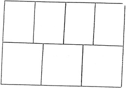
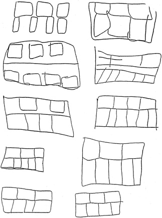
One can suspect an NLD by giving the child a simple visual memory test in the office just before cycloplegia is instilled. It requires only 1 to 2 minutes to administer (Fig. 2).41 NLD is often overlooked as a reason for poor school performance because of the nature of the deficits and progression of problems during development. A well-structured inquiry of the parents regarding the child's history would reveal such behavior to the examining ophthalmologist (problems with writing, copying, scissors skills, possibly artwork).

Fig. 2. A. This design is to be drawn from memory after a 5-second presentation. B. These drawings are attempts by individuals with nonverbal learning disabilities to reproduce the stimulus design from memory. Reprinted with permission from Koller HP: An ophthalmologist's approach to visual processing/learning differences. In Clinical Practice Guidelines Manual. Bethesda, MD: The Interdisciplinary Council on Developmental and Learning Disorders, 2000.

NEUROPATHOLOGY OF NONVERBAL LEARNING DISORDERS AND RELATED CONDITIONS

Rourke has described NLD as a developmental condition in which children are born with primary neuropsychologic deficits including bilateral tactile perception, usually more on the left side, visual perception, complex psychomotor skills, and the ability to deal with novel material. These primary deficits lead to secondary deficits including poor attention to tactile and visual stimuli and ultimately tertiary deficits including poor tactile, visual memory, reasoning, concept formation, and math skills. Rourke's original work hypothesized a right hemisphere basis for these problems. He further stated that the child's primary assets in verbal skills were the result of normal development of the left hemisphere and then ultimately reliance on these skills to the point in which they became possibly advanced.42

Rourke43 later reconceptualized his model and began to discuss NLD in what he called the “white matter model.” He indicated that the white matter functions, connecting the two hemispheres, projecting to the diencephalon, brainstem, and spinal cord, were impaired in these children and that in healthy children there is more white matter in the right hemisphere than left. Therefore, the underlying pathology is lack of development of the white matter, which affects the right hemisphere more so than the left.

There are several conditions that have been associated with NLD. They include Asperger's syndrome, Williams syndrome, and early hydrocephalus.44 Additional disorders that are hypothesized to disrupt white matter and cause NLD include Turner syndrome,45 agenesis of the corpus callosum,46 and velocardial facial syndrome.47

TREATMENT

The diagnosis of NLD tends to occur later in a child's academic life. As opposed to reading problems, which are diagnosed in the first and second grades (when the child fails to develop reading skills), NLD tends to be diagnosed in later elementary years and junior high school. The reason for this is unclear and may be a combination of academic expectations (i.e., math is harder, some people do not “get” math, and this is more acceptable than the inability to acquire basic reading skills; when comprehension of written text is not required until later in an academic career). These children also can be compartmentalized in that they receive services for motor or sensory problems as younger children but not work on cognitive problems/social difficulties until later. In addition, the need for higher order concept formation and social skills increases in junior high school, and thus the problems may not be observed until then.

Well-developed verbal and reading skills, however, are often observed in these children and adults. Occupational therapy is the most effective treatment for early developmental problems in NLD. Frequently, therapy must be coordinated with a homework tutor, reading specialist, and/or a physical therapist if other, more complex, aspects of NLD are identified. “Numerous treatment strategies have been tried over the years but utilizing current repetitive, standard rehabilitation science techniques seem, at present, to be best.”48

Optometric vision training techniques are at times effective in patients with NLD. These therapies are partially based on occupational and physical therapy principles as well as standard rehabilitation science. Adults, after strokes, as well as patients of all ages after accidental closed-head trauma, and brain tumor surgery are taught to use the uninjured functioning parts of their brain to compensate for loss of normal use of an injured or damaged brain region despite the fact that the area of the brain assuming the new function was never naturally intended to do so. Patients who have had a stroke thus relearn to read, write, talk, and walk again in the presence of permanent brain damage to the regions previously genetically programmed to maintain those specific functions. A similar model exists for children with naturally occurring brain dysfunction, such as is present in NLD.

Laterality training, eye movement coordination strategies to help create smooth pursuits, and hand-eye coordination exercises all help improve, to some degree, performance of those functions. To what direct extent learning itself, in school or at home, is improved in all cases has yet to be shown through evidence-based science using a masked or double-masked controlled study. Published studies reviewed by these authors49 and others50 are anecdotal or retrospective/prospective reviews of measurement or performance, often while other remedial methods are carried out at the same time.

Some optometric vision training exercises are designed to improve visual information processing.51 Initially the first therapeutic goal is to develop the individual's motor-awareness ability by performing isolated, simultaneous, and sequential movements involving both sides of the body. The “vision therapist” is trying to develop motor awareness, then motor planning, motor integration, motor execution, and finally motor memory of the differences between the right and left sides of the body (similar to that in occupational therapy). These strategies apparently work by repetitive, positive psychologic reinforcement and one-on-one patient-therapist interaction as much as a direct influence on the cerebral pathologic learning process itself (i.e., procedural memory). Because NLD involves higher cognitive learning centers in which abstract thinking, visual memory, and spatial recognition are the main deficits, repetitive training makes it possible for the individual to learn to perform certain tasks better. In theory, if a patient with an NLD is trained to enhance motor awareness, motor planning, motor integration, motor execution, and motor memory, and this improvement can then be transferred to the academic sphere, the benefits of optometric vision training could be realized. Tutoring in math as well as organization and conceptualization becomes more important in treatment as the children get older.

The second component of optometric vision training involves orthoptic techniques to improve fusional amplitudes and create diplopia awareness. This may be of some benefit in cases of convergence insufficiency exophoria/tropia by diminishing reading fatigue, but in no way does this mode of vision training help or enhance the ability to actually read or comprehend written material. Many individuals are confused about these two divisions of optometric vision therapy.48

The largest treatment difficulty for children with NLD, especially as they get older, is the psychosocial deficits. Social-skills training (literally training interpersonal behavioral skills through role modeling and role-playing) is one technique.52 Psychologic support and psychiatric consultation for medications have also been used to ameliorate depression and anxiety. Goldberg et al53 reported that approximately 40% of a sample of children with NLDs have a Diagnostic and Statistical Manual of Mental Disorders, Fourth Edition (DSM-IV) diagnosis in adolescence.

Back to Top
ATTENTION DEFICIT/HYPERACTIVITY DISORDERS
The diagnosis of attention deficit disorder and ADHD is established by the criteria listed in the DSM-IV.54 They are as follows:
  1. Either (1) or (2):
    1. Six (or more) of the following symptoms of inattention have persisted for at least 6 months to a degree that is maladaptive and inconsistent with developmental level:
      Inattention
      1. often fails to give close attention to details or makes careless mistakes in schoolwork, work, or other activities
      2. often has difficulty sustaining attention in tasks or play activities
      3. often does not seem to listen when spoken to directly
      4. often does not follow through on instructions and fails to finish schoolwork, chores, or duties in the workplace (not because of oppositional behavior or failure to understand instructions)
      5. often has difficulty organizing tasks and activities
      6. often avoids, dislikes, or is reluctant to engage in tasks that require sustained mental effort (such as schoolwork or homework)
      7. often loses things necessary for tasks or activities (e.g., toys, school assignments, pencils, books, or tools)
      8. is often easily distracted by extraneous stimuli
      9. is often forgetful in daily activities

    2. Six (or more) of the following symptoms of hyperactivity-impulsivity have persisted for at least 6 months to a degree that is maladaptive and inconsistent with developmental level:
      Hyperactivity
      1. often fidgets with hands or feet or squirms in seat
      2. often leaves seat in classroom or in other situations in which remaining seated is expected
      3. often runs about or climbs excessively in situations in which it is inappropriate (in adolescent or adults, may be limited to subjective feelings of restlessness)
      4. often has difficulty playing or engaging in leisure activities quietly
      5. is often “on the go” or often acts if “driven by a motor”
      6. often talks excessively

      Impulsivity
      1. often blurts out answers before questions have been completed
      2. often has difficulty awaiting turn
      3. often interrupts or intrudes on others (e.g., butts into conversations or games)
      4. some hyperactive-impulsive or inattentive symptoms that caused impairment were present before age 7 years
      5. some impairment from the symptoms is present in two or more settings (e.g., at school [or work] and at home)
      6. there must be clear evidence of clinically significant impairment in social, academic, or occupational functioning


Controversy, however, still exists in some circles, concerning the real existence and definition of these two diagnoses as a separate category of learning disorders.55 The American Academy of Pediatrics published the clinical practice guidelines for ADHD in 2000.56 The ophthalmologist can identify patients with suspected ADHD in the office setting by noting the patient's behavior during the eye examination and referring the child or adult to the appropriate professional for definitive diagnosis and effective treatment. The patient will often manifest inattention, impulsivity, and hyperactivity, making the examination more difficult. A child may continually get out of the examination chair, open drawers, or press buttons on the instrument control panel. A history from the family of similar behavioral difficulties in school or at work is also highly suggestive. Drug and psychologic therapies are both appropriate treatments if the diagnosis of ADHD is made.

NEUROANATOMY OF THE DISORDER

The neuroanatomic underpinnings of ADHD have been generally reported as reduced activation of the right neostriatum and possible right hemisphere dysfunction.57 Heilman et al58 also proposed that frontal deficits are involved because of the lack of response inhibition and motor restlessness. Lou et al59 reported that SPECT studies supported this hypothesis because there was hypoperfusion in the right striatal regions of children with ADHD.

Several studies have suggested that the dopaminergic and noradrenic systems are dysfunctional in children with ADHD. This is likely a result of studies designed at looking at these two systems and the response of many of the children with ADHD to stimulant medication (shown to affect both neurotransmitter systems). In addition, genetic studies have shown an increased risk of ADHD in children who have parents with ADHD, thus supporting the biologic cause of the condition.59,60

TREATMENT

Pharmacologic Interventions

Psychostimulants have been traditionally incorporated as the primary line of intervention for children with ADHD. The three most prescribed medications for ADHD are methylphenidate, pemoline, and dextroamphetamine.61 Methylphenidate is the most frequently prescribed of the three approximately 71% of the time.62 The stimulant medications are believed to increase the release of catecholamines and to block their reuptake. Methylphenidate, in particular, affects the dopamine transporter system.63 Volkow et al64 took positron emission tomography (PET) scan images of children taking methylphenidate and discovered the highest concentration of the medication in the striatum after delivery.

Stimulant medications have also been shown to improve behaviors at home. Musten et al64 reported that children with ADHD who take stimulants showed improved behavioral functioning in the home including compliance, on-task behavior, and reduced activity levels.

PSYCHOLOGIC INTERVENTIONS

Family training and/or parent training have been demonstrated to be effective psychosocial treatment interventions for children with ADHD. Pelham et al65 reviewed a number of studies that supported the use of behavioral parent training and found them to be effective. Strayhorn and Weidman66 performed a longitudinal study in which family training took place 2 years before the study (it was a follow-up to their original treatment study) and found that the parent–child interaction training continued to have behavioral benefits in the home (not in the school). Similarly, Erhardt and Baker67 found that a family-based intervention that included management training and education on the nature of ADHD and the associated behavior problems lead to management improvement in the home, parental self-confidence, and increased knowledge of the condition. Barkley et al68 also found positive results in family treatment in terms of reduced behavioral symptomology in the home, reduced conflicts, negative communications, and maternal feelings of depression.

SCHOOL-BASED INTERVENTIONS

Several of the academic suggestions that should be considered include placing the child in a smaller classroom to have more individualized access to the teachers. This general academic modification will allow the teacher–child interaction time to increase and therefore permit the possibility of increased behavioral management opportunities. In addition, using a structured classroom setting is generally believed to be effective.69 Preferential seating in the front of the classroom and away from distracting classroom situations (window, pencil sharpener, free play area, disruptive children) may be useful. The teacher may also develop a nonverbal cue to signal the child in a nonpunitive manner that he or she is not focusing. Similarly, “checking in” with a child may be helpful to make certain he or she is following the assignment (e.g., “we are now on page five”). Providing the child extended time on examinations may be useful because the literature has supported reduced processing speed times with tasks.70 Allowing the child to get up and move around during the school day can be useful to release some of the motor restlessness (this can be done productively by having the child be in charge of taking lunch money to the school office or other errands). Similarly, behavioral interventions such as rewarding appropriate behaviors, task completion, and self-control may be helpful.69

Back to Top
PERVASIVE DEVELOPMENTAL DISORDERS
Often, the ophthalmologist can identify an individual with pervasive developmental disorders (PDD; previously called autism spectrum disorders) through history and observation of the patient's behavior in the examination chair. Lack of eye contact is often the initial complaint and reason for referral to the ophthalmologist.71 From the family history, the ophthalmologist can learn that the patient has been observed to exhibit a lack of social relatedness and normal peer interaction. A limited but intense range of interest and repetitive, stylized behaviors are also characteristics of autistic spectrum disorders of which the ophthalmologist should be aware. It is the ophthalmologist's responsibility to detect and physical abnormality in the visual system that would cause lack of eye contact or other PDD sign or symptom. Beyond that, the ophthalmologist should direct the family to the appropriate experts for further evaluation, diagnosis, and therapy.

NEUROANATOMY OF AUTISM

Recent research has shown that children with autism have larger brain volumes at 2 to 3 years of life and abnormal head circumferences at the same age.73 Courchesne et al72 found a developmental pattern of brain growth in their sample of autistic children; these children were born with smaller head circumferences and had an accelerated rate of growth between the ages of 1 to 2 months and 6 to 14 months. The excessive brain size was the result of increased white matter volumes in the cerebellum and cerebrum, and increased gray matter primarily in the frontal lobes.

Additional neuroanatomic findings have been reported in the literature. The literature has reported cerebellar and limbic system abnormalities.73,74 Reduced cerebellar size has been one consistent finding along with increased neuronal density in the limbic system.75 In addition, PET imaging studies confirmed lower activation levels in frontoparietal regions as well as in the neostriatum and thalamus.76

The incidence rate of autism has been reported to be increasing during the last several years.77 The reason for this remains elusive. The genetic transmission of autism seems to be multifactorial, and there are cases that involve autosomal recessive genes, X-linked genes, and chromosal abnormalities.31 Fragile X syndrome and untreated phenylketonuria have also been shown to cause autism.78 Chromosome 9 has been explored as possibly being involved in the transmission of the condition.79 The incidence rate of autism in the healthy population ranges from 2 to 4 per 10,00080 to 1 per 1000,54 whereas several studies have shown that there is approximately a 3% chance of having an autistic sibling when another sibling has been diagnosed with the condition.80,81

COGNITIVE/BEHAVIORAL MANIFESTATIONS

Approximately 75% of autistic individuals are mentally retarded with lower verbal scores than scores on “hands-on” type tests, which may even approximate average. High-level autistic children may also show adequate word recognition, spelling, and reading fluency, and perform within normal academic limits in the early years of school (usually stand out socially). Subsequently, they begin to struggle later in school with math concepts and reading comprehension that involves reasoning and organization of material as the demand for these skills increase. Moreover, social skills differences continue to grow with age, as do needs for more executive functions (i.e., organization, mental flexibility, problem solving). These cognitive skills are typically more limited for these patients, even in “higher level” cases. The literature has shown that there are more males impacted by the disease (3:1 ratio) but that females tend to have lower intellectual abilities and more behavioral disturbances.82

TREATMENT

Treatment of the autistic individual has been controversial. The essence of the argument lies in whether an autistic individual can be treated to become or approximate “normal” versus treatments that are focused on developing compensatory mechanisms for the individual to cope with the disorder.

In 1987, Ivar Lovaas82 published an article documenting 19 children with autism diagnoses who were treated with a behavior modification treatment called applied behavior analysis. His group trained the children using discrete trial learning for desired behaviors and to decrease undesired behaviors (i.e., self-stimulation, aggressiveness). The methodology was extremely intensive requiring support 40 hours per week over the course of at least several years. The concept was to have the child's behavior directed during all, or at least most, hours of waking. The treatment involves parents, clinicians, teachers, and trained technicians. Lovaas reported that 47% of his sample after several years of treatment (the children started treatment on average at ∼34 months of age) and between the ages of 6 and 7 years old (when the children theoretically would have completed the first grade) attained “normal intellectual and academic functioning.” This number represented a substantial improvement over the 2% of the control group at the same time interval. It should be noted that 40% of the children in the experimental group were mildly retarded and placed in special education classes of the language delayed. This “second” level result was commensurate with the control group's 45%.83 There have been several critiques against Lovaas' methodology.82 However, McEachin et al84 looked at the same experimental group, aged between 9 and 19 years old, and found continued performance in the “normal” range in most of the subjects they were able to reevaluate.

In contrast with the discrete trial learning approach, there are several approaches that have focused on developing compensatory methods within the child (and the child's immediate support system–family, teachers, and therapists). These methods typically do not attempt to “normalize” the child but rather help the child maximize his or her potential. One such program is the Treatment and Education of Autistic and Related Communication Handicapped Children (TEACCH) founded by Shopler and colleagues85 at the University of North Carolina at Chapel Hill. The TEACCH program was founded in 1971 and provides parent training in interactions with their autistic children, evaluations of the children's strengths/weaknesses, coordinated community-based services including residential programming and training in daily living skills, academic support, and vocational training through a collaborative approach. The TEACCH program helps the individuals with physical organization, schedules, work systems, and task organization.86 It is difficult to evaluate the TEACCH program because of its long-reaching extensions and individually tailored programs, but several studies have reported effectiveness in home training.87,88

Pharmacologic interventions are also commonly applied in children with autism. Medications are typically designed to reduce the undesired behaviors associated with autism including aggressiveness and self-injurious behavior (i.e., head banging and picking/scratching body parts). Neuroleptics have been used effectively to reduce aggressive behaviors and hyperactivity89,90 in several studies. Antidepressants, primarily the selective serotonin reuptake inhibitors, have been incorporated to diminish stereotypic behaviors, compulsions, and rituals.91,92

LEGAL RIGHTS OF CHILDREN WITH LEARNING DISABILITIES

Children with LD diagnoses are protected under the IDEA, which provides free, appropriate public education for all children. Each child with an LD diagnosis (and/or any medical/psychiatric condition that might affect learning such as blindness) is covered under this law from infancy (i.e., early intervention programs) to the age of 21 years or graduation from high school. IDEA lists 13 classifications for exceptionality (qualifying the individual for special education services). The categories are as follows:

  1. Autism
  2. Deaf-blindness
  3. Deafness
  4. Emotional disturbance
  5. Hearing impairment
  6. Mental retardation
  7. Multiple disabilities
  8. Orthopedic impairment
  9. Other health impairment
  10. Specific LD
  11. Speech and/or language impairment
  12. Traumatic brain injury
  13. Visual impairment93

Schools are held responsible for identifying and evaluating these children, and, when a determination of “exceptionality” is made, there is a provision of a free public education in the least restrictive environment. The child's educational program that has been developed is called an Individual Educational Plan.

IDEA is called an “entitlement law” in that children are permitted these services under the law, which is enforceable. It should be noted that IDEA no longer holds when a child transitions into a college setting. College students with disabilities, however, are protected under the ADA of 1990 that is a civil right law. This law was activated to ensure that “no otherwise qualified person with a disability is denied access to, benefits of, or is subject to discrimination solely on the basis of disability.”94 This law protects students with needs but does not provide for free, appropriate public education. This can be very confusing for students and parents of individuals with LDs because the legal standards are less restrictive for college students (i.e., what is enforceable in terms of what accommodations for these students), and thus many students who receive support in the public school system do not receive the same support in college (which is legally accurate). Thus, students who are pursuing college will need to spend more time exploring the legal supports and the individual support that are in place in their college (because there is no legal requirement to provide support services for students with LD, and even if a college does, there is no mandate regarding the nature of the support program).

Back to Top
CONCLUSION
As mentioned earlier, the ophthalmologist should be aware of the four neuropsychologic categories of learning disorders, their underlying physiologic neuropathology, and the available strategies to treat and rehabilitate individuals so affected. Many of the conditions are inherited, and parents of affected children often manifest some of the characteristics of the particular disorder present in the patient themselves. Keep in mind that there is often comorbidity. Many individuals have more than one type of LD.

Koller has emphasized that “having the ophthalmologist aware of all of the medical and nonmedical conditions and situations that could affect learning in a child or older individual increases the potential for a child with LDs to receive appropriate, effective, and timely remedial treatment.”95 Table 3 is a list of all specialties, both medical and nonmedical, that could be involved in any case of LD. Table 4 is a list of support groups and organizations that parents and families can contact for additional information on learning disorders. Because the ophthalmologist is often the first expert to whom the primary care physician refers the individual with a suspected LD, he or she must possess the knowledge, motivation, passion, and resources necessary to refer the patient to the appropriate specialists. The public perceives us to be experts in visual processing and perception as well as visual function and ocular pathology. Ophthalmology must embrace this field and be a leading partner in the interdisciplinary science of learning disorders, along with all of the specialists listed, for the benefit of our patients.

TABLE 3. Role of the Pediatric and Comprehensive Ophthalmologist Concerning Individuals with Learning Disabilities

  1. Identify and treat any eye or eye-related systemic disease or abnormality.
  2. By observation and careful history, identify which broad category of learning differences the patient likely has and convey that impression to the pediatrician, the family, and any other interested parties. These children have often been through many psychologic tests, tutoring, and other attempts at remediation without a definitive diagnosis or combination of diagnoses. Suggest a comprehensive neuropsychologic educational evaluation from a qualified, credentialed neuropsychologist when all interested parties agree.
  3. Help the family by giving them a direction in which to proceed so that the child with nonocular learning differences can start to achieve his or her full potential. Not every individual with learning disabilities requires every possible specialist. These professionals include the following:
    1. Pediatric neurologist
    2. Pediatric psychiatrist
    3. Pediatric developmental specialist
    4. Pediatric endocrinologist
    5. Pediatric geneticist
    6. Pediatric otolaryngologist
    7. Pediatric ophthalmologist
    8. Speech and language pathologist
    9. Neuropsychologist
    10. Educational psychologist
    11. Educator with special education credentials
    12. Reading tutor
    13. Physical therapist
    14. Occupational therapist
    15. Pediatric social worker
    16. School placement expert (educator)
    17. Disabilities attorney
    18. Family physician or general pediatrician
  4. A specialized attorney is often beneficial for helping families receive federal learning disabled benefits to which they are entitled under the terms of the Individuals with Disabilities Education Act (IDEA) and Americans with Disabilities Act (ADA).
  5. When the answer to the question, “Is the child's reading and/or learning problem in school because of his eyes?” is “No,” the ophthalmologist must explain why it is not and offer a positive and constructive method to direct those families toward obtaining the proper and appropriate care. The public still believes the eye specialists are knowledgeable in the field of visual perception as well as visual function.


TABLE 4. Patient Support Groups for Individuals Who Are Learning Disabled


A. CHADD (Children and Adults with ADD) CHADD.org
B. CEC (Children's Educational Counsel) CEC.org
C. ASLHA (American Speech, Language, and Hearing Association) ASLHA.org
D. LDAA (Learning Disabilities Association of America) LDAA.org
E. PERC (Parents Educational Resource Center) PERC.org
F. The Orten Dyslexia Society
G. AHA (American Hyperplexia Association) hyperplexia.org
H. HALO (Health Achievement Learning Opportunities Centers) HALOCENTERS.com


Back to Top
REFERENCES

1. Attention deficit hyperactivity disorder in adults. Medical Crossfire 4;2:30, February 2002

2. The Optometric Extension Program Foundation 1996 Annual Report. Available at: http://www.healthy.net/oep/annual.htm. Accessed 5/31/2005.

3. Scheiman MM, Rouse MW: Optometric Management of Learning-Related Vision Problems. St. Louis, Mo: C.V. Mosby, 1994

4. Koller HP: How does vision affect learning? Part II. J Ophthalmic Nurs Technol 18(1):12, 1999

5. Lyon GR: Research initiatives in learning disabilities: Contributions from scientists supported by the National Institute of Child Health and Human Development. J Child Neurol 10:S120–S126, 1995

6. Hoyt CS: Visual training and reading. Am Orthopt J 49:23, 1999

7. Picard EM, Del Dotto JE, Breslau N: Prematurity and low birthweight. Central nervous system dysfunction in other disorders. In Yeates KO, Ris MD, Taylor HG, eds:. Pediatric Neuropsychology: Research, Theory, and Practice. New York: Guilford Press, 2000

8. Taylor HG, Minich N, Bangert B, et al: Long term neuropsychological outcomes of very low birth-weight: Associations with early risks for periventricular brain insults. J Int Neuropsychol Soc 10:987, 2004

9. Koller HP, Goldberg KB: A guide to visual and perceptual learning disabilities. Curr Concepts Ophthalmol 7:24, 1999

10. Phillips PH, Newman NJ: Mitochondrial diseases in pediatric ophthalmology. J AAPOS 1(2):115, 1991

11. Groom M, Kay MD, Corrent GE: Optic neuropathy in familial dysautonomia. J Ophthalmol 17(2):101, 1997

12. Koller HP: An ophthalmologist's approach to visual processing/learning differences. J Pediatr Ophthalmol Strabismus 39(3):140, 2002

13. Miller NR: Chapter 56. In Miller NR, Newman NJ, eds: Walsh and Hoyt's Clinical Neuro-Ophthalmology, Fifth Edition. Baltimore: Williams & Wilkins, 1998:3664

14. Dorland's Illustrated Medical Dictionary, 28th Edition. Philadelphia: W. B. Saunders, 1994

15. Troost BT: Migraine. In Duane TD, Jaeger EA, eds.: Clinical Ophthalmology. Vol. 2, Chapter 19. Philadelphia: Harper & Row, 1988

16. Miller NR: Chapter 56, Table 56-3. In Miller NR, Newman NJ, eds: Walsh and Hoyt's Clinical Neuro-Ophthalmology, Fifth Edition. Baltimore: Williams & Wilkins, 1998

17. O'Hara MA, Koller HP: Migraine in a pediatric ophthalmology practice. J Pediatr Ophthalmol Strabismus 35:203, 1988

18. Van Stavern GP: Chapter 26. Headache and facial pain. In Miller NR, Biousse V, Newman NJ, Kerrison JB, eds. Walsh and Hoyt's Clinical Neuro-Ophthalmology, Sixth Edition. Baltimore: Williams & Wilkins, 2005:1282–1293

19. O'Hara MA, Koller HP: Migraine in a pediatric ophthalmology practice. J Pediatr Ophthalmol Strasbismus 35:203, 1988

20. Miller NR: Chapter 56. In Miller NR, Newman NJ, eds: Walsh and Hoyt's Clinical Neuro-Ophthalmology, Fifth Edition. Baltimore: Williams & Wilkins, 1998:3666

21. National Joint Committee for Learning Disabilities (NJCLD). Learning disabilities: Issues on definition. A position paper. Journal of Learning Disabilities 20:107, 1987

22. Gaddes WH, Edgell D: Learning Disabilities and Brain Function. 3rd ed. New York: Springer, 1993

23. Baker L, Cantwell DP: Comparison of well, emotionally disordered, and behaviorally disordered children with linguistic problems. J Am Acad Child Adolesc Psychiatry 26:193, 1987

24. Hynd GW, Obrzut JE, Hayes F, Becker MG: Neuropsychology of childhood learning disabilities. In Wedding D, Horton AM, Webster J, eds. The Neuropsychology Handbook. New York: Springer Publishing, 1986:456–485

25. American Psychiatric Association. Diagnostic and Statistical Manual of Mental Disorders, Fourth Edition. Washington, DC: American Psychiatric Association, 1994

26. Commonwealth of Pennsylvania. Chapter 14: Special education services and programs state regulations. Harrisburg, PA: Department of Education; 2001

27. Grimes J: Responsiveness to interventions: The next step in special education identification, service, and exiting decision making. In Bradley R, Danielson L, Hallahan D, eds. Identification of Learning Disabilities: Research to Practice. Mahwah, NJ: Lawrence Erlbaum Associates, Inc., 2002:531–547

28. Shaywitz S: Overcoming Dyslexia. New York: Alfred A. Knopf, 2003

29. Kolb B, Wishaw IQ: Fundamentals of Human Neuropsychology. 5th edition. New York: W. H. Freeman and Company, 2003

30. Foder JA: The Modularity of Mind. Cambridge, MA: MIT Press, 1983

31. Pennington BF: Diagnosing Learning Disorders: A Neuropsychological Framework. New York: The Guilford Press, 1991

32. Welsh LW, Welsh JJ, Healy M, Cooper B: Cortical, subcortical and brainstem dysfunction: A correlation in dyslexic children. Am J Rhinolaryngol 9(3):310, 1982

33. Welsh LW, Welsh JJ, Healey MP: Central auditory testing and dyslexia. Laryngoscope XC(6):972, 1980

34. Shaywitz BA, Fletcher JM, Shaywitz SE: Defining and classifying learning disabilities and attention-deficit/hyperactivity disorder. J Child Neurol 10(suppl 1):S50, 1995

35. Shaywitz SE: Dyslexia: Current concepts. N Engl J Med 338:307, 1998

36. Pennington BF, Bender B, Puck M, Salbenblatt J, Robinson A: Learning disabilities in children with sex chromosome anomalies. Child Dev 53:1182, 1982

37. Lovett M: Developmental reading disorders. In Feinberg T, Farah M, eds. Behavioral Neurology and Neuropsychology. New York: McGraw Hill, 1997:773–787

38. Shaywitz BA, Shaywitz SE, Pugh KR, et al: Disruption of posterior brain systems for reading in children with developmental dyslexia. Biol Psychiatry 52:101, 2002

39. Goldberg E: Gradiential approach to neocortical functional organization. J Clin Exp Neuropsychol 11(4):489, 1989

40. Kolb B, Wishaw IQ: Fundamentals of Human Neuropsychology. 5th edition. New York: W. H. Freeman and Company, 2003

41. Foss JM: Nonverbal learning disabilities and remedial interventions. Ann Dyslexia 41:128, 1991

42. Rourke BP: Nonverbal Learning Disabilities: The Syndrome and the Model. New York: Guilford Press, 1989

43. Rourke BP: Syndrome of Nonverbal Learning Disabilities: Neurodevelopmental Manifestations. New York: Guilford Press, 1995.

44. Rourke BP: Frequently Posed Questions About the Syndrome of NLD. Philadelphia: 2003

45. Lyon GR, Rumsey JM: Neuroimaging: A Window to the Neurological Foundations of Learning and Behavior in Children. Baltimore: Brookes, 1996

46. Smith LA, Rourke BP: Callosal agenesis in syndrome of nonverbal learning disabilities. In Rourke BP, ed. Syndrome of Nonverbal Learning Disabilities: Neurodevelopmental Manifestations. New York: Guilford Press, 1995:351–378

47. Van Amelsvoot T, Daly E, Robertson D, et al: Structural brain abnormalities associated with deletion at chromosome 22q11: Quantitative neuroimaging study of adults with velo-cardio-facial syndrome. Br J Psychiatry 178:412, 2001

48. Koller HP: An ophthalmologist's approach to visual processing/learning differences. J Pediatr Ophthalmol Strabismus 39(3):133, 2002

49. Koller HP, Goldberg KB: Working with visually impaired children and their families. In Nelson LB, Olitsky SE, eds. Harley's Pediatric Ophthalmology, 5th edition. Philadelphia: W. B. Saunders, 2005

50. Reinecke RD: Role of the ophthalmologist in reading disorders. Chapter 25. In Nelson LB, ed. Harley's Pediatric Ophthalmology, 4th edition. Philadelphia: W. B. Saunders, 1998, 499–507

51. Scheiman MM, Rouse MW: Optometric Management of Learning-Related Vision Problems. St. Louis: C.V. Mosby, 1994.

52. Thompson S: Nonverbal learning disorders. The Gram 1, 2000

53. Goldberg KB, Moss E, Dadario B, Corsey R: Information Processing and Personality in Children and Adolescents with Nonverbal Learning Disabilities. Philadelphia: Society for Personality Assessment, 2001

54. American Psychiatric Association: Diagnostic and Statistical Manual of Mental Disorders, Fourth Edition. Washington, DC: American Psychiatric Association, 1994

55. Carey WB: Problems in diagnosing attention and activity. Pediatrics 103(3):664, 1999

56. American Academy of Pediatrics: Clinical Practice Guidelines: Diagnosing and evaluation of the child with attention deficit/hyperactivity disorder. Pediatrics 105(5):1158, 2000

57. Mesulam MM: Principles of Behavioral and Cognitive Neurology. 2nd Edition. New York: Oxford University Press, 2000

58. Heilman KM, Voeller KKS, Nadeau SE: A possible pathophysiologic substrate of attention deficit hyperactivity disorder. J Child Neurol 6(suppl):S76, 1991

59. Lou HC, Hendrikson L, Bruhn P: Striatal dysfunction in attention deficit and hyperkinetic disorder. Arch Neurol 46:48, 1989

60. Zametkin AJ: The neurobiology of attention-deficit hyperactivity disorder: A synopsis. Psychiatr Ann 19(11):584, 1989

61. Greenhill LL: Childhood attention deficit hyperactivity disorder: Pharmacological treatment. In Nathan PE, Gorman JM, eds. A Guide to Treatments That Work. London: Oxford University Press, 1998:42–62

62. Greenhill LL, Halperin JM, Abikoff H: Stimulant medications. J Am Acad Child Adolesc Psychiatry 38(5):503, 1999

63. Volkow ND, Ding YS, Fowler JS, et al: Is methylphenidate like cocaine? Arch Gen Psychiatry 52:456, 1995

64. Musten LM, Firestone P, Pisterman S, Bennett S, Mercer J: Effects of methylphenidate on preschool children with ADHD: Cognitive and behavioral functions. J Am Acad Child Adolesc Psychiatry 36:1407, 1997

65. Pelham WE, Wheeler T, Chronis A: Empirically supported psychosocial treatments for attention deficit hyperactivity disorder. J Clin Child Psychol 27(2):190, 1998

66. Strayhorn JM, Weidman CS: Follow-up one year after parent-child interaction training: Effects on behavior of preschool children. J Am Acad Child Adolesc Psychiatry 30:138, 1991

67. Erhardt D, Baker BL: The effects of behavioral parent training on families with young hyperactive children. J Behav Ther Exp Psychiatry 21:121, 1990

68. Barkley RA, Guevremont DC, Anastopoulos AD, Fletcher KE: A comparison of three family therapy programs for treating family conflicts in adolescents with attention-deficit hyperactivity disorder. J Consult Clin Psychol 60(3):450, 1992

69. McGoey KE, Eckert TL, DuPaul GJ: Early intervention for preschool-age children with ADHD: A literature review. J Emotion Behav Disord 10(1):14, 2002

70. Kennedy R, Glassman R: Subtypes in ADHD: A newsmaker interview with Mary Solanto, Ph.D; 2003. Available at http://www.medscape.com/viewarticle/463181. Accessed 9/04.

71. Koller HP: Visual perception and learning differences: An ophthalmologist's view. Am J Orthop 49:175, 1999

72. Courchesne E, Carper R, Akshoomoff N: Evidence of brain overgrowth in the first year of life in autism. J Am Med Assoc 290(3):337, 2003

73. Courchesne E: Autism. In Lamb HR, Peschel E, Peschel R, Howe CW, Howe JW, eds.: New Directions for Mental Health Services: No. 54 Neurobiological Disorders in Children and Adolescents. San Francisco, CA: Jossey-Bass Publishers, 1992:19–23

74. Gaffney GR, Kuperman S, Tsai LY, Minchin S: Forebrain structure in autism. J Am Acad Child Adolesc Psychiatry 28:534, 1988

75. Courchesne E, Yeung-Courchesne R, Press GA, Hesselink JR, Jernigan TL: Hypoplasia of cerebellar vermal lobules VI and VII in autism. N Engl J Med 318:1349, 1988

76. Horowitz B, Rumsey J, Grady C, Rappaport S: The cerebral metabolic landscape in autism. Arch Neurol 45:749, 1988

77. Kabot S, Masi W, Segal M: Advances in the diagnosis and treatment of autism spectrum disorders. Prof Psychol Res Pr. 34(1):26, 2003

78. Folstein S, Rutter ML: Autism: Familial aggregation and genetic implications. J Autism Dev Disord 18:3, 1988

79. Smalley SL, Asarnow RF, Spence MA: Autism and genetics: A decade of research. Arch Gen Psychiatry 45:953, 1988

80. Happe F, Frith U: The neuropsychology of autism. Brain 119:1377, 1996

81. Konstantareas MM, Homatidis S, Busch J: Cognitive, communication and social differences between autistic boys and girls. J Appl Dev Psychol 10:411, 1989

82. Lovaas OI: Behavioral treatment and normal educational and intellectual functioning in young autistic children. J Consult Clin Psychol 55(1):3, 1987

83. Gresham FM, MacMillan DL: Early intervention project: Can its claims be substantiated and its effects replicated? J Autism Dev Disord 28:5, 1998

84. McEachin JJ, Smith T, Lovaas O: Long-term outcome for children with autism who received early intensive behavioral treatment. Am J Ment Retard 97(4):359, 1993

85. Mesibov GB: A comprehensive program for serving people with autism and their families: The TEACCH model. In Matson, JL, ed. Autism in Children and Adults: Etiology, Assessment, and Intervention. Pacific Grove, CA: Brooks/Cole Publishing Company, 1994:85–97

86. Gresham FM, Beebe-Frankenberger ME, MacMillan DL: A selective review of treat ments for children with autism: Description and methodological considerations. School Psych Rev 28(4):559, 1999

87. Ozonoff S, Cathcart K: Effectiveness of a home program intervention for young children with autism: A review. J Autism Dev Disord 28:25, 1998

88. Schopler E, Reichler RJ, Bashford A, Lansing MD, Marcus LM: Individualized Assessment and Treatment for Autistic and Developmentally Disabled Children (vol. 1): Psychoeducational Profile Revised. Austin, TX: PRO-ED, 1990

89. Malek-Ahmadi P, Simonds JF: Olanzapine for autistic disorder with hyperactivity (letter to the editor). J Am Acad Child Adolesc Psychiatry 37:902, 1998

90. Harteveld EM, Buitelaar JK: Autism: Role of drug treatment and a guide to its use. CNS Drugs 8:227, 1997

91. Bower B: Promising addition to autism treatment. Science News 141:164, 1992

92. McDougle CJ: Psychopharmacology. In Cohen DJ, Volkmar FR, eds. Handbook of Autism and Pervasive Developmental Disorders (2nd edition). New York: John Wiley & Sons, Inc., 1997

93. Commonwealth of Pennsylvania. Chapter 14: Special education services and programs state regulations. Harrisburg, PA: Department of Education, 2001

94. Americans with Disabilities Act of 1990, www.eeoc.gov/policy/ada.html

95. Greenspan SI, ed: Clinical Practice Guidelines, The Interdisciplinary Council on Developmental and Learning Disorders. Bethesda, MD: ICDL Press, 2000:257

Back to Top