Chapter 60
Pathogenesis and Control of Blinding Trachoma
CHANDLER R. DAWSON
Main Menu   Table Of Contents

Search

ETIOLOGY, PATHOLOGY, AND PATHOGENESIS
CLINICAL DISEASE, CLASSIFICATION, AND DIAGNOSIS
EPIDEMIOLOGY
TREATMENT AND PREVENTION
COMMENTARY
REFERENCES

Trachoma is a chronic infectious keratoconjunctivitis that is a major cause of blindness in many developing countries. It presents initially as a follicular conjunctivitis, with superficial keratitis and corneal vascularization, and gradually progresses to conjunctival scarring and lid distortion. Damage to the cornea occurs from the chronic inflammatory disease and later from exposure and trauma from the distorted lids and inturned lashes.

Endemic trachoma is still a major cause of blindness in poorer rural communities of developing countries, especially in arid areas.1 Trachoma affects about 600 million people, with active infectious disease in 150 million and operable trichiasis and entropion in 11 million.2 There are an estimated 6 million blind from trachoma and even more have partial loss of vision. Clinical criteria can provide an estimate of the current and future impact of the disease on affected communities and thus the need for intervention. It is also possible to distinguish communities with “blinding trachoma” from communities with “nonblinding trachoma.”

Trachoma is a chronic inflammation of the conjunctiva and cornea that progresses to conjunctival scarring and corneal vascularization. Chlamydia trachomatis is the specific and unique etiologic agent of trachoma but other pathogenic microorganisms often contribute to the disease process.3 In its communicable inflammatory phase, the conjunctiva in trachoma is characterized by the presence of lymphoid germinal centers (lymphoid follicles) surrounded by intense and inflammatory diffuse infiltration and hyperemia (papillary hypertrophy) and by the growth of superficial blood vessels over the superior cornea (vascular pannus).

Trachomatous inflammation may undergo spontaneous resolution or progress to severe conjunctival scarring, which leads to distortion of the lids with trichiasis or entropion, corneal surface defects, and corneal opacity. Thus, trachoma may heal with no permanent damage or in its severe forms produce permanent damage to the cornea. The final visual acuity may range from normal vision to total blindness.1

Back to Top
ETIOLOGY, PATHOLOGY, AND PATHOGENESIS

ETIOLOGY

Chlamydia trachomatis eye infections occur in two distinct epidemiologic settings. The first pattern is the potentially blinding disease of developing countries that is spread by eye-to-eye transmission of infection, defined as endemic trachoma. It is caused by C. trachomatis, invariably the serotypes A, B, Ba, or C.4

The second pattern is infection of the eye by sexually transmitted C. trachomatis strains (usually serotypes D, E, F, G, H, I, J, or K), which produce an eye disease that can be similar to the early inflammatory phases of endemic trachoma. Mild cases are usually called inclusion conjunctivitis but the term paratrachoma has been used to describe the whole spectrum of disease resulting from eye infections with sexually transmitted C. trachomatis strains. Sexually transmitted C. trachomatis also infects newborns during delivery and can cause conjunctivitis, pneumonia, and gastrointestinal infection. Although the sporadic eye infections associated with sexually transmitted C. trachomatis strains rarely produce permanent visual loss, the respiratory tract infections in infants and genital tract infections in adults are important health problems in affected populations.

The A through K strains of C. trachomatis were formerly referred to collectively as TRIC (TRachoma-Inclusion Conjunctivitis) agents because they share most characteristics, particularly replication limited to columnar epithelium. The lymphogranuloma venereum biovar of C. trachomatis (strains L1, L2 and L3) initially infects the mucosal epithelium but also invades the deeper lymphoid tissues. Lymphogranuloma venereum biovar only rarely involves the eye, where it can cause Parinaud's oculoglandular conjunctivitis with diffuse inflammatory conjunctivitis and massive enlargement of the preauricular and submandibular lymph nodes.5 The other chlamydial species (Chlamydia psittaci and Chlamydia pneumoniae) can also cause chronic follicular conjunctivitis.6

Communities with trachoma often have a high rate of bacterial and viral conjunctivitis, which is closely associated with large populations of eye-seeking flies.7,8 These factors escalate nonblinding trachoma into blinding trachoma. Furthermore, in such communities, bacterial corneal ulceration is common and constitutes an acute pathway to blindness.

PATHOLOGY

Conjunctiva

Chlamydial infection in trachoma is limited to the epithelium but the major inflammatory and cicatricial changes appear in the subepithelial connective tissues. In the inflammatory phase, attachments between conjunctival epithelial cells become loosened, so the epithelial cells are often separated in conjunctival smears rather than in attached cell sheets. The epithelium thickens from three to four cell layers to six or eight layers9 except directly over follicles, where it is considerably thinned.10 The conjunctival epithelial cells become irregular in size and may form multinucleated giant cells.11 The conjunctiva is also infiltrated heavily with lymphocytes and polymorphonuclear leukocytes.

In the subepithelial tissues, there is marked vascular dilation and infiltration of the subepithelial tissue by lymphocytic cells and other inflammatory cells. This infiltration elevates the conjunctiva into flat summits with deep folds of epithelium between to form papilla.9,10 Over the tarsal plates where the conjunctiva is bound down to connective tissue, there is only moderate elevation; in the fornices and at the edge of the upper tarsal plate, rolls of elevated hyperemic tissue develop.

The pathognomonic lesion of trachoma is the lymphoid follicle, which forms in the subepithelial tissue. Small follicles consist only of masses of lymphoid cells.9 In larger follicles, the center consists of large lymphocytic cells, with abundant cytoplasm and a few macrophages. Surrounding this is a zone of small lymphocytes, which may extend to the layer immediately below the basement membrane of the epithelium. The cells in the center of these larger follicles sometimes appear to degenerate; with slight pressure, the contents may be expressed onto the conjunctival surface (mature follicles).10 The center follicles appear clinically avascular but dilated vessels are often seen at their periphery. These vessels are often friable and bleed easily when large follicles rupture under slight pressure.

Fibrosis in the subconjunctival connective tissue occurs by fibroblast formation from blood vessels and by cicatrization of necrotic follicles.9 This scar formation often forms a network, leaving islands of relatively unscarred conjunctiva that protrude above the depressed scars. Scarring also obliterates the layer of subconjunctival vessels, grossly deranging the normal vascular architecture.

Folds of epithelium between papilla may become trapped between by the scar tissue to form deep pseudoglands or pseudocysts in the new connective tissue. Epithelium is sloughed into these cystic structures, which appear clinically as yellow concretions or subepithelial lesions that discharge their contents onto the conjunctival surface and are referred to clinically as posttrachomatous degeneration.10 The greasy material from these cysts can cause irritation and form calcified concretions, which may abrade the cornea.

The bulbar conjunctiva undergoes similar inflammatory and cicatricial changes but to a lesser extent. Bulbar follicles occur frequently in children with active trachoma. Scarring of the episcleral tissue results in adhesions between the conjunctiva and sclera, so that during surgical procedures involving separating conjunctiva from the sclera, numerous fibrous bonds must be cut, with resultant bleeding.

Cornea

Chlamydia trachomatis may infect corneal epithelial cells but to a lesser extent than conjunctiva. The cornea, however, is constantly exposed to the infectious agent and to inflammatory mediators from the tears and palpebral conjunctiva. Thus, keratitis in trachoma comprises focal, anterior, infiltrates in the limbal stroma and corneal vascularization, which is more extensive at the upper limbus. The keratitis in trachoma due directly to C. trachomatis infection is limited to the epithelium and anterior stroma. It is likely that infection with other ocular pathogens also contributes to this process.3,11

The epithelium during infectious trachoma becomes edematous with focal collections of polymorphonuclear leukocytes. Near the limbus, inflammatory cells are present in Bowman's membrane and superficial stroma, and this inflammation may cause destruction of the corneal stem cells at the limbus. Superficial vascularization occurs from the vessels of the conjunctival limbus in conjunction with lymphocytic infiltrates. This vascular pannus consists of new vessels and connective tissue and lies between the epithelium and Bowman's membrane. In some cases, it appears to replace Bowman's membrane.12

Lymphoid follicles occur in the conjunctival limbus and cause the absorption of the underlying connective tissue. When these limbal follicles regress, they leave circular depressions (Herbert's pits) in the scleral overhang at the limbus, so the clear cornea is apparent beneath.12 These follicles can also form in the pannus where it overlies the cornea, so that some patients have a double row of Herbert's pits. Epithelium may fill these depressions, so that the surface is smooth, but loss of connective tissue is still apparent on clinical examination.9

In milder cases, the pannus is not extensive and the overlying epithelium is relatively normal. With extensive pannus, however, the surface epithelium can become rough, thickened, and irregular—probably due to replacement of corneal epithelium by conjunctival epithelium after destruction of the corneal stem cells at the limbus. In patients with healed trachoma, the pannus vessels become attenuated and lose their blood column, appearing as ghost vessels.

The lacrimal gland can be infiltrated during active trachoma. There also may be inflammatory involvement of the canaliculus and lacrimal duct by follicles and scar formation, with blockage of tear outflow and subsequent episodes of dacryocystitis.

PATHOGENESIS

Chlamydial Infection

Trachoma agent cannot be identified in all cases of clinically active trachoma. With the highly sensitive and specific DNA amplification technology (polymerase chain reaction [PCR], ligase chain reaction [LCR]), C. trachomatis is found most frequently in severe intensity cases (80% positive), less often in moderate cases (60%), but also in clinically inactive cases (15% to 30%). A study of extraocular chlamydial infection determined by isolation in cell culture showed that agent was present in the nasopharynx or rectum of 15% of young children with active trachoma in Egypt.13 After local or systemic antibiotic treatment, the rate of chlamydial eye infection is substantially reduced (2% to 15%), although the clinical signs of trachoma activity continue at high levels (40% to 60%).

Immunology

Immunologic studies of trachoma and other chlamydial infections have had two main goals: (1) understanding the pathology of the disease, and (2) identifying protective mechanisms to develop effective vaccines. Both humoral and cell-mediated immune responses to chlamydiae occur in trachoma but consensus is that protective immunity to chlamydia is a type 1 helper T-cell (Th-1)-mediated response.14

The antibody response measured by the microimmunofluorescence (micro-IF) test may be to a broad range of C. trachomatis serotypes or limited to the infecting serotype.15–17 In one hyperendemic population, 80% of children had immunogammaglobulin G (IgG) antibody to C. trachomatis.15 In most studies of chronic trachoma, serum IgG antibodies are present at the highest titers; when they appear, immunogammaglobulin M (IgM) antibodies indicate recent infection and occur rarely in trachoma cases.18

In human and experimental C. trachomatis infections, the serum antibody appears to have a protective role but it cannot eliminate the infection. The main immunogenic protein in the chlamydial outer membrane is the major outer membrane protein (MOMP or ompl).19 Antibody to C. trachomatis strains that vary by one amino acid only may lose its ability to neutralize infectivity, however.20 Thus, vaccines based only on a single epitope of MOMP are unlikely to be effective.

Tear antibodies also have been extensively studied. In trachoma, IgG antibody in tears occurs only rarely in the absence of serum antibody; immunogammaglobulin A (IgA, secretory) antibody is found more often in tears than in serum but almost always with tear IgG antibody.15 Tear antibodies (IgG) reflect trachoma intensity accurately; in one study of endemic trachoma, 80% of severe cases, 31% of moderate cases, and 17% of mild cases had tear antibody but serum IgG antibody varied from 81% to 88% in all three groups.15 The titers of both serum and tear antibody are highest in severe cases and lowest in those that are mild. Tear antibody also correlates more highly with presence of the agent than does serum antibody.15 Thus, tear antibody reflects more closely the intensity of the disease and the presence of infective agent.

Tears from patients with trachoma also have a low molecular weight substance (less than 10,000 d) that inhibits C. trachomatis growth in tissue culture.21 Other studies showed that this inhibition is probably caused by γ-interferon, which inhibits growth of C. trachomatis in cell culture and speeds recovery from experimental infection in mice.22,23 In primary human conjunctival cell cultures infected with a type B (trachoma) serovar of C. trachomatis, γ-interferon (but not α- or β-interferon) inhibited growth of C. trachomatis; this effect was reversed by adding tryptophan.24

Other studies have shown that a chlamydial vaccine capable of inducing γ-interferon-secreting T cells in mucosal tissue was highly associated with resistance to challenge infection by C. trachomatis.25 Several other studies have also confirmed that resistance to chlamydial infection is mainly attributable to cell-mediated immunity.14 The consensus of studies of human and experimental chlamydial infections is that the protective response is mainly type I helper T-cell (Th1) response.14,26

Infection of cultured endocervical (genital) epithelial cells by C. trachomatis (serotypes L2, D, I and C. psittaci) induces the prolonged secretion of γ-interferon and other proinflammatory cytokines (IL-6, IL-8, GRO alpha and granulocyte-macrophage colony-stimulating factors).27 In contrast, exposure of these cells to other bacterial cells causes them to have a quick but transient secretion of γ-interferon. The secretory response to chlamydial infection, however, develops more slowly and is sustained as long as chlamydial protein is being synthesized (50 to 100 hours). The mRNA transcripts for inflammatory cytokines (IL1-β, TNF-α, IFN-γ) were found in inflammatory trachoma but only transcripts for IL1-β were present in scarred trachoma.28 It is probable that this sustained cytokine response causes the chronic conjunctival inflammation in trachoma, the subsequent scarring, and other complications.

Most lymphocytes in conjunctival smears from patients with active trachoma are T lymphocytes (associated with cell-mediated immunity) and there are fewer B cells. Helper, suppressor-cytoxic, and natural killer T cells have been identified in the conjunctival infiltrates. If these smears reflect the cell population in the conjunctiva, the predominant inflammatory response supports the idea that the immunity is predominantly mediated by T-cell mechanisms.

Host Susceptibility

It is likely that susceptibility to infection or tendency to develop scarring in trachoma, intercurrent infections (measles), or other causes of a decreased immune response (e.g., vitamin A deficiency) may increase the susceptibility of individuals to infection. Because trachoma clusters in families, there is the possibility that host genetic factors may also affect susceptibility to chlamydial infection. In Gambia, those with the A*6802 allele of HLA-A28 (class I) had significantly more trachomatous scarring.29 Scarring from trachoma in Gambia was also associated with a polymorphism in the TNF-α genes promoter, independent of the HLA type.30

In Oman, blinding trachoma (trichiasis and corneal opacity) was significantly associated with HLA-DR16 (a DR2 subtype).31 Blinding trachoma was significantly lower in patients in the HLA-DR53 group. Thus, it appears that there are multiple genetic factors that affect susceptibility to infection and the risk of developing scarring and visual loss in trachoma.

Associated Bacterial Infections

Laboratory tests for bacteria are very useful in communities with trachoma because they provide information on other causes of conjunctival inflammation. The more important ocular bacterial pathogens include Haemophilus influenza (particularly the Haemophilus aegyptius subtype), Streptococcus pneumoniae, Neisseria species, Moraxella species, and Staphylococcus aureus; other pathogens such as gram-negative rods and beta streptococci are less frequent.3 Bacterial pathogens occur in higher frequency in the eyes of children with trachoma who have a significantly greater inflammatory response (trachoma intensity); however, bacterial pathogens are also present in lower numbers in mild and inactive cases of trachoma.3 Infection with these bacteria may contribute to the overall degree of inflammation and to the apparent persistence of inflammatory signs (follicles and papillary reaction) when the chlamydial infection has been reduced or eliminated by antibiotic treatment.32

Back to Top
CLINICAL DISEASE, CLASSIFICATION, AND DIAGNOSIS
The onset of trachoma is gradual, and symptoms of active infection are often less apparent than would be expected from the clinical findings. Young children with active trachoma may have photophobia because of the associated keratitis and prefer to stay indoors and squint when outside. Patients with trichiasis and entropion, however, experience constant pain and discomfort from the inturned lashes that abrade the cornea. Corneal ulceration that develops as a complication of the inturned eyelashes produces severe pain and marked photophobia.

In the early stages, trachoma presents as a follicular conjunctivitis, with papillary hypertrophy and diffuse inflammatory infiltration involving the whole conjunctiva, including the upper tarsal conjunctiva.1 As trachoma progresses, cicatrization of the conjunctiva appears as fine linear and small stellate scars in mild cases and as broader confluent deep scars in severe cases.

In communities with hyperendemic trachoma, children acquire chlamydial eye infection in the first year of life. These young children usually develop a mucopurulent or papillary conjunctivitis without apparent follicle formation. In communities with endemic trachoma, concomitant eye infections with other bacterial and viral agents are common and appear to contribute to the inflammatory intensity of trachoma and to the keratitis.3,32 With active infectious trachoma, there is usually only a minimal to moderate amount of mucopurulent discharge, except during episodes of bacterial conjunctivitis, when the discharge is more abundant.

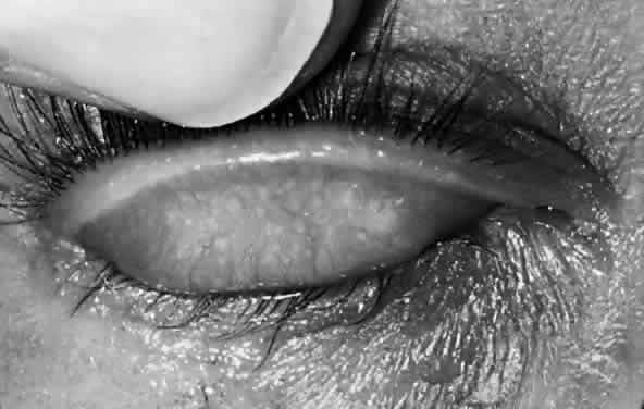
After the first year of life, lymphoid follicles are a regular feature of trachoma (Figs. 1 and 2). These conjunctival lesions are flat or elevated, opaque or clear, have a yellow or gray color, and appear avascular. They vary in diameter from 0.2 to 2.0 mm. The entire conjunctiva is involved by the inflammation but follicle formation in the conjunctiva of the upper tarsus is one of the cardinal signs of trachoma. Follicles may become necrotic, so that they rupture on slight pressure, extruding their contents onto the conjunctival surface. In such cases, the conjunctiva is extremely friable, so small blood vessels rupture on everting the tarsal plate, resulting in hemorrhage into follicles or into the adjacent subepithelial conjunctiva. Not all cases—even those that are severe—develop these large necrotic follicles.

Fig. 1. Early trachoma in a 5-year-old child. The conjunctiva of the everted upper lid margin has numerous lymphoid follicles, which appear as small elevated avascular lesions in the midst of the hyperemic, infiltrated conjunctiva (F3 or TF) (see Table 4). The distribution of fillicles in the central tarsal plate and diffuse infiltration (papillary hypertrophy; P2) (see Table 1) indicates this case is of moderate intensity.

Fig. 2. Severe active trachoma, with marked inflammatory infiltration of the conjunctiva. The lymphoid follicles are visible but partially covered by the infiltrated conjunctiva (F3, P3 or TF, TI (see Table 4).

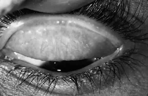
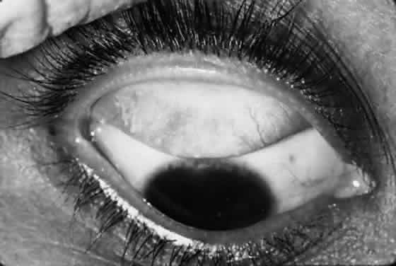
Scars of the conjunctiva may form in a stellate pattern at the site of necrotic follicles. Other scarring appears as fine short lines or broader confluent scars (Figs. 3 and 4). Larger deep scars appear to form in cases with particularly severe trachoma and may not be due simply to necrosis of follicles (Fig. 5). In some cases, a distinctive horizontal scar (line of von Arlt) forms in the upper tarsus, where the ascending and descending subconjunctival vessels meet about a third of the distance from the lid margin to the upper tarsal border.

Fig. 3. Active trachoma in an Egyptian child with conjunctival scarring. The small round avascular follicles and diffuse conjunctival inflammation indicate on-going disease activity (F2, P2 or TF; see Tables 1 and 4). The horizontal scar near the lid margin (C2 or trachomatous scarring) (see Table 1) is characteristic of trachoma in north Africa and can produce entropion of the upper lid early in life.

Fig. 4. Mild conjunctival scarring in a case of healed (inactive) trachoma. The scars appear as fine white lines (C1) (see Table 1). The yellow aggregations are subconjunctival cysts. These cysts (known as posttrachomatous degeneration) are formed by epithelium trapped below the surface by scars. The yellow cheesey material in the cysts is formed by desquamated epithelium, which often extrudes onto the conjunctival surface. The dark notches in the upper limbus are Herbert's pits, the depressions left by limbal follicles in childhood.

Fig. 5. Marked conjunctival scarring in healed trachoma (C3) (see Table 1). Distortion of the lid has produced an apparent inward displacement of the meibomian orifices because conjunctival epithelium has grown over the lid margin. These scars continue to contract throughout life, eventually leading to trichiasis and entropion. Herbert's pits are present at the corneal limbus.

This scar formation also affects the canaliculi and lacrimal duct with obstruction and cutaneous fistula.33 Occlusion of the tear outflow can cause tearing and often leads to the development of dacryocystitis in adult life.

Linear scars that form just adjacent to the lid margin are particularly damaging because of the lid distortion that they cause. Cicatricial changes occur throughout the conjunctiva but are most apparent and damaging on the tarsal conjunctiva. One result of extensive scarring is that destruction of goblet cells occurs in severely scarred eyes, with a loss of mucus secretion. The loss of mucus and distortion of the upper lid lead to inadequate surface wetting of the cornea.

The drooping or half-closed lids in active trachoma have been attributed to the weight of the lid, to inflammatory involvement of Müller's muscle in the upper lid, and to reflex blepharospasm caused by keratitis-induced photophobia. In late cicatricial trachoma, there may also be destruction of Müller's muscle.

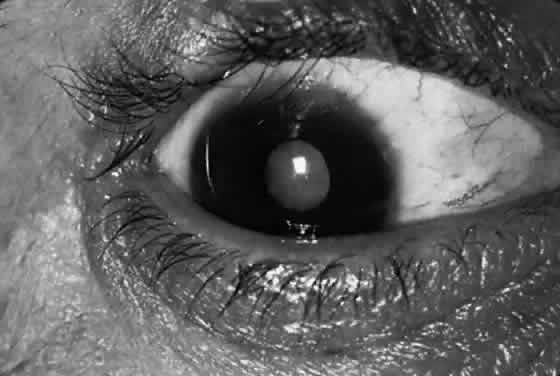
The major potentially blinding sequela of trachoma are distortion of the lids, particularly the upper lid; trichiasis (misdirection of individual lashes); and entropion (inward deformation of the lid margin; Fig. 6).1 Severe conjunctival scarring from trachoma can lead to defects in lid closure and loss of the mucus in tears, with inadequate surface wetting of the cornea. The abrasion of the cornea by wiry lashes—especially when aggravated by even minor foreign-body injury or by inadequate wetting of the cornea—can further predispose the cornea to traumatic and infectious damage.

Fig. 6. Trichasis due to trachoma in a 51-year-old man with mature cataract (T/E 3 or TT) (see Table 4). The inturned lashes usually occur in adult life many years after the acute inflammation has resolved. The constant corneal abrasion by the lashes and inadequate tears can produce corneal erosions, ulceration, and opacity, which constitute the major pathway to blindness in trachoma. In this case, there is notching of the upper lid due to previous lid surgery; this can produce corneal exposure and further corneal damage.

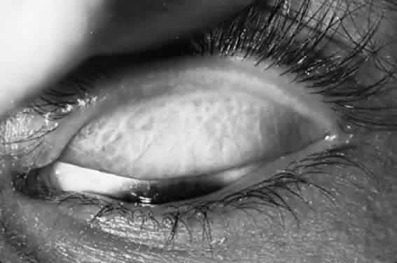
Corneal lesions in trachoma include focal inflammatory infiltrates of the epithelium and anterior stroma, with the formation of a superficial fibrovascular membrane (vascular pannus; Fig. 7). There may be lymphoid follicles at the superficial limbus, which resolve leaving characteristic depressions—Herbert's pits.12

Fig. 7. Moderate corneal scarring in a Tunisian man (CC2) (see Table 1). The vascularized opacity with vessels extending from the lower limbus probably occurred after a corneal ulcer. There is trichiasis of the superior lid. A pterygium extends onto the peripheral cornea from the medial limbus.

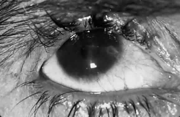
The early keratitis in trachoma presents as diffuse epithelial keratitis, and small infiltrates can form in the epithelium and anterior stroma, usually at the limbus but occasionally in the central cornea. This is soon followed by extension of the superficial vessels from the limbus. These vessels form an even advancing border as they extend onto the cornea (vascular pannus). During active inflammation, there is often a hazy infiltrate around the vessels and discrete inflammatory infiltrates in advance of the pannus. Permanent superficial scarring occurs between the vessels. Particularly severe cases may have marked infiltration and swelling of the pannus, with abundant vascularization (pannus crassus). Pannus ulcers are horizontal, oval, epithelial defects that appear in the upper conjunctiva in front of the vascular pannus and resemble the epithelial ulcers in vernal catarrh.

Pannus formation is always more extensive at the upper limbus but may involve the entire limbus. Deep corneal vessels or irregular extensions of the pannus are usually caused by other lesions, such as bacterial corneal ulcers after trauma. The inturned lashes and exposure also may cause diffuse anterior stromal scarring of the cornea. The bluish-grey elevated lesions of Salzmann's nodular dystrophy are a common finding in adults with trachoma pannus and appear to be related to corneal vascularization, not to lid distortion. In older adults, there may be marked lipid deposition in the pannus, forming a lipoidal degeneration of the upper limbus.

In older patients with extensive scarring of the upper limbus, the corneal epithelium appear to be roughened with diffuse fluorescein staining. This may be caused by replacement of limbal stem cells by conjunctival epithelium.

The extent of pannus formation in children does not appear to be directly related to the disease intensity or extent of conjunctival scarring. In younger patients, marked pannus formation can occur with relatively mild lid scarring, but lesser degrees of pannus may accompany even severe intensity conjunctival inflammation.

CLINICAL CLASSIFICATION

Clinical signs of the upper tarsal conjunctiva and cornea have been used to describe clinical trachoma in the whole eye. To provide a more precise description of inflammatory and cicatricial disease in individual cases, a detailed system was developed, based on the scoring of lymphoid follicles (F), diffuse inflammation and papillary hypertrophy (P), conjunctival scarring (C), lid distortion and inturned lashes (T/E), and corneal opacities (CC) (Table 1).1 The intensity of inflammatory trachoma includes four categories—severe, moderate, mild, and insignificant—which are of prognostic value in the individual case (Table 2). As noted, the prevalence of chlamydial infection parallels this clinical intensity scale.


TABLE 1. Clinical Scoring of Inflammatory Signs for Detailed Epidemiologic and Therapeutic Studies of Trachoma1


Scores for Upper Tarsal Follicles (F)
F0No follicles
F1Follicles present but no more than five in zones 2 and 3 together
F2More than five follicles in zones 2 and 3 together but fewer than five in zone 3
F3Five or more follicles in each of the three zones
Scores for Upper Tarsal Papillary Hypertrophy and Diffuse
Infiltration (P)
P0Absent: normal appearance
P1Minimal: individual vascular tufts (papillae) prominent, but deep subconjunctival vessels on the tarsus not obscured
P2Moderate: more prominent papillae, and normal vessels appear hazy, even when seen by the naked eye
P3Pronounced: conjunctiva thickened and opaque, normal vessels on the tarsus are hidden over more than half of the surface
Conjunctival Scarring (C)
C0No scarring on the conjunctiva
C1Mild: fine scattered scars on the upper tarsal conjunctiva, or scars on other parts of the conjunctiva
C2Moderate: more severe scarring but without shortening or distortion of the upper tarsus
C3Severe: scarring with distortion of the upper tarsus
Trichiasis or Entropion (T/E)
T/E0No trichiasis or entropion
T/E1Lashes deviated towards the eye but not touching the globe
T/E2Lashes touching the globe but not rubbing on the cornea
T/E3Lashes constantly rubbing on the cornea
Corneal Scarring (CC)
CC0Absent
CC1Minimal scarring or opacity but not involving the visual axis and with clear central cornea
CC2Moderate scarring or opacity involving the visual axis, with the pupillary margin visible through the opacity
CC3Severe central scarring or opacity with the pupillary margin not visible through the opacity


TABLE 2. Intensity of Inflammatory Trachoma


IntensityKey SignFolliclesPapillae
SevereP3F3 (or F2 or F1)*P3†
ModerateF3F3†P2
MildF2F2†P0, P1, or P2
Trivial (insignificant or absent)F2 or F1F0 or F1P0, P1, or P2

*The follicles may be obscured by severe papillary hypertrophy and diffuse infiltration.
†Key sign.


Subsequently, a simplified grading was developed for use in trachoma control efforts based on primary health care workers (Table 3).34 This simplified system records the presence or absence of the same signs as the detailed grading system in Table 1. The grading schemes are compared in Table 4.34


TABLE 3. Simplified Grading of Trachoma Currently Recommended for Trachoma Control Programs

  TT—trichiasis/entropion
  TF—5 or more follicles on the central tarsal conjunctiva
  TI—intense papillary reaction of the tarsal conjunctiva
  TS—trachomatous scarring
  CO—central cornea opacities


TABLE 4. Comparison of Detailed and Simplified Trachoma Grading Systems


 Simplified SignsDetailed Scoring
TrichiasisTTT/E 1, 2, 3
Tarsal folliclesTFF 2, 3
Infiltration and papillary hypertrophyTIP 3
Conjunctival scarringTSC 1, 2, 3
Corneal scarringCOCC 2, 3


This simplified system is in widespread use but can result in overdiagnosis of trachoma. Two examples occurred in Nepal: in Eastern Nepal, TF (five or more follicles on the central tarsal conjunctiva) was diagnosed in 22% of children and TI (intense papillary reaction of the tarsal conjunctiva) in 6.7% but no trachomatous scarring could be detected. The investigators correctly judged that without scarring, there was no need to intervene for trachoma.35 Our group found a similar situation in Western Nepal, with 7% of children with TF and 1% with TI; despite the occurrence of severe scarring and trichiasis in older adults, no chlamydial agent was found by a sensitive DNA amplification test, indicating that the follicular conjunctivitis was not active trachoma (T. Lietman, personal communication). In such situations, other signs (conjunctival scarring or Herbert's pits) are needed to definitively diagnose trachoma.

In the older MacCallan classification, trachoma cases are classified in stages by the findings in the conjunctiva alone.1 This classification describes the evolution of the disease but does not have prognostic value. Trachoma stage I and stage II are the early and established phases of inflammatory trachoma, with lymphoid follicles and papilla but without conjunctival scarring. Stage IIA is characterized by mature (i.e., large necrotic follicles) and stage IIB by papillary hypertrophy that has obscured the follicles and underlying tarsal vessels (P3 or severe intensity). In stage III, conjunctival scarring is present with the conjunctival signs of stages I or II. In stage IV, the acute inflammatory signs of the conjunctiva found in stages I and II have resolved, leaving scars in the conjunctiva. If sufficiently severe, these scars can contract further to produce lid distortion and trichiasis. In stage IV, the disease was thought to be noninfectious but the newer diagnostic tests frequently identify chlamydial infection in these putatively healed cases. The MacCallan classification does not describe the degree of inflammation or identify cases with visually disabling lesions and hence is not as useful for prognosis as the newer classification.

Communities with blinding trachoma can be recognized by the presence of those with severe visual loss due to corneal opacity and a substantial prevalence of potentially disabling trachomatous lesions, particularly trichiasis or entropion. These irreversible changes appear as the long-term outcome of prolonged or recurrent inflammatory disease of moderate or severe intensity. Communities with nonblinding trachoma may have a low prevalence of potentially blinding lesions but do not have a substantial prevalence of trachomatous visual loss. In some areas where living standards have improved, trachomatous trichiasis and corneal scarring affect many adults but active inflammatory trachoma in children is relatively mild. In this setting, trachoma control programs should emphasize surgical correction of inturned lids but the need for community-wide antibiotic treatment should be based on the prevalence and severity of conjunctival scarring in children younger than 10 years, in addition to the prevalence of inflammatory trachoma.

In communities with trachoma, infection by C. trachomatis is always present but other ocular microbial pathogens appear to contribute significantly to the intensity of trachoma and probably to the lesions that impair vision.3,7,8

CLINICAL DIAGNOSIS

If there is uncertainty concerning the presence or absence of trachoma in a given community or area, it is essential to use strict diagnostic criteria to identify those with trachoma.1 For this purpose, individual cases must have at least two of the following signs:

  1. Follicles on the upper tarsal conjunctiva
  2. Limbal follicles or their sequela, Herbert's pits
  3. Typical conjunctival scarring
  4. Vascular pannus most marked at the superior limbus.

If there is still a question about the presence of active, infectious trachoma in a community, the presence of infection with C. trachomatis should be required in a substantial proportion of tested children to confirm the diagnosis.

Once the presence of endemic trachoma has been established in a community, the occurrence of one of these signs in an individual case is sufficient to establish the diagnosis in surveys to measure endemicity. In many communities where blinding trachoma was once prevalent, improved conditions have resulted in a reduction in the severity or disappearance of infectious trachoma in children, although trichiasis and corneal opacity are still common in adults.8 Because many trachoma programs limit examinations of children to the diagnosis of tarsal follicles (TF) and severe tarsal inflammation (TI) but not trachomatous scarring (TS), the rate of active trachoma may be exaggerated because of a high prevalence of follicular and papillary conjunctivitis attributable to other causes.

MICROBIOLOGIC DIAGNOSIS

Laboratory tests in trachoma control programs may be used for several reasons: to support the clinical diagnosis of the disease in individual cases, to measure the presence and prevalence of agent in communities where trachoma is endemic or thought to be endemic, to monitor individuals or communities for the effect of therapy, to estimate the total exposure of a population to chlamydial infection, to monitor shifts in serotypes in a given population that may indicate introduction of an agent from outside the community or possible transmission of nonocular serotypes.

C. trachomatis infections are diagnosed by identifying the agent in clinical specimens or by a rising antibody titer. The microscopic examination of Giemsa-stained smears was the mainstay of diagnosis from the first decade of this century and is still a useful tool for field studies but has a low sensitivity.11,36 The first isolation of C. trachomatis strains was reported by T'ang and associates37 in 1957 from Peking, who recovered trachoma strains by inoculating the yolk sacs of fertile hen's eggs. Isolation techniques use specially treated cell cultures for isolation. With the availability of cultivated chlamydial strains, standardized (commercial) procedures have become available.38

To identify infection of individuals with C. trachomatis, the available procedures include:

  1. Morphologic identification of chlamydial inclusions by Giemsa-stained smears; rarely by iodine staining
  2. Isolation of agent in cell cultures
  3. Direct fluorescein-conjugated monoclonal antibodies (DFA) to species-specific (MOMP) or group-specific antigen (lipopolysaccharide— LPS)
  4. Identification of chlamydial antigen by enzyme-labeled immunoassay
  5. Probe for the nucleic acid of the chlamydia or the associated plasmid of C. trachomatis
  6. DNA amplification techniques
    1. PCR
    2. LCR

  7. Detection of antibody to C. trachomatis in serum, tears, or other secretions.

Cytology

For C. trachomatis infections of the eye, several cytologic techniques are available: staining of conjunctival smears with Giemsa stain, iodine, and fluorescein or peroxidase-conjugated monoclonal antibodies.

In Giemsa-stained smears, chlamydial organisms are seen as clusters of distinct particles (inclusions) in the cytoplasm of conjunctival epithelial cells. With an oil-immersion lens (40× to 100× ), the elementary bodies stain reddish-purple and the larger initial bodies a deep blue, like most bacteria.11 The common sources of misdiagnosis of cytoplasmic chlamydial inclusion in Giemsa-stained smears include pigment granules, keratin, nuclear extrusions, goblet cells, eosinophilic granules, and bacteria. In trachoma, the accompanying conjunctival cytology can be used as a guide to screening smears: inclusions are usually found only when polymorphonuclear leukocytes and separated epithelial cells are present; the prevalence of chlamydial inclusions increases progressively with the presence of other inflammatory cells (lymphocytes, plasma cells, macrophages, and multinucleated giant cells).

Iodine staining with Lugol's iodine identifies the glycogen matrix of inclusions. Direct iodine staining of conjunctival smears is insensitive but the technique was once used to identify inclusions in cell culture.

Staining of conjunctival smears with fluorescein-conjugated polyclonal antibody produced in rabbits was done in several research laboratories in the 1960s and 1970s.16 DFA to C. trachomatis MOMP or to chlamydial LPS are widely available to detect chlamydial agent in smears.39 With the DFA, individual elementary bodies are identified as brightly fluorescing extracellular particles but intracellular inclusions are not well identified. For trachoma, DFA is 80% as sensitive as culture and highly specific.36,40 The technique requires a fluorescent microscope and an experienced microscopist who can recognize the typical morphology of free elementary bodies. Commercial DFA reagents are widely available for the diagnosis of genital infections, however.

Isolation

Isolation of C. trachomatis in cell culture has been the definitive way to identify the agent. Although this procedure was the gold standard used to judge other tests for detecting C. trachomatis, there are still only a few laboratories prepared to perform chlamydial isolation.38,40

Identification of Antigen

Many immunoassay procedures to detect chlamydial antigen in clinical specimens are available.38,40 Test kits for single specimens that use this technology are available for office and small hospital laboratories but are less sensitive.

DNA Amplification Techniques

Polymerase chain reaction identifies DNA of the plasmid carried by all C. trachomatis strains (Chlamydia Amplicor, Roche). The LCR (LCX, Abbott) uses a different technique of DNA amplification for the same plasmid DNA. Both tests are 90% to 95% sensitive and have more than 99% specificity.38,40

Serologic Diagnosis

Detection of antibody in serum or tears usually is not useful for the diagnosis of ocular chlamydial infections in patients because the infections are chronic and endemic in populations with trachoma.40 Because patients are rarely tested during the early phases of infection, serologic testing is usually done with a single serum or tear specimen rather than paired specimens. Testing for antibodies has been applied in epidemiologic studies, however.

Serologic Tests

The two major serologic tests are the complement fixation test, which detects antibody to LPS chlamydial group antigen (including C. psittaci and C. pneumoniae strains) and the indirect immunofluorescent technique (micro-IF), which identifies antibody to C. trachomatis antigens. In most cases of trachoma, the microIF test detects IgG antibody, indicating infection at some time in the past.

Comparison of Procedures

In endemic trachoma, the PCR and LCR DNA amplification tests are more sensitive (more than 90%) than DFA and enzyme-labeled immunoassay (60% to 80%).40 Giemsa-staining of smears is clearly less sensitive in detecting chlamydial agent; isolation in cell culture is less available but has good sensitivity and specificity; micro-IF testing for antibodies may be useful for epidemiologic studies but is rarely useful for individual cases.38,40

Back to Top
EPIDEMIOLOGY
Trachoma has a worldwide distribution, and blinding trachoma is still a major public health problem in subSaharan Africa and the Nile Valley, parts of the Middle East, the Indian subcontinent, in Burma and Vietnam, probably Central Asia, some parts of China, and in limited areas of Latin America, Australasia, and the Pacific Islands. Nonblinding trachoma is present in a larger region of these same areas that include many of the drier subtropical and tropical countries.2

Trachoma was once prevalent and severe in many countries of Europe, North America, and northern Asia but it regressed and disappeared with rising living standards that accompanied industrialization and economic development. Under the living conditions prevailing in developed countries and in most urban communities of developing countries, trachoma is rarely transmitted; if acquired, it is mild. In patients with healed trachoma, however, there may be recurrences of active disease but this recurrent adult disease usually does not present a significant public health risk.

In the most heavily affected communities, most children are infected by the age of 1 or 2 years; starting at age 5, the prevalence of active disease declines steadily but some adults continue to have signs of active disease. Because children constitute such a large proportion of the population in hyperendemic trachoma areas, those with active disease are the chief reservoir of trachomatous infection in the community.7,8

Blinding lesions (trichiasis, entropion, corneal scarring) are the outcome of earlier severe or moderate-intensity inflammatory disease. These are generally observed in adults but may occur in childhood because of severe inflammatory disease.

TRANSMISSION

Trachoma is associated with poverty. Economic development appears to eliminate or reduce the severity of the disease.8 Transmission of the disease is by direct or indirect contact with infected material (e.g., hands, clothing, towels). Among the environmental and behavioral features of greatest importance are the presence of young children with dirty faces and nasal discharge, crowding, the absence of latrines, and the unavailability of safe water for household use.

In these areas, the disposal of human and animal wastes contributes to an increase in the fly population. The flies that cluster on children's eyes to feed on ocular discharges transfer these discharges to the eyes of other children in the same family within 15 to 30 minutes.7 Children with endemic trachoma harbor C. trachomatis in the upper respiratory and gastrointestinal tracts,13 so transmission also may occur by respiratory droplet spread or fecal contamination.

Back to Top
TREATMENT AND PREVENTION

TRACHOMA CONTROL PROGRAMS

The primary goal of public health programs for the control of trachoma is the prevention of blindness. World Health Organization recommendations for trachoma control programs are based on the SAFE strategy:

  Surgery for inturned lids and lashes
  Antibiotics for active inflammatory disease, either topical tetracyclines or oral azithromycin
  Face washing and improved cleanliness of young children
  Environmental improvement, including better disposal of animal and human feces and provision of water.

Surgery

The surgical correction of trichiasis or entropion (lid deformities) has an immediate impact on preventing blindness. Simple lid procedures can be performed in affected communities by mobile teams. Where ophthalmologists are not available, general physicians or auxiliary personnel can be trained to do the lid operations. In small trials, lid rotation procedures have been most successful.41

Keratoplasty has a poor outcome in patients with trachoma. Even if these patients have access to good postsurgical management, the reduced flow of aqueous tears, loss of mucus from goblet cell destruction, and poor mechanical surfacing by distorted lids all contribute to a high rate of postoperative complications and graft opacification. Peripheral optical iridectomy, however, may be useful in restoring vision for patients with discrete central corneal scarring. It is also possible that removal of superficial corneal scars by phototherapeutic procedures may be useful in some patients.

Both antibiotic treatment and economic development may result in a rapid reduction in the prevalence of the inflammatory disease. In communities where adults still have a substantial amount of potentially disabling trachomatous conjunctival scarring, however, new cases of trichiasis-entropion will continue to appear. Continuing surveillance is therefore necessary for many years after active inflammatory trachoma has been controlled.

Antibiotic Treatment

In communities with severe blinding trachoma, the objectives of chemotherapy are:

  1. Reduction in the intensity of trachoma and thereby the risk of blindness in the individual
  2. Reduction in transmission of infection from one individual to another.

Sulfonamides, tetracyclines, erythromycin, other macrolides and azalides, and rifampicin are effective in treating active trachoma. Previous trachoma control programs have been based on the mass application of locally applied tetracycline ointment.1 The new erythromycin derivative, azithromycin, has particular advantages for treatment of trachoma: it persists in tissues for extended periods after oral doses, with bacteriocidal levels persisting up to 14 days in the conjunctiva after a single oral dose;42 the drug can be administered to children as young as 4 months, is well tolerated orally, and has few side effects. Although it is relatively expensive, the World Health Organization recommends a single oral dose of azithromycin (20 mg/kg in children) to control infectious trachoma.

Controlled studies of children with active trachoma have shown that a single dose of azithromycin is as effective as 30 to 40 doses of topical tetracycline.43–45 Subsequently, a multi-country trial—Azithromycin in Control of Trachoma (ACT)—compared treatment of one or more villages with azithromycin (one dose weekly for 3 weeks) to all members of the community with similar mass treatment with topical tetracycline eye ointment (once daily for 42 days). The ACT trial involved endemic trachoma areas in Egypt, Gambia, and Tanzania.46 Among children with active trachoma, azithromycin was highly effective in reducing the prevalence of chlamydial infection in Egypt and Tanzania from 73% and 49% pretreatment, respectively, to 2.2% and 2.5% at 2 to 4 months after treatment, compared with tetracycline ointment with 63% and 56% pretreatment to 15% and 18% (respectively) after treatment.46,47 In Gambia, results were less striking, with azithromycin reducing infection from 60% pretreatment to 17% at 2 months and tetracycline producing a decrease from 57% to 20%; however, there was a substantial loss to follow-up and migration in and out of the Gambian villages.48 Multivariate analysis revealed that the clinical response and laboratory response was significantly better in older children and those who were chlamydia-positive before treatment.

In the ACT trial, the response in the tetracycline group was unexpectedly good in all three countries but ointment treatment required a considerable effort and expense for daily visits to households by medical aides. Because most trachoma treatment programs rely on household treatment by family members, compliance with daily applications of ointment is likely to be inadequate under ordinary conditions. In contrast, good compliance with azithromycin is particularly easy to achieve because only a single dose is needed and its side effects are negligible.

Initial intensive large-scale topical chemotherapy is intended to reduce the ocular reservoir of chlamydia in the population. One possible strategy would be to follow mass azithromycin treatment with family-based azithromycin treatment to control further eye-to-eye transmission. Systemic antibiotic treatment is more effective than topical treatment in children with severe-intensity infectious trachoma and should eliminate the extraocular reservoirs of C trachomatis. The migration of substantial numbers of untreated families and individuals into trachoma-endemic areas is a major problem that requires the development of new strategies.

Face Washing

Improved cleanliness of young children is an integral part of any trachoma program. This must be accomplished by educating and motivating women with young children regarding the importance of cleanliness in children and particularly in daily washing of children's faces. This measure has been shown to have a significant effect in reducing the prevalence of active trachoma.49

Environmental Improvement

Better disposal of animal and human waste and the use of screening materials in houses is a critical step in producing lower densities of eye-seeking flies and thus reducing the prevalence of trachoma.50 Other methods of fly control include covering composted material (manure and household waste). The provision of potable water for household use improves cleanliness and reduces the workload of women with young children. The use of pit latrines with covers and fly screens is also effective.

Back to Top
COMMENTARY
The World Health Organization has launched a concerted campaign to control trachomatous blindness. This effort is organized through the Global Elimination of Trachoma 2020, a consortium of nongovernmental development agencies, donor organizations, experts in the field, and World Health Organization staff. The goal of this group is to reduce the prevalence and severity of infectious trachoma in children by antibiotic treatment and to organize surgical services to correct lid deformities, thus eliminating new cases of trachomatous blindness by the year 2020.

The technical developments that have made this possible include the development of the SAFE strategy; availability of azithromycin, which is effective as a single dose against infectious trachoma; the simplified system of trachoma assessment to identify cases needing antibiotic treatment or surgery; and the development of DNA amplification techniques to detect chlamydial infections. This effort is part of World Health Organization's larger program for prevention of easily curable and treatable blinding conditions, with special emphasis on cataract surgery, trachoma, onchocerciasis, vitamin A deficiency, glaucoma, and childhood blindness.

Donations of azithromycin for trachoma control in national programs and some modest funds are being coordinated through a new organization, the International Trachoma Initiative (ITI), which will review applications and distribute azithromycin provided by Pfizer International. The activities of ITI will be reviewed after its initial phase (1998 to 2000). ITI is supported initially by the Edna McConnell Clark Foundation and Pfizer International. This convergent effort to fight trachoma should achieve its goal to eliminate blinding trachoma by 2020.

Back to Top
REFERENCES

1. Dawson CR, Jones BR, Tarizzo M: Guide to trachoma control. World Health Organization, Geneva, 1981

2. Thylefors B, Negrel AD, Pararajasegaram R, Dalzic KY: Global data on blindness. Bull World Health Organ 73: 115, 1995

3. Vastine DW, Dawson CR, Daghfous T et al: Severe endemic trachoma in Tunisia. I. The effect of topical chemotherapy on conjunctivitis and ocular bacteria.Br J Ophthalmol 58: 833, 1974

4. Wang S-P, Grayston TP: Immunological relationships between genital TRIC, lymphogranuloma venereum and related infections in a new microtiter indirect immunofluorescence test. Am J Ophthalmol 70:367, 1970

5. Curth W, Curth HO, Sanders M: Chronic conjunctivitis due to the virus of venereal lymphogranuloma venereum. JAMA 115:445, 1941

6. Lietman T, Brooks D, Moncada J et al: Chronic follicular conjunctivitis associated with Chlamydia psittaci or Chlamydia pneumoniae. Clin Infect Dis 26:1335, 1998

7. Jones BR: The prevention of blindness from trachoma. Trans Ophthal Soc UK 95:16, 1975

8. Dawson CR, Daghfous T, Messadi M, Hoshiwara I, Schachter J: Severe endemic trachoma in Tunisia. Br J Ophthalmol 60:245, 1976

9. Dhermy P, Coscas G, Nataf R, Levaditi JC: Histopathologie des follicules au cours du trachome et de conjonctivitie folliculaire. Rev Int du Trachome 4:295, 1967

10. Badir G, Wilson RP, Maxwell-Lyons F: The histopathology of trachoma. Bull Egypt Ophthalmol Soc 46:96, 1953

11. Yoneda C, Dawson CR, Daghfous T et al: Cytology as a guide to the presence of chlamydial inclusions in Giemsa stained conjunctival smears in severe endemic trachoma. Br J Ophthalmol 59:116, 1975

12. Zscheile FP: Herbert's Pits. Arch Ophthalmol 73:827, 1965

13. Malaty R, Zaki S, Said ME et al: Extraocular infections in children in areas with endemic trachoma. J Infect Dis 143: 853, 1981

14. Rasmussen S: Chlamydial immunity. Curr Opin Infect Dis 11:37, 1998

15. Treharne JD, Dwyer RStC, Darougar S, Jones BR, Daghfous T: Antichlamydial antibody in tears and sera, and serotypes of Chlamydia trachomatis isolated from school-children in Southern Tunisia. Br J Ophthalmol 62:509, 1978

16. Hanna L, Jawetz E, Briones O et al: Antibodies to TRIC agents in matched human tears and sera. J Immunol 110: 1464, 1973

17. Wong S-P, Grayston TP, Kuo CC et al: Serodiagnosis of Chlamydia trachomatis infection with the microimmunofluorescence test. In Hobson D, Holmes KK (eds): Nongonococcal Urethritis and Other Infections, pp 237–248. Washington DC, Am Soc For Microbiol, 1977

18. Jawetz E, Dawson CR, Schachter J et al: Immunoglobulin nature of antibodies in chlamydial infections. In Nichols R (ed): Trachoma and Related Disorders Amsterdam, pp 233–232. Excerpta Medica, Amsterdam, 1971

19. Caldwell H, Kromhout J, Schachter J: Purification and partial characterization of the major outer membrane protein of Chlamydia trachomatis. Infect Immunol 31:1161, 1981

20. Larpe MF, Wong KE, Kuehl LM, Stamm WE: Chlamydia trachomatis major outer membrane variants escape neutralization by both monoclonal antibodies and human immune sera. Infect Immunol 65:317, 1997

21. Mahmoud ED, Elhassan AE, Babikir HE et al: Antichlamydial activity of lacrimal fluid in patients with trachoma. Int Ophthalmol 18:87, 1994

22. Shemer Y, Sarov I: Inhibition of the growth of Chlamydia trachomatis by human gamma interferon. Infect Immunol 48:592, 1985

23. Perry LL, Feilzer K, Caldwell HD: Immunity to Chlamydia trachomatis is mediated by T helper 1 cells through IFN-gamma-dependent and-independent pathways. J Immunol 158:3344, 1997

24. Rapoza PA, Tahiju SG, Carlin JP et al: Effect of interferon on a primary conjunctival epithelial cell model of trachoma. Invest Ophthalmol Vis Sci 32:2919, 1991

25. Igietseme JU, Uriri IM, Kumar SN et al: Route of infection that induces a high intensity of gamma interferon-secreting T cells in the genital tract produces optimal protection against Chlamydia trachomatis infection in mice. Infect Immunol 66:4030, 1998

26. Williams DM, Grabbs BG, Park E et al: Humoral and cellular immunity in secondary infection due to murine Chlamydia trachomatis. Infect Immunol 65:2876, 1997

27. Rasmussen SJ, Eckmann L, Quayle AJ et al: Secretion of proinflammatory cytokines by epithelial cells in response to chlamydial infection suggests a central role for epithelial cells in chlamydial pathogenesis. J Clin Invest 99:77, 1997

28. Bobo L, Novak N, Mkocha H et al: Evidence for a predominant pro-inflammatory conjunctival cytokine response in individuals with trachoma. Infect Immunol 64:3273, 1996

29. Conway DJ, Holland MJ, Campbell AE et al: HLA class I and II polymorphisms and trachomatous scarring in a Chlamydia trachomatis-endemic population. J Infect Dis 174: 643, 1996

30. Conway DJ, Holland MJ, Bailey RE et al: Scarring trachoma is associated with polymorphism in the tumor necrosis factor (TNF-α) gene promoter and with elevated TNF-α levels. Infect Immunol 65:1003, 1997

31. White AG, Bogh J, Lehany W et al: HLA antigens in Omanis with blinding trachoma: markers for disease susceptibility and resistance. Br J Ophthalmol 81:431, 1997

32. Dawson CR et al: Response to treatment in ocular chlamydial infections (trachoma inclusion conjunctivitis): analogies with nongonococcal urethritis. In Hobson D, Holmes KK (eds): Nongonococcal Urethritis and Related Infections, pp 135–139. Washington DC, Am Soc Microbiol, 1997

33. Tabbara KF, Bobb AA: Lacrimal system complications in trachoma. Ophthalmology 87:298, 1980

34. Thylefors B, Dawson CR, Jones BR et al: A simple system for the assessment of trachoma. Bull World Health Organ 65:477, 1987

35. Katz J, West KP, Khatry SK et al: Prevalence and risk factors for trachoma in Sarlahi district in Nepal. Br J Ophthalmol 80:1037, 1996

36. Schachter J, Moncada J, Dawson CR et al: Nonculture methods for diagnosing chlamydial infection in patients with trachoma: a clue to the pathogenesis of the disease? J Infect Dis 158:1347, 1988

37. T'ang F-F, Chang H-L, Huang Y-T, Wang K-C: Studies on the etiology of trachoma with special reference to isolation of the virus in chick embryo. Chinese Med J 75:429, 1957

38. Black CM: Current methods of laboratory diagnosis of Chlamydia trachomatis infections. Clin Microbiol Rev 10:160, 1997

39. Nowinski RC, Tam MR, Goldstein LC et al: Monoclonal antibodies for diagnosis of infectious diseases in humans. Science 219:637, 1983

40. Schachter J: Diagnosis of human chlamydial infections. In Stephens R et al (eds): Chlamydial Infections, pp 577–586. San Francisco, International Chlamydia Symposium, 1998

41. Reacher MH, Munoz B, Alghassany A et al: A controlled trial of surgery for trachomatous trichiasis of the upper lid. Arch Ophthalmol 110:667, 1992

42. Tabbara KF, Al-Kharashi SA, Al-Mansouri SM et al: Ocular levels of azithromycin. Arch Ophthalmol 116:1625, 1998

43. Bailey RL, Arullendran P, Whittle HC, Mabey DC: Randomised controlled trial of single-dose azithromycin in treatment of trachoma. Lancet 342:453, 1993

44. Tabbara KF, El-Asa Ama, Al-Oma D et al: Single dose azithromycin in the treatment of trachoma. A randomized controlled study.Ophthalmology 103:842, 1996

45. Dawson CR, Schachter J, Sallam S et al: A comparison of oral azithromycin with topical oxytetracycline/polymyxin for treatment of trachoma in children. Clin Infect Dis 24:363, 1997

46. Schachter J, West S, Mabey D et al: Azithromycin in control of trachoma. 3. Effect of treatment on Chlamydia trachomatis infection in trachoma. In Stephens et al (eds): Chlamydial Infections, pp 347–350. San Francisco, International Chlamydial Symposium, 1998

47. Dawson CR, Sheta A, Sallam S et al: Azithromycin in the control of trachoma (ACT): #5. Oral azithromycin compared to topical oxytetracycline for active trachoma in Egypt. In Stephens R et al (eds): Chlamydial Infections, pp 355–358. San Francisco, CA 94110, San Francisco, International Chlamydial Symposium, 1998

48. Mabey D, Bailey R, Faal H et al: Azithromycin in control of trachoma. 2. Community based treatment of trachoma with oral azithromycin: a one year follow-up study in The Gambia. In Stephens R et al (eds):Chlamydia Infections, pp 351–354. San Francisco, International Chlamydia Symposium, 1998

49. West S, Munoz B, Lynch M et al: Effect of face washing on trachoma in Kongwa, Tanzania. Lancet 345:155, 1985

50. Courtright P, Sheppard J, Lane S et al: Latrine ownership as a protective factor in inflammatory trachoma in Egypt. Br J Ophthalmol 75:322, 1991

Back to Top