Chapter 9
Ophthalmic Viscosurgical Devices (OVDs): Physical Characteristics and Clinical Applications
Stephen S. Lane
Main Menu   Table Of Contents

Search

RHEOLOGIC AND PHYSICAL PROPERTIES
COHESION AND DISPERSION
DESIRED PROPERTIES OF OVDs
VISCOELASTIC COMPOUNDS, THE BUILDING BLOCKS OF COMMERCIALLY AVAILABLE VISCOELASTIC MATERIALS
COMMERCIAL OVD PREPARATIONS
CLINICAL USES OF OVDs
COMPLICATIONS OF OVD USE
SUMMARY
REFERENCES

The introduction of viscoelastic agents (now termed ophthalmic viscosurgical devices [OVDs] by the International Standards Organization [ISO]) for uses in ophthalmic intraocular procedures has had a significant impact on the practice of ophthalmology. OVDs possess a unique set of properties, based on their chemical structure, that enable them to protect the corneal endothelium from mechanical trauma and to maintain an intraocular space, even in the face of an open incision. Viscosurgery,1 a term used to designate the procedures and manipulations performed with OVDs, has been used in a broad spectrum of ophthalmic procedures. The use of OVDs has become commonplace in anterior segment surgery, and it is likely that the widespread use and availability of these materials facilitated the transition first in the conversion from intracapsular to planned extracapsular surgery, and then to phacoemulsification.

The physical properties of OVDs are the result of chain length and intrachain, and interchain molecular interactions. It is important to realize that the diverse rheologic properties of any given OVD have a direct impact on the clinical characteristics of that particular material. A thorough understanding of these properties will allow ophthalmic surgeons the opportunity to choose an OVD that is task specific. For example, a specific substance may be selected because of its space maintenance qualities, its corneal endothelial protection qualities, or its coating qualities.

Back to Top
RHEOLOGIC AND PHYSICAL PROPERTIES
The rheologic characteristics of OVDs that are most relevant when considering their usefulness in ophthalmic surgery are viscoelasticity, viscosity, pseudoplasticity, and surface tension (Tables 1 and 2).


TABLE 1. Physical Properties of Viscoelastic Substances


  Healon Healon GV Amvise Amvise Plus Chondroitin Sulfate Viscoat
Resting viscosity (cps)*>200,0002,000,000100,000102,00017,000 at 50%41,000
Dynamic viscosity (cps)=40,000–64,00080,00040,00055,00030 at 20%40,000
ColorClearClearClearClearYellowClear
Pseudoplasticity+++++++++++++No++
Contact angle60°=60°==52°
  Occucoat Vitrax ProVisc Healon 5 Cellugel
Resting viscosity (cps)*=40,000=7 million 
Dynamic viscosity (cps)=400030,00039,00060,000–80,00030,000
ColorClearClearClearClearClear
Pseudoplasticity+++++++++++++
Contact angle52°=== 

*At shear rate of zero, Data from Arschinoff S, Personal communication.
+|At shear rate of 2/s, 25°C.
= Not available.
Pseudoplasticity key: + = slight; +++ = good; ++ = fair; ++++ = excellent.
Modified from Liesegang TJ: Viscoelastic substances in ophthalmology, Surv Opththalmol 34 p. 268–293, 1990


TABLE 2. Physical Properties of Various Viscoelastic Materials


  Healon 5 Healon GV Healon Vitrax Amvisc Amvisc Plus
SourceRooster combsRooster combsRooster combsRooster combsRooster combsRooster combs
Manufacturer Molecular mass (daltons)Pfizer/PharmaciaPfizer/Pharmacia 5 × 10⁶Pfizer/Pharmacia 2.5-3.8 × 10⁶Advanced Medical Optics 5 × 10⁵Bausch & Lomb 2 × 10⁶Bausch & Lomb 2 × 10⁶
Content2.3% sodium Hyaluronate1.4% sodium hyaluronate1% sodium hyaluronate3% sodium hyaluronate1% sodium hyaluronate1.6% sodium hyaluronate
pH Buffer solvent7.0–7.5 phosphate-buffered saline7.0–7.5 phosphate-buffered saline7.0–7.5 phosphate-buffered saline7.0–7.5 physiological BSS6.5–7.2 physiological saline7.2 physiological saline
Osmalality (mOsm/kg H2O)320302309310318340
Concentration (mg/mL)231410301016
  Viscoat Occucoat ProVisc Cellugel
SourceBacterial fermentation (sodium hyaluronate): shark fin cartilage (sodium chondroitin sulfate)Wood pulpBacterial fermentationWood pulp
ManufacturerAlconBausch & LombAlconAlcon
Molecular mass (daltons)500 × 103; 25 × 10386 × 1031.9 × 1063 × 105
Content3% sodium hyaluronate; 4% sodium chondroitin sulfate2% HPMC1% sodium hyaluranate2% HPMC
pH Buffer solvent7.0–7.5 physiological phosphate buffer7.2 BSS and variable buffers7.25 physiological sodium chloride phosphate bufferN/A
Osmalality (mOsm/kg H2O)360319310315
Concentration (mg/mL)Sodium hyaluronate 30; Chondroitin sulfate 402010N/A


Viscoelasticity

Elasticity refers to the ability of a solution to return to its original shape after being stressed. The rheologic property of viscoelasticity is the essence of the usefulness of these materials as surgical tools in ophthalmology. Elasticity allows the anterior chamber to reform after deformation by depression on the cornea when external forces are released. A nonelastic solution, such as balanced salt solution (BSS), will show no such reformation after release of forces.

The terms viscosity and viscoelasticity are not synonymous. Viscosity, viscoelasticity, and pseudoplasticity are, however, interrelated. The amount of elasticity of an elastic compound increases with increasing molecular weight and greater chain length of the molecules. Unfortunately, comparison of the different OVDs with regard to elasticity is not easily made because of the different ways and nonuniform expression of values by the various manufacturers.

Viscosity

Viscosity (Table 1) reflects a solution's resistance to flow, which is, in part, a function of the molecular weight of the substance. Viscosity of OVDs is measured in centipoise (cPs) or centistokes (cSt), which are measures of the resistance to flow relative to a given shear force. Liquid solutions are generally considered to have viscosities of less than 10,000 cSt at rest, whereas solutions with resting viscosities greater than 100,000 cSt are gel-like. The higher the solution's molecular weight, the more it resists flow. Molecular weight, on the other hand, reflects the size of the solution's molecules. Viscosity is dependent on the degree of movement of a solution, which is also known as the shear rate, and varies inversely with temperature. The viscosity of a solution can be increased by increasing either the concentration or the molecular weight of the solution.

To facilitate optimal intraocular manipulation, an OVD should maintain space and protect tissues (possess a high viscosity at low shear rates), allow movement of instruments, aid in IOL (Intraocular Lens) implantation (possess a moderate viscosity at medium shear rates), and allow easy introduction into the eye through a small cannula (possess a low viscosity at high shear rates).2 At the present time, no single OVD fulfills all of these requirements optimally.

Pseudoplasticity

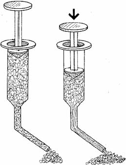
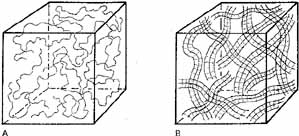
Pseudoplasticity refers to a solution's ability to transform when under pressure, from a gel-like substance to a more liquid substance. The more pseudoplastic a material is, the more rapidly it changes from being highly viscous at rest to a thin, watery solution at high shear rates. A change in molecular structure accompanies this pseudoplastic behavior. In clinical terms, a high molecular weight, high viscosity OVD at rest (zero shear force) acts as an excellent lubricant and coats tissues and maintains space very well. When under the influence of stress (i.e., a high shear rate), however, the OVD will become an elastic molecular system behaving as an excellent shock absorbing gel. The highest shear rates occur when a solution is passed through a cannula, and viscosity becomes independent of molecular weight. When the molecules align themselves in the direction of flow, the viscosity is determined solely by the concentration. Pseudoplastic solutions, therefore, have a low viscosity at high shear rates and can be extruded easily through a small diameter cannular (27 or 30 gauge) (Fig. 1) It is important to emphasize that the viscosity of a viscoelastic substance at rest (0 shear rate) is a function of concentration, molecular weight, and the size of the flexible molecular coils of the material (Figs.2A and 2B). At high shear rates, the viscosity is independent of molecular weight and is determined mainly by the concentration.3

Fig. 1 When shear is applied (flow through cannula), the large, randomly entangled coils begin to uncoil, allowing for flow. With increasing shear (more pressure on the syringe plunger) unfolding increases, entanglement drops, and the viscous solution lows easily through the cannula.

Fig. 2 A. Sodium hyaluronate molecules in low concentration at rest (zero shear). In solution, the NaHa chain unfolds on itself and forms a long, loose, randomly arranged coil. B. As the concentration of these large NaHa molecules is increased, the individual molecular coils start to overlap and become compressed. This crowding of the chains increases the chances for various noncovalent chain–chain interactions. This, in turn, increases the viscosity of the solution; it also increases the elasticity of the solution.

Surface Tension

The coating ability of an OVD is not only determined by the surface tension of the material itself, but also by the surface tension of the contact tissue, surgical instrument, or IOL. By measuring the angle formed by a drop of the OVD on a flat surface (the contact angle), the coating ability of a substance can be estimated. Lower surface tension and lower contact angle indicate a better ability to coat. In this respect, a solution of sodium hyaluronate has a significantly higher surface tension and contact angle than does a solution of chondroitin sulfate, sodium hyaluronate/chondroitin sulfate in combination, or HPMC, thus indicating these latter solutions provide superior coating.3 A comparison of the various physical properties of OVDs is summarized in Tables 1 and 2.

Back to Top
COHESION AND DISPERSION
In an attempt to help us better understand the interaction between these various rheologic properties and their clinical usefulness, Arshinoff 4 has divided OVDs into two categories: viscocohesive OVDs and Viscodispersive OVDs. ViscoCohesive OVDs are characterized by high viscosity materials that adhere to itself through intramolecular bonds or intermolecular entanglement and resists breaking apart. In general, OVDs with long molecular chains will be more cohesive because the molecules become entangled. Cohesive OVDs possess a high molecular weight, a high degree of pseudoplasticity, and high surface tension.

ViscoDispersive OVDs exhibit opposite characteristics. They possess lower viscosity and adhere well to external surfaces, for instance, tissues and instruments. These materials tend to break apart easily compared to cohesive materials, exhibit lower molecular weight, lower surface tension, and lower pseudoplasticity.

Although cohesiveness and dispersiveness are not measurable rheologic properties in themselves, they are useful constructs when considering the clinical behavior of OVDs.

Recently, a new descriptive term has been introduced by Arshinoff, “viscoadaptive.” This term refers to the ability of an OVD to adapt its behavior to the intended surgical task without the surgeon having to do anything except perform the task at hand. Unlike devices that fit one or the other of the previously mentioned categories, the viscoadaptive agent ideally functions as both, adapting its behavior to changing parameters in its environment. That changing parameter under most circumstances is the degree of turbulence present.

Back to Top
DESIRED PROPERTIES OF OVDs
It is apparent from the previous discussion that the interplay between the various rheologic properties is responsible for the clinical characteristics we desire in performing ophthalmic surgery. As a corollary, the degree to which we can maximize these desirable clinical characteristics is, for the most part, based upon our ability to optimize the unique rheologic and physical properties each OVD possesses. The desired properties of an ideal OVD are listed in Table 3.


TABLE 3. Desired Properties of an Ideal OVD


Ease of infusion
Retention under positive pressure in the eye
Retention during phacoemulsification (turbulence)
Easy removal/no removal required
Does not interfere with instruments or IOL placement
Protects the endothelium
Nontoxic
Does not obstruct aqueous outflow
Clear


Back to Top
VISCOELASTIC COMPOUNDS, THE BUILDING BLOCKS OF COMMERCIALLY AVAILABLE VISCOELASTIC MATERIALS

Sodium Hyaluronate

Sodium hyaluronate (NaHa) is a biopolymer occurring in many connective tissues throughout the body, including both the aqueous and vitreous humors. Its basic structural unit is a disaccharide, joined by a β1–4 glucosidic bond, which is linked in a repeating fashion with β glucosidic bonds to form a long unbranched chain. This mucopolysaccharide chain subsequently forms a random coil when placed in a solution, such as physiologic saline. As the concentration of large sodium hyaluronate molecules is increased (>0.5 mg/mL), individual molecular coils start to overlap and are compressed (Fig. 2). This crowding of the chains increases the chances for various noncovalent chain–chain interactions. This, in turn, causes a considerable increase in the viscosity of the solution. For example, the kinematic viscosity of a 2 mg/mL concentration of sodium hyaluronate in physiologic buffer is only in the 100-cSt range: at 10 mg/mL, it is in the 100,000 cSt range. Therefore, a fivefold increase in the concentration causes a 1000-fold increase in the viscosity of the solution. With this increase in viscosity, the elastic properties of the solution also increase. This forms the rationalization of Amvisc Plus and Healon GV. The elastic behavior of a concentrated (>0.5mg/mL) sodium hyaluronate solution is greatly dependent on the mechanical energy (shear force) applied to the solution. On a molecular level, this means that under the imposed strain, the polysaccharide coils slip by each other, and conformational and configurational rearrangements occur while the solution exhibits viscous flow (Fig. 2). The hyaluronate acid fraction (NIF-NaHa)5 used for ophthalmic procedures has a high molecular weight (2–5 million d), a low protein content (<0.5%), and carries a single negative charge per disaccharide unit.

This fraction is highly purified and has been shown to be sterile, nontoxic, nonantigenic,6 noninflammatory,7 and pyrogen-free. In primate vitreous humors, sodium hyaluronate has a half-life of approximately 72 days.8 In primate aqueous humors the half-life is 2 to 7 days depending on the viscosity.9 Clinical observations in humans have supported this result.

Chondroitin Sulfate

Chondroitin sulfate (CDS) is another biopolymer possessing viscoelastic characteristics and has been identified as one of the three major mucopolysaccharides in the cornea. Its structure is similar to hyaluronic acid, consisting of the same repeating disaccharide unit. CDS is of medium molecular weight in the range of 50,000 d. Chondroitin sulfate, like sodium hyaluronate, does not appear to be metabolized but is eliminated from the anterior chamber in approximately 24 to 30 hours.

Hydroxypropyl Methylcellulose

Hydroxypropyl Methylcellulose (HPMC) is yet another viscoelastic material used for intraocular procedures.10 Unlike the previous two compounds, it does not occur naturally in animals but is distributed widely as a structural substance in plant fibers, such as cotton and wood. It is a cellulose polymer composed of D-glucose molecules linked together by β-glycosidic bonds.

Special care must be taken in the filtration of this material to ensure a highly purified preparation, as Rosen et a1.11 noted the presence of vegetable fibers and other contaminates in samples they examined. Methylcellulose is a nonphysiologic compound and, as such, does not appear to be metabolized intraocularly but is eliminated from rabbit anterior chambers in approximately 3 days. It is also quite hydrophilic and hence can be irrigated from the eye.

Back to Top
COMMERCIAL OVD PREPARATIONS

Healon®, Healon GV®, Healon 5®

The first commercially available sodium hyaluronate, Healon®, was developed by Balazs,12 who sold the rights to Pharmacia. In 1958 Balazs suggested the use of hyaluronic acid as a vitreous substitute and, subsequently, two different ophthalmic preparations were developed. Etamucaine (Laboratories Chibert, Clermount-Ferrand, France), a bovine hyaluronic acid preparation of low viscosity and concentration, was found to be well tolerated as a vitreous replacement, despite a mild intravitreal inflammatory response.13

The second preparation, Healon®, truly initiated the age of viscosurgery. This high viscosity, high molecular weight sodium hyaluronate derived primarily from roosters' combs was developed and purified by Balazs et al.7,8,12,14 to produce a specific noninflammatory fraction. In 1972, the first human intraocular injections of Healon into the vitreous and anterior chambers were reported, and its use in surgical procedures varying from vitreoretinal diseases to cataract extraction and keratoplasty was suggested. These suggestions have been pursued actively and, subsequently, OVDs have become an invaluable tool in a broad array of applications.

By increasing both the molecular weight and concentration, Pharmacia introduced Healon GV® (greater viscosity) in 1992. With a resting viscosity of 2,000,000 cSt (at least 10 times more viscous than most other viscoelastics), Healon GV® provides superior viscosity for particularly demanding surgery.15 Despite positive vitreous pressure, Healon GV® is able to create and maintain a deep anterior chamber where other OVDs may fail. (Three times the resistance to pressure as Healon® in the presence of high positive vitreous pressure). Procedures dealing with small pupils, pediatric cataract extraction, and capsulorhexis during positive vitreous pressure are facilitated with the use of Healon GV®. However, because of its highly cohesive nature, Healon GV® leaves the anterior chamber very quickly during irrigation/aspiration or phacoemulsification, leaving the corneal endothelium susceptible to compromise.

Pharmacia and Upjohn developed a new OVD, which they believe possesses all of the best properties of Healon GV® and yet is retained in the anterior chamber throughout the phacoemulsification procedure. Healon 5® is the result of this effort and has been described as the first viscoadaptive OVD by the manufacturer. When exposed to low flow rates, it behaves as a super-viscous cohesive device, like an enhanced Healon GV®. However, as flow rates increase, Healon 5® begins to fracture into smaller pieces, making its behavior mimic some of the properties of dispersive OVDs. The properties of Healon 5® allow this OVD to also provide excellent capacity to mechanically dilate and maintain dilation of the pupil during intraocular maneuvers. It was released for sale in the United States in early 2001.

Amvisc®, Amvisc Plus®

Amvisc® is another sodium hyaluronate product extracted from rooster combs. It was first distributed by Precision-Cosmet and is now distributed by Bausch and Lomb Surgical. Released in 1983, Amvisc® is slightly less viscous than Healon®. Amvisc Plus, a 1.6% sodium hyaluronate product with a higher viscosity than Amvisc®, is also available. The viscosity of Amvisc Plus® is 55,000 cps compared with Amvisc® (40,000 cps). This higher viscosity obtained by increasing the total concentration (16 mg/mL) and using a sodium hyaluronate molecule of lower molecular weight allows for improved space maintenance, tissue manipulation, and tissue immobilization when compared to Amvisc.16

AMOVitrax®

AMOVitrax® (Advanced Medical Optics) is a low–molecular weight OVD preparation of a highly purified, noninflammatory fraction of sodium hyaluronate dissolved in BSS. Despite the relatively low molecular weight, AMOVitrax® is highly concentrated, which provides for a significantly viscous material. Unlike other sodium hyaluronic compounds, AMOVitrax® requires no refrigeration with a shelf life of 18 months. AMO Vitrax® (like Viscoat®) possesses a lower viscosity than Healon® at rest (0 shear) but maintains its viscosity under medium shear, whereas Healon® decreases sharply in a linear fashion.

ProVisc®

ProVisc® (Alcon Surgical, Inc.) is a sterile nonpyrogenic, high molecular weight, noninflammatory, highly purified fraction of sodium hyaluronate dissolved in physiologic sodium chloride phosphate buffer. The hy-aluronate material is obtained from microbial fermentation by a purified proprietary process. In this respect, it is similar to the process involved in the sodium hyaluronate fraction of Viscoat®. Clinical testing demonstrates that ProVisc® is equal to Healon® in its efficacy for protecting the corneal endothelium and in its level of safety.17 Like Viscoat®, ProVisc® requires refrigeration.14a

Viscoat®

Viscoat® is a 1:3 mixture of 4% chondroitin sulfate and 3% sodium hyaluronate manufactured by Alcon Surgical, Inc. The sodium hyaluronate fraction, like ProVisc®, is produced by bacterial fermentation through genetic engineering techniques. The chondroitin sulfate is obtained from shark fin cartilage. This combination of the compounds combines the higher viscosity and chamber-maintaining properties of sodium hyaluronate with the coating and cell protection properties of chondroitin sulfate. Koch, in a prospective randomized study, compared the endothelial protective effect of Healon® and Viscoat® during iris-plane phacoemulsification and posterior chamber phacoemulsification.18 In the iris-plane phacoemulsification group, Viscoat® provided greater corneal endothelial cell protection than Healon®. In the posterior chamber phacoemulsification group, however, no significant differences in cell protection were noted with both materials exhibiting excellent endothelial cell protection.

Ocucoat®

Ocucoat® (Bausch and Lomb Surgical) is a highly purified synthetic, nonprotein, nontoxic preparation of 2% HPMC. Ocucoat® has been marketed as a visco-adherent rather than a viscoelastic because of its significant coating ability and its relative lack of elastic properties. The reader must be aware that formulations produced by individual hospital pharmacies are not consistent proprietary products and can contain various solid particles, mainly vegetable matter remaining from the raw material.11 Ocucoat® is manufactured from the highest pharmaceutical grade HPMC and is subjected to a special dual-filtration process. A study presented in 1988 verified that Ocucoat® is as free of particulate debris as Healon® (Smith SG, European Intraocular Implant Council Meeting, 1988). Because of its poor elastic properties, a larger bore cannula and increased infusion pressure are necessary for injection. Unlike other OVDs, Ocucoat® can be sterilized by autoclaving and stored at room temperature. Because the raw materials are ubiquitous, there is potential for decreased cost. The complex biotechnical processes required to ensure purity, however, may limit this.

Cellugel®

Cellugel® (Alcon Surgical, Inc.) is a sterile, nonpyrogenic, noninflammatory, single-use, OVD, of highly purified 2% HPMC supplied in a disposable syringe delivering 1.0 mL, packaged in a sterile peel pouch, and is terminally sterilized by autoclaving. Like Ocucoat®, Cellugel® can be stored at room temperature. Unlike Ocucoat®, Cellugel® has a 10-fold greater viscosity and 4-fold higher molecular weight. As a result, the ability to maintain space is greater with Cellugel® than with Ocucoat®, despite both being 2% HPMC.

Back to Top
CLINICAL USES OF OVDs
Anterior segment surgery by its very nature induces corneal damage as has been documented by specular microscopy and pachymetry studies. Endothelial damage may occur at any stage in even routine procedures from the opening of the anterior chamber with manipulation of the cornea to insertion of an IOL after cataract extraction. Hence, the introduction of OVDs to anterior segment surgery can be easily appreciated from the perspective of corneal endothelial protection alone. Additional applications of these materials, however, have quickly become manifest, and the field of viscosurgery has broadened rapidly. Alpar19 outlined some of the applications that have been used in anterior segment surgery, many of which were developed specifically for cataract surgery. OVDs can be applied externally to provide corneal and conjunctival epithelial protection throughout the procedure without impairing visibility.20 Both intraocularly and extraocularly, OVDs can be used to form a mechanical barrier to control hemorrhage. Maintenance of the anterior chamber while fashioning the surgical wound and during intraocular manipulations can be accomplished with the injection of an OVD through a small incision. The iris and other intraocular tissues can be manipulated with the “soft instrument” effect of OVDs even in the face of increased vitreous or orbital pressure, and they may contribute to greater surgical control in the case of an expulsive hemorrhage. Finally, the use of OVDs may help to decrease the incidence of postoperative cystoid macular edema by appropriate maintenance of intraocular pressure and alteration of the refraction of the operating microscope light (Table 4).


TABLE 4. Clinical Uses of OVDs


Cataract Surgery
Corneal surgery/penetrating keratoplasty
Glaucoma surgery
Anterior segment reconstruction as a result of trauma
Posterior segment surgery


A review of each of the OVD's attributes should make it apparent that the use of a single agent during most ophthalmic intraocular procedures is accompanied by compromises in surgical suitability. In phacoemulsification, for example, the ideal single OVD would offer a combination of cohesive and dispersive characteristics that would fulfill the range of needs through the course of the phacoemulsification procedure (Table 5).


TABLE 5. OVD Requirements During Phacoemulsification


Surgical Task Primary Viscoelastic Function Required Properties Agent Category
CapsulorrhexisMaintain deep anterior chamberHigh viscosity at low shear rates; elasticityCohesive
Emulsify nucleusStay in eye to cushion and coat tissues, especially corneal endotheliumLow molecular weight; low surface tension; high viscosity at high shear ratesDispersive
Remove cortexEndothelial coatingLow surface tensionDispersive
Open bag, insert IOLMaintain deep anterior chamber and capsular bagHigh viscosity at low shear rates; elasticityCohesive
Remove OVDRemove quickly and completelyHigh molecular weight; high surface tensionCohesive


Although a combination of agents can fulfill both cohesive and dispersive needs, the use of multiple agents may be cost prohibitive. Therefore, by employing a needs-specific approach that takes into account the surgical needs (requirements), surgeons can more astutely match agent with task to improve clinical outcomes. Healon 5® viscoadaptive is an attempt to provide both cohesive and dispersive properties in a single agent. Continued experience with this agent will determine whether this claim holds true.

Back to Top
COMPLICATIONS OF OVD USE
Despite the many positive attributes of OVDs, their drawbacks and complications must also be given careful consideration. Most important is the elevation of intraocular pressure noted postoperatively after use in cataract surgery. First noted with the use of Healon,21 the elevation is especially severe and prolonged, if the material is not thoroughly removed at the conclusion of surgery,22 giving rise to what has been termed, Healon-block glaucoma. This increase in intraocular pressure is dose-related and of a transient nature, occurring in the first 6 to 24 hours and typically resolving spontaneously within 72 hours postoperatively. It is presumed that this ocular hypertensive effect is the result of large molecules of the OVD creating mechanical resistance in the trabecular meshwork and hence decreasing outflow facility.

The clearance of an OVD through the trabecular meshwork is believed to be dependent upon a combination of the material's viscosity and molecular weight.22 Theoretically, materials possessing lower viscosities and lower molecular weights clear the eye faster, thereby creating less intraocular pressure elevation.

Recently, our attention has been directed at the importance of early (1–8 hours) postoperative intraocular pressure measurements when evaluating the effects of Healon23 and other OVDs21,24 on postoperative intraocular pressure. Significant intraocular pressure spikes can be missed if only 24-hour postoperative measurements are taken.

Lane et al.25 compared the early postoperative intraocular pressures after the use of Healon®, Viscoat®, and Ocucoat® in ECCE and IOL implantation. In this study, the Viscoat® and Ocucoat® groups were further randomized into subgroups in which the material was either retained at the conclusion of surgery or removed with irrigation/aspiration. The results of this study showed significant increases in intraocular pressure in all groups at the 4±1 hour postoperative period. At 24 hours all groups except for the Viscoat®-removed group showed significant elevations in intraocular pressures from baseline values. More recently, Ranier et al.25a confirmed this, noting a significant IOP rise over baseline for both Healon 5 and Viscoat in the early postoperative period.

In an attempt to blunt the postoperative intraocular pressure rise, diluting, removing, and/or aspirating the OVD from the eyes at the conclusion of cataract surgery has been advocated by many.13,21,26–30 It must be stressed, however, that this procedure has been shown only to shorten or reduce the incidence rather than to eliminate the elevation of intraocular pressure.28,29,31 Others recommend the use of pharmacologic prophylactic treatment in minimizing postoperative intraocular pressure rises. Acetazolamide,32 intracameral miotics, β-blockers, such as timolol24,33 or levobunolol,34 and/or pilocarpine 4% gel35 have all been shown to be effective in reducing postoperative intraocular pressure.

It must be stressed, however, that all OVDs as well as the surgical procedure alone are capable of increasing the intraocular pressure in the early postoperative period. Removal of the viscoelastic substance and use of acetazolamide as well as other glaucoma medications, blunt intraocular pressure elevations but not in a predictable fashion. It is important to realize that the intraocular pressure response in any given individual after cataract surgery is, only in part, caused by which OVD is used.

Several other disadvantages of viscosurgery deserve brief mention. Because of the viscous nature and electrostatic charge of these materials, inflammatory and red blood cells may remain suspended in the anterior chamber after surgery, giving the appearance of a plastic anterior uveitis. Intraocular hemorrhage may also be trapped between the vitreous space and the OVD within the anterior chamber and hence mimic the appearance of a vitreous hemorrhage.36

Calcific band keratopathy has occurred as a complication peculiar to the initial formulation of chondroitin sulfate-containing OVDs. Several investigators noted that postoperative subepithelial corneal deposits identified histochemically as calcium phosphate precipitates were associated with the use of the intracameral Viscoat.28,37–39 Since the reformulation of Viscoat, this complication has not recurred.

Finally, OVD induced capsular distension syndrome was described by Davison40 and Masket.41 In this condition OVD is retained at the conclusion of uneventful cataract surgery and becomes entrapped behind the IOL. As the anterior capsulorhexis adheres to the anterior surface of the IOL for 360 degrees and the entrapped OVD denatures, the IOL is forced anteriorly by the pressurized capsule, and the posterior capsule is distended posteriorly. Clinically, the patient complains of decreased distance, visual acuity, and improved near acuity due to the induced myopia from the forward shift of the IOL. By slit lamp examination a large space is noted between the IOL and posterior capsule while the anterior chamber is slightly shallowed. Intraocular pressure is usually normal, despite the shallow anterior chamber. Treatment usually involves YAG laser application to the anterior capsule from the edge of the capsulorhexis out peripherally to the edge of the IOL optic, which allows the OVD to escape anteriorly. Alternatively, the posterior capsule may undergo laser capsulotomy with escape of OVD posteriorly.

Back to Top
SUMMARY
OVDs have found applications within ophthalmology and have become indispensable tools in a variety of ophthalmic surgical procedures. The viscoelastic properties of these materials enable them to protect the corneal endothelium and epithelium effectively, maintain intraocular spaces, manipulate intraocular tissues, and control intraocular hemorrhage. Viscosurgery has helped to decrease the amount of corneal damage sustained during surgery and to facilitate difficult and delicate intraocular manipulations. At present, we have a choice of 10 commercially available substances, 7 sodium hyaluronate materials (Healon®, Healon GV®, Healon 5®, Amvisc®, Amvisc Plus®, AMO Vitrax®, and ProVisc®), a combination of sodium hyaluronate and chondroitin sulfate (Viscoat®), and two HPMC products (Ocucoat® and Cellugel®).

Widespread success in clinical situations has been achieved with the pure hyaluronate and combination hyaluronate sodium chondroitin sulfate material. Ocucoat® and Cellugel® possess the potential advantages of lower cost, no requirement of refrigeration, and a larger quantity of the material per unit (1 mL) while maintaining absolute purity because of the extensive refinement and filtration process. Because of the success of all of these products, a great deal of interest has been stimulated to develop other OVDs. We would expect that a number of new materials may become available in the coming years.

At the present time, no single OVD is ideal under all circumstances. For any particular surgical task, the surgeon should consider the multiple physiochemical characteristics of each OVD available as well as their desirable and undesirable clinic effects, and then choose the most appropriate substance. With our current armamentarium of OVDs the ophthalmic surgeon can now optimize the surgical result by selecting the OVD most appropriate for the procedure. As new materials are developed, and as our knowledge of the physical properties, clinical effects, and surgical indications are better defined, the selection process for choosing the best product should improve.

Back to Top
REFERENCES

1. Balazs EA, Miller D, Stegmann R: Viscosurgery and the use of Na hyaluronate in intraocular lens implantation. Presented at the International Congress and First Film Festival on Intraocular Implantation. Cannes: France, 1979

2. Bothner H, Wik O: Rheology of intraocular solutions. Viscoelastic Materials 2:53–70, 1986

3. Bothner H, Wik O: Rheology of hyaluronate. Acta Otolaryngol (Stockh) 442(suppl):25–30, 1987

4. Arshinoff S: The safety and performance of ophthalmic viscoelastics in cataract surgery and its complications. In Arshinoff S (ed): Proceedings of the Sixth Annual National Ophthalmic Speakers Program 1993. Montreal:Medicopea International, 1994:21–28

5. Balazs EA: Sodium hyaluronate and viscosurgery. In Miller D, Stegman R (eds): Healon—A Guide to Its Use in Ophthalmic Surgery. New York: John Wiley, 1983:5–28

6. Richter AW, Ryde EM, Zetterstrom EO: Non-immunogenicity of a purified sodium hyaluronate preparation in man. Int Arch Allergy Appl Immunol 59:45–48, 1979

7. Denlinger JL, Eisner G, Balazs EA: Age-related changes in the vitreous and lens of rhesus monkeys (Macaca mulatta): I. Initial biomicroscopic and biochemical survey of free-ranging animals. Exp Eye Res 31:67–69, 1980

8. Denlinger JL, Balazs EA: Replacement of the liquid vitreous with sodium hyaluronate in monkeys: I. Short-term evaluation. Exp Eye Res 31:81–99, 1980

9. Schubert H, Denlinger JL, Galzs EA: Na-hyaluronate injected into the anterior chamber of the owl monkey: Effect on IOP and range of disappearance (Abstract). ARVO 9:118, 1981

10. Fechner PU, Rechner MU: Methylcellulose and lens implantation. Br J Ophthalmol 67:259–263, 1983

11. Rosen ES, Gregory RPF, Barnett F: Is 2% hydroxy propylmethylcellulose a safe solution for intraoperative clinical applications? J Cataract Refract Surg 12:679–684, 1986

12. Balazs EA: Physiology of the vitreous body. In Schepens CL (ed): Importance of the Vitreous Body in Retina Surgery with Special Emphasis on Reoperations. St. Louis: CV Mosby, 1960:29–48, 144–146

13. Girod P, Rouchy JP: L'acide hyaluronique dans la chirurgie du corps vitre: Reflexions a propos de 24 cas. Annals Oculist 203:25–40, 1970

14. Denlinger JL, El-Mofty AAM, Balazs EA: Replacement of the liquid vitreous with sodium hyaluronate in monkeys: II. Long-term evaluation. Exp Eye Res 30:101–117, 1980

15. Hutz WW, Exkhardt B, Kohnen T: Comparison of viscoelastic substances used in phacoemulsification. J Cataract Refract Surg 22:955–959, 1996

16. Probst L, Nichols B: Endothelial and intraocular pressure changes after phacoemulsification with AmVisc Plus and Viscoat. J Cataract Refract Surg 19:725–730, 1993

17. Lehman R, Brint S, Stewart R, et al: Clinical comparison of ProVisc and Healon in cataract surgery. J Cataract Refract Surg 21:543–547, 1995

18. Koch DD, Liu JF, Glasser DB, et al: A comparison of corneal endothelial changes after use of Healon or Viscoat during phacoemulsification. Am J Ophthalmol 115:188–201, 1993

19. Alpar JJ: Viscoelastic surgery. Ann Ophthalmol 19:350–353, 1987

20. Norn MS: Preoperative protection of cornea and conjunctiva. Act Ophthalmol 59:587–594, 1981

21. Barron BA, Busin M, Page C, et al: Comparison of the effects of Viscoat and Healon on postoperative intraocular pressure. Am J Ophthalmol 100:377–384, 1985

22. Pape LG: Intracapsular and extracapsular technique of lens implantation with Healon. J Am Intraocul Implant Soc 6(4):342–343, 1980

23. Henry JC, Olander K: Comparison of the effect of four viscoelastic agents on early postoperative intraocular pressure. J Cataract Refract Surg 22:960–966, 1996

24. Cherfan GM, Rich WJ, Wright G: Raised intraocular pressure and other problems with sodium hyaluronate and cataract surgery. Trans Ophthalmol Soc UK 103:227–279, 1983

25. Lane SS, Naylor DW, Kullerstrand LJ, et al: Prospective comparison of the effects of Occucoat, Viscoat, and Healon on intraocular pressure and endothelial cell loss. J Cataract Refract Surg 17:21–26, 1991

25a. Rainer G, Menapace R, Findl O, et al: Intraocular pressure after small incision cataract surgery with Healon 5 and Viscoat. J Cataract Refract Surg 26:271–276, 2000

26. Choyce DP: Healon in anterior chamber lens implantation. J Am Intraocul Implant Soc 7:138–139, 1981

27. Miller D, Stegmann R: The use of Healon in intraocular lens implantation. Int Ophthalmol Clin 22:177, 1982

28. Nevyas AS, Raber IM, Eagle RC, et al: Acute band keratopathy following intracameral Viscoat. Arch Ophthalmol 105:958–964, 1987

29. Olivius E, Thorburn W: Intraocular pressure after cataract surgery with Healon. J Am Intraocul Implant Soc 11:480–482, 1985

30. Kohnen T, vonEhr M, Schutte E, Koch DD: Evaluation of intraocular pressure with Healon and Healon GV in sutureless cataract surgery with foldable lens implantation. J Cataract Refract Surg 22:227–237, 1996

31. Glasser DB, Matsuda M, Edelhauser HF: A comparison of the efficacy and toxicity of and intraocular pressure response to viscous solutions in the anterior chamber. Arch Ophthalmol 104:1819–1824, 1986

32. Lewen R, Insler MS: The effect of prophylactic acetazolamide on the intraocular pressure rise associated with Healon-aided intraocular lens surgery. Ann Ophthalmol 17:315–318, 1985

33. Percival P: Complications from use of sodium hyaluronate (Healonid) in anterior segment surgery. Br J Ophthalmol 66:714–716, 1982

34. Weidle EG, Lisch W, Thiel HJ: Excision of the posterior lens capsule without damaging the anterior vitreous face. Fortschr Ophthalmol 82:256–258, 1985

35. Ruiz RS, Wilson CA, Musgrove KH: Management increased intraocular pressure after cataract extraction. Am J Ophthalmol 103:487, 1987

36. Nirankari VS, Karesh J, Lakhanpal V: Pseudo vitreous hemorrhage: A new intraoperative complication of sodium hyaluronate. Ophthalmic Surg 12:503–504, 1981

37. Binder PS, Deg JK, Kohl S: Calcific band keratopathy after intraocular chondroitin sulfate. Arch Ophthalmol 105:1243–1247, 1987

38. Coffman MR, Mann PM: Corneal subepithelial deposits after use of sodium chondroitin. Am J Ophthalmol 102:279–280, 1986

39. Ullman S, Lichtenstein SB, Heerlein K: Corneal opacities secondary to Viscoat. J Cataract Refract Surg 12:489–492, 1986

40. Davison JA: Capsular bag distension after endophacoemulsification and posterior chamber intraocular lens implantation. J Cataract Refract Surg 16:99–108, 1990

41. Masket S: Postoperative complications of capsulorhexis. J Cataract Refract Surg 19:721–724, 1993

Back to Top