Chapter 49 LASIK ROGER F. STEINERT and ANN Z. McCOLGIN Table Of Contents |
BACKGROUND PREOPERATIVE EVALUATION OPERATIVE TECHNIQUE OUTCOMES RETREATMENT WAVEFRONT-GUIDED PRK AND LASIK OUTCOMES COMPLICATIONS CONCLUSIONS REFERENCES |
The term keratomileusis comes from the Greek words for “cornea” and “to carve.” Laser in situ keratomileusis (LASIK) combines keratomileusis with the accuracy of the excimer laser and is used worldwide for correction of a broad range of refractive abnormalities. The safety and efficacy of the procedure combined with the quick visual recovery and minimal patient discomfort have made LASIK the most popular refractive procedure for the treatment of all but the highest levels of myopia and astigmatism, and for low to moderate hyperopia.1 This chapter summarizes the background, preoperative evaluation, technique, clinical outcomes, and potential complications of LASIK. |
BACKGROUND | |
Jose I. Barraquer first described corneal lamellar surgery for the correction
of refractive error in 1949.2 In the late 1950s and early 1960s Barraquer introduced two landmark contributions
in keratorefractive surgery.3,4 The first innovation was a manually advancing electric microkeratome for
creating a corneal cap. The microkeratomes in use today employ many
of the same general principles as Barraquer's original design. The
second was the use of a cryolathe that froze the corneal cap for precise
lenticular reshaping of corneal tissue. The cryolathe was technically
difficult to use. The freezing process and subsequent suturing often
resulted in irregular astigmatism and loss of best corrected vision. Swinger, Krumeich, and Cassiday5 later developed a technique for removing corneal tissue without the complex cryolathe. In their system, after the corneal cap was cut by the microkeratome, it was stabilized with suction in the inverted position on a forming die while the refractive cut was made by a second pass of the microkeratome on the stromal side of the corneal cap. The reshaped free cap was then sutured back onto the patient's corneal stromal bed. This procedure avoided the technical difficulties encountered with the cryolathe but unpredictability and irregular astigmatism remained as major obstacles. In the late 1980s Ruiz and Rowsey6 introduced the concept of tissue removal from the stromal bed rather than from the free corneal cap, referred to as in situ keratomileusis. In this technique, the microkeratome removed the corneal cap in a first pass, but then the suction ring was changed in order to excise a free lenticel of tissue with a second pass of the microkeratome. The dimensions of the ring used in the second pass determined the thickness, and thus the dioptric power, of the lenticel removed. Although this technique involved less trauma to the corneal cap, the results remained suboptimal secondary to unpredictable refractive changes and irregular astigmatism. To improve the predictability of the microkeratome cut, Ruiz developed an automated microkeratome in the late 1980s.7 Automated advancement of the microkeratome head, coupled with development of an adjustable suction ring for control of the second microkeratome cut, represented major advances in performing the refractive keratectomy. The lack of predictable tissue removal in the second pass and the small optical zone of the excised refractive lenticel often left the patient with unacceptable optical aberrations, however. In 1990 Pallikaris7 performed the first LASIK procedure when he used the excimer laser instead of the second microkeratome pass for the removal of tissue to induce the refractive change. The excimer laser produced better optical results for three reasons: (a) the excimer laser ablates tissue with submicron accuracy, (b) the laser does not deform the tissue during the refractive reshaping, and (c) larger optical zones are achieved. The final critical step in achieving consistently excellent results with lamellar refractive surgery was the modification in the microkeratome to stop the pass just short of creating a full free cap. By leaving a narrow hinge of tissue, the outer cornea becomes a flap that is reflected out of the way during the laser exposure, then returned to its original position and allowed to adhere through natural corneal dehydration to the underlying stromal bed (Fig. 1). Repositioning of the flap in its original position and avoiding the distortion induced by sutures are critical in reducing irregular astigmatism. Ongoing improvements in microkeratomes continue to make them safer and easier to use. Some surgeons feel that femtosecond scanning lasers represent an advance in creation of the LASIK flap that will replace the blade microkeratome.
INSTRUMENTS Although many variations of the mechanical microkeratome exist, the basic principles of the microkeratome cutting head and the role of the suction ring are the same. The suction ring has two functions: (a) to adhere to the globe, providing a stable platform for the microkeratome cutting head; and (b) to raise the pressure in the eye to a high level, which firms the cornea so that the cornea cannot move away from the cutting blade. The dimensions of the suction ring determine the diameter of the flap and size of the stabilizing hinge. The suction ring is connected to a vacuum pump that typically is controlled by an on-off footpedal. The cutting head has several key components: (a) a highly sharpened disposable cutting blade. The blade is discarded after each patient, either after a single eye or bilateral treatment; (b) an applanation plate that flattens the cornea in advance of the cutting blade. The length of blade that extends beyond the applanation plate is a principal determinant of flap thickness; and (c) a motor, either electrical or gas-driven turbine, which oscillates the blade rapidly, typically 6,000 to 15,000 cycles per minute. The same motor or a second motor often is used to mechanically advance the cutting head, attached to the suction ring, across the cornea. In several models, the surgeon manually controls the advance of the cutting head. The hinge initially was located nasally in the Barraquer-designed microkeratomes. A sliding microkeratome has easiest access to the cornea when approaching from the temporal side, leaving the nasal area the last to be cut. Subsequently, several popular microkeratomes were introduced that allow the microkeratome head to pivot on a post, resulting in an arcing path that leaves the superior zone the last to be cut.10 A superior hinge was touted as a more desirable location because it tended to resist any disturbance of the flap by the wiping motion of the upper eyelid. A nasal hinge did not have this advantage, and also had the drawback that many pupils are located somewhat nasal of the corneal center. As a result, the nasal hinge, even if shifted maximally to the nasal limbus, might impinge on a large treatment zone. Some surgeons have found evidence that a nasal hinge location spares some innervation of the flap from the nasal long ciliary nerves as they enter the cornea, and as a result there may be less tendency for postoperative dry eye in LASIK patients with nasal hinge flaps compared to superior hinge flaps. In contrast, however, one study found more rapid recovery of corneal sensation in patients with superior hinges compared to nasal hinges, although all patients recovered sensation to the preoperative levels within 6 to 12 months after surgery.11 An alternative newer methodology for creating flaps is now in clinical practice.12 The IntraLase employs femtosecond Nd-YAG laser pulse technology to create a lamellar dissection within the stroma. Each laser pulse creates a discrete area of photodisruption of the collagen. Thousands of adjacent pulses are scanned across the cornea in a controlled pattern that results in a flap where the computer is programmed for flap diameter, depth, and hinge location and size. Advocates of this expensive and complex technology point to the potential for better depth control, avoiding complications such as buttonhole perforations and epithelial defects, and affording precise control of flap dimensions and location. Outcomes of randomized trials comparing femtosecond scanning lasers to metal blade microkeratomes are pending. |
PREOPERATIVE EVALUATION |
The preoperative evaluation for patients prior to LASIK is very similar
to the evaluation prior to photorefractive keratectomy (PRK). This
chapter is limited to the differences in evaluating the LASIK
patient. As with PRK, careful attention must be given to pre-existing systemic or ocular conditions that may interfere with healing. However, because LASIK violates the corneal surface less than PRK and does not provoke a lengthy healing response of the epithelium and superficial keratocytes, a history of keloid formation is not necessarily a contraindication to LASIK. In addition, a history of quiescent herpes simplex keratitis or autoimmune disease is less of a contraindication to LASIK compared to PRK. Many reports indicate that postoperative dry eye difficulties are more common with LASIK than PRK. Assessment of history, the tear meniscus, Rose Bengal, or lissamine green staining, and Schirmer testing (if indicated) are particularly important. Patients with dry eyes preoperatively should have aggressive artificial tear supplementation, and, as needed, be considered for more aggressive treatment such as topical cyclosporine and prophylactic placement of occlusive punctal plugs placed prior to LASIK, in addition to artificial tear supplementation, in order to limit keratopathy that can lead to irregularity of the flap postoperatively. When evaluating the cornea prior to LASIK, it is particularly important to look for signs of an anterior basement membrane dystrophy that could predispose to epithelial defects with the microkeratome pass. These patients usually are best served having PRK or creating the LASIK flap by a femtosecond scanning laser. Corneal topography must be performed to quantitate corneal cylinder and rule out the presence of forme fruste keratoconus,13 the early stages of frank ectasia such as the easily missed pattern of pellucid marginal degeneration,14 or contact lens–induced corneal warpage. Keratometry is important because corneas steeper than 48 D are more likely to have thin flaps or frank “buttonholes” (central perforation) and corneas flatter than 40 D are likely to have a smaller diameter flap and are at increased risk for free caps because of transection of the hinge. When there is increased risk of a thin flap, the surgeon should be mindful that reuse of the microkeratome blade in the second eye typically results in creation of a second flap that is 10 to 20 μ thinner than the first flap. These issues may be reduced or eliminated by the use a femtosecond laser to create a lamellar flap. Measurement of corneal thickness using pachymetry is especially important prior to LASIK because an adequate stromal bed must remain after subtracting both the thickness of the lamellar flap and the ablated tissue in order to avoid corneal ectasia.15 Most practitioners use a guideline of a minimum residual corneal bed thickness of 250 μ and at least 50% of the original corneal thickness, although this is a clinically derived figure and no absolute determinations of this figure have been made. Even 250 μ remaining in the stromal bed after ablation does not guarantee that postoperative corneal ectasia will not develop. Most cases of “unexpected ectasia” despite these precautions have been associated with LASIK flaps thicker than the nominal expected thickness. As a result, an increasing number of surgeons are using intraoperative pachymetry to determine the actual flap thickness. In calculating the likely residual stromal thickness, the surgeon must use the value for the intended total correction, not the value of the nomogram-adjusted refractive error that is placed in the computer. The true tissue ablation is closer to the value needed to achieve the refractive shift. A nomogram adjustment to a lower refractive error that becomes the input to the laser does not mean that there is less tissue removed, but rather reflects consistent overcorrection in the absence of the nomogram adjustment. The nomogram reduction of the programmed input does not mean less tissue is removed, however! In addition, after the LASIK procedure, excess corneal flattening reduces the quality of the optical performance of the eye and increases aberrations; as a rule of thumb, postoperative central power below about 33 to 35 D should be avoided because of the high amount of spherical aberration associated with an excessively flat cornea, a difficulty exacerbated by a large pupil under mesopic and scotopic conditions. Ideally, all patients are measured preoperatively with a wavefront error detection device and, where appropriate, the laser ablation is modified to reduce postoperative aberrations. |
OPERATIVE TECHNIQUE | |||||||||||
PREPARATION Prior to the LASIK procedure, the excimer laser beam is tested for proper homogeneity and fluence in an identical fashion to PRK preparation. The microkeratome and vacuum unit are assembled, carefully inspected, and tested to ensure proper function. The importance of meticulous maintenance of the microkeratome cannot be overemphasized. A new blade is placed within the microkeratome and, in models that permit different heads for different thickness flaps, the desired head size verified. The patient may receive a mild sedative such as oral diazepam 5 to 10 mg approximately 30 minutes prior to the procedure. Topical anesthetic drops are instilled and the skin is prepped with povidone iodine or another skin antiseptic. Some surgeons drape the skin or lashes with a plastic drape or Steri-Strips, whereas others feel this is unnecessary and a potential source of material to jam the microkeratome. A lid speculum is placed that has a configuration to accommodate the suction device and the path of the microkeratome (Fig. 2). The cornea may be marked with an optical zone marker to assist in proper centration of the suction ring and one or more periradial lines usually are placed to ensure proper realignment of the flap (Fig. 3).
CREATION OF THE FLAP With a microkeratome, the suction ring usually is centered over the entrance pupil, but if using a suction ring that creates a flap of less than 9.5 mm in diameter some surgeons prefer to skew it toward the hinge to ensure that the hinge will not be located within the laser treatment zone. The ring is selected to create a diameter larger than the ablation zone; this is particularly important for hyperopic corrections and astigmatic corrections, as well as wavefront-guided treatments, all of which typically involve large areas of ablation, sometimes as much as a 10-mm diameter. Once the ring is properly positioned, suction is activated. The intraocular pressure should be raised to over 65 mm Hg and verified, preferably with a pneumotonometer (Fig. 4) rather than the less precise plastic Barraquer applanator, because low pressure can result in a poor quality, thin, or incomplete flap. Prior to making the lamellar cut, the surface of the cornea is moistened with proparacaine containing glycerin or with nonpreserved artificial tears. Balanced salt solution is avoided at this point because of the possibility of creating mineral deposits within the microkeratome that can interfere with its proper function. The microkeratome then is placed on the suction ring with the cornea moistened (Fig. 5) and its path is checked to assure that it is free of obstacles such as speculum, drape, or overhanging eyelid. The microkeratome then is activated, passing over the cornea (Fig. 6) until halted by the hinge-creating stopper, after which the microkeratome head is reversed off the cornea. Depending on the model, epithelial defects may be reduced by lowering the vacuum or discontinuing the suction during the reversal; other models require vacuum to remain at full pressure during reversal.
With the IntraLase femtosecond laser, the docking platform suction ring also is typically centered over the pupil. After docking the cone and applanating the cornea, the surgeon can further shift centration electronically to the desired position. The laser scanning process then is activated, and the patient is verbally reassured during the process to maintain a steady upward gaze. THE EXCIMER LASER ABLATION SYSTEM The excimer laser system then is focused and centered over the pupil and the patient is asked to look at the fixation light. The flap is reflected (Fig. 7) and the patient is asked to continue to fixate. If the surgeon wishes to check bed thickness, an ultrasonic pachymeter is applied at this point (Fig. 8). The lights in the room and laser may need to be adjusted in order to allow the patient to be able to continue to visualize the fixation light through the irregular stromal surface after the flap has been lifted. If moist, the stromal bed is dried with a microsurgical debris-free sponge (Fig. 9). The tracking system, if present, usually is activated at this point, followed by application of the excimer laser ablation. With or without a tracking device, the surgeon must monitor the patient to assure that the patient's fixation is maintained (Fig. 10). It is important to initiate the stromal ablation promptly, before excessive stromal dehydration has taken place. However, if centration is lost the ablation should be halted and fixation regained prior to finishing the treatment. The amount of ablation is dependent upon each surgeon's individual nomogram developed based upon monitoring outcomes. Major variables that some but not all surgeons find to be important include: specific laser, individual surgeon, amount of correction, gender, and age.
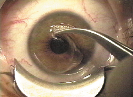
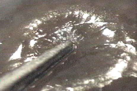
FLAP REPLACEMENT After the ablation is completed, the flap is reflected (Fig. 11). The interface is irrigated (Fig. 12) until any interface debris is eliminated, best visualized with oblique rather than coaxial illumination. The surface of the flap is stroked with a smooth instrument such as the irrigation cannula from the hinge to the periphery to assure that wrinkles are eliminated and that the flap settles into its original position, as indicated by the radial marks made earlier and illustrated by the application of prednisolone acetate 1% suspension (Fig. 13). The endothelial pump will begin to secure the flap in position within several minutes. If a significant epithelial defect is present, a bandage contact lens should be placed. Once the flap is adherent, the lid speculum is carefully removed, taking care not to move the flap. Most surgeons place a drop of antibiotic and steroid over the eye at the conclusion of the procedure followed by placement of a clear shield or protective goggles. Some surgeons recheck the flap at the slit-lamp approximately 15 to 30 minutes later to assure it has remained in proper alignment.
Many surgeons instruct their patients to use topical antibiotics and steroids postoperatively for approximately 5 days. In addition, it is very important to keep the surface of the flap well lubricated in the early postoperative period. Patients are examined 1 day after surgery to assure that the flap has remained in proper alignment and that there is no evidence of infection or excessive inflammation. In the absence of complications, the next examinations are typically approximately 1 week; 1 month; and 3, 6, and 12 months postoperatively. |
OUTCOMES |
LASIK studies vary considerably in the techniques employed, degree of refractive
error treated, length that patients are followed, and variables
analyzed. This section summarizes the findings of some of the larger
LASIK studies using modern techniques for the treatment of different
degrees of myopia, as well as for astigmatism and hyperopia. As was
the case with PRK results, this section cites only a few historically
significant studies and emphasizes the typical range of outcomes encountered
in current clinical practice. Low Myopia Because PRK already had been judged adequate for patients with low to moderate myopia and did not include the risk of creating a corneal flap, LASIK was initially conceived as a procedure for myopia greater than 6 D. However, as LASIK techniques and microkeratome designs improved, many studies reported safety and efficacy of LASIK at a level at least as high as PRK for correction of low myopia. Series of eyes treated with LASIK for low myopia16–19 report 45% to 83% achieve uncorrected visual acuity of 20/20 or better, 85% to 100% achieved 20/40 or better, and 73% to 100% of patients obtain a postoperative refraction within 1.0 D of the intended refraction. In most studies no eyes lose two or more lines of best corrected vision. Several early studies directly comparing LASIK to PRK for low myopia have revealed similar safety and predictability but with higher patient satisfaction after LASIK. El-Maghraby and coauthors20 found that, in a study of patients who had LASIK in one eye and PRK in the other to correct −2.5 to −8.0 D, almost twice as many were highly satisfied with their LASIK eye as compared to their PRK eye 1 year after treatment. In addition to having less postoperative pain and more rapid visual recovery, the LASIK eyes were more likely to achieve an uncorrected visual acuity of 20/20 or better and had a more regular postoperative corneal topography. As techniques and technology continue to develop, reported results also have improved.21 Nevertheless, results of PRK often are reported to be equal to or superior to LASIK for low myopia.22 MODERATE MYOPIA In patients with moderate myopia (approximately −6.0 to −12.0 D), large published series23–29 report that 26% to 57% of eyes achieve an uncorrected postoperative visual acuity of 20/20 or better, 55% to 85% reach at least 20/40, and 41% to 72% are within 1.0 D of intended correction. The percentage of patients losing two or more lines of best-corrected vision ranges from 0% to 3.5%. Studies directly comparing LASIK to PRK for moderate myopia generally reveal that the long-term results are remarkably similar. However, LASIK eyes have faster visual recovery with better uncorrected visual acuity at 1 month. Also, two reports from a large multicenter randomized prospective trial17,18 showed that, although average outcomes appear similar between PRK and LASIK, more PRK eyes lost best-corrected vision than LASIK-treated eyes. HIGH MYOPIA High myopia is most often defined as myopia greater than −12.0 D, with several studies reporting results of patients treated with up to −29.0 D. In this range the predictability of the procedure is markedly reduced, with 26% to 50% achieving at least 20/40 uncorrected vision and 32% to 53% attaining a postoperative refraction within 1.0 D of the intended correction.30–32 In addition, when treating such high degrees of myopia, there was a higher incidence of loss of best corrected vision than in the correction of lower levels of myopia. However, patients with high myopia often gain best-corrected vision after LASIK, probably because of decreased image minification compared to preoperative spectacles. In contrast to LASIK, studies of PRK for high myopia generally report unacceptable haze and regression of effect.33 As experience with LASIK has accumulated; however, an increasing number of surgeons feel that LASIK and PRK are rarely, if ever, appropriate for corrections above −12 D for several reasons. The required ablation depths for high corrections may leave an inadequate stromal bed (less than 250 μ at a minimum) for long-term structural stability of the cornea. High corrections have poor predictability of achieving good high contrast uncorrected visual acuity. In addition, high corrections have an unacceptably high level of vision compromises, including glare, halo, and loss of contrast sensitivity. These difficulties are attributable to the induction of high-order aberrations, particularly spherical aberration, by high corrections that, of necessity, markedly flatten the central cornea compared to the midperipheral cornea. MYOPIA WITH ASTIGMATISM Overall, the results of toric ablations34–36 have not been as predictable as for spherical LASIK ablations. Most often, the procedure undercorrects the cylinder, which may simply indicate the need for improved nomograms or may indicate an inaccuracy of the axis ablated. The introduction of scanning spot lasers has coincided with improved results, possibly because a scanning laser can achieve the desired complex ablation pattern more readily without excess tissue removal.37 Fewer studies are available on the efficacy of toric LASIK. Most published studies include a limited number of subjects with a wide range of refractive errors, sometimes combining results of patients having toric ablations with those having spherical ablations, making the results difficult to interpret. Zaldivar and coauthors reported a series of 84 eyes with 3 to 6 months follow-up after being treated with toric ablations for spherical equivalent refractions ranging from −5.5 to −11.5 D with less than 4.0 D of cylinder.38 In this study, 22% of eyes achieved at least 20/20 uncorrected vision, 77% achieved at least 20/40, and 83% were within 1.0 D of intended correction. El Danasoury and coauthors found that 12 months postoperatively 55% of 56 patients attained 20/20 vision and 91% were within 1.0 diopter of intended correction after undergoing toric ablations for spherical equivalents ranging from −2.25 to −5.0 D with 0.5 to 3.0 D of astigmatism.39 Fraenkel and coauthors reported 43 patients with a preoperative spherical equivalent of −1.5 to −15.0 and astigmatism ranging from 0.75 to 7.0 D.40 In this study, 35% of patients attained uncorrected vision of at least 20/20 postoperatively, whereas 79% attained 20/40 and 91% were within 1.0 D of intended correction. HYPEROPIA In contrast to PRK or LASIK for myopia, where the central cornea is flattened, in treating hyperopia the central cornea is steepened by preferentially ablating the midperiphery. Initial problems with hyperopic treatments included diminished predictability and stability in comparison to myopic treatments as well as loss of best corrected visual acuity, partially secondary to decentrations with small ablation zones.41,42 With enlargement of both the optical zone and peripheral blend zone, as well as improved centration with the assistance of tracking devices, studies of hyperopic LASIK with longer follow-up periods have shown improved outcomes.43 Predictability and stability were better for corrections of +1.0 to +4.0 D than when treating higher degrees of hyperopia.44 Hyperopic corrections also may be improving with the increased use of scanning spot lasers rather than broad-beam lasers, because scanning spots can readily create the correct exposure pattern without use of masks, parallel blades, or other beam manipulators.45,46 HYPEROPIC ASTIGMATISM Treatment of hyperopic astigmatism also has been encouraging. Arbelaez and Knorz47 reported the results of patients treated for spherical hyperopia (60 eyes) and hyperopic astigmatism (50 eyes) with 12 months of follow-up. Ninety-one percent of patients with low spherical hyperopia (+1.0 to +3.0 D) and 83% of low toric ablations attained a postoperative refraction within ±1.0 D of the intended correction. Of eyes treated for +3.1 to +5.0 D, 83% of spherical ablation and 58% of toric ablations achieved refraction within 1.0 D of intended correction. Of eyes with more than 5.0 D of hyperopia, 50% of spherical and 17% of toric ablations achieved a postoperative refraction within 1.0 D of intended correction 12 months postoperatively. MIXED ASTIGMATISM Mixed astigmatism is defined as refractive error with cylinder greater than sphere and of opposite sign. LASIK has been FDA approved for mixed astigmatism of up to 6.0D of sphere and cylinder, with the cylinder greater than the sphere and of opposite sign. The outcomes of LASIK for mixed astigmatism are similar to the results for hyperopia and hyperopic astigmatism. |
RETREATMENT |
Although LASIK reduces refractive error and improves uncorrected visual
acuity in almost all cases, some patients have residual refractive error
that requires retreatment. The degree of refractive error that warrants
retreatment varies depending on the patient's lifestyle and
expectations. Retreatment rates vary from 5.5% to 36%, depending
partially on the degree of myopia being treated, the laser and
nomograms used, and the expectations of the patient population.48–50 One study showed that rates of retreatment are higher with higher initial
corrections, residual astigmatism, and patients older than 40 years. One
advantage of LASIK compared to PRK is that refractive stability
generally occurs earlier, allowing earlier enhancements, typically within
the first half year after LASIK, whereas in PRK the ongoing activation
of keratocytes and risk of haze after enhancement usually requires
at least 6 months before an enhancement PRK can be performed safely. Typically, retreatment
rates are higher in hyperopia and higher myopia
than other indications. Retreatment after LASIK usually is performed by lifting the pre-existing lamellar flap and applying additional ablation to the stromal bed.51,52 In many cases the flap can be lifted even several years after the original procedure. Alternatively, if a strong Bowman's layer scar has formed, a new flap can be created with the microkeratome. When lifting a pre-existing flap it is important to minimize epithelial disruption. A jeweler's forceps or 27-gauge needle can be used to localize the edge of the previous flap. The edge of the flap can be seen more easily at the slit-lamp than with the diffuse illumination of the operating microscope of the laser. Therefore, a flap lift is most easily begun at the slit-lamp and then completed at the excimer laser. A circumferential epithelial dissection is then performed so that the flap can then be lifted without tearing the epithelial edges (Fig. 14A). Alternatively, with experience a surgeon can disclose the location of a healed flap edge by placing pressure on the midperiphery, where a slight discontinuity in the corneal tension causes an arc-shaped light reflection at the flap edge, allowing the flap to be lifted cleanly with the diffuse illumination of the operating laser microscope (Fig. 14B). Once the ablation is performed the flap is repositioned and the interface is irrigated as in initial LASIK procedures. Special care must be taken to assure that no loose epithelium is trapped beneath the edge of the flap that would lead to epithelial ingrowth. In retreatments performed with flap lifting, the usual complications of laser ablation apply, as detailed in the following. In addition, flap lifting is associated with increased risk of epithelial ingrowth into the interface and risk of developing flap striae. Occasionally PRK is considered to enhance a previous primary LASIK treatment. PRK on a LASIK flap in the past was believed to carry increased risk of haze formation and irregular astigmatism.53 Recent experience has called this concern into question.54 Furthermore, PRK is an appealing alternative when the residual stromal bed is insufficient for further ablation, or in other situations such as a buttonhole flap. Recently, some surgeons have investigated the concomitant use of antiscarring agents such as mitomycin-C to improve the results of PRK performed on LASIK flaps.55 This may not be necessary on a routine basis. A final note of caution: Many refractive surgeons have performed enhancements for apparent myopic regression after LASIK when the true cause was subtle early myopic shifting because of nuclear cataract!56 |
WAVEFRONT-GUIDED PRK AND LASIK OUTCOMES | |
Wavefront error measurement technology can be employed diagnostically, to
quantitatively define aberrations affecting the quality of a patient's
vision.57 In addition, this technology is employed to create ablation profiles customized
for an individual patient, in an effort to reduce pre-existing
aberrations and to reduce the induction of new aberrations. In addition
to addressing higher-order aberrations that cause symptoms such as
loss of contrast sensitivity and night-time haloes and glare,58 wavefront-guided treatments are reported to improve the accuracy of correcting
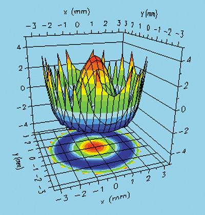
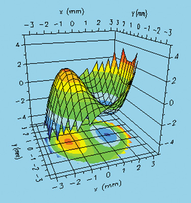
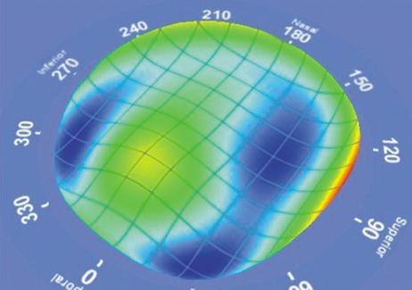
the low-order aberrations of spherical error and astigmatism.59,60 Aberrations are most commonly expressed mathematically as Zernicke polynomials, whose terms can be arranged in a pyramidal fashion (Fig. 15). Two of the most important aberrations are spherical aberration (Fig. 16), which commonly causes a perception of glare and halo, particularly at night, and coma, which causes a smear in a direction like the tail from a comet (Fig. 17).
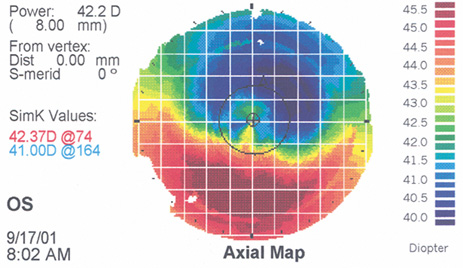
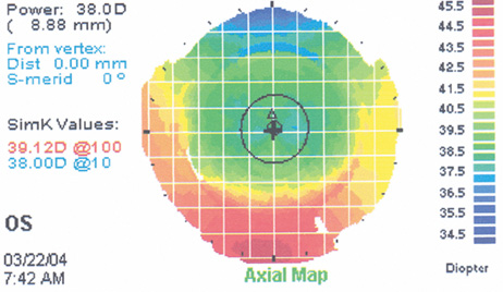
In its premarket approval application to the Food and Drug Administration, Alcon Autonomous compared the effectiveness of 139 eyes with wavefront-guided Custom ablations to 47 conventionally treated eyes at 6 months after surgery. All eyes had a preoperative myopic manifest refractive error under −7.0 D sphere and under 0.5 D cylinder. Seventy-nine point nine percent of wavefront-treated eyes achieved UCVA of 20/20 or better, 91.5% 20/25 or better, and 98.6% 20/40 or better. The manifest refraction spherical equivalent (MRSE) was ±0.5 D in 74.8% and ±1.0 D in 95.7%. Aberrations were smaller at 6 months than preoperatively for 38% of Custom eyes compared to 14% of a healed flap edge by RMS increased by 0.08 μ (20%) in the Custom group and 0.33 μ (82%) in the conventional group. Spherical aberrations increase by 0.04 μ (22%) for Custom eyes and 0.23 μ (108%) for conventional eyes. No Custom patients reported “significantly worse” glare or haloes postoperatively and only one patient reported significantly worse night driving difficulty. Mean contrast sensitivity for Custom eyes improved by 0.1 to 0.2 log units relative to conventional eyes. Best spectacle-corrected vision (BSCVA) and low-contrast acuity were both slightly better for Custom eyes than conventionally treated eyes. More Custom eyes showed clinically significant increase than decreases, whereas more conventional eyes showed clinically significant decreases than increases in both of these measures. A single site comparison of the Alcon Custom Cornea and the Bausch and Lomb Zyoptix systems, reporting results at 1 month, showed similar excellent results, such as 93% and 90% rates of 20/20 UCVA and 80% and 70% 20/16 UCVA, respectively.61 A single site comparison of Alcon Custom Cornea and VISX Custom Vue for eyes under −8 D and cylinder under 1.5 D yielded 98% and 95%, respectively, within 0.5 D of target.62 In a contralateral eye study, after a nomogram adjustment, the Alcon Custom Cornea and VISX Custom Vue results were 80% and 100%, respectively, within 0.5 D of goal.63 The use of wavefront-guided treatments is FDA approved for primary treatments only, but this technology is attractive for enhancements of patients with vision disturbances attributable to aberrations.64 As an example, Figure 18A shows the corneal topography of a decentered ablation with loss of BCVA and complaints of glare. The wavefront measurement showed a large amount of coma, consistent with the decentration (Fig. 18B). The patient received a wavefront-guided enhancement. Marked improvement occurred, with improved BCVA, resolution of most vision complaints, and improvement in the corneal topography (Fig. 18C) and reduction in the amount of aberration (Fig. 18D). |
COMPLICATIONS | |||||||
In the past, the more severe complications associated with LASIK have been
related to problems with the microkeratome. Advances in microkeratome
technology have significantly reduced the incidence of severe, sight-threatening
complications. However, it is still imperative that meticulous
care is taken in the cleaning and assembly of the microkeratome
in order to assure that a smooth, uninterrupted keratectomy is performed. MICROKERATOME COMPLICATIONS An overview of microkeratome complications is gained from a synopsis of 47,094 LASIK cases.65 Epithelial Defects The most common microkeratome complication is the development of epithelial defects created by the friction of the microkeratome applanation plate sliding across the surface of the cornea that has been stiffened by the elevated intraocular pressure. Even in the absence of visible anterior basement membrane disease preoperatively, patients may have inadequate epithelial adhesion structures to resist the frictional stress. As a result, a central or inferior defect may result, with either frank sloughing of a sheet of epithelium (Fig. 19) or development of a stretched loosened epithelium, often called an “epithelial slider.” The other type of epithelial defect occasionally seen is a localized abrasion near the hinge where the flap is maximally compressed at the end of the cut; this type of defect less commonly results from basement membrane adhesions and more commonly the structure of the microkeratome itself.
Prevention of epithelial defects is preferable to treatment. Key steps are: (a) careful preoperative screening for anterior basement membrane disease; (b) avoidance of toxic damage to the epithelium from anesthetics and drying; and (c) frequent administration of lubricants, particularly immediately prior to the microkeratome pass (Fig. 5). If a defect occurs, application of a bandage soft contact lens after completing the laser treatment and repositioning the flap is usually the best course. Administration of a potent broad spectrum antibiotic as infection prophylaxis and intense topical steroids to discourage diffuse lamellar keratitis (DLK) is advisable. The bandage contact lens is retained until the epithelial defect heals. For patients with a “slider” area of loose but intact epithelium, the surgeon is guided by the size and location of the sliding sheet to determine whether it is better to debride the loosened sheet or allow it to remain and try to re-establish adhesion. The patient with an epithelial defect needs to be informed about the condition and the expected slower recovery of vision. In occasional cases more intensive treatment is needed to re-establish integrity of the epithelium, even with the application of PTK (see Chapter XX). Free Cap Sporadically the microkeratome transects the hinge, creating a free cap rather than the desired flap. The two principal causes of free caps are either surgeon error in selecting the incorrect settings for the “stop” that controls the flap creation or an unusually small or flat cornea. A flat corneal curvature is a risk factor for development of a free cap.37 When the preoperative keratometry reading is 41 D or less in the meridian of the hinge, or the corneal diameter is under 11 mm, the surgeon should consider adjusting the microkeratome to create a slightly larger hinge or smaller diameter flap. If a free cap does occur, the most important initial action is to recognize that the flap is absent and stop the assistant from taking the microkeratome; the free cap is almost always adherent to the microkeratome head and the cap must be identified and recovered before it is lost (Fig. 20). The cap is placed in a sterile container, preferably closed to reduce desiccation. The laser procedure is completed if the exposed stromal bed is large enough for the laser ablation, and then the cap is repositioned. The use of gentian violet ink alignment marks aids in identification of the epithelial side as well as correct repositioning of the flap. Although some surgeons have success simply allowing a free cap to adhere naturally, many surgeons feel more secure placing a suture to perform the function of the hinge. Usually a 10-0 nylon suture is employed as a single interrupted stitch. The suture must not be tight; tension distorts the flap and creates irregular astigmatism. If the flap is secure on the first postoperative day it is usually safe to remove it at that visit. Some surgeons also use a bandage soft contact lens to assist in stabilizing the cap.
Irregular, Thin, and Buttonhole Flaps Defects within the blade, poor suction, or uneven progression of the microkeratome across the cornea can produce an irregular, thin, or buttonholed flap (Fig. 21) that is a potential source of irregular astigmatism with loss of best corrected vision. A steep corneal curvature is a risk factor for the development of these intraoperative flap complications.66 If a thin or buttonholed flap is recognized, it should be replaced without performing the ablation.67,68 A bandage soft contact lens is applied to stabilize the flap, typically for several days to 1 week. A new flap can be cut after at least 3 months of healing, preferably with a different microkeratome head designed to produce a deeper cut, and the ablation can be applied at that time. Another option to consider is the application of PRK over the flap for correction of the residual optical deficit, with or without topical mitomycin C to discourage scar formation.69,70
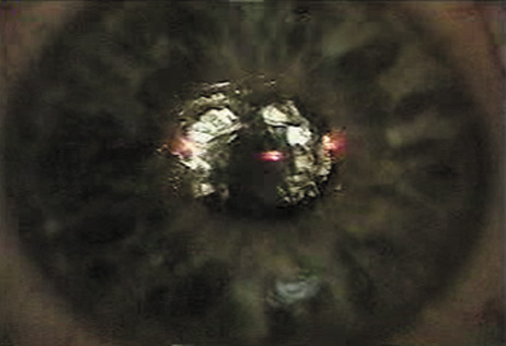
Corneal Perforation Corneal perforation is a rare but devastating intraoperative complication that can occur if the microkeratome is not properly assembled, or if the depth plate of the microkeratome is not properly placed in an older model microkeratome. Therefore, it is imperative that the surgeon double check that the microkeratome has been properly assembled prior to proceeding. Most microkeratomes are now made with a prefixed depth plate so that this source of error is eliminated. Corneal perforation also can occur when LASIK is performed on an excessively thin cornea.71 Therefore, corneal thickness must be measured with pachymetry prior to performing LASIK, especially in patients undergoing retreatment. DRY EYE AND CORNEAL SENSATION The occurrence and etiology of dry eye syndromes after laser ablation procedures, particularly LASIK, is discussed in the preceding in the preoperative evaluation. The surgeon must carefully monitor the patient postoperatively for signs of punctate keratitis or more severe manifestations of neurotrophic epitheliopathy. Intensive tear film support with nonpreserved artificial tears, gels and ointments, and the addition of punctal occlusive plugs or cyclosporine drops if lubricants are inadequate, are the mainstay of therapy while awaiting the return of innervation.72 Both the surgeon and patient must remain aware that many patients seeking laser vision correction do so because of contact lens intolerance, and dry eyes are one of the most common reasons for that intolerance.73 That dryness persists, of course, and is transiently worsened during the recovery from the denervation of the flap.74 The patient may perceive the long-term persistent dryness to have worsened and blame the LASIK procedure. Dry eyes are a common cause of transient loss of BCVA after LASIK.75 STRIAE Postoperatively, the flap can slip, resulting in prominent wrinkles and a widened peripheral gutter (Fig. 22). The earlier the flap malposition is recognized and treated, the easier it is to remove potentially visually significant folds.
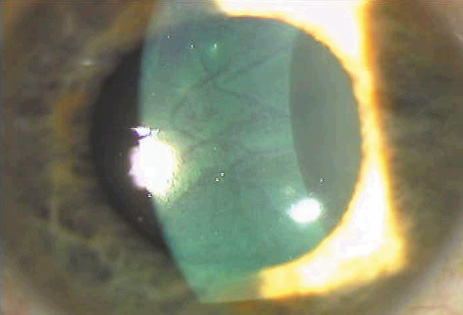
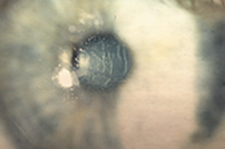
Current approaches to smoothing the flap and avoiding striae at the end of the procedure vary widely. No matter which technique is employed, however, the surgeon must carefully examine for the presence of striae once the flap is repositioned. Coaxial and oblique illumination should be used at the operating microscope. Checking the patient at the slit-lamp 15 to 30 minutes postoperatively is important to detect early flap slippage. If present, striae should be immediately corrected by refloating and stroking the flap smooth. A protective plastic shield often is used for the first 24 hours to discourage touching of the eyelids and inadvertent disruption of the flap. Macrostriae represent full-thickness, undulating stromal folds (see Fig. 22). These invariably occur because of initial flap malposition or postoperative flap slippage. Careful examination should disclose a wider gutter on the side where the folds are most prominent. Flap slippage should be rectified as soon as it is recognized, because the folds rapidly become fixed. A lid speculum is placed under the operating microscope or at the slit-lamp, the flap is lifted, copious irrigation used in the interface, and the flap is stroked repeatedly until the striae resolve. Using hypotonic saline or sterile distilled water as the interface irrigating solution swells the flap and may initially reduce the striae, but swelling reduces flap diameter, which widens the gutter, delays flap adhesion because of prolonged endothelial dehydration time, and may leave worse striae after the flap dehydrates. If the macrostriae have been present for more than 24 hours, reactive epithelial hyperplasia in the valleys and hypoplasia over the elevations of the macrostriae tends to fix the folds into position. In that case, the central 6 mm of the flap over the macrostriae should be de-epithelialized in order to remove this impediment to smoothing of the wrinkles. A bandage soft contact lens always should be employed to stabilize the flap and protect the surface until full re-epithelialization occurs. In severe cases of intractable macrostriae, a tight 360-degree antitorque running 10-0 nylon suture or multiple interrupted sutures may be placed for several days, but irregular astigmatism may be present after suture removal.76 Microstriae are fine, hairlike optical irregularities best seen on red reflex illumination or light reflected off the iris. Microstriae are fine folds in the Bowman's layer. This anterior location of the microstriae accounts for the disruption of best-corrected visual acuity they cause (Fig. 23A). Computer topography color maps usually do not show these fine irregularities; however, disruption of the surface contour usually is heralded by disruption of the regularity of the topographer's placido mires. In addition, application of dilute fluorescein reveals so-called “negative striae,” in which the elevated striae disrupt the tear film with loss of fluorescence over the elevated striae (Fig. 23B). A few striae may not be significant visually. In addition, mild loss of acuity or other optical symptoms such as ghost images usually improve over time as the epithelial thickness adjusts to the folds and restores a more regular anterior tear film. The ocular surface should be supported with frequent administration of nonpreserved artificial tears or a bandage soft contact lens to encourage remodeling of a smooth corneal surface.
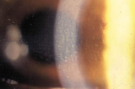
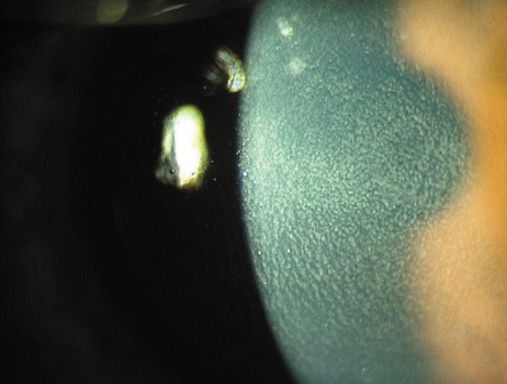
Persistent optically significant striae must be addressed, nonetheless. Many interventions have been recommended with variable results. Some surgeons advocate hydration of Bowman's layer. Although this eventually occurs with prolonged stroking of the epithelial surface with a moistened surgical spear sponge or irrigating cannula, microstriae usually disappear within minutes of deliberate de-epithelialization of the area over the microstriae, followed by several drops of sterile distilled water. Hypotonic solution applied directly to the Bowman layer speeds the disappearance of the microstriae. If the striae persist, then the flap should be lifted and the interface irrigated with balanced salt solution to allow the flap to reposition. In severe cases, traction with fine tooth forceps or suturing as described for macrostriae also may be helpful. Care must be taken not to tear the fragile flap. A bandage soft contact lens then is applied with antibiotic and mild steroid until re-epithelialization is established. An alternative to these procedures is phototherapeutic keratectomy (PTK). Pulses from a broad-beam laser, set to a maximal diameter of 6.5 mm, are applied initially to penetrate the epithelium in about 200 pulses. The epithelium acts as a masking agent, exposing the elevated striae before the valleys between the striae. After the transepithelial ablation, additional pulses are applied with the administration of a thin film of medium viscosity artificial tears every five to 10 pulses, up to a maximum of 100 additional pulses. If these guidelines are followed, little to no haze results and an average hyperopic shift of under +1 diopter occurs, because of the minimal tissue removal. Steinert, Ashrafzadeh, and Hersh demonstrated significant improvement in BCVA and lack of optically significant haze with this technique.77 The principles and techniques of PTK for reduction of corneal surface irregularities are discussed in more detail elsewhere in this volume. CENTRAL ISLANDS AND DECENTRATION Irregular astigmatism can be induced by complications related to the laser ablation. The fluence and homogeneity of the laser must be checked before the procedure to assure that there is a smooth ablation profile without “hot spots” or “cold spots.” Central islands can occur after LASIK just as they do after PRK, but the incidence after LASIK is significantly lower. The etiology of central islands after LASIK is not fully understood but evidence points to vortex currents in the center of broad beam ablation, central accumulation of fluid or debris during the ablation, or patient healing responses.78,79 Central islands can be avoided sometimes with the use of anti-island software in which more pulses are delivered to the center of the ablation zone. If a central island does occur and does not resolve on its own, it can be treated with the excimer laser by lifting the flap and applying a central ablation to the elevated island. Another potential source of irregular astigmatism related to the laser application is a decentered ablation. Mild decentrations often are asymptomatic, but larger decentrations can lead to significant optical aberrations that can be difficult to treat. Until recently, decentration has been addressed by further ablation with opposite decentration,80–82 or through arcuate keratotomies and limbal relaxing incisions.83 Wavefront measurements may be able to more directly and precisely direct enhancement retreatments to improve the optical disruptions caused by decentrations, which particularly induce the aberration known as coma.84 (See the earlier discussion on outcomes and enhancements in the preceding.) INTERFACE INFLAMMATION Sterile interface inflammation has been referred to as “Sands of the Sahara,”85 diffuse lamellar keratitis (DLK),86 or, perhaps most accurately, diffuse interface keratitis (DIK).87 This syndrome can range from asymptomatic interface haze near the edge of the flap to marked diffuse haze with diminished best-corrected vision. DLK is clinically staged depending on severity. Stage 1 is a barely visible fine dusting of white blood cells in the interface, often just in the periphery. In stage 2, the cells are more easily seen, and often accumulate in the ridges caused by microkeratome blade chatter, giving the characteristic parallel lines that look like windswept desert sand (Fig. 24A). Stage 3 is reached when the density of inflammatory cells begins to interfere with visualizing intraocular details (Fig. 24B). If the release of collagenase occurs with haze, softening, or loss of stroma and onset of large wrinkles, DLK is in stage 4 (Fig. 24C).
The inflammation generally resolves on its own without sequelae, but severe cases can lead to scarring or flap melting; therefore, many surgeons treat the inflammation with aggressive topical steroids in stages 1 and 2; or, in the more severe cases of stages 3 and 4, irrigation of the interface with balanced salt solution and intense topical steroids. In highly threatening cases a brief course of systemic steroids also may be employed. Although initial attempts to identify a single cause of interface keratitis centered on debris or solution on microkeratome blades, subsequent cases documented inflammation after flap-lift retreatments without the use of a microkeratome. In one case, interface inflammation was observed after epithelial disruption without lifting the flap at all. Most likely, the inflammation is a nonspecific reaction to corneal trauma and the presence of an interface becomes a potential space in which white blood cells can aggregate. When a cluster of DLK occurs, a common source is a toxic biofilm that builds up in the reservoir of the steam sterilizer.88 INFECTIOUS KERATITIS It is important to differentiate sterile interface inflammation from potentially devastating infectious inflammation. Infection within the interface can lead to flap melting, severe irregular astigmatism, and corneal scarring requiring penetrating keratoplasty. If infection is suspected, the flap should be lifted and the interface cultured and irrigated with antibiotics.89 The most common infections are from gram-positive organisms, followed closely in frequency by atypical mycobacteria.90 Infection from virtually all organisms has been reported; fungal infection is particularly difficult to treat. If an infection does not respond, amputation of the flap may be necessary to improve antibiotic or antifungal penetration. Fourth-generation fluoroquinolones, gatifloxacin, and moxifloxacin have excellent efficacy against most of the bacteria causing post-LASIK infections, including mycobacteria. An infected LASIK flap after a recurrent abrasion is shown in Figure 20. EPITHELIAL INGROWTH Epithelial ingrowth into the lamellar interface can first be recognized anytime from 2 days to 12 months postoperatively (Fig. 25A).91 Isolated nests of epithelial cells that are not advancing do not need to be treated. However, if the epithelium is advancing toward the visual axis (Fig. 25B), is associated with irregular astigmatism, or triggers overlying flap melting, it should be removed by lifting the flap and scraping the epithelium from the underside of the flap as well as from the stromal bed prior to repositioning the flap. Two series have reported an incidence of epithelial ingrowth requiring intervention after approximately 2% of LASIK procedures. The incidence of epithelial ingrowth is greater in patients who develop an epithelial defect at the time of the procedure and in those undergoing a retreatment with lifting of a pre-existing flap.57,58 Therefore, in these instances, special care should be taken to assure that no epithelium becomes caught under the edge of the flap when it is repositioned. Placement of a bandage contact lens at the conclusion of the procedure also may decrease the incidence of epithelial ingrowth for patients at higher risk of developing this complication. In a few cases of recurrent ingrowth in the same area, placement of a suture may help seal the peripheral flap and discourage recurrent ingrowth.92 In some cases, PRK may be a good option when epithelial ingrowth is challenging and recurrent.93
INTERFACE DEBRIS Debris in the interface occasionally is seen postoperatively. The principal indication for intervention, with flap lifting, irrigation, or manual removal of debris, occurs when the foreign material elicits an inflammatory reaction. Small amounts of lint, nondescript particles, or tiny metal particles from stainless steel surgical instruments usually are well tolerated. On the other hand, although a small amount of blood that has oozed into the interface from transected peripheral vessels may be tolerated, any significant amount of blood usually elicits an inflammatory cell response and should be irrigated from the interface. Use of a topical vasoconstrictor such as epinephrine to facilitate coagulation at the time of replacing the flap helps to minimize this problem. ECTASIA The importance of an adequate residual stromal bed to prevent structural instability and postoperative corneal ectasia is discussed in the section on preoperative evaluation. Current standards recommend a minimum residual stromal bed of at least 250 μ after completion of the ablation.94 Although keratectasia usually is associated with LASIK performed for higher myopic corrections; in thin corneas or patients who have had multiple laser ablations, cases of ectasia have been reported in corrections as low as −4 D where the residual stromal was believed to be thicker than 250 μ.95 In many of these cases, later examinations have shown that the microkeratome created a flap thicker than expected, resulting in a thinner residual stromal bed.96 In other cases, preoperative subtle keratoconus or other ectasias may have been present. In many cases, good vision can be restored with a rigid gas permeable contact lens. The use of implanted intrastromal polymethylmethacrylate segments to reduce the irregular astigmatism is under investigation.97 Penetrating keratoplasty may be required in extreme cases. |
CONCLUSIONS |
LASIK has emerged as the procedure of choice among patients and surgeons
for most levels of refractive error. The limits of the procedure, particularly
in treating higher levels of myopia (greater than −12.0) and
hyperopia (greater than +4.0) must be
respected and alternative refractive options considered. Although LASIK has rapidly surpassed PRK in popularity for treatment of many refractive errors, visual results actually are very similar among the procedures. A study of patients treated for myopia of −1.00 to −9.50 D showed equal refractive outcomes. Another study of treatment of myopia between −6.00 and −15.00 D showed slightly less tendency for postoperative optical symptoms in LASIK eyes compared to PRK eyes. Preference for LASIK often relates more to the comfort level and early recovery of vision than it does to ultimate visual acuity and function. The incidence of complications in LASIK decreases with surgeon experience and, as with any surgical procedure, complications are best managed with avoidance. However, when recognized and properly treated, the majority of complications do not result in loss of best corrected vision. If visual acuity is diminished, it is most often secondary to irregular astigmatism. Often, epithelial hyperplasia and hypoplasia occur over time; they can smooth the corneal surface, reducing irregular astigmatism and improving visual function. However, if significant symptoms persist, the patient may benefit from selective application of the laser to smooth the corneal surface. New technology in the form of topography or wavefront optics–based custom ablations will be helpful in the treatment of these patients. |
REFERENCES |
1. Duffey RJ, Leaming D: Trends in refractive surgery in the United States. J Cataract Refract Surg 30:1781, 2004 2. Barraquer Moner JI: Refractive keratoplasty. Est Inf Oftal 10:2, 1949 3. Barraquer JI: Method of cutting lamellar grafts in frozen corneas. New orientations for refractive surgery. Arch Soc Am Ophthalmol 1:237, 1958 4. Barraquer JI: Method of cutting lamellar grafts in frozen corneas. New orientations for refractive surgery. Arch Soc Am Ophthalmol 1:237, 1958 5. Swinger CA, Krumeich JH, Cassiday D: Planar lamellar refractive keratoplasty. J Refract Surg 2:17, 1986 6. Ruiz LA, Rowsey JJ: In situ myopic keratomileusis. Invest Ophthalmol Vis Sci 29:S39, 1988 7. Ruiz LA: Cheratomileusi automatizzata in situ. In: Chirurgia della Miopia Assile Mediante Cheratomileusi. Milan: CAMO, 1993:137 8. Pallikaris IG, Papatzanaki ME, Stathi EZ, et al: Laser in situ keratomileusis. Lasers Surg Med 10:463, 1990 9. Pallikaris IG, Papatzanaki ME, Siganos DS, et al: A corneal flap technique for laser in situ keratomileusis. Human study. Arch Ophthalmol 145:1699, 1991 10. Lee KW, Joo CK: Clinical results of laser in situ keratomileusis with superior and nasal hinges. J Cataract Surg 29:457, 2003 11. Kumano Y, Matsui H, Zushi I, et al: Recovery of corneal sensation after myopic correction by laser in situ keratomileusis with a nasal or superior hinge. J Cataract Refract Surg 29:757, 2003 12. Nordan LT, Slade SG, Baker RN, et al: Femtosecond laser flap creation for laser in situ keratomileusis: 6 month follow-up of initial U.S. clinical series. J Refract Surg 19:8, 2003 13. Seiler T, Quurke AW: Iatrogenic kerectasis after LASIK in a case of forme fruste keratoconus. J Cataract Refract Surg 24:1007, 1998 14. Fogla R, Rao SK, Padmanabhan P: Keratoectasia in 2 cases with pellucid marginal corneal degeneration after laser in situ keratomileusis. J Cataract Refract Surg 29:788, 2003 15. Ambrosio R Jr, Klyce SD, Wilson SE: Corneal topographic and pachymetric screening of keratorefractive patients. J Refract Surg 19:24, 2003 16. Wang Z, Chen J, Yang B: Comparison of laser in situ keratomileusis and photorefractive keratectomy to correct myopia from −1.25 to −6.00 diopters. J Refract Surg 13:528, 1997 17. El Danasoury MA, El-Maghraby A, Klyce SD, Mehrez K: Comparison of photorefractive keratectomy with excimer laser in situ keratomileusis in correcting low myopia (from −2.00 to −5.50 diopters). A randomized study. Ophthalmology 106:411, 1999 18. Salah T, Waring III GO, El Maghraby A, Moadel K, Grimm SB: Excimer laser in situ keratomileusis under a corneal flap for myopia of 2 to 20 diopters. Am J Ophthalmol 121:143, 1996 19. Maldonado Bas A, Onnis R: Results of laser in situ keratomileusis in different degrees of myopia. Ophthalmology 105:606, 1998 20. El-Maghraby A, Salah T, Waring III GO, Klyce S, Ibrahim O: Randomized bilateral comparison of excimer laser in situ keratomileusis and photorefractive keratectomy for 2.50 to 8.00 diopters of myopia. Ophthalmology 106:447, 1999 21. McDonald MB, Carr JD, Frantz JM, et al: Laser in situ keratomileusis for myopia up to −11 diopters with up to −5 diopters of astigmatism with the Summit Autonomous LADARVision excimer laser system. Ophthalmology 108:309, 2001 22. Pop M, Payette Y: Photorefractive keratectomy versus laser in situ keratomileusis: A control-matched study. Ophthalmology 107:251, 2000 23. Pesando PM, Ghiringhello MP, Tagliavacche P: Excimer laser in situ keratomileusis for myopia. J Refract Surg 13:521, 1997 24. Knorz MC, Wiesinger B, Liermann A, Seiberth V, Liesenhoff H: Laser in situ keratomileusis for moderate and high myopia and myopic astigmatism. Ophthalmology 105:932, 1998 25. Knorz MC, Liermann A, Seiberth V, Steiner H, Wiesinger B: Laser in situ keratomileusis to correct myopia of −6.00 to −29.00 diopters. J Refract Surg 12:575, 1996 26. Steinert RF, Hersh PS, the Summit Technology PRK-LASIK Study Group: Spherical and aspherical photorefractive keratectomy and laser in-situ keratomileusis for moderate to high myopia: Two prospective, randomized clinical trials. Trans Am Ophthalmol Soc XCVI:197, 1998 27. Hersh PS, Brint SF, Maloney RK, et al: Photorefractive keratectomy versus laser in situ keratomileusis for moderate to high myopia. A randomized prospective study. Ophthalmology 105:1512, 1998 28. Guell JL, Muller A: Laser in situ keratomileusis (LASIK) for myopia from −7 to −18 diopters. J Refract Surg 12(2):222, 1999 29. Tsai RJ: Laser in situ keratomileusis for myopia of −2 to −25 diopters. J Refract Surg 13:S427, 1997 30. Perez-Santonja JJ, Bellot J, Claramonte P, Ismail MM, Alio JL: Laser in situ keratomileusis to correct high myopia. J Cataract Refract Surg 23:372, 1997 31. Marinho A, Pinto MC, Pinto R, Vaz F, Neves MC: LASIK for high myopia: one year experience. Ophthal Surg Lasers 27:S517, 1996 32. Kim HM, Jung HR: Laser assisted in situ keratomileusis for high myopia. Ophthal Surg Lasers 27:S508, 1996 33. Williams DK: Multizone photorefractive keratectomy for high and very high myopia: Long-term results. J Cataract Refract Surg 23:1034, 1997 34. El Danasoury MA, Waring III GO, El Maghraby A, Mehrez K: Excimer laser in situ keratomileusis to correct compound myopic astigmatism. J Refract Surg 13:511, 1997 35. Condon PI, Mulhern M, Fulcher T, Foley-Nolan A, O'Keefe M: Laser intrastromal keratomileusis for high myopia and myopic astigmatism. Br J Ophthalmol 81:199, 1997 36. Salchow DJ, Zirm ME, Stieldorf C, Parisi A: Laser in situ keratomileusis for myopia and myopic astigmatism. J Cataract Refract Surg 24:175, 1998 37. McDonald MB, Carr JD, Frantz JM, et al: Laser in situ keratomileusis for myopia up to −11 diopters with up to −5 diopters of astigmatism with the Summit Autonomous LADARVision excimer laser system. Ophthalmology 108:309, 2001 38. Zaldivar R, Davidorf JM, Oscherow S: Laser in situ keratomileusis for myopia from −5.50 to −11.50 diopters with astigmatism. J Refract Surg 14:19, 1998 39. El Danasoury MA, Waring GO, El Maghraby E, Mehrez K: Excimer laser in situ keratomileusis to correct compound myopic astigmatism. J Refract Surg 13:511, 1997 40. Fraenkel GE, Webber SK, Sutton GL, Lawless MA, Rogers CM: Toric laser in situ keratomileusis for myopic astigmatism using an ablatable mask. J Refract Surg 15:111, 1999 41. Ibrahim O: Laser in situ keratomileusis for hyperopia and hyperopic astigmatism. J Refract Surg 14:S179, 1998 42. Ojeimi G, Waked N: Laser in situ keratomileusis for hyperopia. J Refract Surg 13:S432, 1997 43. Davidorf JM, Eghbali F, Onclinx T, Maloney RK: Effect of varying the optical zone diameter on the results of hyperopic laser in situ keratomileusis. Ophthalmology 108:1261, 2001 44. Goker S, Er H, Kahvecioglu C: Laser in situ keratomileusis to correct hyperopia from +4.25 to +8.0 diopters. J Refract Surg 14:26, 1998 45. Reviglio VE, Bossana EL, Luna JD, et al: Laser in situ keratomileusis for myopia and hyperopia using the LaserSite 200 laser in 300 consecutive cases. J Refract Surg 16:716, 2000 46. Salz JJ, Stevens CA, LADARVision LASIK Hyperopia Study Group: LASIK correction of spherical hyperopia, hyperopic astigmatism, and mixed astigmatism with the LADARVision excimer laser system. Ophthalmology 109:1647, 2002 47. Arbelaez MC, Knorz MC: Laser in situ keratomileusis for hyperopia and hyperopic astigmatism. J Refract Surg 15:406, 1999 48. Durrie DS, Aziz AA: Lift-flap retreatment after laser in situ keratomileusis. J Refract Surg 15:150, 1999 49. Perez-Santonja JJ, Ayala MJ, Sakla HF, Ruiz-Moreno JM, Alio JL: Retreatment after laser in situ keratomileusis. Ophthalmology 106:21, 1999 50. Hersh PS, Fry KL, Bishop DS: Incidence and associations of retreatment after LASIK. Ophthalmology 110:748, 2003 51. Davis EA, Hardten DR, Lindstrom M, Samuelson TW, Lindstrom RL: LASIK enhancements: a comparison of lifting to recutting the flap. Ophthalmology 109:2308, 2002 52. Netto MV, Wison SE: Flap lift for LASIK retreatment in eyes with myopia. Ophthalmology 111:1362, 2004 53. Carones F, Vigo L, Carones A, Brancato R: Evaluation of photorefractive keratectomy retreatments after regressed myopic laser in situ keratomileusis. Ophthalmology 108:1732, 2001 54. Weisenthal RW, Salz J, Sugar A, et al: Photorefractive keratectomy for treatment of flap complications in laser in situ keratomileusis. Cornea 22:399, 2003 55. Ane HA, Swale JA, Majmudar PA: Prophylactic use of mitomycin-C in the management of buttonholed LASIK flaps. J Cataract Refract Surg 29:390, 2003 56. Soong HK, Dastjerdi MH: Lenticular myopia from oil-droplet cataract: A cautionary note in laser in situ keratomileusis. J Cataract Refract Surg 30:2438, 2004 57. Lee YC, Hu FR, Wang IJ: Quality of vision after laser in situ keratomileusis. Influence of dioptric correction and pupil size on vision function. J Catarct Refract Surg 29:769, 2003 58. Boxer Wachler BS: Effect of pupil size on visual function under monocular and binocular conditions in LASIK and non-LASIK patients. J Cataract Refract Surg 29:275, 2003 59. Nuijts RM, Nabar VA, Hament WJ, Eggink FA: Wavefront-guided versus standard laser in situ keratomileusis to correct low to moderate myopia. J Cataract Refract Surg 28:1907, 2002 60. Phusitphoykai N, Tungsiripat T, Siriboonkoom J, Vongthongsri A: Comparison of conventional versus wavefront-guided laser in situ keratomileusis in the same patient. J Refract Surg 19:S217, 2003 61. Durrie DS, Stahl J: Randomized comparison of custom laser in situ keratomileusis with the Alcon CustomCornea and the Bausch & Lomb Zyoptix systems: One month results. J Refract Surg 20:S614, 2004 62. Awwad ST, El-Kateb M, Bowman RW, Cavanagh HD, McCulley JP: Wavefront-guided laser in situ keratomileusis with the Alcon CustomCornea and the VISX Custom Vue: Three month results. J Cataract Refract Surg 20:S606, 2004 63. Slade S: Contralateral comparison of Alcon CustomCornea and VISX Custom Vue wavefront-guided laser in situ keratomileusis: one month results. J Refract Surg 20:S601, 2004 64. Chalita MR, Xu M, Krueger RR: Alcon CustomCornea wavefront-guided retreatments after laser in situ keratomileusis. J Refract Surg 20:S624, 2004 65. Nakano K, Nakano E, Oliveira M, Portellinha W, Alvarenga L: Intraoperative microkeratome complications in 47,094 laser in situ keratomileusis surgeries. J Refract Surg 20:S723, 2004 66. Gimbel HV, Anderson Penno EE, van Westenbrugge JA, Ferensowicz M, Furlong MT: Incidence and management of intraoperative and early postoperative complications in 1000 consecutive laser in situ keratomileusis cases. Ophthalmology 105:1839, 1998 67. Stulting RD, Carr JD, Thompson KP, et al: Complications of laser in situ keratomileusis for the correction of myopia. Ophthalmology 106:13, 1999 68. Lin RT, Maloney RK: Flap complications associated with lamellar refractive surgery. Am J Ophthalmol 127:129, 1999 69. Lane HA, Swale JA, Majmudar PA: Prophylactic use of mitomycin-C in the management of a buttonholed LASIK flap. J Cataract Refract Surg 29:390, 2003 70. Chalita MR, Roth AS, Krueger RR: Wavefront guided surface ablation with prophylactic use of mitomycin C after a buttonhole laser in situ keratomileusis flap. J Refract Surg 20:176, 2004 71. Joo CK, Kim TG: Corneal perforation during laser in situ keratomileusis. J Cataract Refract Surg 25:1165, 1999 72. Battat L, Macri A, Dursun D, Pflugfelder SC: Effects of laser in situ keratomileusis on tear production, clearance, and the ocular surface. Ophthalmology 108:1230, 2001 73. Michaeli A, Slomovic AR, Sakhichand K, Rootman DS: Effect of laser in situ keratomileusis on tear secretion and corneal sensitivity. J Refract Surg 20:379, 2004 74. Calvillo MP, McLaren JW, Hofge DO, Bourne WM: Corneal reinnervation after LASIK: Prospective 3-year longitudinal study. Invest Ophthalmol Vis Sci 45:3991, 2004 75. Tanaka M, Takano Y, Dogru M, et al: Effect of preoperative tear function on early functional visual acuity after laser in situ keratomileusis. J Cataract Refract Surg 30:2311, 2004 76. Jackson DW, Hamill MB, Koch DD: Laser in situ keratomileusis flap suturing to treat recalcitrant flap striae. J Cataract Refract Surg 29:264, 2003 77. Steinert RF, Ashrafzadeh A, Hersh PS: Results of phototherapeutic keratectomy in the management of flap striae after LASIK. Ophthalmology 111:740, 2004 78. Noack J, Tonnies R, Hohla K, Birngruber R, Vogel A: Influence of ablation plume dynamics on the formation of central islands in excimer laser photorefractive keratectomy. Ophthalmology 104:823, 1997 79. Oshika T, Klyce SD, Smolek MK, McDonald MB: Corneal hydration and central islands after excimer laser photorefractive keratectomy. J Cataract Refract Surg 24:1575, 1998 80. Alkara N, Genth U, Seiler T: Diametral ablation: A technique to manage decentered photorefractive keratectomy for myopia. J Refract Surg 15:436, 1999 81. Lim-Bon-Siong R, Williams JM, Steinert RS, Pepose JS: Retreatment of decentered excimer photorefractive keratectomy ablations. Am J Ophthalmol 123:122, 1997 82. Talamo JH, Wagoner MD, Lee SY: Management of ablation decentration following excimer laser photorefractive keratectomy. Arch Ophthalmol 113:706, 1995 83. Pallikaris I, Siganos D: LASIK complications management. In The Eximer Manual. Boston: Little, Brown, 1997:227 84. Steinert RF The: 2004 Binkhorst Lecture. The pursuit of perfect vision: Ophthalmology's Holy Grail? J Cataract Refract Surg 2005, in press 85. Kaufman SC, Maitchouk DY, Chiou AGY, Beuerman RW: Interface inflammation after laser in situ keratomileusis. Sands of the Sahara syndrome. J Cataract Refract Surg 24:1589, 1998 86. Smith RJ, Maloney RK: Diffuse lamellar keratitis. A new syndrome in lamellar refractive surgery. Ophthalmology 105:1721, 1998 87. Steinert RF, McColgin AZ, White A, Horsburgh GM: Diffuse interface keratitis after LASIK: A non-specific syndrome. Am J Ophthalmol 129:380, 2000 88. Holland SP, Mathias RG, Morck DW, Chiiu J, Slade SG: Diffuse lamellar keratitis related to endotoxins released from sterilizer reservoir biofilms. Ophthalmology 107:1227, 2000 89. Karp CL, Tuli SS, Yoo SH, et al: Infectious Keratitis after LASIK. Ophthalmology 110:503, 2003 90. Freitas D, Alvarenga L, Sampaio J, et al: An outbreak of Mycobacterium chelonae infection after LASIK. Ophthalmology 110:276, 2003 91. Asano-Kato N, Toda I, Hori-Komai Y, Takano Y, Tsubota K: Epithelial ingrowth after laser in situ keratomileusis: Clinical features and possible mechanisms. Am J Ophthalmol 134:801, 2002 92. Rojas MC, Lumba JD, Manche EE: Treatment of epithelial ingrowth after laser in situ keratomileusis with mechanical debridement and flap suturing. Arch Ophthalmol 122:997, 2004 93. Vajpayee RB, Gupta V, Sharma N: PRK for epithelial ingrowth after LASIK. Cornea 22:259, 2003 94. Rad AS, Jabbarvand M, Saifi N: Progressive keratectasia after laser in situ keratomileusis. J Reefract Surg 20:S718, 2004 95. Randleman JB, Russell B, Ward MA, Thompson DP, Stulting RD: Risk factors and prognosis for corneal ectasia after LASIK. Ophthalmology 110:267, 2003 96. Ou RJ, Shaw EL, Glasgow BJ: Keratectasia after laser in situ keratomileusis (LASIK): Evaluation of the calculated residual stromal bed thickness. Am J Ophthalmol 134:771, 2002 97. Pokroy R, Levinger S, Hirsh A: Single Intacs segment for post-laser in situ keratomileusis keratectasia. J Cataract Refract Surg 30:1685, 2004 |