Chapter 8
Cornea
MICHAEL K. SMOLEK and STEPHEN D. KLYCE
Main Menu   Table Of Contents

Search

SURFACE ANATOMY
TEAR FILM
EPITHELIUM
ANCHORING STRUCTURES
BOWMAN'S LAYER
STROMA
DESCEMET'S MEMBRANE
ENDOTHELIUM
CONCLUSION
ACKNOWLEDGMENT
REFERENCES

As a portion of the ocular tunic, the cornea protects the delicate intraocular contents with its tough, yet pliable, collagen structure. It is remarkable that a tissue with this ability to resist injury can provide the essential optics and transparency to focus an image on the retina. In this chapter the gross, microscopic, and ultramicroscopic anatomic structure of the cornea is described. Corneal physiology is not discussed, except when structural features can be more clearly defined by a description of their physiologic significance. An effort has been made to cite references primarily for normal adult human corneas; references to other species are made when human information is lacking or ambiguous. Citations to embryonic development are included when they provide better insight into the adult anatomy of the cornea.
Back to Top
SURFACE ANATOMY
The anterior and posterior surfaces of the human cornea are often approximated in schematic eye calculations by radii of curvature of 7.8 and 6.5 mm, respectively, compared to the external surface of the scleral globe, which has a radius of approximately 11.5 mm.1 The anterior corneal surface becomes slightly flatter in the periphery, giving the overall cornea a naturally prolate shape. If the cornea were perfectly spherical, it would suffer from considerable spherical aberration in which the rays passing through the peripheral parts of an optical system do not come to the same focal point as the rays near the central axis of the system. Peripheral corneal flattening reduces, but does not entirely eliminate, the corneal contribution of spherical aberration to the optical system of the eye. The crystalline lens provides additional correction of the residual spherical aberration of the cornea, depending on the accommodative power of the lens. The radius of curvature of the anterior surface translates into a vergence power of approximately 48.8 diopters (D), which accounts for roughly three quarters of the total refractive power of the eye's optical system. The posterior corneal surface adds negative power so that the total power of the typical, normal cornea is approximately 43 D.

The difference in curvature between the relatively flatter anterior surface and the relatively steeper posterior surface is associated with the central cornea being thinner than the periphery. Maurice reported a central thickness value of 0.52 mm and a peripheral value of approximately 0.65 mm in adult humans when measured with an optical pachometer.2 Abnormal tissue thinning may be indicative of a corneal dystrophy, such as keratoconus, Terrien's marginal degeneration, or pellucid marginal degeneration.

The cornea protrudes slightly beyond the limits of the scleral globe because of the difference in curvature between the relatively steep cornea and the relatively flat globe. A shallow sulcus is formed at the intersection of the corneal and scleral surfaces, which roughly demarcates a region called the limbus. The limbus is typically defined in one of two ways. The histologic limbus is the full-thickness annular interface that separates the optically transparent corneal stroma from the opaque sclera. The surgical limbus is the annular region bound by a line from the anterior surface termination of Bowman's layer to the posterior surface termination of Descemet's membrane and by a line oriented perpendicular to the external scleral surface that intersects Schlemm's canal in the angle of the anterior chamber.

Externally, the cornea appears elliptical with its vertical chord shorter than its horizontal chord (10.6 versus 11.7 mm for males and 9.6 versus 10.7 mm for females).2 This difference arises from opaque scleral tissue extending over the anterior corneal margin slightly more along its superior and inferior aspects. When viewed from within the dissected globe, the posterior cornea appears circular with a diameter of 11.7 mm. The external corneal surface area is approximately 120 mm2, or one-fourteenth the total area of the ocular globe.3 Surprisingly, the surface area is not significantly increased by the localized distension found in keratoconus, although keratoglobus conditions do appear to have a greater surface area.

Back to Top
TEAR FILM
The precorneal tear film is approximately 7 μm thick with a volume of 6.2 ± 2 μL during normal tear production.4 Tear fluid is typically produced at a rate of 1.2 μL/minute, with a major portion drained from the palpebral fissure through the nasolacrimal duct and a smaller volume lost through evaporation from the ocular surface (approximately 3 μL/hour at 30% relative humidity).5 Tear chemistry is complex; ingredients include various electrolytes, metabolites, proteins, enzymes, and lipids.6 The functional significance of the tear film is broad. It provides lubricating qualities and a smooth optical interface with the air. It also protects the epithelium from airborne contaminants and provides natural immunity to infectious agents through secretory immunoglobulin molecules.7

The tear film is composed of the cellular exudates of separate structures of the ocular adnexa and has been difficult to measure precisely because of its delicate fluid nature.8 The anterior-most layer is the lipid or oily layer derived from secretions of the meibomian glands located in the eyelid and caruncle and is between 0.1 and 0.5 μm thick. The aqueous lacrimal tear layer is at least 5 μm thick and often thicker, depending on tear production by the lacrimal glands located in the superiotemporal margin of the orbit. The posterior mucous layer is approximately 1 μm thick and is derived from secretions of conjunctival goblet cells. The hydrophilic nature of mucus substantially reduces surface tension and provides a smooth, wettable surface for the aqueous tear layer. The separation between the middle lacrimal tear layer and the posterior mucous layer is actually indistinct, making definitive thickness measurements of the two components difficult. It is highly likely that these two components form a graded mixture, with the posterior mucous component gradually blending into the anterior aqueous lacrimal tear component.

Back to Top
EPITHELIUM

The corneal epithelium is the anterior-most cell layer of the cornea (Fig. 1). It is typically several cell layers thick, consisting of the apical cell squamous layer, the multilayered, polygonal-shaped wing cells beneath the apical layer, and the posterior-most layer of basal cells (Fig. 2). The wing cell layer is two or three cells thick in the central cornea, but tends to be four to five cells thick in the periphery. In total, the epithelium is approximately 50 μm thick in the central human cornea.

Fig. 1. Full-thickness view of the normal human cornea. From top: epithelium, Bowman's layer (arrow), stroma, Descemet's membrane (arrow), and endothelium (hematoxylin-eosin stain, 80×). (Courtesy of Drs. Rodrigues, Waring, Hackett, and Donohoo.)

Fig. 2. Full-thickness electron micrograph of corneal epithelium. Note the cell shape change with depth, the variation of cell membrane interdigitation, and the intracellular differences between cell types. S, apical surface cells; W, wing cells; and B, basal cells. Also note the microvilli seen along the apical membrane of the surface cells (3,620×). Inset: Epithelium (E) overlies a thin, dense basement membrane (arrow) with no discernible laminar appearance (periodic acid-Schiff [PAS] stain, 330×). (Courtesy of Drs. Rodrigues, Waring, Hackett, and Donohoo.)

The epithelium is in a constant state of turnover, with exfoliating apical cells being replaced by underlying wing cells. During normal exfoliation, desquamating cells are released only after the replacement cell has established new tight junctions with neighboring cells and the new apical membrane is capable of maintaining continuity of the tear film.9 Studies of induced exfoliation of a monolayer of epithelial cells with a biologic detergent indicate recovery of the paracellular barriers and transepithelial electric resistance in approximately 1 hour.10 The epithelium completely turns over in approximately 7 days.11 Once injured, a high degree of motility ensures coverage of a denuded area by adjacent basal cells, followed by replacement of the normal complement of cell layers. Basal cells are the only epithelial cells capable of mitosis; however, many epithelial cells originate as the progeny of limbal stem cells and migrate centripetally to supplement or replace cells lost through normal desquamation or injury.12–14 Using immunohistochemical staining for antibodies to keratins, Wiley and associates found regional heterogeneity indicating that the superior corneal periphery and limbus have the greatest numbers of stem cells producing replacement epithelial cells.15 Limbal stem cell deficiency may result in conjunctival epithelium invasion of the cornea, leading to vascularization, the appearance of goblet cells, and an irregular or unstable epithelium that reduces visual acuity and may produce pain or discomfort.16

The epithelium is known to chemically interact with keratocyte cells of the stroma. These interactions appear to be dominated by cytokines such as interleukin-1 (IL-1) and soluble Fas ligand that are released by injured epithelial cells. It would appear that IL-1 is a master regulator for corneal wound healing given its effect on keratocyte apoptosis and the modulation of matrix metalloproteinase and growth factors such as keratinocyte growth factor (KGF) and hepatocyte growth factor (HGF). The Fas ligand system is known to influence the immune privileged state of the cornea. In addition to the epithelial-to-keratocyte communication, keratocytes influence the state of the epithelium via HGF and KGF, which affect cell turnover, motility, and proliferation.17

APICAL CELLS

Apical surface cells appear broad and flattened: 4 to 5 μm thick and 40 to 50 μm in diameter. Freshly emerged surface cells appear bright during specular microscopy and have relatively small numbers of microvilli covering their apical membrane (Fig. 3).18

Fig. 3. Scanning electron micrograph of corneal epithelium surface. Note the numerous microvilli and the cellular margins (12,900×).(Courtesy of Drs. Rodrigues, Waring, Hackett, and Donohoo.)

As the cell matures, its specular microscopic appearance tends to darken (Fig. 4). This may be due to changes in surface texture, because microvilli densely cover the apical membrane at this stage of development.4,19 Prior to cell exfoliation, apical surface margins tend to appear smooth, with microplicae clustered only near the center. In its final stages the biomicroscopic appearance of the cell surface appears darker than in its earlier stages (Fig. 5).

Fig. 4. Confocal microscopic transverse image of the human surface epithelium in vivo. The squamous cell nuclei and some cell margins are visible (500×). (Courtesy of Nidek Technologies.)

Fig. 5. Scanning electron micrograph of the anterior surface of the corneal epithelium. Note that the darker cells have fewer microvilli or microplicae near the cell margins (1,500×). (Courtesy of Drs. Rodrigues, Waring, Hackett, and Donohoo.)

The 300-nm thick glycocalyx (buffy cell coat) of the apical membrane can be preserved intact for histologic evaluation.7,8,19 The glycocalyx is composed of glycoprotein material, and numerous separate fine filaments become visible on the apical surface after tannic acid staining (Fig. 6). These filaments cover the tips and sides of the microplicae and microvilli extensively, inserting into the cell membrane. Angular bends and filament branching are evident, as well as a beaded substructure.19 The shortest filaments are 150 nm in length in the central cornea, while filaments 300 nm long are found on the conjunctival cell surfaces.19 The glycocalyx binds loosely with the overlying mucous layer and provides binding sites for immunoglobulin present in the tears.7

Fig. 6. Transmission electron micrograph showing the apical membrane of the surface epithelium and intercellular junctions along the lateral membranes. Arrows indicate fine, branching filaments of the glycocalyx emerging from the surface microvilli. A tight junction (box) and densely staining desmosomes are seen along the lateral membranes. Glycogen is evident (circle), as well as numerous vesicles (23,000×). Inset: Arrows point to microvilli from two adjacent surface cells (64,500×). (Courtesy of Drs. Rodrigues, Waring, Hackett, and Donohoo.)

The margins of the apical cell membrane possess the important tight junctions surrounding the cell circumference near the apical margin.20 This junction complex is the correlate of the paracellular pathway of high resistance to ion flow. The lateral and basal membranes of the apical cells have gap junctions, numerous desmosomal junctions, and numerous membrane-bound vesicles. The cytoplasm contains a flattened nucleus (which probably disintegrates prior to desquamation), few organelles, and a notable increase in tonofilaments compared with the underlying cells. There are some aggregates of glycogen granules, small mitochondria, sparsely distributed free ribosomes, and poorly developed Golgi's complexes. Cytoplasmic vesicles are fairly numerous.21

WING CELLS

Wing cells are distinguished by a variety of polygonal shapes and by their large ovoid nuclei. The cells are roughly 12 to 15 μm in diameter, and their cytoplasm contains few rough endoplasmic reticulum cisternae, mitochondria, or Golgi's complexes. The large numbers of cytoskeletal tonofilaments are approximately 8 nm in length, and numerous interdigitations exist along the cell membranes.21 Desmosomal and gap junctions are seen between adjacent wing cells and between basal and apical cells (Fig. 7).

Fig. 7. Transmission electron micrograph of elaborate interdigitations between epithelial wing cell membranes. Desmosomes are seen along the cell walls, as well as intracellular tonofilaments (arrow). Intracellular rough endoplasmic reticulum, scattered mitochondria, and ribosomes are seen in addition to the prominent nuclei (13,800×). Inset: Desmosome junction (49,500×). (Courtesy of Drs. Rodrigues, Waring, Hackett, and Donohoo.)

BASAL CELLS

Basal cells appear as elongated polygonal cells approximately 10 μm in width and 15 to 20 μm in height, with prominent ovoid nuclei. The polygonal nature can be readily discerned in confocal microscopy images of the basal cell layer (Fig. 8). Buschke and associates22 showed that cell mitosis occurs in only 1 of 250 basal cells of the rat epithelium. In their study, mitosis occurred in irregular clumps of three to six cells, and mitotic cells were much more numerous in the periphery. The basal cell cytoplasm appears similar to that of the wing cells, as do the anterior and lateral cell membranes with their complement of desmosomal attachments. However, the basal membrane is notable for the presence of hemidesmosomes, which are discussed below.

Fig. 8. Confocal microscopic transverse image of the human basal epithelium in vivo. The cells are outlined by a fine polygonal mesh which is the source of haloes around bright lights at night (Sattler's veil) that occurs with epithelial edema (500×). (Courtesy of Nidek Technologies).

Back to Top
ANCHORING STRUCTURES

HEMIDESMOSOMES

The epithelial basal cell hemidesmosome is a multilayered junctional structure associated with the basal membrane, with intracellular keratin filaments (intermediate filaments) entering along its cytoplasmic aspect. As basal epithelial cells migrate in response to wound healing, hemidesmosome junctions are observed to disassemble and their integrin components diffusely distribute across the basal cell membrane.23 In vitamin A-deficient rat models, the severity of epithelial sloughing was found to correspond to the size and frequency of residual hemidesmosomes.24

Kurpakus and colleagues25 found a monoclonal antibody, mAb6A5, that was directed against a 200-kd polypeptide component on the cytoplasmic exposure of the hemidesmosome. This component may be associated with the electron-dense plate that is seen crossing through the intermediate filaments just proximal to the hemidesmosome plaque.26 Short filaments pass from the dense plate into the plaque. Polypeptides of 180 and 230 kd were isolated by Owaribe and co-workers26 and localized to the hemidesmosome plaque. The 230-kd component was found to be the bullous pemphigoid antigen (BPA), while the 180-kd component was heavily glycosylated, making it a candidate for a transmembrane glycoprotein. In addition, major polypeptides of 120, 200, and 480 kd were isolated from the region and may be hemidesmosome structural components as well.26

Stepp and associates,27 using direct immunofluorescence of frozen sections, found β1, β4, and β5 integrin subunits and α3, α5, α6, and αv subunits localized in the epithelium. Specifically, α5, α6, and β4 subunits of integrin heterodimers were found to be localized on the basal membrane side of the hemidesmosome site, while β1, β5, α2, α3, and αv were localized to sites associated with cell-t-cell contact.27 A 125-kd polypeptide was isolated to the basal side of the hemidesmosome by Kurpakus and co-workers28 with the monoclonal antibody mAbHD. This polypeptide may be a protein associated with the anchoring filaments that span the lamina lucida from the hemidesmosome plaque to the basal lamina.

Fluorescence confocal imaging of the integrin subunits α6 and β4 indicated that the α6 subunits were localized to both the basal and lateral surfaces of the basal epithelial cell, but the β4 subunits were present only along the basal floor of the cell. The number and distribution of hemidesmosomes do not appear to vary with age; however, discontinuous staining for the α6 subunits in corneal epithelium of individuals over the age of 70 has been noted.29

BASAL LAMINA

Basement membrane is secreted extracellularly by the epithelial cells and forms one of several structural components associated with cell adhesion to Bowman's layer or stroma. With light microscopy, a densely stained basement membrane approximately 75 to 100 nm thick is visible.21 Under the high magnifications of electron microscopy, this layer appears distinctly laminated. Anteriorly, the lamina lucida appears as a clear, electron-lucent zone approximately 23 nm thick, while the lamina densa that lies apposed to Bowman's layer is an electron-dense region approximately 48 nm thick.21 It has not been established how basal lamina adheres to Bowman's layer. A third component of the basement membrane is the reticular lamina, which lies distal to the lamina densa and is within Bowman's layer. This region includes the anchoring fibrils and plaques and other electron dense materials associated with the lamina densa (Fig. 9).

Fig. 9. Transmission electron micrograph of epithelial basal lamina in the human cornea, Bowman's layer is seen below, with basal epithelial cells above. Note the desmosome junction seen centrally between two basal cells (arrowhead), Magnified view appears in upper right box. Multiple hemidesmosomes are seen along the basal lamina (curved arrows) with platelike features seen within the lamina lucida. Magnified view of a hemidesmosome is seen in the box, lower right, Bar = 0.5 μm. (Courtesy of Roger Beuerman, Ph.D., New Orleans, Louisiana.)

Numerous electron-dense, fine filaments are observed clustered within the lamina lucida; they course from the hemidesmosomes of the basal epithelium and insert into neighboring regions on the lamina densa. Within the lamina lucida, a dense plate often can be seen interposed along the filaments and lying parallel to the hemidesmosome plaque. The precise composition of these filaments and the dense plate is uncertain, but the 125-kd polypeptide described previously may be involved.28 In addition, the antihuman monoclonal antibody, 19-DEJ-l, was found to be reactive to the antigenic epitope of either the anchoring filaments or the dense plate through the filaments.30

ANCHORING FIBRILS AND PLAQUES

Within the anterior aspect of Bowman's layer, fine filaments travel distally from the lamina densa near the proximal insertion of the anchoring filaments and immediately coalesce to form striated anchoring fibrils approximately 0.15 μm in width (Fig. 10).31 These anchoring fibrils then course posteriorly to an average depth of 0.60 μm in the adult human and terminate in electron-dense regions called anchoring plaques.31,32 Maximum penetration depths of 2.05 μm were reported for the anchoring fibrils in humans. Some anchoring fibrils may terminate among the type I collagen fibrils31 or return proximally to reinsert into the lamina densa at a neighboring site.32 There also appear to be plaque-to-plaque anchoring fibrils. Anchoring fibrils found in Bowman's layer appear similar to those found in the stroma of the rabbit, a species that lacks Bowman's layer.31

Fig. 10. Transmission electron micrograph of the epithelial basal lamina in rabbit cornea. Large micrograph shows anchoring filaments within the lamina lucida, oriented between the hemidesmosomes of the basal epithelial cells (HD) and the lamina densa. Anchoring fibrils (AF) travel distally (large arrows) from the lamina densa to insert into electron-dense anchoring plaques (small arrows) (73,900×). Inset right: Cross-banding on the anchoring fibril. Inset left: An anchoring fibril inserting into two adjacent lamina densa sites. (From Gipson IK, Spurr-Michaud SJ, Tisdale AS: Anchoring fibrils form a complex network in human and rabbit cornea. Invest Ophthalmol Vis Sci 28:212, 1987.)

Anchoring fibrils were found to be type VII collagen filaments in bovine32 and human corneas31 using immunogold labeling and immunofluorescence staining techniques, respectively. Type VII collagen is a dimer of high molecular weight.33 Each monomer has a helical domain and a globular, carboxyl domain. The dimer is created by disulfide bonds linking the tails of the monomers together, and striated fibrils are formed by the nonstaggered arrangement of the helical filaments. The globular domains were found localized within the anchoring plaques, which also label for type IV collagen.30 Gipson and co-workers31 and Keene and colleagues32 presented similar models for the architecture of the anchoring fibril network and indicated that the striated type I collagen fibrils in Bowman's layer must be interwoven and trapped by the anchoring fibril network (Fig. 11).

Fig. 11. Schematic representation of the anchoring fibril network. Separate collagen filaments emerge from the lamina densa, coalesce into banded anchoring fibrils (arrows), and travel distally to insert into anchoring plaques (AP). Note the anchoring filaments between the lamina densa and the hemidesmosomes (HD) with a dense plate feature lying across the filaments within the lamina lucida. Large type I collagen fibrils are interwoven with the anchoring fibril network. (From Gipson IK, Spurr-Michaud SJ, Tisdale AS: Anchoring fibrils form a complex network in human and rabbit cornea. Invest Ophthalmol Vis Sci 28:212, 1987.)

BASEMENT MEMBRANE CONTROVERSY

Basement membranes throughout the body are formed primarily from type IV collagen, yet evidence for type IV collagen within corneal lamina densa remains sketchy and controversial. Marshall and associates were unable to demonstrate the presence of type IV collagen in the human basal lamina using immunoelectronmicroscopy labeling with colloidal gold.34 Indirect immunofluorescence labeling for anti-type IV antigens was shown within a subepithelial band by Konomi and colleagues,35 but the layer did not appear strongly labeled. Newsome and co-workers36 indicated strong immunofluorescence labeling, but the corneal location was not specified. Nakayasu and associates37 showed strong immunofluorescence labeling of a subepithelial layer near the limbus but did not report type IV labeling for the concomitant immunoelectronmicroscopy portion of the study.

In a project designed to resolve this controversy, Kolega and colleagues38 found that basement membrane type IV collagen was differentially stained according to location in the adult human. There was no immunofluorescent staining for type IV collagen in the central cornea using three different monoclonal antibodies, and only weak staining in the corneal periphery. However, there was intense staining in the conjunctiva and across the limbal region. Cleutjens and colleagues39 and others40,41 also found no type IV collagen in the central cornea of the adult but did find it in the periphery. Furthermore, type IV collagen was found to be strongly labeled in the fetal central cornea.39

Linsenmayer and associates42 had previously discussed findings of heterogeneous type IV collagen distribution in a variety of basement membranes throughout the body, including the crystalline lens capsule.42 Thus, it would be unusual, but not unexpected, for corneal epithelial basement membrane to exhibit a heterogeneous distribution. If type IV collagen is missing, binding mechanisms in the lamina densa would have to be reconsidered. This also would have implications for how laminin, heparan sulfate proteoglycan, and fibronectin binding operate in the absence of type IV collagen.

It is possible that the type IV collagen present in the human cornea does not react to any anti-type IV antibodies known to date.43 A second possibility may be that type IV collagen is masked to specific immunolabels by proteoglycans surrounding the collagen.40,41,44 Finally, type IV collagen that is seen in the embryologic basement membrane may diminish with maturation of the tissue. Cleutjens and co-workers39 have stated that the underlying Bowman's layer, because it is acellular, may not provide the appropriate signals to the epithelium to stimulate type IV production. Any type IV that is present could be either remnant embryonic material or produced by progeny cells of the limbal stem cells. This would explain the differential localization of type IV and indicate that progeny cells of the stem cells lose their ability to produce type IV collagen during migration toward the central cornea.

To further complicate the issue, Cleutjens and colleagues45 noted an absence or thinning of lamina densa within the central human adult cornea, despite electron-dense plaques, anchoring fibers, and so forth, being present. They speculate that there may be a correlation between epithelial migration, the thinned appearance of the basal lamina, and the missing type IV collagen. However, Kolega and associates38 found that the loss of type IV labeling was not spatially correlated with the labeling of the monoclonal antibody AE-5, a marker of 64-kd basic keratin differentiation in epithelium. Finally, it has been suggested that type VII collagen anchoring fibrils may not even need a complete basal lamina structure as part of the anchoring complex.45

Back to Top
BOWMAN'S LAYER
When viewed with electron microscopy, Bowman's layer appears as a felt-like composite of randomly oriented, striated collagen fibrils dispersed throughout an amorphous matrix (Fig. 12). In an adult, this layer is approximately 8 to 12 μm thick, being slightly thicker in the corneal periphery.21 Bowman's layer is acellular, except for nerve axons coursing toward the epithelium.46 Historically, electron microscopy has suggested a lack of keratocyte (fibroblast) cells within Bowman's layer, which called into question its ability to regenerate after injury or that keratocytes could migrate into Bowman's layer. In confocal microscopy views of the living eye, numerous keratocytes are observed at the level of Bowman's layer (Fig. 13).

Fig. 12. Bowman's layer of the human cornea. Stromal collagen fibers (S) insert into the posterior aspect of Bowman's layer (B). The anterior aspect of Bowman's layer appears smooth but slightly irregular. The lamina densa appears as a continuous, dense band adjacent to numerous hemidesinosomes along the basal epithelium (E). Bar = 2 μm. (Courtesy of Roger Beuerman, Ph.D., New Orleans, Louisiana.)

Fig. 13. Confocal microscopic transverse image of the human Bowman's layer in vivo. Nerve axons course freely through the structure and keratocyte nuclei appear numerous and more refractile than deeper in the stroma. (500×). (Courtesy of Nidek Technologies.)

Bowman's layer exists in primates and a few additional species (most notably in birds). This is of interest, particularly in terms of the biomechanical strength and stiffness that Bowman's layer appears to contribute to the cornea. Nonprimate animals are poor models compared to primates when predicting clinical results with keratorefractive surgical procedures. Biomechanical computer modeling of the cornea with anisotropic material properties is still in its infancy, but recent efforts may be extended for a fuller appreciation of the contribution of Bowman's layer to corneal shape after surgery.47

Bowman's layer is composed of collagen types I, III, V, and VI as shown by immunoelectronmicroscopy34,37,48 and immunofluorescence microscopy,35,36 with type I constituting the bulk of collagen present. Type IV and VII collagens are present in association with the anchoring complexes described in the previous section. While type I collagen is absent from the amorphous background matrix,34 it has been localized with immunogold labeling to striated fibrils having uniform diameters of 20 to 25 nm and 67-nm banding.46 Use of immunofluorescence microscopy for antigens specific to type I collagen has proven less reliable, because there have been reports of both its presence36 and absence37 in Bowman's layer.

The existence of type III collagen anywhere in the eye has been controversial, but recent immunoelectronmicroscopy findings support its presence in Bowman's layer in the normal human adult.34,36 Type III collagen was confirmed in the embryonic chick cornea with immunofluorescence labeling, but was found lacking in adult avian tissue.49 It has been found in diseased and wounded human corneas.36,37 The function of type III collagen has been linked to control of fibril diameter and uniformity when hybridized with type I collagen.37

Type V collagen also has been implicated as a fibril diameter-controlling collagen in heterotypic association with type I in the embryonic chick cornea.50,51 However, type V labeling sites on the predominantly type I fibril could be found only after considerable mechanical disruption of these fibrils. Conversely, immunofluorescence labeling for type V collagen occurred throughout Bowman's layer in the human adult without mechanical disruption37 and was confirmed by immunoelectronmicroscopy.37,48 The dichotomy between species may be explained by labeling sites being more exposed on the human fibrils than on avian fibrils, and this also may depend on masking by the surrounding proteoglycans.

Using immunogold labeling, type VI collagen was found abundantly distributed as fine filaments throughout Bowman's layer.48 It was not associated with type I collagen striated fibrils. As with many other collagens, the precise function of type VI collagen is unknown, but it is ubiquitous in connective tissues throughout the body33 and may contribute to the collagen matrix in which striated collagens are embedded. It is not found in the primary stroma of the embryonic avian eye until after fibroblasts arrive to lay down the secondary stroma.52

The anterior surface of Bowman's layer is sharply defined by its interface with the lamina densa of the overlying basal lamina; however, this interface is not necessarily smooth.46 When viewed with scanning electron microscopy, the surface appears undulating, with a woven texture and occasional pores. These pores are 0.5 to 1.5 μm in diameter and are likely to be the channels through which corneal nerve axons travel to enter the epithelium.46

In the posterior aspect of Bowman's layer, striated collagen fibrils from the underlying stroma become contiguous with Bowman's layer. Individual fibrils splay outward into the amorphous matrix.46 Occasionally, relatively large bundles of stromal fibrils course obliquely from the midstromal regions and terminate eventually in Bowman's layer.53 These fibril bundles presumably provide considerable cohesion between the underlying stroma and Bowman's layer; Bowman's layer cannot be stripped away from stroma as a continuous sheet as can Descemet's membrane. As a consequence of the random insertions by the stromal fibrils, there is only a roughly defined interface between the two layers when viewed under high magnification.

Back to Top
STROMA
The bulk of the cornea consists almost entirely of the corneal stroma, a fibrous tissue layer approximately 450 μm thick in the central cornea (Fig. 14). As determined by biochemical and immunohistologic methods, the stroma is composed predominantly of type I collagen with types III, V, and VI also in evidence.34,37,48 Immunogold labeling was intense for collagen type VI, which is associated with the interfibrillar matrix only and not localized to the striated fibrils.48 Type III and V collagens are codistributed with striated type I collagen. This codistribution is similar to the codistribution of types III and V in Bowman's layer. Type V and III collagens also have been labeled at the interfacial matrix separating stroma and Descemet's membrane.34,48

Fig. 14. Stromal collagen fibrils (c) in uniform spatial arrangement within orthogonal lamellae. A portion of a keratocyte is seen within the interlamellar space. Granular material (asterisk) is visible adjacent to the cell body (30,000×). (Courtesy of Drs. Rodrigues, Waring, Hackett, and Donohoo.)

Type I collagen is organized into striated fibrils 25 to 35 nm in diameter with periodic banding according to electron microscopy.46 Type V collagen appears colocalized with type I in the striated fibers.54 Using atomic force microscopy, corneal collagen was reported to have a D-periodicity of 63.9 to 68.5 nm.55,56 Scleral periodicity banding occurs at 67-nm intervals. Fibrils are composed of 4-nm microfibril components arranged in a right-hand helix and tilted at 15 degrees to the fibril axis in the cornea and 5 degrees in the scleral. This striated banding is the result of the offset, stacked arrangement of individual procollagen triple-helix molecules into units of five (the microfibril), which are further combined to form the basic unit of the collagen fibril. The 67-nm axial repeat was shown to correspond to the N– and C- telopeptids and the D-band periodic gap zone.57

These fibrils are combined into highly ordered, sheet-like bundles called lamellae, which lie essentially parallel to the corneal surfaces. Lamellae vary in width and thickness throughout the stroma, with a tendency to have smaller dimensions anteriorly (0.5 to 30 μm wide and 0.2 to 1.2 μm thick) and larger dimensions posteriorly (100 to 200 μm wide and 1 to 2.5 μm thick).46 Hundreds of individual lamellae can be discerned in a cross-sectional slice of the full-thickness central cornea.

LAMELLAR ORGANIZATION

Fibrils within a given stromal lamellae appear to run without interruption along the length of the lamellae presumably to become contiguous with scleral fibrils at the limbus. Normally occurring fibril terminations are observed only in association with extracellular compartments on the processes of fibroblasts in which collagen molecules are organized into fibril strands during stromal remodeling.58

Fibrils in adjacent lamellae tend to be oriented at highly oblique angles relative to one another. The orientation of lamellae as a function of depth has been well studied in the developing chick cornea in which the orientation is nearly orthogonal.59 There have been reports of nonrandom orientations of lamellae in the human. At midcentral stroma, lamellae tend to orient along the vertical and horizontal axes.60 In the far periphery evidence exists for circumferential annular orientation that is parallel to the limbus.61 Because it is impractical to track single bundles of fibrils for any great distance in the stroma, these fibrils may actually be organized with swirling arc-like orientations near the limbus that collectively appear to be a continuous belt of fibrils.62

Cross-sectional views of the central cornea give a first impression of a highly ordered structure with little interweaving among lamellae. Closer examination of the histologic evidence, combined with mechanical strength tests of lamellar organization, indicates that a much more complex network of interweaving and lamellar bifurcations does exist.63 In particular, the anterior one-third of the stroma appears more disorganized than the posterior two-thirds when seen with light microscopy. Similarly, the peripheral regions of the stroma are more disorganized than the central regions in the human, as shown by interlamellar cohesive strength tests and concomitant histology.63 Polarized light micrographs of relatively large expanses of the stroma in cross section reveal the extent of interweaving and bifurcation more readily.53

Some collagen fibril bundles are not truly lamellar but are oriented at oblique angles to the surface-parallel lamellae.53,63 These bundles also appear to bifurcate frequently, with portions becoming contiguous with surface-parallel lamellae at various depths. A measure of cohesive strength that structurally ties the anterior stroma to more posterior regions of stroma is imparted by these depth-varying fibril bundles. These oblique bundles have not been observed in rabbit stroma, which may account for the greater stiffness and shearing strength seen with isolated human stroma compared with the rabbit.2 However, when swollen stroma is examined in thick sections, fine collagen bundles are found between the lamellar bundles in the rabbit, but not in the human. In the corneas of the elasmobranchs, suture fibers oriented perpendicularly to the corneal surface are seen. These observations of depth-varying collagen in diverse species may indicate analogous structural features that resist shearing forces, but they are not homologous structures.

PROTEOGLYCANS

The hydrophilic mucopolysaccharide ground substance in which collagen fibrils are embedded takes the form of the proteoglycan, polypeptide protein cores to which glycosaminoglycans (GAGs) are covalently bonded. GAGs are large polysaccharide groups consisting of repeating disaccharide units, with glucosamine or galactosamine on the first monosaccharide and galactose, glucuroninc acid, or iduronic acid on the second monosaccharide. Proteoglycans can assume a wide variety of forms depending on the number and types of GAGs per molecule, as well as the inclusion of additional side chains such as oligosaccharides. Some of the GAGs found in the stroma are keratan sulfate, dermatan sulfate, chondroitin sulfate, chondroitin, and the atypical, noncovalently bound hyaluronic acid. A discussion about the complexity and subtle species distinctions of corneal proteoglycans is beyond the scope of this chapter, but the subject has been reviewed elsewhere.64,65

The most abundant corneal stromal proteoglycans are lumican with keratan sulfate GAG side chains and decorin with chondroitin/dermatan sulfate GAG side chains. In vitro analysis of fibril formation indicates that both lumican and decorin appear to have an inhibitory effect on collagen fibrillogenesis because of the core proteins of the PGs and not the GAG side chains.66

Keratan sulfate and dermatan sulfate GAGs appear to bind along the collagen fibrils at regularly spaced sites and tend to be oriented perpendicularly to the fibril. The less prevalent chondroitin sulfate and hyaluronic acid are localized within interfibrillar spaces without evidence of binding to the fibril collagen.67 In the developing cornea, proteoglycan-GAG complexes vary in distribution and orientation with time.67 The primary role of GAGs appears to be the maintenance of interfibrillar spacing.

GAGs are heterogeneously dispersed throughout the cornea. Castoro and co-workers found dermatan sulfate to be more prevalent in the anterior portion of the bovine stroma, and keratan sulfate more prevalent in the posterior portion.68 This distinction was based on differential water content within the stroma. It was theorized that more total water was found in the posterior stroma because keratan sulfate readily absorbs and releases water. Conversely, the anterior stroma contained relatively less extractable water, presumably because dermatan sulfate GAGs bind to water molecules more tightly than do keratan sulfate GAGs. However, Klyce and Russell have shown that this anterior-posterior hydration gradient can be predicted entirely by consideration of the transport and permeability characteristics of the epithelium and endothelium.69 Borcherding and associates63 found a reduction of keratan sulfate at the corneal limbus of the human stroma with a corresponding rise in dermatan sulfate. Dermatan sulfate was not found centrally but was a major GAG within the sclera. Chondroitin was found centrally but not peripherally, while chondroitin sulfate was found only in the periphery and limbus.70

TRANSPARENCY

Recently, Müller and colleagues71 reported that the distance between adjacent collagen fibrils is 20 nm on average, and given a fibril diameter of 23 nm, the interfibrillar spacing by her protocol using fresh, unswollen tissue is only 43 nm on average. They also found that the ring-like structures of proteoglycans surrounding each fibril (when viewed in cross section) have a mean diameter of 45 nm, while the distance between proteoglycans along the axial length of each fibril is 42 nm. Each proteoglycan has an average length of 54 nm based on her measurements and a thickness of 11 nm.71

The small, regular diameter of the individual collagen fibrils and their highly ordered spatial arrangement with interfibrillar spacing much smaller than that of visible light (400 to 700 nm) has been implicated as the basis for corneal transparency.72 In contrast, the sclera has collagen fibrils that are not uniformly arranged and have varying diameters, which results in an opaque tissue.

Maurice72 recognized that because of the difference in refractive index between the stromal collagen fibrils and ground substance, up to 94% of incident light on the cornea would be scattered, making the cornea virtually opaque. He proposed that collagen fibrils are in a lattice-like arrangement with perfect dimensional order and that each fibril scatters an individual wavelet of light. By the process of destructive wavefront interference, the light scatter from individual fibrils would be cancelled by one another, and the cornea would remain transparent as long as the lattice arrangement was maintained. This theory is an example of long-range spatial order in which even fibrils relatively distant from one another are precisely spaced in accordance with the spatial dimensions of the lattice.

Histologic views of the stromal fibrils in the normal cornea suggest that they possess a quasi-regular spatial arrangement with short-range interfibrillar order but not long-range order.73 It is generally believed that short-range order must be accounted for in any description of corneal transparency and light scatter because of the unique arrangement of the fibrils. Of the short-range order models, that of Hart and Farrell is among the most rigorous.73

In the case of physiologically induced light scatter, Benedek74 noted the histologic appearance of regions devoid of fibrils, which he called "lakes." Light scatter could be generated by a difference in the local refractive index of the stroma, for example by a pocket of localized edema, as compared to the surrounding tissue. Lakes were suspected to be artifacts of histologic processing, but Farrell and associates have shown that these lakes theoretically could induce light scatter equivalent to observed empiric data.75 This supports the theory that edematous light scatter is generated by lakes with critical dimensions approaching one-half wavelength of light and not merely by a random disruption of fibrillar order. However, models are subject to limitations, and edema may induce a continuum of disruption, including lakes and short-range order disruption. Also, Bowman's layer, which has randomly distributed fibrils, is transparent. The mechanisms responsible for this clarity appear to be the lack of scattering elements spatially arranged with dimensions that approach the critical distance of one-half wavelength of light and the fact that the overall thickness of the layer is relatively small compared to the stroma.

KERATOCYTES

Occupying 3% to 5% of the total stromal volume, keratocytes are interspersed throughout the corneal stroma and form a communicating network through their branching stellate processes. With confocal microscopy, keratocytes appear to be less densely dispersed than in Bowman's layer (Fig. 15). In the deep stroma, keratocytes appear spindle-shaped and more numerous than in the midstroma (Fig. 16).

Fig. 15. Confocal microscopic transverse image of the human mid-stroma in vivo. The keratocyte nuclei appear less dense than in Bowman's layer and less refractile. Note the highly refractile nucleus in the upper part of the image. This may be an activated keratocyte (500×). (Courtesy of Nidek Technologies.)

Fig. 16. Confocal microscopic transverse image of the human deep-stroma in vivo. Many of the keratocyte nuclei at this level are spindle-shaped and appear dense in numbers compared to the central cornea (500×). (Courtesy of Nidek Technologies.)

Tight junctions have been seen between tips of processes, although usually a 20-nm intercellular space is seen.21 Gap junctions are functional based on microinjections of fluorescent dyes that pass from cell to cell.76 Occasionally, portions of keratocyte processes appear to lie within a lamellar bundle; however, the majority of keratocytes reside within the potential interlamellar spaces. The stromal density of the keratocytes demonstrates a linear loss as a function of age, which tends to parallel the loss of endothelial cells over time.77

The keratocyte is approximately 2 μm thick at the cell body with a disproportionately large nucleus compared to its cytoplasmic volume. Free ribosomes, rough endoplasmic reticulum, and Golgi's complexes are found in the cytoplasm. Immunoelectronmicroscopic antigen labeling specific for types III, V, and VI collagen was found localized to keratocytes; however, type III labeling was weak.34,48 Keratocytes have a remarkable presence of a network of fenestrations on their cell walls that appear to be functionally related to the diffusion and mechanical attachment of collagen fibers to the cell body.78

Keratocytes are normally spatially quiescent, albeit constantly maintaining the extracellular matrix. They appear to have a clockwise, corkscrew-like appearance in terms of their distribution with depth in the stroma. It is speculated this has some function with respect to communication in depth, while maintaining optimum corneal transparency.78

However, keratocytes have a considerable degree of mobility; when activated they are able to migrate into wound margins rapidly to synthesize new collagen and glycoproteins for tissue repair. In vitro results indicate that a wide variety of growth factors increase keratocyte chemotaxis.79 A major difference between activated and quiescent keratocytes is the organization of the contractile cytoskeletal proteins. In the quiescent form, contractile proteins appear to be associated with f-actin for the maintenance of cell shape and interconnectivity. In the activated form, a putative contractile apparatus comprised of f-actin, myosin, and α-actinin is organized into muscle-like stress fiber bundles.80 Chondroitin sulfate and collagen types V and VI were shown to have an inhibitory effect on activated keratocyte migration, while fibronectin increased migration significantly. Integrins had a mediating effect on migration, presumably affecting cell–matrix interactions.81

Keratocytes can vacate the anterior stroma rapidly after de-epithelialization.82 The signal for this response is unknown, but the observation underscores the potential responsiveness of this usually unremarkable cell. When keratocytes do respond to a stromal insult, they are more appropriately termed fibroblasts to reflect the diminution in their stellate appearance and their increased ability to generate procollagen for subsequent fibril construction. There is strong circumstantial evidence that keratocyte activation is correlated to a higher level of reflected light scatter from the stroma, specifically from the cell bodies themselves, and this may constitute much of the haze observed after photorefractive keratectomy (PRK).83 It has been well documented that tears are cytotoxic to keratocyte cells, and it has been shown that apoptosis is reduced by surgical removal of the extraorbital lacrimal gland, although some compensatory cytotoxicity appears to be derived from the intraorbital lacrimal gland in the mouse model.84

Birk and Trelstad85 investigated the mechanisms and regulation of fibrillogenesis in embryonic chicken cornea. They found that extracellular fibroblast cell surface compartmentalization was responsible for the organization of the fibrils into lamellar bundles. Furthermore, the orthogonal organization of lamellae in the chick was due to the orthogonality of the surface compartments of the fibroblast processes. Fibroblasts apparently orient themselves during the deposition of the secondary stroma by taking spatial cues existing in the primary stroma. Thus, a single fibroblast cell is able to dispense bundles of collagen fibrils along axes that are oriented at or near 90 degrees to one another; these bundles then form into lamellae.

Back to Top
DESCEMET'S MEMBRANE
Descemet's membrane can be thought of as the basal lamina of the endothelium, and it varies in thickness in the human from approximately 3 μm at birth to 8 to 10 μm in adulthood (Fig. 17).86 The age-related growth and renewal of the membrane after injury indicate that it is an extracellular secretion of the endothelium. Descemet's membrane is stratified into distinct layers according to histologic appearance and immunohistochemical labeling: a thin, unbanded zone immediately adjacent to the interfacial matrix of the stroma (approximately 0.3 μm); a banded anterior zone (2 to 4 μm); and an unbanded posterior zone that may be as much as two-thirds the total thickness of Descemet's membrane in adults (> 4 μm). The banded and unbanded zone thicknesses vary, depending on the age of development and the species.66 Because the unbanded posterior zone is laid down by the endothelium over the course of a lifetime, it is a temporal record of the physiologic state of the endothelium. For example, unusual striated collagen banding occurs in the posterior zone in association with corneal disease such as Fuchs' dystrophy.87

Fig. 17. Full-thickness view of Descemet's membrane. Stromal collagen (C) with a keratocyte (F) is seen at top. Endothelium (E) is seen at bottom. Large box indicates anterior banded zone of 100-nm spaced collagen. Smaller boxes indicate occasional foci in the amorphous posterior unbanded zone. Arrows point to vesicles on the endothelial basal membrane. (Courtesy of Drs. Rodrigues, Waring, Hackett, and Donohoo.)

As noted previously, Descemet's membrane does not adhere strongly to the stroma, and it can be surgically dissected as a sheet. The randomly oriented collagen fibers in the interfacial matrix of the stroma have a densely matted appearance,40 and 22-nm thick fibers arising from the matrix penetrate Descemet's membrane to a depth of only 0.16 to 0.21 μm.88

The banded anterior zone is composed of numerous precisely aligned sheets forming a three-dimensional array or lattice.21 Each sheet of the array is constructed from triangular elements with densely staining nodes at the apices. The separation between nodes is approximately 100 to 110 nm. The highly ordered arrangement of the nodes causes the banded appearance seen in cross section. In the posterior unbanded zone, the appearance is of a homogeneous, fibrillogranular region. The anterior unbanded zone also has a fibrillogranular appearance but is extremely difficult to discern visually (Fig. 18).86

Fig. 18. Descemet's membrane is a composite of a nonbanded posterior portion with a granular appearance (DM) and an anterior portion with a banded appearance (arrows). At the interface between the stromal collagen (above) and Descemet's membrane is a narrow, unbanded, granular-appearing region. The endothelium (E) is seen below (37,000×). (Courtesy of Drs. Rodrigues, Waring, Hackett, and Donohoo.)

The composition of Descemet's membrane is primarily collagen. Marshall and colleagues34,48 found immunogold labeling for collagen types III and IV in the posterior unbanded zone, type IV in the anterior banded zone, and types V and VI in the anterior unbanded zone and at the interfacial matrix of the stroma. In addition, long striated collagen V labeled filaments were seen within Descemet's membrane immediately adjacent to the endothelial cells. These filaments may be the same as those occasionally seen with specular microscopy2 and by electron microscopy86 but do not appear to be associated with the extensive striated distribution found with disease-related states. Tamura and associates89 also found collagen type VIII strongly labeled in the anterior banded zone using immunohistochemical localization for monoclonal antibodies.

Back to Top
ENDOTHELIUM
The human corneal endothelium is a single layer of 400,000 to 500,000 cells. Confocal microscopy provides views of this cell layer that surpass the details seen under specular microscopy (Figs. 19 and 20). Cells are 4 to 6 μm in height and 20 μm in width, and their posterior surfaces are predominantly hexagonal when viewed under specular microscopy (Fig. 21). Cross-sectional views with electron microscopy show that cell lateral walls are extremely tortuous and interdigitate with extensive folds and finger-like projections. It has been estimated that the total paracellular path length may be 10 times longer than the total height of the cell.90 Numerous gap junctions along the lateral membranes provide cell-to-cell cytoplasmic communication as evidenced by the presence of connexin 43 and the spreading of fluorescent dye from an injected cell to surrounding cells (Fig. 22).91

Fig. 19. Confocal microscopic transverse image of the human corneal endothelium in vivo. In the young normal cornea, the majority of the cells will have a hexagonal outline and they will be fairly uniform in size. The dark spots near the center in many of the cells may represent the central endothelial cilium (500×). (Courtesy of Nidek Technologies.)

Fig. 20. Confocal microscopic transverse image of the human corneal endothelium in vivo after penetrating keratoplasty. Note cellular enlargement, polymegathism, and polymorphism. Cell nuclei are clearly visible. Normally non-dividing, endothelial cells enlarge slowly with age to compensate for cell loss, maintaining a continuous lining on Descemet's membrane. Cataract surgery and transplants generally exhibit cell loss. Below about 400 cells per mm2 endothelial decompensation can occur with ensuing edema (500×). (Courtesy of Nidek Technologies.)

Fig. 21. Scanning electron micrograph of corneal endothelium. Note the regular hexagonal arrangement of the cells (1,170×). (Courtesy of Drs. Rodrigues, Waring, Hackett, and Donohoo.)

Fig. 22. Scanning electron micrograph of endothelial cell intracellular junctions (8,100×). (Courtesy of Drs. Rodrigues, Waring, Hackett, and Donohoo.)

The apical portion of the lateral cell membranes facing the anterior chamber possesses small flap-like or leaflet-like features that overlap adjacent cells and harbor segmented tight junctions that form an incomplete seal around the apical cell margin (Fig. 23). Megamolecules (greater than 80 kd) are essentially prevented from penetrating the paracellular pathway, but molecules such as horseradish peroxidase and lanthanum are able to cross the apical junctions.92,93 While these tight junctions appear dimensionally larger than those found in the apical cells of the epithelium, they are not as efficient.90 Nevertheless, these junctions, combined with the closely apposed paracellular pathway, prevent excessive passive transport of anterior chamber fluid into the stroma. Any normal leakage that may occur around the endothelial cells tends to be counterbalanced by the active ion transport processes in the cell membranes. With pathologic cellular damage or substantial reduction in cell density from a normal value between 1,400 and 2,500 cells/mm2 to a critical value of approximately 400 to 700 cells/mm2, the endothelial transport capability becomes overwhelmed, and chronic stromal edema can ensue.90 Endothelial cell density normally increases from the center to peripheral cornea by approximately 10%, with the superior peripheral quadrant increasing by nearly 16%.94

Fig. 23. Extensively interdigitated lateral membranes of endothelial cells. Near the apical margin, a tight junction is formed (arrow). Bar = 0.5 ×m. (Courtesy of Drs. Rodrigues, Waring, Hackett, and Donohoo.)

The normal central corneal endothelium has been shown to have a reduction in the cell density over a 10-year period. Repeated specular microscopy in the same individuals over a 10-year period with the same camera and processing method indicates that cell density decreased from a mean of 2,715 ± 301 to 2,539 ± 284 cells per square millimeter (p < 0.0001). The percentage of hexagonal cells dropped over the same time period from 67% to 64% (p = 0.003), while the coefficient of variation increased from 0.26 ± 0.05 to 0.29 ± 0.06 (p < 0.001).95

A single cilium often can be seen on the apical endothelial membrane in a location geographically associated with the intracellular centriole pair.96 It is unclear whether this cilium is a vestigial artifact or if it has an important regulatory or receptive function. Small numbers of short microvilli also are seen on the apical surface.

It has often been reported that the endothelial cell monolayer does not proliferate in vivo. However, endothelium does possess the ability to proliferate, but this capacity is normally turned off in the natural state due to the arrest of the G1 phase of the cell cycle. The endothelial cells appear unable to respond to autocrine or paracrine stimulation even though they express mRNA and the protein for growth factors and their receptors. Importantly, cell-to-cell contact appears to inhibit endothelial cell proliferation during development and later maintains the nonproliferative monolayer state of the adult cornea. Ethylenediaminetetraacetic acid (EDTA) releases cell-to-cell contact inhibition and promotes proliferation in cell cultures of older corneas when exposed to appropriate growth factors.97–99

It was once believed that the basal cell surface facing Descemet's membrane was irregular in appearance due to the complexity suggested by the lateral walls. In recent views provided by scanning electron micrographs of stripped bovine and human endothelium, the cell mosaic facing Descemet's membrane appeared hexagonal in a manner similar to the mosaic facing the anterior chamber, although the cell outlines appeared broader and less distinct.100 The cell margins adjacent to Descemet's membrane were ruffled, with overlapping, flattened processes, some of which appeared to insert into adjacent cell membranes, while many were free and damaged presumably during histologic preparation.

It has been shown that the glycosaminoglycans keratan sulfate (KS) and chondroitin sulfate (CS) are associated with the endothelial cell surface in a variegated pattern (some cells have high levels of KS and adjacent cells may have none, while CS levels tend to show an inverse relationship to KS levels) and likely play a role in the ability of the cells to adhere and migrate across the basal lamina.101

The intracellular contents of the endothelial cell are dominated by a large nucleus and numerous organelles, including mitochondria, smooth and rough endoplasmic reticulum, and Golgi's apparatus.21 The obvious metabolic activity of these amitotic cells indicated by their morphology is related to the energy requirement of the endothelial ion transport system that plays a major role in the regulation of corneal hydration.

INNERVATION

Sensory innervation of the cornea occurs primarily through the ophthalmic branch of the trigeminal nerve, which inserts into the posterior globe as the long ciliary nerve fibers and as a portion of the short ciliary nerves through the ciliary ganglion. The limbal region is supplied by these nerve fibers passing anteriorly within the suprachoroidal space with possible branching connections between the fibers of the long and short ciliary fibers.90 These fiber trunks begin branching near the ora serrata region to form a circumferential plexus near the corneoscleral junction. Branches from this plexus travel anteriorly to innervate the adjacent conjunctival and limbal epithelium.102 Some minor innervation of the peripheral cornea also occurs through superficial branches in the episcleral and subconjunctival regions.

Primary branching from the plexus occurs as 60 to 70 nerve fibers radially enter the midstromal cornea with approximately half containing 15 to 30 axons and the remainder having fewer than 15 axons per fiber.103 Most fibers lose their myelin sheaths 2 to 3 mm within the cornea; however, a few branches may retain a Schwann cell covering for some distance into the stroma. Loss of myelin sheathing would aid in making the cornea optically transparent. Considerable branching, including recurrent branching, occurs among the midstromal fibers. Relatively few nerve fibers have been found to branch into the posterior third of the stroma, and no innervation of the endothelium or Descemet's membrane has been found in humans.104

As the midstromal fibers travel toward the central cornea, axons become finer, and beaded features are seen along the filaments prior to their termination. Collaterals from the midstromal fibers branch anteriorly at 90 degrees to create an extensive sub-Bowman's layer plexus. Fibers from the plexus travel anteriorly through Bowman's layer toward the epithelium, where they again turn at 90 degrees and travel parallel to the corneal surface just posterior to the epithelium. Based on confocal microscopy, the majority of fibers in the subbasal plexus of the central cornea appear to be oriented along the superior-inferior axis. There are approximately 5400 to 7200 nerve bundles in the subbasal plexus; because each bundle may contain several axons, the total number of axonal fibers may be as high as 44,000.105 Each fiber immediately forms numerous, elaborate leashes with up to several dozen beaded axon terminals per leash and some additional unbeaded fibers. The beaded appearance was at one time considered an artifact of tissue processing or trauma; however, confocal microscopy has shown these to be genuine features (Fig. 24). The unbeaded straight terminals are not seen entering the epithelium; however, the beaded terminals of the leashes diverge vertically and obliquely among the epithelial cells in a complex manner before terminating at the level of the apical cells (Fig. 25).102 The diameter of the individual nerve fibers in the subbasal plexus are between 0.05 and 2.5 μm with most being in the range of 0.1 to 0.5 μm.

Fig. 24. Confocal microscopic transverse image of the human basement epithelial membrane in vivo demonstrates unmyelinated beaded nerve leashes. These send off nerve terminals within the corneal epithelium (500×; courtesy of Nidek Technologies).

Fig. 25. Nerve axons (N) are visible in the basal cell layer of the epithelium (49,500×). (Courtesy of Drs. Rodrigues, Waring, Hackett, and Donohoo.)

Sympathetic innervation of the cornea occurs through fibers with cell bodies in the superior cervical ganglion.106,107 It has been demonstrated in some species such as the rabbit by using neuroanatomic mapping methods and observing the functional changes in epithelial ion transport induced by sympathectomy. Sympathetic innervation is considered rare in all primates, including humans. Parasympathetic innervation has not been demonstrated in humans.

The corneal epithelium is one of the most highly innervated structures in the body. Sensitivities are 300 to 600 times that of skin, and a corneal surface area of 0.01 mm2 may contain as many as 100 terminal endings.102 For the human cornea with a surface area of approximately 120 mm2, there may be as many as 1.2 million terminal endings.

Back to Top
CONCLUSION
The understanding of corneal anatomy is being refined continuously. Studies of epithelial wound healing have led to a clearer picture of the hemidesmosome-anchoring fibril complex. Similarly, refractive surgery has generated the need for more information on stromal collagen structure and fibroblast activity. The use of ophthalmic lasers and the continued efforts to improve corneal storage media have stimulated more research in endothelial cell morphology. Clinical research has been, and will continue to be, a significant motivator in the study of corneal anatomy. It is likely that the relationship between physiologic function and anatomic diversity will become increasingly important as attempts are made to alter the cornea in very subtle ways to ultimately improve clinical results.
Back to Top
ACKNOWLEDGMENT
This study was supported in part by National Institutes of Health grants EY-014162 (M.K.S.), EY-13311 (S.D.K.), and EY-02377 (L.S.U.) from the National Eye Institute, Bethesda, Maryland.
Back to Top
REFERENCES

1. Tripathi RC, Tripathi BJ: Anatomy, orbit, and adnexa of the human eye. In Davson H (ed): The Eye. 3rd ed. New York: Academic Press, 1984:1–268

2. Maurice DM: The cornea and sclera. In Davson H (ed): The Eye. 3rd ed. New York, Academic Press, 1984: 1–158

3. Smolek MK, Klyce SD: Is Keratoconus a true ectasia? An evaluation of corneal surface area. Arch Ophthalmol 118:1179, 2000

4. Mishima S, Gassett A, Klyce SD: Determination of tear volume and tear flow. Invest Ophthalmol 5:264, 1966

5. Rolando M, Refojo MF: Tear evaporimeter for measuring water evaporation rate from the tear film under controlled conditions in humans. Exp Eye Res 36:25, 1983

6. Van Haeringen NJ: Clinical biochemistry of tears. Surv Ophthalmol 26:84, 1981

7. Gipson IK, Yankauchas M, Spurr-Michaud SJ, et al: Characteristics of a glycoprotein in the ocular surface glycocalyx. Invest Ophthalmol Vis Sci 33:218, 1992

8. Nichols BA, Chiappino ML, Dawson CB: Demonstration of the mucous layer of the tear film by electron microscopy. Invest Ophthalmol Vis Sci 26:464, 1985

9. Hazlett LD, Wells P, Spann B, et al: Epithelial desquamation in the adult mouse cornea: a correlative TEM-SEM study. Ophthalmic Res 12:315, 1980

10. Wolosin JM: Regeneration of resistance and ion transport in rabbit corneal epithelium after induced surface cell exfoliation. J Membr Biol 104:45, 1988

11. Hanna C, Bicknell DS, O'Brien JE: Cell turnover in the adult human eye. Arch Ophthalmol 65:695, 1961

12. Davenger M, Evenson A: Role of the pericorneal papillary structure in renewal of corneal epithelium. Nature 229:560, 1971

13. Alldredge OC, Krachmer JH: Clinical types of corneal transplant rejection. Their manifestations, frequency preoperative correlates, and treatment. Arch Ophthalmol 99:599, 1981

14. Thoft RA, Friend J: The X, Y, Z hypothesis of corneal epithelial maintenance. Invest Ophthalmol Vis Sci 24:1442, 1983

15. Wiley L, SunderRaj N, Sun TT, et al: Regional heterogeneity in human corneal and limbal epithelia: an immunohistochemical evaluation. Invest Ophthalmol Vis Sci 32:594, 1991

16. Dua HS, Azuara-Blanco A: Limbal stem cells of the corneal epithelium. Surv Ophthalmol 44:415, 2000

17. Wilson SE, Liu JJ, Mohan RR: Stromal-epithelial interactions in the cornea. Prog Retin Eye Res 18:293, 1999

18. Doughty MJ: Morphometric analysis of the surface cells of rabbit corneal epithelium by scanning electron microscopy. Am J Anat 189:316, 1990

19. Nichols B, Dawson CR, Tongi B: Surface features of the conjunctiva and cornea. Invest Ophthalmol Vis Sci 24:570, 1983

20. McLaughlin BJ, Cadwell RB, Sasaki Y, et al: Freeze-fracture quantitative comparison of rabbit corneal epithelial and endothelial membranes. Curr Eye Res 4:951, 1985

21. Hogan MJ, Alvarado JA, Weddell E: Histology of the Human Eye. Philadelphia: WB Saunders, 1971

22. Buschke W, Friedenwald JS, Fleischmann W: Studies on the mitotic activity of the corneal epithelium: methods- The effects of colchicine, ether, cocaine, and ephedrin. Bull Johns Hopkins Hosp 73:143, 1943

23. Gipson IK, Spurr-Machaud S, Tisdale A, et al: Redistribution of the hemidesmosome components alpha 6 beta 4 integrin and bullous pemphigoid antigens during epithelial wound healing. Exp Cell Res 207:86, 1993

24. Shams NB, Hanninen LA, Chaves HV, et al: Effect of vitamin A deficiency on the adhesion of rat corneal epithelium and the basement membrane complex. Invest Ophthalmol Vis Sci 34:2646, 1993

25. Kurpakus MA, Jones JC: A novel hemidesmosomal plaque component tissue distribution and incorporation into assembling hemidesmosomes in an in vitro model. Exp Cell Res 194:139, 1991

26. Owaribe K, Nishizawa Y, Franke WW: Isolation and characterization of hemidesmosomes from bovine corneal epithelial cells. Exp Cell Res 192:622, 1991

27. Stepp MA, Spurr-Michaud S, Gipson IK: Integrins in the wounded and unwounded stratified squamous epithelium of the cornea. Invest Ophthalmol Vis Sci 34:1829, 1993

28. Kurpakus MA, Stock EL, Jones JCR: Analysis of wound healing in an in vitro model: Early appearance of laminin and a 125 × 103 Mr polypeptide during adhesion complex formation. J Cell Sci 96:651, 1990

29. Trinkhaus-Randall V, Tong M, Thomas P, et al: Confocal imaging of the alpha 6 and beta 4 integrin subunits in the human cornea with aging. Invest Ophthalmol Vis Sci 34:3103, 1993

30. Fine JD, Horiguchi Y, Jester J, et al: Detection and partial characterization of a midlamina lucida-hemidesmosome-associated antigen (19-DEJ-1) present within human skin. J Invest Dermatol 92:825, 1989

31. Gipson IK, Spurr-Michaud SJ, Tisdale AS: Anchoring fibrils form a complex network in human and rabbit cornea. Invest Ophthalmol Vis Sci 28:212, 1987

32. Keene DR, Sakai L Y, Lunstrum GP et al: Type VII collagen forms an extended network of anchoring fibrils. J Cell BioI 104:611, 1987

33. Burgeson RE: Type VII collagen. In Mayne R, Burgeson R (eds): Structure and Function of Collagen Types. New York: Academic Press, 1987:145–172

34. Marshall GE, Konstas AG, Lee WR: Immunogold fine structural localization of extracellular matrix compounds in aged human cornea: I. Types I–IV collagen and laminin. Graefes Arch Clin Exp Ophthalmol 229:157, 1991

35. Konomi H, Hayashi T, Nakayasu K, et al: Localization of type V collagen and type IV collagen in human cornea, lung, and skin. Am J PathoI 116:417, 1984

36. Newsome DA, Foidart JM, Hassell JH, et al: Detection of specific collagen types for normal and keratoconus corneas. Invest Ophthalmol Vis Sci 20:738, 1981

37. Nakayasu K, Tanaka M, Konomi H, et al: Distribution of types I, II, III, IV, and V collagen in normal and keratoconus corneas. Ophthalmic Res 18:1, 1986

38. Kolega J, Manabe M, Sun T-T: Basement membrane heterogeneity and variation in corneal epithelial differentiation. Differentiation 42:54, 1989

39. Cleutjens JP, Havenith MG, Kasper M, et al: Absence of type IV collagen in the centre of the corneal epithelial basement membrane. Histochem J 22:688, 1990

40. Fujikawa LS, Foster CS, Gipson IK, et al: Basement membrane components in healing rabbit corneal epithelial wounds: Immunofluorescence and ultrastructural studies. J Cell Biol 98:128, 1984

41. Scheinman JI, Tsai C: Monoclonal antibody to type IV collagen with selective basement membrane localization. Lab Invest 50:101, 1984

42. Linsenmayer TF, Fitch JM, Mayne R: Basement membrane structure and assembly: Inferences from immunological studies with monoclonal antibodies. In Trelstad R (ed): The Role of Extracellular Matrix in Development. New York: Alan R Liss, 1984:145–172

43. Fitch JM, Linsenmayer TF: Monoclonal antibody analysis of ocular basement membranes during development. Dev Biol 95:137, 1983

44. Yurchenco PD, Schittny JC: Molecular architecture of basement membranes. FASEB J 4:1577, 1990

45. Cleutjens JP, Havenith MG, Vallinga M, et al: Monoclonal antibodies to native basement membranes reveal heterogeneous immunoreactivity patterns. Histochemistry 92:407, 1989

46. Komai Y, Ushiki T: The three dimensional organization of collagen fibrils in the human cornea and sclera. Invest Ophthalmol Vis Sci 32:2244, 1991

47. Hanna KD, Jouve FE, Waring GO: Preliminary computer simulation of the effects of radial keratotomy. Arch OphthalmoI 107:911, 1986

48. Marshall GE, Konstas AG, Lee WR: Immunogold fine structural localization of extracellular matrix compounds in aged human cornea: II. Collagen types V–VI. Graefes Arch Clin Exp Ophthalmol 229:164, 1991

49. Von der Mark K, Von der Mark H, Timpl R, et al: Immunofluorescent localization of collagen types I, II, and III in the embryonic chick eye. Dev Biol 59:75, 1977

50. Birk DE, Fitch JM, Linsenmayer TF: Organization of collagen types I and V in the embryonic chicken cornea. Invest Ophthalmol Vis Sci 27:1470, 1986

51. Fitch JM, Birk DE, Mentzer A, et al: Corneal collagen fibrils: dissection with specific collagenases and monoclonal antibodies. Invest Ophthalmol Vis Sci 29:1125, 1988

52. Linsenmayer TF, Bruns RR, Mentzer A, et al: Type VI collagen: immunohistochemical identification as a filamentous component of the extracellular matrix of the developing avian corneal stroma. Dev Biol 118:425, 1986

53. Tripathi RC, Bron AJ: Secondary anterior crocodile shagreen of Vogt. Br J Ophthalmol 59:59, 1975

54. Linsenmayer TF, Gibney E, Igoe F, et al: Type V collagen: molecular structure and fibrillar organization of the chicken alpha 1 (V) NH2 -terminal domain, a putative 
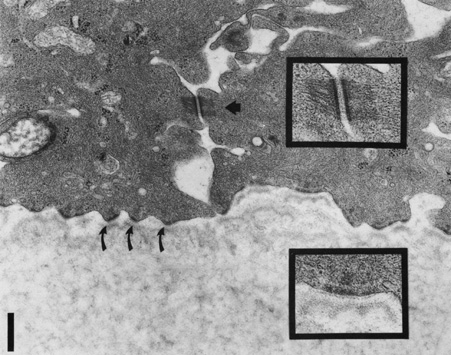
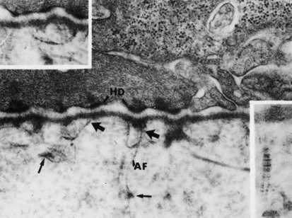
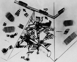
regulator of corneal fibrillogenesis. J Cell Biol 121:1181, 1993

55. Yamamato S, Hashizume H, Hitome J, et al: The subfibrillar arrangement of corneal and scleral collagen fibrils as revealed by scanning electron and atomic force microscopy. Arch Histol Cytol 63:127, 2000

56. Meller D, Peters K, Meller K: Human cornea and sclera studied by atomic force microscopy. Cell Tissue Res 288:111, 1977

57. Holmes DF, Gilpin CJ, Baldock C, et al: Corneal collagen fibril structure in three-dimensions: structural insights in fibril assembly, mechanical properties, and tissue organization. Proc Natl Acad Sci USA 98:7307, 2001

58. Ploetz C, Zycband EI, Birk DE: Collagen fibril assembly and deposition in the developing dermis: segmental deposition in extracellular compartments. J Struct Biol 106:73, 1991

59. Trelstad RL, Coulombre AJ: Morphogenesis of the collagenous stroma in the chick cornea. J Cell Biol 50:840, 1971

60. Meek KM, Blamires T, Elliott GF, et al: The organisation of collagen fibrils in the human corneal stroma: a synchrotron x-ray diffraction study. Curr Eye Res 6:841, 1987

61. Meek KM, Newton RH: Organization of collagen fibrils in the corneal stroma in relation to mechanical properties and surgical practice. J Refract Surg 15:695, 1999

62. Maurice DM: Mechanics of the cornea. In Cavanagh HD (ed): The Cornea: Transactions of the World Congress on the Cornea III. New York: Raven Press, 1988:1987–1993

63. Smolek MK, McCarey BE: Interlamellar adhesive strength. Invest Ophthalmol Vis Sci 31:1087, 1990

64. Scott JE: Proteoglycan-fibrillar collagen interactions. Biochem J 252:313, 1988

65. Scott JE, Bosworth TR: A comparative biochemical and ultrastructural study of proteoglycan-collagen interactions in corneal stroma. Biochem J 270:491, 1990

66. Rada JA, Cornuet PK, Hassell JR: Regulation of corneal collagen fibrillogenesis in vitro by corneal proteoglycan (lumican and decorin) core proteins. Exp Eye Res 56:635, 1993

67. Cintron C, Covington HI: Proteoglycan distribution in developing rabbit cornea. J Histochem Cytochem 38:675, 1990

68. Castoro JA, Bettelheim AA, Bettelheim FA: Water concentration gradients across bovine cornea. Invest Ophthalmol Vis Sci 29:963, 1988

69. Klyce SD, Russell SR: Numerical solution of coupled transport equations applied to corneal hydration dynamics. J Physiol 292:107, 1979

70. Borcherding MS, Blacik LJ, Sittig RA, et al: Proteoglycans and collagen fibre organization in human corneoscleral tissue. Exp Eye Res 21:59, 1975

71. Muller LJ, Pels E, Schurmans LRHM, et al: A new three-dimensional model of the organization of proteoglycans and collagen fibrils in the human corneal stroma. Exp Eye Res 78:493, 2004

72. Maurice DM: The structure and transparency of the cornea. J Physiol 136:263, 1957

73. Hart RW, Farrell RA: Light scattering in the cornea. J Opt Soc Am 59:766, 1969

74. Benedek GB: The theory of transparency of the eye. Appl Optics 10:459, 1971

75. Farrell RA, McCally RL, Tatham PER: Wavelength dependencies of light scattering in normal and cold swollen rabbit corneas and their structural implications. J Physiol 233:589, 1973

76. Watsky MA: Keratocyte gap junctional communication in normal and wounded rabbit corneas and human corneas. Invest Ophthalmol Vis Sci 36:2568, 1995

77. Moller-Pedersen T: A comparative study of human corneal keratocyte and endothelial cell density during aging. Cornea 16:333, 1997

78. Muller LJ, Pels L, Vrensen GF: Novel aspects of the ultrastructural organization of human corneal keratocytes. Invest Ophthal Vis Sci 36:2557, 1995

79. Andresen JL, Ehlers N: Chemotaxis of human keratocytes is increased by platelet-derived growth factor-BB, epidermal growth factor, transforming growth factor-alpha, acidic fibroblast growth factor, insulin-like growth factor-I, and transforming growth factor-beta. Curr Eye Res 17:79, 1998

80. Jester JV, Barry PA, Lind GJ, et al: Corneal keratocytes: in situ and in vitro organization of cytoskeletal contractile proteins. Invest Ophthalmol Vis Sci 35:730. 1994

81. Andresen JL, Ledet T, Hager H, et al: The influence of corneal stromal matrix proteins on the migration of human corneal fibroblasts. Exp Eye Res 71:33, 2000

82. Crosson CE: Cellular changes following epithelial abrasion. In Beuerman RW, Crosson CE, Kaufman HE (eds): Healing Processes in the Cornea, Vol 1. Advances in Applied Biotechnology Series. Houston, TX: Gulf Publishing, 1989: 3–14

83. Moller-Pedersen T: Keratocyte reflectivity and corneal haze. Exp Eye Res 78:553, 2004

84. Zhao J, Nagasaki T: Lacrimal gland as the major source of mouse tear factors that are cytotoxic to corneal keratocytes. Exp Eye Res 77:297, 2003

85. Birk DE, Trelstad RL: Extracellular compartments in matrix morphogenesis: collagen fibril, bundle, and lamellar formation by corneal fibroblasts. J Cell Bioi 99:2024, 1984

86. Johnson DH, Bourne WM, Campbell RJ: The ultrastructure of Descemet's membrane. I. Changes with age in normal cornea. Arch Ophthalmol 100:1942, 1982

87. Bourne WM, Johnson DH, Campbell RJ: The ultrastructure of Descemet's membrane. III. Fuchs' dystrophy. Arch Ophthalmol 100:1952, 1982

88. Binder PS, Rock ME, Coalwell Schmidt K, et al: High-voltage electron microscopy of normal human cornea. Invest Ophthalmol Vis Sci 32:2234, 1991

89. Tamura Y, Konomi H, Sawada H, et al: Tissue distribution of type VIII collagen in human adult and fetal eyes. Invest Ophthalmol Vis Sci 32:2636, 1991

90. Klyce SD, Beuerman RW: Structure and function of the cornea. In Kaufman HE, Barron BA, McDonald MB, Waltman SR (eds): The Cornea. New York: Churchill Livingstone, 1988:3–54.

91. Williams K, Watsky M: Gap junctional communication in the human corneal endothelium and epithelium. Curr Eye Res 25:29, 2002

92. Waring GO, Bourne WM, Edelhauser HF, et al: The corneal endothelium: Normal and pathological structure and function. Ophthalmology 89:531, 1982

93. Kreutziger GO: Lateral membrane morphology and gap junction structure in rabbit corneal endothelium. Exp Eye Res 23:285, 1976

94. Amann J, Holley GP, Lee SB, et al: Increased endothelial cell density in the paracentral and peripheral regions of the human cornea. Am J Ophthalmol 135:584, 2003

95. Bourne WM, Nelson LR, Hodge DO: Central corneal endothelial cell changes over a ten-year period. Invest Ophthalmol Vis Sci 38:779, 1997

96. Gallagher B: Primary cilia of the corneal endothelium. Am J Anat 159:475, 1980

97. Senoo T, Obara Y, Joyce N: EDTA. a promoter of proliferation in human corneal endothelium. Invest Ophthal Vis Sci 41:2930, 2000

98. Senoo T, Joyce NC: Cell cycle kinetics in corneal endothelium from old and young donors. Invest Ophthal Vis Sci 41:660, 2000

99. Joyce N: Proliferative capacity of the corneal endothelium. Prog Retin Eye Res 22:359, 2003

100. Sherrard ES, Ng YL: The other side of the corneal endothelium. Cornea 9:48, 1990

101. Rae JL, Watsky MA: Ionic channels in corneal endothelium. Am J Physiology 270:975, 1996

102. Rozsa AJ, Beuerman RW: Density and organization of free nerve endings in the corneal epithelium of the rabbit. Pain 14:105, 1982

103. Zander E, Weddell G: Observations on the innervation of the cornea. J Anat 85:68, 1951

104. Mawas MJ: L'innervation de la cornee humaine. Bull Soc Ophtalmol Paris 2:162, 1951

105. Muller LJ, Vrensen GF, Pels L, et al: Architecture of human corneal nerves. Invest Ophthalmol Vis Sci 38:985, 1997

106. Klyce SD, Beuerman RW, Crosson CE: Alteration of corneal epithelial ion transport by sympathectomy. Invest Ophthalmol Vis Sci 26:434, 1985

107. Klyce SD, Jenison GL, Crosson CE, et al: Distribution of sympathetic nerves in the rabbit cornea. ARVO Abstract. Invest Ophthalmol Vis Sci 27:354, 1986

Back to Top