Chapter 38
Eye Movements
R. JOHN LEIGH and JANET C. RUCKER
Main Menu   Table Of Contents

Search

THE OCULAR MOTOR PERIPHERY: THE EXTRAOCULAR MUSCLES AND THEIR INNERVATION
BRAINSTEM CONTROL OF HORIZONTAL GAZE
BRAINSTEM CONTROL OF VERTICAL AND TORSIONAL GAZE
BRAINSTEM CONTROL OF VERGENCE EYE MOVEMENTS
CEREBELLAR INFLUENCES ON EYE MOVEMENTS
CEREBRAL CONTROL OF EYE MOVEMENTS
A SCHEME FOR INTERPRETING NYSTAGMUS AND SACCADIC INTRUSIONS
REFERENCES

A grasp of the properties of eye movements is indispensable to practicing ophthalmologists. Abnormal eye movements often provide diagnostic clues and sometimes cause visual complaints that the ophthalmologist is well qualified to manage. Thanks to an ongoing basic research effort, it is now possible to define the neural substrate for normal eye movements and to provide credible explanations for most gaze disorders and their visual consequences.1

The overall purpose of eye movements is to guarantee that our view of the world is clear and stable. In order to provide optimal viewing conditions during a variety of visual tasks, several functional classes of eye movements have evolved (Table 1).2 Although the visual system can tolerate some slip of images on the retina,3 if this slip exceeds a threshold that is determined by the spatial frequency of the object under view (approximately 5 degrees per second for higher spatial frequencies),4 then visual acuity declines and an illusion of movement of stationary objects (oscillopsia) may result. Normally this is prevented by gaze-holding eye movements. The first of these is the fixation mechanism, which depends on the visual system's ability to detect and respond to excessive slip of images on the retina. Visual fixation requires directed visual attention and responds within 100 ms. The second is the vestibulo-ocular reflex (VOR), which depends on motion detectors in the inner ear and generates eye rotations to compensate for head perturbations at short latency (less than 15 ms) so that vision remains clear during locomotion.5,6 Thus, the vestibulo-ocular reflex acts much more promptly than visually mediated fixation. Loss of the vestibulo-ocular reflex caused, for example by aminoglyoside antibiotics, causes oscillopsia during locomotion,7 because the visual system cannot act promptly enough to compensate for the head vibrations that occur with each footstep. During sustained (lower frequency) head movements, however, the visual system can contribute to holding gaze steady by generating optokinetic eye movements. When the eyes are turned out to an eccentric position in the orbits, then the brain must take into account the mechanical pull of the orbital suspensory tissues (such as Tenon's capsule) and program movements accordingly. This eccentric gaze-holding function, depends on the ability of the brain to generate a tonic contraction of the extraocular muscles; when faulty, the result is gaze-evoked nystagmus, one of the most common abnormalities of eye movements.

TABLE 1. Functional Classes Of Eye Movements*


Class of eye movementMain function
Visual fixationHolds the image of a stationary object on the fovea
Vestibulo-ocular reflexHolds images of the seen world steady on the retina during brief head rotations
OptokineticHolds images of the seen world steady on the retina during sustained and low frequency head rotations
Smooth pursuitHolds the image of a moving target close to the fovea
Nystagmus quick phasesReset the eyes during prolonged rotation, and direct gaze towards the oncoming visual scene
SaccadesBring images of eccentrically located objects of interest onto the fovea
VergenceMoves the eyes in opposite directions so that images of a single object are placed simultaneously on both foveas during gaze shifts in depth

* Adapted from Leigh RJ, Zee DS: The Neurology of Eye Movements, 3rd ed. New York: Oxford University Press, 1999.


A second group of eye movements is concerned with pointing the fovea at an object of interest. These gaze-shifting eye movements are more recently evolved than the gaze-holding mechanisms, and are absent in animals lacking a well developed fovea, such as the rabbit. Saccades are rapid movements by which we place the image of an object of interest on fovea. They are important for visual searches of our environment. Saccades can be generated voluntarily, or in response to visual stimuli (presented in the periphery of vision), auditory, somatosensory, or vestibular inputs. The last of these are the quick phases of nystagmus, which reset the eye during sustained gaze-holding movements. Smooth pursuit eye movements hold the image of a moving target close to the fovea, and also allow sustained visual fixation of a stationary target while we are in motion. Vergence eye movements turn the eyes in opposite directions so that the images of a single object can be place simultaneously on both foveas.

It should be realized that under natural conditions there is always interaction and cooperation between different classes of eye movements; thus, vergence is usually associated with fast (saccadic) or slow (pursuit) version movements, because natural gaze shifts are seldom strictly confined to changes in depth (requiring pure vergence) or direction (requiring pure version). For didactic purposes, however, it is useful to separate them. Each functional class of eye movements has distinctive properties that suit it to its specific aims and some of these are summarized in Table 1. Knowledge of these properties will guide the clinical examination. In addition, each functional class of eye movements has a distinct neural substrate, which is discussed below. Familiarity with the neural substrate for each functional class of eye movements aids topological diagnosis.

Our approach will be bottom-up: first to review the extraocular muscles and their innervations, and then examine how neural commands for each functional class of eye movements reaches the ocular motoneurons that reside in the third, fourth, and sixth cranial nerve nuclei. For clarity, we first describe the control of horizontal eye movements, which resides in the pons and medulla, and then control of vertical and torsional eye movements, which depends mainly on the midbrain. We then summarize the cerebellar contributions to control of eye movements, and review cerebral influences on gaze. We conclude by presenting an approach for interpreting nystagmus and saccadic intrusions by applying this scheme for gaze control. As we proceed from extraocular muscles to cerebral cortex, we review normal structure and function and, at each stage, describe classic effects of disease; our account of the latter cannot be comprehensive, and the reader is referred to current texts.1,8–10

Back to Top
THE OCULAR MOTOR PERIPHERY: THE EXTRAOCULAR MUSCLES AND THEIR INNERVATION

ANATOMY OF THE ORBITAL CONTENTS AND ROTATIONS OF THE EYES

The eyeball is suspended in the orbit by fascia, the main component of which is Tenon's capsule. Each eye is rotated by six muscles: four rectus muscles and two oblique muscles. The directions of pulling action of the extraocular muscles depend on the starting position of the eye. The primary action of the muscle refers to the axis around which the eye principally rotates when that muscle contracts; the secondary and tertiary actions refer to the axes around which there are lesser rotations. The pulling actions of each muscle are summarized in Table 2. Concepts of the way that the extraocular muscles attach to the eye and move it have undergone a revolutionary change in the past decade, based on magnetic resonance imaging (MRI) in volunteers and dissection of cadaver orbits.11,12 These studies have demonstrated fibroelastic sleeves for the extraocular muscles that act as pulleys and, for example, stop slide-slip of the horizontal rectus muscles during vertical eye movements. It is well known that the extraocular muscles have two layers, but it now appears that the outer orbital layer inserts into the pulley of the muscle while the inner global layer passes through the pulley to insert onto the globe. Different populations of ocular motoneurons may supply the global and orbital layers, and their proprioception may differ.13 At first sight, such an arrangement raises many complications about the control of eye movements, because it suggests that separate populations of ocular motoneurons will be needed to contract the orbital layer to position the pulley (which is the functional origin of the muscle) and contract the global layer to move the eye. However, this active pulley hypothesis may simplify certain aspects of movements.12 For example, the pulleys may govern rotations of the eyes during saccades and pursuit so that they obey Listing's law.14 Listing's law states that when the eye rotates to a tertiary position, this is achieved by rotation around a single axis perpendicular to the primary position of gaze and to the new position of gaze. Together these axes form Listing's plane, which is approximately frontoparallel. Donder's law states that every tertiary eye position is associated with a specific torsional rotation. The noncommutative properties of rotations pose a computational challenge to the brain (the order of rotations must be specified), but the pulleys may simplify that problem mechanically in the orbit. It also seems possible that certain forms of congenital strabismus may be caused by misplacement of pulleys in the coronal plane.12 Precise measurements of eye rotations in all three directions,15 and high-definition imaging of the extraocular muscles are likely to clarify the role played by the extraocular muscle pulleys further.

TABLE 2. Actions Of The Extraocular Muscles With The Eye In Primary Position*


MusclePrimary actionSecondary actionTertiary action
Medial rectusAdduction
Lateral rectusAbduction
Superior rectusElevationIntorsionAdduction
Inferior rectusDepressionExtorsionAdduction
Superior obliqueIntorsionDepressionAbduction
Inferior obliqueExtorsionElevationAbduction

*Adapted from Leigh RJ, Zee DS: The Neurology of Eye Movements, 3rd ed. New York: Oxford University Press, 1999.


PROPERTIES OF THE EXTRAOCULAR MUSCLES

Extraocular muscles differ anatomically and physiologically from limb muscles.16,17 The former have fibers that are smaller and more richly innervated; some extraocular muscle fibers are among the fastest contracting and yet are relatively fatigue-resistant. The extraocular muscles contain twitch fibers that have a single endplate per fiber and generate action potentials. In addition, there are nontwitch fibers that cannot generate action potentials but show graded contractions to trains of electrical pulse stimuli. These tonic fibers are capable of a smoothly modulated muscle contraction, which may be important for maintaining steady gaze. The extraocular muscles are selectively vulnerable to some disorders (e.g., myasthenia gravis) but resistant to others (e.g., Duchenne's dystrophy). Furthermore, the appearances of diseases that affect the muscle are different when they involve the extraocular muscles. Both central global and peripheral orbital muscle layers contain fibers more suited for either sustained contraction or brief rapid contraction. However, the orbital layer contains many fatigue-resistant twitch fibers and the global zone contains twitch fibers with variable degrees of fatigue resistance; the different muscle types receive different innervation.13

Six types of fibers have been defined in the extraocular muscles.16,17 (i) In the orbital layer, approximately 80% of fibers are singly-innervated, have fast-type myofibrillar adenosine triphosphatase (ATPase), and high oxidative activity (with numerous mitochondria in dense clusters). They are very fatigue-resistant. (ii) The remaining orbital fibers are multiply innervated fibers, with multiple nerve terminals. They have twitch capacity near the center of the fiber, and nontwitch activity proximal and distal to end plate band. (iii) In the global layer, approximately 33% of fibers are red, singly innervated, fast-twitch, and fatigue-resistant. (iv) The global layer contains intermediate, singly innervated fibers (approximately 25%) with fast-twitch properties, numerous mitochondria, and intermediate level of fatigue resistance. (v) Global, pale singly innervated fibers (approximately 33%) have fast-twitch properties but contribute only sporadically because of their low fatigue resistance. (vi) Global multiply innervated fibers (approximately 10%) have slow-twitch, slow tonic properties that exhibit slow, graded, nonpropagated responses to neural or pharmacologic activation; this fiber type is unparalleled in any other human skeletal muscle. It should be noted that levator palpebrae lacks multiply innervated fibers; this may reflect the importance of this fiber type for fixation and smooth eye movements. Unlike other skeletal muscles, in which embryonic myosin is transformed to adult isoforms, extraocular muscles preserve their embryonic myosin in the proximal and distal portions of both types of orbital layer fibers.16 This may underlie the remarkable capacity of extraocular muscles to adapt to changes in innervation and disease states.

Ocular motoneurons and muscle fibers are the final common pathway for all ocular motor systems. The contribution that different fiber types make to different types of eye movements was clarified in classic experiments by Scott and Collins,18 who used miniature electrode needles with multiple recording sites. They reported that fibers are differentiated functionally by the amount of work performed and that the electromyographic (EMG) activity of a given unit correlates with an eye position, irrespectively of the type of movement. (It should be noted, however, that fibers that do not generate action potentials will not be evident on EMG). Orbital layer fibers are recruited before the global, but both types of fibers participate in every movement. Scott and Collins proposed a division of labor such that orbital fibers are active throughout nearly the entire range of movement but during fixation, global fibers are recruited only as the eye is called into the field of action of that muscle. The classic scheme of Scott and Collins is being reinterpreted in the light of the discovery of pulleys for the extraocular muscles and proprioceptive innervation of the extraocular muscles (discussed below).12,13

The different structure of the extraocular muscles apparently determines their differential involvement in neuropathic and myopathic pathologic processes. One example is myasthenia gravis (MG), an autoimmune condition with antibodies against acetylcholine receptors, in which extraocular muscles are preferentially affected.16,19 This may be accounted for by the difference in structure of the acetylcholine receptor in the extraocular muscles, in which the embryonic 2β type is preserved at the neuromuscular junction of multiply innervated fibers (in contrast to the adult 2β type of the receptor in the skeletal muscles).16,20 However, because multiply innervated fibers are absent from the levators, this explanation cannot account for frequent ptosis in myasthenic patients.21 Generally normal saccadic metrics in the presence of the affected ductions again argue for the preferential involvement of the multiply-innervated fibers, with preservation of the fast-twitch singly innervated fibers. Recently, it has been shown that injection of a standard dose of edrophonium (Tensilon) increased peak velocity-amplitude relationship in patients with MG while decreasing it in normal controls or patients with ocular palsies of other causes.22 Because clinical improvement in the amplitude is not specific for MG, the difference in the velocity response may prove to be of higher diagnostic value. The effect in normal controls may be the result of subclinical cholinergic excess (depolarizing blockade-like) and suggests that 10 mg is too high a dose.

Duchenne's Muscular Dystrophy

In this systemic myopathy, as in most others, eye movements are spared.23 This correlates with the absence of necrosis in the extraocular muscles despite the deficiency of a subsarcolemmal protein, dystrophin.24 In other muscles, the initial pathology is increase in intracellular free calcium because of disruption of sarcolemmal integrity.16 It has been suggested that higher capacity of extraocular muscles to scavenge free radicals caused by higher levels of superoxide dismutase might account for this selective preservation of function.25

Although it has long been known that the extraocular muscles contain end-organs necessary for proprioception, their role in the control of eye movements has remained moot until recently.13 In part this has been because vision exerts much more powerful and immediate feedback control on eye movements than any non-visual signal. In addition, experimental studies in monkey have supported a role for the other extraretinal signal - efference copy or corollary discharge of eye movement commands;26 this internal neural signal is consistent with Helmholtz's idea that the brain monitors its own effort of will. One conceptual reason to doubt that proprioception has any role in the control of eye movements is that no external loads are applied to the extraocular muscles, and evidence exists to refute the presence of a stretch reflex in the extraocular muscles.27

In extraocular muscles, the main sources of proprioceptive input are muscle spindles, which lie mainly in the orbital layer; myotendinous cylinders (palisade endings), which are associated with the global layer; and Golgi tendon organs that lie in the peripheral layer.28 It has been hypothesized that only twitch fibers play a substantial role in eye movement, whereas the global layer nontwitch muscle fibers adjust the tension on the palisade endings and modulate the afferent proprioceptive signal.28 Extraocular proprioceptors project to the brain via the ophthalmic branch of the trigeminal nerve and the Gasserian ganglion, to the spinal trigeminal nucleus (pars interpolaris and pars caudalis).29 From the trigeminal nucleus, proprioceptive information is distributed widely to structures involved in oculomotor control: superior colliculus, vestibular nuclei, nucleus prepositus hypoglossi, cerebellum, frontal eye fields, and also structures involved in visual processing: lateral geniculate body, pulvinar, visual areas 17 and 18.30 It has been shown that extraocular proprioception contributes to the development of visual binocularity, aspects of pattern recognition, and formation of visuospatial maps.30

Studies have suggested that ocular proprioception is important for adaptive recalibration, such as occurs after ocular motor palsies. Thus, after experimentally induced, unilateral extraocular palsies, it was shown that proprioceptive deafferentation of the paretic eye produced gradual worsening of both static alignment and saccadic conjugacy.31 Taken together, this evidence argues that proprioception contributes to long-term adaptive mechanisms responsible for eye alignment during fixation and saccades.

ANATOMY OF THE CRANIAL NERVES

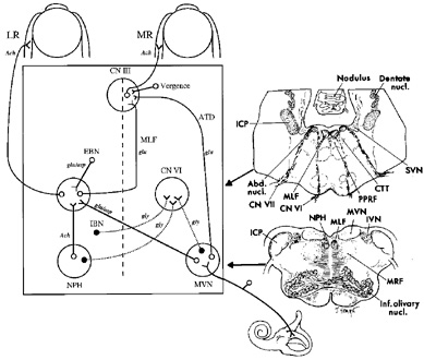
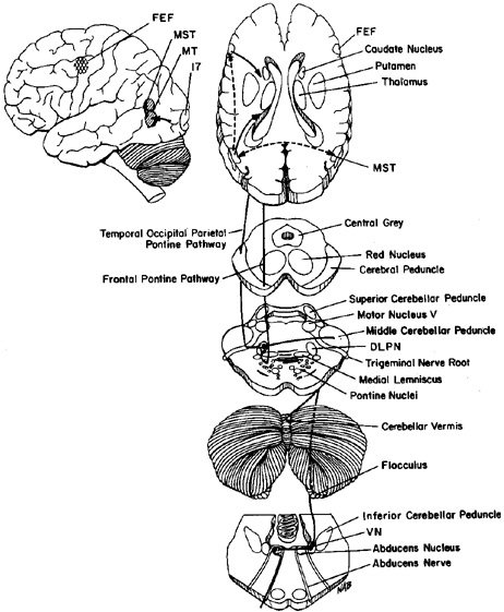
The ocular motor nuclei are located in the brainstem, close to the midline. The intracranial courses of the ocular motor nerves are summarized in Figure 1.

Fig. 1. The intracranial courses of the third, fourth, and sixth cranial nerves. Superior view. Lig. of Gruber: petroclinoid ligament. (From Warwick R. Wolff's Anatomy of the Eye and Orbit, 7th ed. Philadelphia: WB Saunders, 1976:295, with permission)

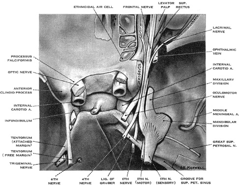
The abducens nucleus lies in the floor of the fourth ventricle, in the lower pons; it is capped by the genu of the facial nerve. As discussed below, the abducens nucleus contains two distinct populations of cells: motor neurons that innervate the lateral rectus muscle and internuclear neurons that innervate, via the medial longitudinal fasciculus, contralateral medial rectus motoneurons. From the medial aspect of the nucleus, fibers destined for the ipsilateral, lateral rectus muscle pass ventrally, laterally and caudally through the pontine tegmentum and medial lemniscus, and lateral to the corticospinal tract, to emerge from the pons at its caudal border. The abducens nerve then courses nearly vertically along the clivus, through the prepontine cistern, and rises to the petrous crest, where it bends forward to penetrate the dura. Here it lies medial to the trigeminal nerve, and passes under the petroclinoid ligament in Dorello's canal. Because the nerve is relatively tethered in the last two locations, it is vulnerable to shear forces; thus, sixth nerve palsy is a nonlocalizing sign in the context of elevated intracranial pressure or trauma. In the cavernous sinus, it lies lateral to the internal carotid artery and medial to the ophthalmic division of the trigeminal nerve. For a short portion, pupillosympathetic fibers run with the sixth nerve as they leave the carotid artery to reach the first division of the trigeminal nerve. Therefore, the combination of the sixth-nerve palsy with Horner's syndrome is suggestive of a cavernous sinus process. The sixth nerve then enters the orbit through the superior orbital fissure, and passes through the annulus of Zinn to innervate the lateral rectus on its inner surface. The most common causes of the sixth-nerve palsy are (peripheral) vascular disease caused by diabetes mellitus or hypertension in an elderly population, and tumors in younger patients.

The trochlear nucleus lies at the ventral border of the periaqueductal gray matter at the level of the inferior colliculus. It innervates the contralateral superior oblique muscle. The trochlear nerve is the longest and the thinnest of cranial nerves; therefore, it is susceptible to even minor head trauma. Its fibers pass dorsolaterally and caudally, around the central gray matter, and decussate completely in the roof of the aqueduct, within the superior medullary velum. Lesions there can produce bilateral fourth nerve palsies. The trochlear nerve emerges from the dorsal aspect of the brainstem, caudal to the inferior colliculus, and close to the tentorium cerebelli, and passes laterally around the upper pons to reach the prepontine cistern. It then runs forward on the free edge of the tentorium before entering the cavernous sinus. Within the lateral wall of the sinus the fourth nerve lies below the third nerve and above the ophthalmic division of the fifth nerve. It crosses over the oculomotor nerve to enter the superior orbital fissure and passes to the medial aspect of the orbit to supply the superior oblique muscle.

The oculomotor nucleus is a paramedian structure that lies at the ventral border of the periaqueductal gray matter. It extends rostrally to the level of the posterior commissure and caudally to the trochlear nucleus. It sends fibers to the medial rectus, superior rectus, inferior rectus and inferior oblique muscles, and to the levator palpebrae superioris. Warwick's anatomic scheme for the oculomotor nucleus32 has been revised with the demonstration that the neurons supplying the medial rectus muscle are distributed into three areas of the oculomotor nucleus.33 The neurons innervating each superior rectus muscle lie next to each other, and their axons decussate in this part of the nucleus. The central caudal nucleus, which supplies both levator palpebrae superioris muscles, is a single structure. All projections from the oculomotor nucleus are ipsilateral except for those to the superior rectus, which are totally crossed, and those to the levator palpebrae superioris, which are bilateral.

The fascicles of the oculomotor nerve pass ventrally through the red nucleus, the substantia nigra, and the medial part of the cerebral peduncle. A topographic organization has been proposed on the basis of the effects of clinical lesions; from lateral to medial, the order is inferior oblique, superior rectus, medial rectus and levator palpebrae, inferior rectus, and parasympathetic pupillary fibers from Edinger-Westphal nucleus.34 The rootlets of the third nerve emerge from the interpeduncular fossa and then fuse and pass between the posterior cerebral artery and superior cerebellar artery into the basal cistern. The third nerve passes lateral to the posterior communicating artery and below the temporal lobe uncus, where it runs over the petroclinoid ligament just lateral to the posterior clinoid process. During its subarachnoid course, parasympathetic pupillary fibers lie in the peripheral, dorsomedial part of the nerve. Their peripheral location, however, is not the only reason for the pupillary involvement with structural lesions of the third nerve; early pupillary involvement also reflects pressure-sensitive nature of these fibers. The oculomotor nerve pierces the dura close to the free edge of the tentorium cerebelli. Within the cavernous sinus, the third nerve lies initially above the trochlear nerve, where it receives sympathetic fibers from the carotid artery. As it leaves the cavernous sinus, it divides into superior and inferior branches; these pass through the superior orbital fissure. The superior branch runs laterally to the optic nerve and ophthalmic artery to supply the superior rectus and levator palpebrae muscles. The inferior branch supplies the medial rectus, inferior rectus and inferior oblique muscles, and the ciliary ganglion. The most common cause of isolated third nerve palsy is vascular disease (in association with diabetes or hypertension); in such cases the pupil is usually spared. The second most common cause are aneurysms, typically of the supraclinoid portion of the internal carotid or posterior communicating artery; such cases characteristically have pupillary involvement; exceptions to this rule, however, have been reported.

Back to Top
BRAINSTEM CONTROL OF HORIZONTAL GAZE

MOTOR COMMANDS FOR CONJUGATE HORIZONTAL MOVEMENTS

The abducens nucleus is of prime importance in the control of horizontal gaze because it governs conjugate movements of both the ipsilateral lateral rectus and the contralateral medial rectus muscles. It contains two populations of neurons: (i) abducens motoneurons, which supply the lateral rectus muscle, and (ii) abducens internuclear neurons, which project up the contralateral medial longitudinal fasciculus (MLF) to synapse on medial rectus motoneurons of the oculomotor nucleus (Fig. 2).35,36 Thus, axons of the abducens nerve, and axons of the abducens internuclear neurons that run in the MLF,37 together encode the conjugate, horizontal eye movement command. In addition, oculomotor internuclear neurons, which lie in the medial rectus subdivision of the oculomotor nucleus, project to the contralateral abducens.38 Together, these two types of internuclear neurons are responsible for yoking of conjugate horizontal movements known as Hering's law. Both abducens motoneurons and abducens internuclear neurons receive similar afferent input from each functional class of eye movement (Table 1).39.40 The abducens nucleus receives excitatory vestibular and optokinetic afferents from the contralateral vestibular nuclei.41,42 Saccadic commands originate from burst neurons of the pontine and medullary reticular formation.43,44 A descending smooth pursuit pathway projects to the abducens nucleus via the vestibular and cerebellar fastigial nuclei (discussed further below). The signals that are necessary for eccentric gaze-holding function reach the abducens nucleus from the nucleus prepositus hypoglossi and the medial vestibular nuclei.45

Fig. 2. Anatomic scheme for the synthesis of signals for horizontal eye movements. The abducens nucleus (CN VI) contains abducens motoneurons that innervate the ipsilateral lateral rectus muscle (LR), and abducens internuclear neurons that send an ascending projection in the contralateral medial longitudinal fasciculus (MLF) to contact medial rectus (MR) motoneurons in the contralateral third nerve nucleus (CN III). From the horizontal semicircular canal, primary afferents on the vestibular nerve project mainly to the medial vestibular nucleus (MVN), where they synapse and then send an excitatory connection to the contralateral abducens nucleus and an inhibitory projection to the ipsilateral abducens nucleus. Saccadic inputs reach the abducens nucleus from ipsilateral excitatory burst neurons (EBN) and contralateral inhibitory burst neurons (IBN). Eye position information (the output of the neural integrator) reaches the abducens nucleus from neurons within the nucleus prepositus hypoglossi (NPH) and adjacent MVN. The medial rectus motoneurons in CN III also receive a command for vergence eye movements. Putative neurotransmitters for each pathway are shown: Ach, acetylcholine; asp, aspartate; glu, glutamate; gly, glycine. The anatomic sections on the right correspond to the level of the arrow heads on the schematic on the left. Abd. nucl., abducens nucleus; CN VI, abducens nerve; CN VII, facial nerve; CTT, central tegmental tract; ICP, inferior cerebellar peduncle; IVN, inferior vestibular nucleus; Inf. olivary nucl., inferior olivary nucleus; MVN, medial vestibular nucleus; MRF, medullary reticular formation; SVN, superior vestibular nucleus. (Modified from Leigh RJ, Zee DS. The Neurology of Eye Movements, 3rd ed. New York: Oxford University Press, 1999)

EFFECTS OF LESIONS OF THE PONS AND MEDULLA ON HORIZONTAL GAZE

Lesions of the abducens nucleus cause paralysis of both the ipsilateral lateral rectus and contralateral medial rectus muscles for all conjugate eye movements,46–48 but vergence movements are spared. Clinical lesions restricted to the abducens nucleus are rare, and often there is also involvement of adjacent structures including the facial nerve fascicle, MLF, and paramedian pontine reticular formation (PPRF).

Lesions of the medial longitudinal fasciculus produce internuclear ophthalmoplegia (INO), which is characterized by paresis of adduction, for conjugate movements, on the side of the lesion49,50; in addition there is usually dissociated nystagmus, characterized by abduction overshoot of the eye contralateral to the lesion. This nystagmus is not an inherent feature of the MLF lesion but probably reflects efforts of central adaptive mechanisms to correct the adduction weakness.51 Thus, dissociated nystagmus is also described with other disorders that cause selective medial rectus weakness. INO is often accompanied by skew deviation, caused by disruption of otolithic-ocular projections that run in the MLF.52 In addition, bilateral INO is usually associated with gaze-evoked vertical nystagmus, impaired vertical pursuit, and decreased vertical vestibular responses.53 Small-amplitude saccadic intrusions may interrupt fixation.54 This could be attributed to the associated lesion of the adjacent omnipause neurons or their connections.55 The most frequent cause of INO in young adults, especially when bilateral, is multiple sclerosis; other causes are summarized in Table 3.

TABLE 3. Causes of Internuclear Ophthalmoplegia*

  1. Multiple sclerosis (commonly bilateral)
  2. Brainstem infarction (commonly unilateral), including complication of arteriography and hemorrhage
  3. Brainstem and fourth ventricular tumors
  4. Arnold-Chiari malformation, and associated hydrocephalus and syringobulbia
  5. Infection: bacterial, viral and other forms of meningoencephalitis; in association with AIDS
  6. Hydrocephalus, subdural hematoma, supratentorial arteriovenous malformation
  7. Nutritional disorders: Wernicke's encephalopathy and pernicious anemia
  8. Metabolic disorders: hepatic encephalopathy, maple syrup urine disease, abetalipoproteinemia, Fabry's disease
  9. Drug intoxications: phenothiazines, tricyclic antidepressants, narcotics, propranolol, lithium, barbiturates
  10. Cancer: either caused by carcinomatous infiltration or remote effect
  11. Head trauma, and cervical hyperextension or manipulation
  12. Degenerative conditions: progressive supranuclear palsy
  13. Syphilis
  14. Pseudointernuclear ophthalmoplegia of myasthenia gravis, and Fisher's syndrome
*Adapted from Leigh RJ, Zee DS. The Neurology of Eye Movements, 3rd ed. New York: Oxford University Press, 1999.
AIDS, acquired immunodeficiency syndrome.


When INO is subtle, it is best identified by comparing the speed and conjugacy of a series of large horizontal saccades, looking for relative slowing of the adducting movement. A series of quick phases of nystagmus, induced with an optokinetic tape, is another way to compare relative speed of adducting and abducting movements. Some care is required in interpreting these signs, however, because abducting saccades are faster in normal subjects, and quantitative comparison of the velocity of the two eyes may be required to make the diagnosis in subtle cases.56,57

Patients with INO may be orthotropic (or exophoric) in primary position without symptomatic diplopia unless accompanied by skew deviation. Some patients with bilateral INO show exotropia (wall-eyed bilateral INO syndrome). The explanation for exotropia is unclear, however, because monkeys with a lidocaine-induced internuclear ophthalmoplegia show an increased accommodative vergence to accommodation ratio, implying that the MLF actually carries signals that inhibit vergence.58 Furthermore, exotropia may occur in patients with INO who have preserved convergence.1 The classification of INO into anterior and posterior types depending on the integrity of the convergence mechanism is not of localizing value in identifying the rostral-caudal location of the MLF lesion. The so-called posterior internuclear ophthalmoplegia of Lutz,59 in which abduction (but not adduction) is impaired is rare. It has been difficult to account for because the abducens contains neurons that innervate both lateral and medial rectus muscles for all conjugate eye movements;60 one possible explanation may be dysfunction of the descending excitatory projections from the third to the sixth nucleus.38

A combined lesion of one MLF and the adjacent abducens nucleus (or its inputs) produces paralysis of all conjugate movements save for abduction of the eye contralateral to the side of the lesion: one-and-a-half syndrome.61,62 When the lesion is acute, the patient may be exotropic63; the deviated eye may show nystagmus and is on the side opposite that of the brainstem lesion. This syndrome occurs with a variety of causes including brainstem infarction, hemorrhage, multiple sclerosis, and pontine glioma.

Discrete lesions of the PPRF,64,65 which mainly corresponds to the nucleus pontis centralis caudalis and contains saccadic burst neurons, cause loss of horizontal saccades and quick phases of nystagmus to the side of the lesion. Bilateral, destructive PPRF lesions may also produce slow vertical saccades. Selective bilateral impairment of horizontal saccades occurs in degenerative conditions, such as variants of olivopontocerebellar atrophy.66 Other causes of slow or absent saccades are summarized in Table 4. Infarction of the paramedian pons may also involve axons conveying vestibular and pursuit inputs to the abducens nucleus.67 Pontine disease may cause a unilateral defect of smooth pursuit by affecting the dorsolateral pontine nuclei and their projections to the cerebellum (see below).68 More rostral brainstem lesions tend to cause ipsilateral smooth pursuit deficits, whereas caudal brainstem and cerebellar lesions cause contralateral deficits.69 Unilateral lesions of the vestibular nerve or labyrinth cause vertigo, nystagmus, and skew deviation (see below).1 Bilateral peripheral vestibular lesions cause impaired vision and oscillopsia during head movements, especially those occurring during locomotion.7 Lesions affecting the central vestibular nuclei also cause an imbalance that is manifest by nystagmus and skew deviation. When such lesions involve the medial vestibular nuclei (MVN) and adjacent nucleus prepositus hypoglossi (NPH), they also impair the ability to hold eccentric gaze and produce gaze-evoked nystagmus. Thus, bilateral, experimental lesions of MVN and NPH abolish the eccentric gaze-holding mechanism for eye movements in the horizontal plane.70 In health, a network of neurons in the brainstem MVN and NPH as well as the vestibular cerebellum generates the eye position command required to hold the eye steady in an eccentric orbital position. Following lesions of this neural network, the eye cannot be held in an eccentric position in the orbit and it drifts back to primary position. Corrective quick phases then produce gaze-evoked nystagmus.

In lateral medullary infarction (Wallenberg's syndrome), there may be spontaneous nystagmus that is usually horizontal or mixed horizontal-torsional with the slow phases directed toward the side of the lesion; the nystagmus may reverse direction in eccentric positions, suggesting coexistent involvement of the gaze-holding mechanism. Skew deviation, with an ipsilateral hypotropia, cyclodeviation (lower eye more extorted), and ipsilateral head tilt (when present together called the ocular tilt reaction) reflect an imbalance of otolithic inputs (see below).71 In addition, patients with Wallenberg's syndrome show a characteristic lateropulsion in which the trajectory of saccades, whether horizontal or vertical, is deviated towards the side of the lesion.72 A hypothetical explanation for lateropulsion is discussed further below.

Back to Top
BRAINSTEM CONTROL OF VERTICAL AND TORSIONAL GAZE

The oculomotor and trochlear nuclei contain the motoneurons for vertical and torsional eye movements. Thus, the substrate for each functional class of eye movements must project to these motoneurons (Fig. 3). On the one hand, vertical and torsional saccades and the mechanism for eccentric vertical gaze-holding are synthesized in the midbrain. On the other hand, vestibular and pursuit signals ascend to the midbrain from the lower brainstem.

Fig. 3. Anatomic schemes for the synthesis of upward, downward, and torsional eye movements. From the vertical semicircular canals, primary afferents on the vestibular nerve (vn) synapse in the vestibular nuclei (VN) and ascend in the medial longitudinal fasciculus (MLF) and brachium conjunctivum (not shown) to contact neurons in the trochlear nucleus (CN IV), oculomotor nucleus (CN III), and the interstitial nucleus of Cajal (INC). The rostral interstitial nucleus of the medial longitudinal fasciculus (riMLF), which lies in the prerubral fields, contains saccadic burst neurons. It receives an inhibitory input from omnipause neurons of the nucleus raphe interpositus (rip), which lie in the pons (for clarity, this projection is only shown for upward movements). Excitatory burst neurons in riMLF project to the motoneurons of CN III and CN IV, and also send an axon collateral to INC. Each riMLF neuron sends axon collaterals to yoke-pair muscles (Hering's law). Projections to the elevator subnuclei (innervating the superior rectus and inferior oblique muscles) may be bilateral due to axon collaterals crossing at the level of the CN III nucleus. Projections of inhibitory burst neurons are less well understood, and are not shown. The INC provides a gaze-holding signal, and projects to vertical motoneurons via the posterior commissure. Signals contributing to vertical smooth pursuit and eye–head tracking reach CN III from the y-group via the brachium conjunctivum and a crossing ventral tegmental tract. Neurotransmitters: asp, aspartate; glu, glutamate; gly, glycine. (Modified from Leigh RJ, Zee DS. The Neurology of Eye Movements, 3rd ed. New York: Oxford University Press, 1999)

In the prerubral fields at the junction of midbrain and diencephalon, lies the rostral interstitial nucleus of the medial longitudinal fasciculus (riMLF),73 which has also been called the nucleus of the prerubral fields and the nucleus of the fields of Forel. It contains burst neurons for vertical saccades and quick phases, and for torsional quick phases. Each riMLF contains neurons that burst for both upward and downward eye movements, but for torsional quick phases in only one direction. For example, the right riMLF discharges for quick phases that are directed clockwise with respect to the subject.74 Each riMLF is connected to its counterpart commissural projections. The riMLF projects to motoneurons innervating elevator muscles bilaterally, but to motoneurons innervating depressor muscles ipsilaterally; therefore each riMLF is responsible for innervation of the contralateral superior oblique and ipsilateral inferior rectus muscles for saccades.75,76 Furthermore, each burst neuron in the riMLF sends axon collaterals to motoneurons supplying yoke muscle pairs; this appears to be part of the neural substrate for Hering's law in the vertical plane.77

The riMLF also projects to the interstitial nucleus of Cajal (INC), which has been shown to play an important role in vertical gaze-holding.78 In addition, the INC also receives inputs from the vestibular nuclei.79 The INC projects via the posterior commissure to motoneurons of the contralateral vertical ocular motor subnuclei.80 The INC also contains neurons that project to motoneurons of the neck and trunk muscles, and appears to coordinate combined torsional-vertical movements of the eyes and head. The neural signals necessary for vertical vestibular and smooth pursuit eye movements ascend to INC from the medulla and pons. The MLF is the most important route for these projections but the brachium conjunctivum and other pathways are also involved.81

EFFECTS OF MIDBRAIN LESIONS ON VERTICAL AND TORSIONAL GAZE

Experimental unilateral lesions of the riMLF cause a mild defect in vertical saccades, because nucleus on each side contains burst neurons for both upward and downward movements. A right riMLF lesion would also have little effect on upward saccades but downward saccades might be slowed82,83; this asymmetry of vertical defects may reflect the bilateral projections of riMLF to elevator muscles but ipsilateral projections to depressor muscles. However, a unilateral riMLF lesion produces a specific defect of torsional quick phases.82,83 For example, with a lesion of the right riMLF, torsional quick phases, clockwise from the point of view of the subject (extorsion of the right eye and intorsion of the left eye) are impaired.

Vertical saccadic deficits with unilateral lesions of the riMLF in humans are rare and often may reflect involvement of the commissural pathways of the riMLF that makes the lesion, in effect, bilateral.82,84 Thus, one reported patient with a unilateral midbrain lesion involving riMLF, but no evidence of commissural involvement, manifested contralesional tonic ocular tilt reaction (see below), torsional nystagmus and impaired vertical gaze, especially for downward saccades that became both limited and slow.85 In general, however, bilateral lesions are required to produce clinically apparent deficits of vertical eye movements.86 Bilateral experimental lesions of the riMLF in monkeys cause a defect of vertical and torsional saccades that may be more pronounced for downward eye movements83,87; vertical gaze-holding, vestibular eye movements and, possibly, pursuit are preserved, as are horizontal saccades. Patients with discrete, bilateral infarction in the region of the riMLF show deficits of either downward or both upward and downward saccades.88 Extensive bilateral PPRF lesions cause slow vertical saccades by impairing the ascending input to the riMLF. Certain metabolic and degenerative disorders may lead to selective slowing or absence of vertical saccades (Table 4).

TABLE 4. Causes Of Slow Saccades*

  1. Olivopontocerebellar atrophy and spinocerebellar degenerations
  2. Huntington's disease
  3. Progressive supranuclear palsy
  4. Parkinson's disease (advanced cases) and diffuse Lewy body disease
  5. Whipple's disease
  6. Lipid storage diseases
  7. Wilson's disease
  8. Drug intoxications: anticonvulsants, benzodiazepines
  9. Tetanus
  10. In dementia: Alzheimer's disease (stimulus-dependent) and in association with AIDS
  11. Lesions of the paramedian pontine reticular formation
  12. Internuclear ophthalmoplegia
  13. Peripheral nerve palsy, diseases affecting the neuromuscular junction and extraocular muscle, restrictive ophthalmopathy
* Adapted from Leigh RJ, Zee DS: The Neurology of Eye Movements, 3rd ed. New York: Oxford University Press, 1999.


Unilateral experimental lesions of the INC are reported to impair gaze-holding function in the vertical plane and reduce the vertical ocular motor range.78 In addition, skew deviation (ipsilateral hypertropia), extorsion of the contralateral eye, intorsion of the ipsilateral eye, and contralateral head tilt occurs. Stimulation near the INC in the monkey produces an ocular tilt reaction that consists of an ipsilateral head tilt and a synkinetic ocular reaction: depression and extorsion of the eye ipsilateral to the stimulation and elevation and intorsion of the contralateral eye.89 This ocular tilt reaction is similar to that produced by stimulation of the contralateral utricular nerve or an ipsilateral lesion of the vestibular nucleus (see Wallenberg's syndrome, above). Bilateral lesions of the medial longitudinal fasciculus (bilateral INO) impair vertical vestibular and pursuit movements but spare vertical saccades.90 In addition, partial loss of the vertical eye position signal causes vertical gaze-evoked nystagmus.

Lesions of riMLF and INC are both often associated with torsional nystagmus. While tonic torsional deviation in both cases is contralesional, the direction of the fast phases for the nystagmus varies: with riMLF lesions, the nystagmus is contralesional whereas with INC lesions, it is ipsilesional.85

Lesions of the posterior commissure are equivalent to bilateral lesions of the INC, and reduce the range of vertical movements, especially upward (Parinaud's syndrome)91; usually all types of eye movement are affected, although the vestibulo-ocular reflex and Bell's phenomenon may sometimes be spared. Experimental inactivation of the posterior commissure with lidocaine impairs vertical gaze-holding function.92 In addition, posterior commissure lesions may cause slowing of vertical saccades below the horizontal meridian, and disorders of convergence including convergence-retraction nystagmus. Attempts at upward saccades evoke convergence-retraction nystagmus that persists as long as the refixation effort is maintained. This is best elicited with a down-moving optokinetic tape stimulating a series of repetitive upward saccades; a convergence or retraction movement replaces each fast phase. These movements may or may not truly be nystagmus but consist of opposed adducting saccades followed by slow divergence movements.93,94 With dorsal midbrain lesions, abduction may be bilaterally impaired, perhaps reflecting increased vergence gain, giving the appearance of pseudoabducens palsy.95 Rarely, a divergence-retraction nystagmus may occur in patients with dorsal midbrain syndrome. Other associated findings with pretectal lesions are pathologic lid retraction (Collier's sign), and middilated pupils that show a smaller reaction to light than to the near stimulus. Pineal area tumors96,97 and midbrain infarction84,88 are the most common causes.

SKEW DEVIATION

Skew deviation consists of a vertical misalignment of the visual axes caused by a disturbance of prenuclear inputs. The hypertropia may be the same in all positions of gaze, may vary with gaze position, or may alternate (e.g., right hypertropia on right gaze, left hypertropia on left gaze).98,99 When skew deviation is nonconcomitant, it can be differentiated from vertical extraocular muscle palsy by the coexistence of signs of central neurologic dysfunction and from trochlear nerve palsy by a negative head-tilt test. An additional sign that may help diagnosis is the direction of cyclorotation: with fourth-nerve palsy, the hypertropic eye is excyclotorted, whereas with skew deviation it is incyclotorted. Skew deviation is a part of the ocular tilt reaction (OTR)52 that in full form includes the triad of head tilt, ocular torsion, and vertical skew, with the torsional deviation being the most sensitive and consistent sign. Present evidence suggests that skew deviation occurs whenever peripheral or central lesions cause an imbalance of otolithic inputs.52,98 OTR in general, and skew deviation in particular, are of localizing value because of our knowledge of the pathways of central otolithic connections, which cross the midline at the lower pontine level and then ascend in the MLF to the contralateral INC. Peripheral vestibular or lower brainstem lesions affecting vestibular nuclei cause ipsilateral OTR, with the ipsilesional head tilt, hypotropia, and excyclotorsion. With more rostral lesions involving MLF and INC, the OTR is contralateral (i.e., there is contralateral head tilt, hypotropia, and excyclotorsion). OTR may be tonic (sustained)100,101 or paroxysmal,102,103 the latter may reflect irritation at the level of INC. OTR patients also show a deviation of the subjective vertical.52 In one patient, stimulation of in the region of INC caused an ocular tilt reaction, with episodes of contralateral hypertropia and ipsilateral head tilt.104

Skew deviation has been reported in association with a variety of disorders of the brainstem and cerebellum,105 and as a reversible finding with phenytoin toxicity and raised intracranial pressure.106 Rarely, skew deviation slowly alternates or varies in magnitude over the course of minutes107; these patients have midbrain lesions. The periodicity of the phenomena is reminiscent of periodic alternating nystagmus (see below) and the two phenomena may coexist.105 Alternating skew deviation, where the side of the hypertropic eye varies depending on the direction of the horizontal gaze has been reported with bilateral brainstem or cerebellar patients; it may represent release of the phylogenetically old righting reflex observed in lateral-eyed animals.109

Back to Top
BRAINSTEM CONTROL OF VERGENCE EYE MOVEMENTS

MOTOR COMMANDS FOR VERGENCE MOVEMENTS

Electrophysiologic studies in monkeys have shown that almost all oculomotor neurons innervating the medial rectus and the majority of neurons in the abducens nucleus discharge for both conjugate (version) and disjunctive (vergence) eye movements, although there is evidence that different neurons play relatively smaller or larger roles in conjugate as opposed to vergence eye movements.110 Premotor commands for vergence have been found on neurons in the mesencephalic reticular formation, 1 to 2 mm dorsal and dorsolateral to the oculomotor nucleus.111,112 Three main types of neurons can be found; cells that discharge in relation to vergence angle, vergence velocity,113 and both vergence angle and velocity.

An important pontine nucleus for vergence is the nucleus reticularis tegmenti pontis (NRTP). It seems important for coordinating pursuit and saccadic movements with vergence, and also for holding the eyes steady at a specified vergence angle.114 Clinical lesions affecting NRTP and other pontine nuclei have been reported to cause defects of slow or rapid vergence movements.115

DISORDERS OF VERGENCE

Midbrain lesions may disrupt the near triad of convergence, accommodation, and pupillary constriction. They often also impair vertical gaze and produce so-called convergence-retraction nystagmus (see above). Disorders of convergence are also reported with pontine lesions.115 Convergence paralysis or paresis may be associated with various organic processes such as encephalitis, multiple sclerosis, and occlusive vascular disease involving the rostral midbrain. Senescence and lack of effort are common causes of impaired convergence. Overall, however, the most common disorders of vergence are congenital, causing strabismus in childhood. In many such patients, abnormality of the accommodative-convergence synkinesis can be demonstrated. Spasm of the near reflex (accommodative or convergence spasm) is usually psychogenic. Affected patients appear to have unilateral or bilateral abducens paresis116 but accompanying pupillary miosis usually indicates the diagnosis. The excessive near effort required to sustain these spasms is uncomfortable and rarely can be sustained. Patients may complain of blurred vision, eyestrain, dizziness, headache, and diplopia. Rarely, a tonic spasm of the near reflex may occur in association with organic disease, including generalized seizures, head trauma, dorsal midbrain syndrome, craniocervical junction abnormalities, intoxications, and Wernicke's encephalopathy.117–119

Insufficiency or paresis of divergence is a rare clinical syndrome characterized by orthophoria at near but relatively concomitant esotropia at distance, even though abduction is normal. Diplopia while viewing distant objects is the principal complaint. A prerequisite for the diagnosis of divergence paresis is the presence of normal-velocity abducting saccades (to exclude abduction weakness), and this has seldom been measured. Often, the issue of decompensation of long-standing esophoria bias is raised, for example, after concussion. Another situation is mild abduction weakness after head trauma or with raised intracranial pressure; in these cases, traction on the abducens nerve may be the cause. Tests of vergence amplitudes and stereopsis may not settle the diagnosis and measurement of saccade velocities is necessary. Divergence insufficiency has been reported in seizure disorders, progressive supranuclear palsy, acute brainstem dysfunction, and after viral infections.120,121 Stern and Tomsak122 reported a case of divergence insufficiency occurring late in the course of a sixth-nerve palsy, with MRI demonstration of a dorsal pontine lesion at the level of the abducens nucleus; they discuss the relationship of divergence paralysis to subtle sixth-nerve dysfunction. Whatever the underlying disorder, most patients do well with base-out prisms incorporated in distant spectacle correction and only rarely is ocular muscle surgery required.

Back to Top
CEREBELLAR INFLUENCES ON EYE MOVEMENTS
The overall role of the cerebellum in the control of eye movements is to optimize them so that they can provide clear and stable vision. Indeed, the cerebellum may be regarded as the repair shop for eye movements. Two distinct parts of the cerebellum play an important role in the control of eye movements: (i) the vestibulocerebellum (flocculus, paraflocculus, nodulus, and ventral uvula) and (ii) the dorsal vermis of the posterior lobe, and the fastigial nuclei.

The flocculi and ventral paraflocculi (tonsils) are paired structures that lie ventral to the inferior cerebellar peduncle and adjacent to the eighth cranial nerve. The flocculus receives bilateral inputs from the vestibular nuclei and the nucleus prepositus hypoglossi, and inputs from the contralateral inferior olivary nucleus.123 The main projections of the flocculus pass to the ipsilateral superior, medial, and y-group of vestibular nuclei.124 The flocculus and ventral paraflocculus contain Purkinje cells that encode gaze velocity during pursuit and smooth, combined eye-head tracking.125 Floccular cells may also contribute to vestibular eye movements during self-rotation.126 The flocculus also plays an important role in the adaptive control of the vestibulo-ocular reflex.127

The nodulus is the midline part of the flocculonodular lobe, lying caudal to the inferior medullary velum, and the adjacent ventral uvula; it receives afferents from the vestibular nuclei and inferior olive.128 Nodulus and uvula project to the vestibular nuclei. Together, these structures govern the temporal response of the vestibulo-ocular reflex, so that a sustained, constant velocity rotation induces nystagmus that outlasts by two or three times the duration of signal from the labyrinthine semicircular canals (a process called velocity storage).

Lobules IV to VII of the vermis receive mossy fiber inputs from the PPRF, NRTP, the dorsolateral pontine nuclei, vestibular nuclei, nucleus prepositus hypoglossi, and the inferior olivary nucleus.129 The projection from NRTP may contain information from the frontal eye fields necessary for the planning of saccades.130 The dorsal vermis projects to the fastigial nuclei, which also receives afferents from the inferior olive.131 The fastigial nucleus sends projections contralaterally (crossing within the nucleus) via the uncinate fasciculus (which runs in the dorsolateral border of the brachium conjunctivum) to the PPRF, and riMLF. Some Purkinje cells in the dorsal vermis discharge before saccades. Stimulation of the vermis produces saccades and a topographic organization is reported: upward saccades are evoked from the anterior part; downward saccades from the posterior part; and ipsilateral, horizontal saccades from the lateral part.132 Neurons in the caudal fastigial nucleus have properties that suggest that they help accelerate contralateral saccades and smooth pursuit.133,134

EFFECTS OF CEREBELLAR LESIONS ON EYE MOVEMENTS

Experimental lesions of the flocculus and paraflocculus in monkeys cause similar findings to those encountered in patients with the Arnold-Chiari malformation.135 This includes impaired smooth tracking and gaze-evoked nystagmus. This gaze-holding deficit reflects the contribution that the cerebellum makes to enhance the network for holding steady eccentric gaze, which includes the medial vestibular nuclei and the nucleus prepositus hypoglossi (see above). Floccular lesions also impair the ability to adapt the properties of the vestibulo-ocular reflex in response to visual demands.127 Thus, patients with disease involving the vestibular cerebellum may have vestibulo-ocular eye movements that are hypoactive or hyperactive, and they may complain of impaired vision and oscillopsia with head movements. Downbeat nystagmus is common finding, and probably reflects an imbalance in central vestibular connections caused by loss of floccular modulation of the vertical vestibulo-ocular reflex (Table 5).

TABLE 5. Ocular Motor Disorders Caused by Lesions of the Cerebellum*

Vestibulocerebellum

Flocculus, Paraflocculus, and Ventral Uvula:
  1. Gaze-evoked nystagmus
  2. Impaired smooth pursuit (and eye-head tracking)
  3. Downbeat nystagmus
  4. Rebound nystagmus
  5. Uncalibrated vestibulo-ocular reflex
Nodulus:
  1. Prolongation of durations of vestibular nystagmus in response to rotational stimuli
  2. Periodic alternating nystagmus (when in darkness)

Dorsal Vermis, Fastigial Nucleus, and their projections

Dorsal Vermis Lesions: Saccadic dysmetria, with hypometria of contralateral saccades
Fastigial Nucleus: Hypermetria of ipsilateral saccades, hypometria of contralateral saccades (ipsipulsion), and impairment of contralateral smooth pursuit
Uncinate Fasciculus and Superior Cerebellar Peduncle: contralateral hypermetria (contrapulsion)

*Adapted from Leigh RJ, Zee DS. The Neurology of Eye Movements, 3rd ed. New York: Oxford University Press, 1999.


In monkeys, lesions of the nodulus and uvula maximize the velocity-storage effect,136 and maneuvers that would usually minimize it, such as pitching the head forward during postrotational nystagmus, become ineffectual.136 Similar effects are seen in patients with midline cerebellar tumors involving the nodulus.137 Furthermore, when monkeys that have nodular lesions are placed in darkness, they develop periodic alternating nystagmus.136 Patients with periodic alternating nystagmus often have lesions involving the nodulus and ventral uvula.138

Experimental lesions of the dorsal vermis produce marked hypometria (overshooting) of ipsilateral saccades and mild hypometria of contralateral saccades.139 Inactivation of the caudal fastigial nuclei with the γ-aminobutyric acid (GABA) agonist muscimol causes hypermetria of ipsilateral saccades and hypometria (undershooting) of contralateral saccades.140 This mechanism of saccadic dysmetria may account for the ipsipulsion of saccades encountered in patients with Wallenberg's syndrome (lateral medullary infarction); loss of input from an infarcted inferior cerebellar peduncle cause an increase in Purkinje cell activity that inhibits the ipsilateral fastigial nucleus.140 Similarly, lesions affecting crossed outputs of the fastigial nucleus via the superior cerebellar peduncle result in contrapulsion, and are seen with midbrain processes. Lesions that involve the fastigial nucleus in humans often cause a severe form of saccadic dysmetria in which the eye may repetitively overshoot a stationary target: macrosaccadic oscillations. The posterior vermis contributes to smooth pursuit and has reciprocal connections with the dorsolateral pontine nuclei. Posterior vermis lesions may impair smooth pursuit.141 Experimental unilateral lesions of the fastigial nucleus may cause a defect of contralateral smooth pursuit, but bilateral lesions may leave smooth pursuit preserved, reflecting directional (acceleration-deceleration) influences of this nucleus on tracking.142

Back to Top
CEREBRAL CONTROL OF EYE MOVEMENTS
Present concepts of how the cerebral hemispheres control eye movements are based on several lines of investigation, each with its own strengths and weaknesses.1,143 Experimental studies in the rhesus monkey have provided substantial insights into how neurons encode and program eye movements. However, caution is required in extrapolating the effects of discrete cortical lesions in monkeys to the effects of disease in humans, because the cortical architecture is significantly different. Proton emission tomography (PET), functional magnetic resonance imaging (fMRI), and transcranial magnetic stimulation have made it possible to suggest homologous cortical areas in humans. Development of special test paradigms have proved successful in identifying specific defects of eye movement control in patients with well delineated cerebral lesions. Thus, the scheme presented here should be viewed as a working hypothesis to be tested by future studies.

THE SACCADIC SYSTEM

Multiple Roles for Saccades

Rapid eye movements serve several distinct purposes.1,143 First, fast phases of nystagmus reset the eyes following vestibular or optokinetic slow-phase movements. Second, spontaneous saccades, at a frequency of about 20 per minute, allow visual search, although they also occur in darkness. Third, reflexive saccades are made in response to new visual, auditory, or tactile cues at short latency. Fourth, voluntary saccades point the eyes at a predetermined location or goal. Voluntary saccades can be made in a predictive fashion if a visual target is moving in a regular pattern: the eye movement anticipates the target jump. Voluntary saccades can also be made toward remembered or imagined target locations, or in response to verbal commands (e.g., “Look left”"). A special case of voluntary saccades is the antisaccade; the subject is instructed to respond to a visual stimulus by looking to the corresponding position in the opposite visual hemifield. Another test that can be conducted in the laboratory setting is to ask subjects to make saccades to the remembered location of a target (a light flashed in darkness) after a delay of several seconds (the memory period). Finally, saccades can be voluntarily suppressed when it is necessary to maintain steady foveal fixation. Although the burst neurons of the PPRF (see above) are ultimately responsible for generating all types of rapid eye movements, different cortical and subcortical areas contribute to programming saccades for different functions. Testing of each type of saccade make it possible to evaluate the separate neural substrates.

Cortical Areas that Contribute to Saccade Generation

In addition to the frontal eye fields (FEF) and parietal eye fields (PEF), several other cortical areas contribute to the control and programming of saccades (Fig. 4), including the supplementary eye field (SEF); the dorsolateral prefrontal cortex (DLPC), lying on the dorsolateral surface of the frontal lobe, anterior to the FEF, occupying approximately the middle third of the middle frontal gyrus; and the posterior parietal cortex (PPC). What is the flow of information that occurs to program a saccade?

Fig. 4. Areas of cerebral cortex and their projections that contribute to generation of saccades. A: Probable location of cortical areas important for eye movements in human brain MST, medial superior temporal visual area; MT, middle temporal visual area (V5); V1, primary visual cortex. B: Block diagram of the major structures that project to the brainstem saccade generator (premotor burst neurons in PPRF and riMLF). Also shown are projections from cortical eye fields to superior colliculus. DLPC, dorsolateral prefrontal cortex; FEF, frontal eye fields; IML, intramedullary lamina of thalamus; NRTP, nucleus reticularis tegmenti pontis; PEF, parietal eye fields; PPC, posterior parietal cortex; SEF, supplementary eye fields; SNpr, substantia nigra, pars reticulata; STN, subthalamic nucleus. Not shown are the pulvinar, which has connections with the superior colliculus and both the frontal and parietal lobes, and certain projections, such as that from the superior colliculus to nucleus reticularis tegmenti pontis (NRTP), and the pathway that conveys efference copy from brainstem and cerebellum, via thalamus, to cerebral cortex. (Modified from Leigh RJ, Zee DS. The Neurology of Eye Movements, 3rd ed. New York: Oxford University Press, 1999)

After visual inputs reach striate cortex (Brodmann area 17, visual area V1), processing of features such as form, color, spatial location and motion of a viewed object occurs largely separately, in different cortical areas.144 Thus, striate cortex projects to several secondary visual areas, each of which is mainly concerned with analysis of certain features of vision. The inferior parietal lobule is important for the planning of saccades, and in monkeys, contributes to localizing an object in space by taking into account the location of a stimulus within the visual field and the direction in which the eyes are pointing145; in humans, the homologue is probably near to the intraparietal sulcus (posterior parietal cortex [PPC]). In the posterior insula lies one of several representations of vestibular cortex (VC), which contributes information of head or body movements.52 Also important for programming eye movements are the middle temporal (MT, or V5) and medial superior temporal (MST) visual areas,146 which, in humans, probably lie at the junction of temporal, parietal, and occipital lobes.147 In monkeys, area MT contains neurons that encode the speed and direction of moving targets; experimental lesions here cause a selective defect of motion vision and associated tracking defects.146,148 Area MT projects to MST, which contains neurons that combine visual motion information with vestibular and eye movement signals.146,149 Areas MT and MST are important for generating smooth pursuit (see below), and also provide important information to cortical areas concerned with saccade generation because saccades are often made toward moving targets.

The PEF in monkeys lies within the intraparietal sulcus, in the lateral intraparietal area (LIP); neurons here discharge before saccades made “reflexively” to visual targets.150 LIP projects to both the frontal eye fields and the superior colliculus, but not directly to the PPRF or riMLF.151 Experimental lesions of LIP delay the initiation of saccades made to visual stimuli.152 In humans, the PEF, which corresponds to the LIP in monkeys, lies in the intraparietal sulcus.143,153 Parietal lesions in humans also cause an increase saccadic latency to visual targets. Unilateral parietal disease may cause bilateral increases in saccadic latency, although the effect is more marked for right hemisphere lesions.154,155 This defect in initiation of saccades often coexists with disorders of hemivisual attention or it may occur alone.154 Parietal lesions impair the accuracy of saccades if the patient is required to respond to two target lights flashed in succession; the second saccade becomes inaccurate.156 In this double-step paradigm, the brain must take account of the location in the visual field of the second target flash (prior to any eye movement) and the size and direction of the final eye movement; both pieces of information must be correct to make an accurate saccade to the remembered location of the second target flash. Thus, PEF seem important for generating saccades to novel visual stimuli.

The frontal eye field (FEF) in rhesus monkeys lies along the posterior bank of the arcuate sulcus, corresponding to part of Brodmann area 8. The FEF receives afferents from visual areas MT and MST, dorsolateral prefrontal cortex, PEF, SEF, and from the intralaminar thalamic nuclei. In monkeys, some FEF neurons discharge before volitional but not reflexive saccades. Furthermore, the FEF seem important in suppressing saccades and maintaining steady fixation. In humans, FEF lies in the precentral gyrus and sulcus, close to the intersection with the superior frontal sulcus.153 The FEF project caudally to the superior colliculus, caudate nucleus, and brainstem reticular formation (see below). Acute pharmacological inactivation of the FEF in monkeys with muscimol produces an ocular motor scotoma, being unable to saccade to targets corresponding to the injection's site movement field.157 On the other hand, acute pharmacologic disinhibition with bicuculline disrupted steady fixation.157 Chronic disease involving the FEF in humans causes increased latency of saccades to visual targets if the previous (fixation) target remains visible (overlap paradigm). If, however, the fixation target disappears before the new target is presented (gap paradigm), then there is no increase in the response time.158 This evidence supports the view that the FEF are important for disengagement of fixation. Furthermore, unilateral FEF lesions cause increased saccadic latency to remembered target locations, bilaterally.159 The ability to generate predictive saccades is also impaired, as are antisaccades that are directed into the visual hemifield opposite to that of the test stimulus.158,160 Thus, the FEF seem important for the programming of saccades concerned with intentional exploration of the visual environment (i.e. volitional saccades).

The SEFs in monkeys lie in the dorsomedial frontal lobes, in the anterior part of the supplementary motor area.161 The SEFs receive inputs from FEF, parietal cortex, and intralaminar thalamic nuclei, and project to the FEF, caudate nucleus, superior colliculus, and brainstem reticular formation.162 In humans, the SEF lies anterior to the supplementary motor area (SMA) in the upper part of the paracentral sulcus.143 Disease involving the SEF in humans does not affect saccades made to visual or remembered targets but saccades made after rotating patients to a new position are inaccurate.159 Thus, one apparent difference between SEF and FEF is that the former, but not the latter, is important for generating saccades to targets at specific spatial (rather than retinal) locations. Another function impaired by disease affecting the SEF, especially on the left side, is the ability to make a sequence of saccades to several target lights that are turned and left on in a specified order.163 Evidence suggests that the SEF is aided by the hippocampus in remembering the chronological sequence of target presentation.164 Thus, the SEF appear important in programming saccades concerned with complex motor behaviors. When FEF, LIP, and SEF activity are compared during target selection and saccade generation, SEF activity appeared most concerned with internally guided target selection based on reward during prior trials.143

The dorsolateral prefrontal cortex (PFC) is crucial for making accurate motor responses (including saccades) to the remembered spatial locations of targets.162,165 Lesions of PFC, including pharmacological blockade of D1 dopamine receptors,160,165 disrupt this working memory area so that if a target is out of sight, it is also out of mind. In addition, patients with DLPC lesions show impaired ability to make predictive saccades.166

CORTICAL PROJECTIONS TO BRAINSTEM SACCADE CIRCUITS

Parallel descending pathways connect the cortical regions described above with brainstem and cerebellar structures concerned with the generation of saccades. Current evidence indicates that no single direct projection exists from cortical neurons to ocular motoneurons; instead, several intermediate structures play important roles, including the caudate nucleus, substantia nigra, superior colliculus and pontine nuclei (Fig. 5).167–169 It appears that for generation of purely vertical saccades, corresponding areas in both hemispheres must discharge simultaneously, thus canceling out the horizontal components.

Fig. 5. A hypothetical scheme for horizontal smooth pursuit. Primary visual cortex (V1) projects to the homologue of the middle temporal visual area (MT) that, in humans, lies at the temporal-occipital-parietal junction. MT projects to the homologue of the medial superior temporal visual area (MST) and also to the frontal eye field (FEF). MST also receives inputs from its contralateral counterpart. MST projects through the retrolenticular portion of the internal capsule and the posterior portion of the cerebral peduncle to the dorsolateral pontine nucleus (DLPN). The DLPN also receives inputs important for pursuit from the frontal eye field; these inputs descend in the medial portion of the cerebral peduncle. The DLPN projects, mainly contralaterally, to the flocculus, paraflocculus and ventral uvula of the cerebellum; projections also pass to the dorsal vermis. The flocculus projects to the ipsilateral vestibular nuclei (VN), which in turn project to the contralateral abducens nucleus. Note that the sections of brainstem are in different planes from those of the cerebral hemispheres. (Modified from Leigh RJ, Zee DS. The Neurology of Eye Movements, 3rd ed. New York: Oxford University Press, 1999)

Parietal area LIP projects to the superior colliculus, and to the FEF.151 The FEF projections run in the internal capsule and clinical lesions involving the anterior limb of the internal capsule and adjacent deep frontal region are reported to increase saccadic latency.155 Below the level of the internal capsule, several separate pathways can be identified: one to the caudate nucleus; a second to the intralaminar thalamic nuclei and the ipsilateral superior colliculus; and a third, pedunculopontine pathway that runs in the most medial aspect of the cerebral peduncle, and projects to the NRTP that, in turn, projects to the cerebellum.167–169 The PPRF (see above) and particularly the midline pontine raphe nuclei that contain saccadic omnipause cells also receive projections from the FEF. A partial ocular motor decussation is hypothesized to occur between the levels of the trochlear and abducens nuclei.167 The SEF also project to the caudate nucleus, where convergence with FEF projections occurs, and to the superior colliculus and pontine omnipause neurons.170,171 The dorsolateral prefrontal cortex projects to parts of the caudate nucleus, and to the superior colliculus.172 The caudate nucleus sends inhibitory projections to the non-dopaminergic, substantia nigra pars reticulata (SNpr), which in turn, inhibits neurons in the superior colliculus. Thus, this basal ganglia system is composed of two serial, inhibitory links: a caudonigral inhibition that is only phasically active and a nigrocollicular inhibition that is tonically active.173 Recent work has implicated the caudate nucleus in reward-related ocular motor behavior.174 If frontal cortex causes caudate neurons to fire, then the nigrocollicular inhibition is removed and the superior colliculus is able to activate a saccade. It seems possible that disease affecting the caudate nucleus could impair the ability to make saccades to complex tasks. On the other hand, disease affecting the SNpr could disinhibit the superior colliculus, so causing excessive, inappropriate saccades. Both deficits have been described in patients with disorders involving the basal ganglia, such as Huntington's disease.175

THE SUPERIOR COLLICULUS

All three frontal areas and parietal area LIP project directly to the superior colliculus (SC). The role of the SC in humans remains undefined but in monkeys, it has been shown to contribute to generation of visually guided saccades. Thus, the SC is essential for programming saccades to visual stimuli short latency (so-called express saccades).176,177 To test for such express saccades, the fixation light is turned off before the target light is illuminated (gap paradigm).

The SC contains a visual, retinotopic map in the cells of its superficial layers and a motor map in the intermediate layers.178 Stimulation at any point on the motor map will produce a saccade of specific amplitude and direction; the smallest saccades are represented in the rostral SC and largest in caudal SC. Although it had been postulated that the SC transforms information about saccade size encoded on its map into the command for a saccade encoded in terms of discharge frequency and duration, current evidence suggests a more complex role.179,180 Nonetheless, the SC does appear to contribute to the control of saccades, as well as fixation and smooth pursuit. Reports of human patients with disease restricted to the SC are rare. In one patient with a unilateral lesion, contralateral saccades were produced at longer latency and were hypometric.181 Involvement of the SC and adjacent mesencephalic reticular formation is an established feature of progressive supranuclear palsy,182 and such patients may show increased latency with the gap paradigm.183 There remains a need to better understand the contribution of the SC to gaze control in humans. Applying specific tests of SC function (such as the gap paradigm), and improving resolution with functional imaging may provide new insights.

The Role of Descending Parallel Projections

The relative importance of descending pathways from cortical and subcortical structures in saccade generation has been elucidated by studies of the effects of discrete experimental lesions. Chronic lesions of the superior colliculus, in monkeys, cause relatively minor deficits such as an increase in saccadic latency.184 In monkeys, chronic FEF lesions also cause minor deficits that affect saccades to remembered targets. In contrast, combined lesions of the FEF and superior colliculi produce a severe and lasting deficit of eye movements, with a restricted range of movement.184 Severe deficits of saccadic and pursuit eye movements also follow combined, bilateral lesions of parietal-occipital and frontal cortex in monkeys.152 With unilateral, combined parietofrontal lesions, saccades to visual targets in contralateral hemispace are impaired185; with hemidecortication, the deficit is more enduring.186 Clinically, this occurs more often with right hemispheric lesions. In humans, combined lesions of frontal and parietal cortex cause loss of ability to make voluntary saccades or ocular motor apraxia.187

PURSUIT SYSTEM

Traditionally, it has been thought that posterior cerebral cortex generates ipsilateral smooth pursuit. As indicated above, neurons in the secondary visual areas MT encode the speed and direction of a moving target.146 MT projects to the adjacent visual area MST, where neurons encode not only visual information, but also a corollary discharge (efference copy) of the eye movement command.188 Parietal cortex also influences smooth pursuit, probably by enhancing attention on the moving target. In addition, the FEF also contribute to both the initiation and maintenance of smooth pursuit.189 From MST, noncrossed projections descend to the dorsolateral pontine nuclei (DLPN)190 and subsequently cross to the contralateral flocculus, paraflocculus, and vermis of the cerebellum.191 The flocculus projects to the vestibular nuclei on the same side,125 which in turn, cross again to the ocular motor nuclei, ipsilateral to the original MT; the vermis projects to the underlying fastigial nucleus.

The effects of experimental or discrete clinical stimuli can be interpreted with reference to this scheme (Fig. 6). Lesions of striate cortex abolish smooth pursuit of targets restricted to the blind hemifield, but pursuit is normal in both directions in the seeing hemifield.192 Lesions of MT do not cause a conventional visual field defect but selectively impair the ability to estimate the speed of a target: both saccades and smooth pursuit are consequently impaired.146,148 Lesions of MST in monkeys cause a disturbance similar to the ipsilateral pursuit deficit encountered with unilateral cortical lesions in humans; horizontal pursuit is impaired for targets moving toward the side of the lesion.148,193 Experimental lesions of DLPN similarly cause impairment of ipsilateral smooth pursuit.194 Lesions of the cerebellar flocculus and dorsal vermis also impair ipsilateral smooth pursuit, but experimental or clinical lesions of the vestibular or cerebellar nuclei produce a defect of contralateral smooth pursuit.69,195 Lesions of the PPRF that impair or abolish saccades may not affect smooth pursuit, unless axons of passage are affected.64,65

Fig. 6. Examples of abnormal eye movements and their effect on vision. A: A pendular type of congenital nystagmus waveform with superimposed quick phases. Note that following each quick phase, foveation periods (indicated by arrows) occur, at which time the eye is close to desired fixation point (0 deg) and eye velocity is low (i.e., the image is on the fovea and image slip is low). This patient experienced no oscillopsia. B: A pendular type of acquired nystagmus in a patient with multiple sclerosis who complained of blurred vision and oscillopsia. Note the presence of both horizontal (HOR) and vetical (VER) components, and the absence of foveation periods. C: An example of saccadic intrusions (square waves) that repeatedly moved the image of regard off the fovea; the patient had progressive supranuclear palsy. D: Sustained saccadic oscillations that interfere with clear vision. The amplitudes of the horizontal and vertical components of this diagonal microsaccadic flutter are small, but the high frequency of these oscillations impaired vision in the patient, who was otherwise well. Clinically, these oscillations could only be discerned with an ophthalmoscope. E: Macrosaccadic oscillations in a patient with brainstem and cerebellar abnormalities. Note how the predominatly horizontal component oscillates across the fixation point. Upward deflections indicate rightward or upward eye movements. (Modified from Leigh RJ, Zee DS: The Neurology of Eye Movements, 3rd ed. New York: Oxford University Press, 1999)

A variety of conditions bilaterally impair smooth pursuit eye movements: old age, sedative drugs, inattention,196 fatigue, and impaired consciousness, as well as diffuse cerebral,197 cerebellar, or brainstem disease. A pursuit defect difficult to distinguish from that of inattention is found in schizophrenia.198

COMMON DISTURBANCES OF GAZE WITH HEMISPHERIC LESIONS

In acute stages of a hemispheric lesion, patients' eyes are often deviated conjugately toward the side of the lesion. Such deviations are more common after large strokes involving right post-Rolandic cortex,199,200 when visual hemineglect often coexists. Acute lesions involving FEF may also result in transient gaze deviation. Conjugate deviation of the patient's eyes away from the side of the lesion (wrong-way deviation) occurs with subfrontal or thalamic lesions.61 The reason for the latter is not clear; wrong-way deviation might result from abnormal efference copy of intended motor command or, alternatively, from impaired processing of the proprioceptive signal of the eye position (both may be expected with thalamic lesions).201

Acutely, patients may not be able to voluntarily direct their eyes toward the side of the intact hemisphere, but vestibular stimulation usually produces a full range of horizontal movement in contrast to gaze palsies associated with pontine lesions.95 Vertical saccades may be dysmetric with an inappropriate horizontal component directed toward the side of the lesion. Because both hemispheres appear to contribute to the generation of a purely vertical saccade, the loss of one hemisphere may be the cause of such oblique saccades.202

Chronically, there usually is no resting deviation of the eyes unless there has been a prior lesion in the contralateral hemisphere.203 Attempted forced eyelid closure may induce contralateral spastic conjugate eye movement, the mechanism of which is not understood.204 In primary position, a low-amplitude nystagmus may be present (evident during ophthalmoscopy), with slow phases directed toward the side of the intact hemisphere205; it may represent an imbalance in smooth pursuit tone, with unopposed pursuit drives directed away from the side of the lesion. Horizontal pursuit is impaired for tracking of targets moving toward the side of the lesion; a convenient way to demonstrate this asymmetry of pursuit is with a hand-held optokinetic drum or tape.206 Though usually referred to as an optokinetic stimulus, the drum is principally testing smooth pursuit rather than optokinetic function. The response is decreased when the stripes are moved toward the side of the lesion. At the bedside, the magnitude of this optokinetic response is usually judged by the frequency and amplitude of quick phases.

Large bihemispheric, especially dorsal posterior cortical lesions, cause a disturbance of ocular motility that has been called acquired ocular motor apraxia.1,187 It is characterized by loss of voluntary control of eye movements, with preservation of reflex movements, including the vestibulo-ocular reflex and quick-phases of nystagmus. Acquired apraxia limited to the vertical plane implies bilateral lesions at the mesencephalic-diencephalic junction.207,208

When acquired ocular motor apraxia is associated with acute optic ataxia (eye–hand uncoordination with impaired reaching) and disturbance of visual attention (simultaneous agnosia), the eponym Balint's syndrome has been applied.187,209 This syndrome is most commonly seen with bilateral lesions of dorsolateral posterior cortex, such as occur in Alzheimer's disease, demyelinative, or neoplastic processes. Occasionally, extensive bilateral frontal lesions can mimic Balint's syndrome. Voluntary movements of the eyes are limited in the horizontal and vertical planes. Gaze shifts are usually achieved by combined movements of eyes and head.187 Slow and quick phases of vestibular nystagmus are both largely preserved, confirming to the intact brainstem mechanisms. Most affected are saccades to novel visual stimuli entering the periphery of the visual field (i.e. reflexive saccades, probably reflecting disruption of the output from the PEFs to the superior colliculus and the frontal eye fields). The frontal eye fields descending projections may also be impaired, thus producing dysfunction of the volitional saccades by depriving the superior colliculus and brainstem reticular formation from their supranuclear inputs; similar results have been produced experimentally in monkey.184 The term spasm of fixation is applied to such patients with difficulties in voluntarily shifting gaze. Holmes210 described a group of such patients and noted that if the visual scene was a homogenous white screen, then voluntary eye movements became possible. As reviewed above, the frontal eye fields are concerned with disengagement of fixation. Thus, it appears that spasm of fixation might have a real physiologic basis but before applying the term to patients, it seems necessary to demonstrate difference in saccadic latency between trials in which the fixation stimulus disappeared before the new visual stimulus appeared (gap paradigm; see above) and trials in which the fixation stimulus remained visible.211

Back to Top
A SCHEME FOR INTERPRETING NYSTAGMUS AND SACCADIC INTRUSIONS
Here we review abnormal eye movements that degrade vision and cause illusory motion of the seen environment (oscillopsia). As indicated at the beginning of this chapter, these symptoms are caused by excessive movement of images of stationary objects upon the retina. In addition, saccades may disrupt steady vision by taking the fovea away from the object of regard. It is now possible to identify the pathophysiology underlying several types of nystagmus and saccadic abnormalities. However, several other forms of nystagmus and saccadic intrusions remain unexplained. Our approach is to apply the scheme that we have developed for the normal control of gaze to those abnormalities that are understood. For a more comprehensive approach, discussion of less understood forms of nystagmus, and measures to treat abnormal eye movements that disrupt vision, the reader is referred elsewhere.1,212,213

Nystagmus is a repetitive, to-and-fro motion (oscillation) of the eyes that is initiated by a slow phase. In health, nystagmus serves a physiologic function during self-rotation. Sustained head-and-body rotation would cause the eyes to lodge at the corners of the orbit, in extreme contraversive deviation, where they could no longer make appropriate movements. This is prevented by quick phases of nystagmus, which reset the eyes so that they can then continue to make compensatory movements. In contrast, pathologic nystagmus causes excessive drift of images of stationary objects on the retina, leading to impaired vision and oscillopsia. The slow phase of pathological nystagmus often indicates which eye movement disorder is responsible.

The pathophysiologic processes that are known to produce nystagmus can be thought of in terms of disorders of the three main types of eye movements that normally act to hold gaze steady: the vestibulo-optokinetic reflexes, the mechanism for holding the eye in an eccentric position in the orbit, and the visual fixation mechanism (Table 1). We summarize the features of nystagmus resulting from each of these mechanisms in turn.

NYSTAGMUS CAUSED BY VESTIBULAR DISTURBANCES

Disorders of either the peripheral or central vestibular system can produce nystagmus because of an imbalance in the level of tonic neural activity in the vestibular nuclei. The effects of peripheral vestibular lesions have been quantified in monkeys during the acute and recovery phase1; nystagmus from acute, peripheral vestibular disease rarely persists because of the brain's ability to recover from these lesions. It has also been possible to induce a central form of vestibular nystagmus—downbeat nystagmus—by lesioning either the cerebellar flocculi and paraflocculi or the midline floor of the fourth ventricle.214,215 This nystagmus can be conceptualized as being caused by a central imbalance of inputs from the vertical semicircular canals. It appears that the vestibular cerebellum inhibits the central projections of the anterior but not the posterior semicircular canals. Thus, following vestibular cerebellar lesions, upward drifts may represent loss of inhibition of the central connections of the anterior semicircular canals, which mediate upward eye movements, whereas inputs from the posterior canals, which mediate downward eye movements, are unaffected. However, more than one mechanism is probably responsible for downbeat nystagmus, including disturbance of the vertical gaze-holding mechanism, and central otolithic imbalance. Although upbeat nystagmus has not been produced experimentally, discrete lesions in humans also make it likely to reflect a specific disruption of the central projections from the semicircular canals,1 and it is reported with medullary lesions. Similarly, torsional nystagmus, which occurs with pontomedullary tegmental lesions and syringobulbia, may also be regarded as a disorder of central vestibular connections.216

Another rare form of nystagmus due to disorders affecting the vestibular cerebellum is acquired periodic alternating nystagmus (PAN). This is a spontaneous horizontal nystagmus, present in primary gaze that reverses its direction about every two minutes. Acquired PAN occurs most commonly with disease involving the midline cerebellum.1 Experimental ablation of the nodulus and uvula of the cerebellum, in monkeys, causes PAN when the animals are put into darkness.136 One function of the nodulus and uvula is to control the time course of rotationally-induced nystagmus (so-called velocity storage). Thus, after ablation of the nodulus and uvula, the duration (velocity storage) of rotationally induced nystagmus is prolonged excessively, and it is postulated that normal vestibular repair mechanisms act to reverse the direction of this nystagmus, so producing the oscillations of PAN.136,217,218 These oscillations would ordinarily be blocked by visual stabilization mechanisms that tend to suppress nystagmus, but disease of the cerebellum that causes PAN usually also impairs these mechanisms. Baclofen is effective treatment for most patients with acquired PAN. A congenital form of PAN, which is characteristically less periodic than the acquired form, does not respond to baclofen.212

NYSTAGMUS CAUSED BY DISTURBANCE OF THE ECCENTRIC GAZE-HOLDING MECHANISM

The medial vestibular nucleus and the adjacent prepositus hypoglossi nucleus are essential for normal eccentric gaze-holding function (sometimes referred to as the neural integrator for eye movements). To hold the eye steady in an eccentric position in the orbit requires a tonic contraction of the extraocular muscles. This is achieved by a step eye position signal that is generated by the neural integration. Experimental lesions of the gaze-holding mechanism cause gaze-evoked nystagmus70; because of a deficient eye position signal, the eyes cannot be maintained at an eccentric orbital position and they drift toward the primary position. Corrective quick phases move the eyes back toward the desired location. Gaze-evoked nystagmus frequently accompanies other vestibular eye signs with central vestibular lesions. It may also be caused by structural or toxic lesions that involve the vestibulocerebellum or its connections with the brainstem nuclei, and is most commonly produced by anticonvulsants, sedatives, and alcohol. A selective lesion of the medial vestibular nucleus and nucleus prepositus hypoglossi causing complete gaze-holding failure has been reported in a patient who died of lithium intoxication.219

Recent studies using the technique of pharmacologic inactivation have provided important information regarding the pharmacological substrate for both gaze-holding and vestibular mechanisms. Of special interest is the role of GABA. Unilateral microinjection of the GABAA agonist, muscimol, into the medial vestibular and prepositus hypoglossi nuclei of monkey and cat produced bilateral gaze-evoked nystagmus.220,221 Other evidence indicates that GABAB is important in modulating the temporal properties (velocity storage) of the vestibulo-ocular reflex.222 Some of these basic findings have guided therapeutic trials.212

NYSTAGMUS CAUSED BY DISTURBANCE OF THE VISUAL FIXATION MECHANISM

Disturbance of visual inputs, either caused by disease of the anterior visual pathways,223 or of secondary cortical areas that are important for motion detection,205 can produce nystagmus. It has also been suggested that disturbance of visual inputs was responsible for acquired pendular nystagmus (APN); an example is shown in Figure 6B. APN is encountered in patients with a variety of conditions affecting brainstem and cerebellum, such as disorders of central myelin (multiple sclerosis, Pelizaeus-Merzbacher disease, toluene intoxication), vascular disease (pontine strokes, the syndrome of oculopalatal myoclonus), and degenerative disorders (spinocerebellar degenerations). Disturbance of either the vestibulo-ocular reflex or the eccentric gaze-holding mechanism do not appear to be the root cause of APN because either may be relatively preserved.1 However, it seems possible that disturbance of the visual fixation was responsible. Thus, in one study of APN in patients with multiple sclerosis (MS), the oscillations were greater in the eye with evidence of more severe optic nerve demyelination;224 this led the authors to postulate that visual delays caused by demyelination of the anterior optic pathways could have led to the oscillations of APN. This hypothesis was tested by electronically manipulating the delay to visual feedback during fixation in patients with APN and normal controls.225 Such manipulation did not change the characteristics of the APN, but did superimpose lower-frequency oscillations similar to those that can be induced in normal subjects by this electronic technique. Thus, disturbance of fixation caused by visual delays could not account for the high-frequency oscillations that often characterize APN; more likely are abnormalities of internal negative feedback circuits, such as the reciprocal connections between brainstem nuclei and cerebellum.225 In cases with a convergent-divergent type of APN, an internal instability in connections between nucleus reticularis tegmenti pontis and cerebellar nucleus interpositus, which are important for vergence control, might be responsible.226

APN is, perhaps, the most visually disabling form of nystagmus. APN in association with MS is often suppressed, with resultant improved vision, by gabapentin or memantine. Patients with APN after brainstem stroke (including the syndrome of oculopalatal myoclonus or tremor) respond less well to these drugs.212,227,228

FORMS OF NYSTAGMUS OF UNCERTAIN ORIGIN

Seesaw nystagmus is characterized by pendular oscillations, one half-cycle of which consists of elevation and intorsion of one eye and synchronous depression and extorsion of the other eye; during the next half-cycle, the vertical and torsional movements reverse.1 In some patients, one half-cycle of seesaw nystagmus alternates with an oppositely-directed quick phase; this has been called hemi-seesaw nystagmus.229 Seesaw or hemi-seesaw nystagmus has been described in patients with discrete lesions involving the interstitial nucleus of Cajal (INC).229 These findings are in accord with experimental studies that implicate a central disturbance of otolithic inputs. Stimulation in the region of the INC in the monkey produces an ocular tilt reaction consisting of extorsion and depression of the eye on the stimulated side and intorsion and elevation of the other eye.89 The INC receives vestibular inputs, which probably include secondary otolithic projections. Thus, seesaw nystagmus could represent a sinusoidal oscillation involving central otolithic connections. Another hypothesis for seesaw nystagmus arises from its frequent association with optic chiasm lesions; such patients lack crossed visual projections that may be important for stabilizing gaze in the frontal plane. It has also been suggested that interruption of subcortical pathways that carry signals to the inferior olive and cerebellar flocculus, and are normally used for adaptive control of vestibular responses, may be important in the genesis of seesaw nystagmus.1,229

The pathogenesis of congenital nystagmus is not understood, although two animal models have now been identified, one induced by visual deprivation230 and the other with congenital anomalous projections of the visual pathways.231 The characteristic features of congenital nystagmus are reviewed elsewhere.1 Affected individuals seldom have visual symptoms (unless there are coexistent visual systems disorders, such as in albinism) because of foveation periods: brief epochs during which the eye is stationary and the fovea is pointing at the visual target. These foveation periods provide a signature that allows congenital nystagmus to be recognized on eye movement records (Fig. 6A). Selected patients may benefit from surgical procedures that aim to move null point (where nystagmus is minimized) to the center of the ocular motor range, to weak the extraocular muscles, or induce convergence, which often suppresses congenital nystagmus.1,232 However, any form of surgery may dampen congenital nystagmus leading to the suggestion that simply detaching and reattaching the extraocular muscles may improve the oscillations233; further trials are required to establish the role of such surgery.

SACCADIC INTRUSIONS

Saccadic intrusions are abnormal gaze-shifting movements that disrupt steady foveal fixation1,213; examples are shown in Figure 6. Individual saccadic intrusions take the image of the object of regard away from the fovea and may, for example, interfere with reading. Sustained saccadic oscillations may cause the complaint of oscillopsia. In addition to nystagmus and saccadic intrusions, there are some other disorders of eye movements that may disrupt clear and stable vision; one example is superior oblique myokymia, which is probably an abnormality of the trochlear motor units, and may respond to several drugs including carbamazepine and gabapentin.212,234 Square wave jerks are involuntary saccades that take the eyes off the target and are followed, after a nearly normal intersaccadic interval (130 to 200 ms), by a corrective saccade that brings the eyes back to the target (Fig. 6D). They may occur in normal individuals, but also in a variety of neurological disorders, most prominently cerebellar disorders and progressive supranuclear palsy.1 Square waves seldom cause visual symptoms, unless large and frequent. Macrosaccadic oscillations are large-amplitude square-wave intrusions that oscillate the eye around the desired fixation point; they occur with cerebellar lesions affecting the fastigial nucleus and possibly with brainstem lesions.1,55 Ocular flutter is a burst of back-to-back saccades without an intersaccadic interval (Fig. 6C). Saccadic oscillations without an intersaccadic interval that have variable horizontal, vertical (and torsional) components are called opsoclonus. There appears to be a continuum between flutter and opsoclonus. Ocular flutter and opsoclonus are usually encountered in patients with signs of brainstem or cerebellar dysfunction (e.g., brainstem encephalitis or paraneoplastic syndromes).1 Occasionally, otherwise normal individuals show intermittent, small-amplitude, high-frequency saccadic oscillations that can only be observed with the ophthalmoscope (macrosaccadic flutter).234 Some normal individuals can generate flutter-like movements (voluntary nystagmus), usually during voluntary convergence. Patients with flutter and opsoclonus frequently complain of oscillopsia, even if the oscillations are of small amplitude; this is because of their high frequency that causes large retinal slip velocities. Although there is no animal model for flutter and opsoclonus, current hypotheses explain these oscillations in terms of the connectivity between the brainstem neurons that generate and regulate saccades burst and omnipause cells.234,235

Back to Top
REFERENCES

1. Leigh RJ, Zee DS: The Neurology of Eye Movements. 3rd ed. New York: Oxford University Press, 1991

2. Walls GL: The evolutionary history of eye movements. Vision Res 2:69, 1962

3. Carpenter RHS: The visual origins of ocular motility. In Cronly-Dillon JR, ed. Vision and Visual Function, Vol 8, Eye Movements. London: MacMillan Press, London, 1991:1

4. Burr DC, Ross J: Contrast sensitivity at high velocities. Vision Res 22:479, 1982

5. Grossman GE, Leigh RJ, Bruce RJ, et al: Performance of the human vestibulo-ocular reflex during locomotion. J Neurophysiol 62:264, 1989

6. Maas EF, Huebner WP, Seidman SH, et al: Behavior of human horizontal vestibulo-ocular reflex in response to high-acceleration stimuli. Brain Res 499:153, 1989

7. LIVING without a balancing mechanism. N Engl J Med 246:458, 1952

8. Kaminski HJ, Leigh RJ: Neurobiology of eye movements—From molecules to behavior. Ann NY Acad Sci 956:1, 2002

9. Glaser JS, ed: Neuro-ophthalmology, 4th ed. Hagerstown, MD: Harper and Row, 2005.

10. Miller NR, Newman NJ, Biousse V, eds: Walsh and Hoyt's Clinical Neuro-Ophthalmology, 6th ed. Philadelphia: Lippincott-Raven, 2005

11. Demer JL, Miller JM, Poukens V, et al: Evidence for fibromuscular pulleys of the recti extraocular muscles. Invest Ophthalmol Vis Sci 36:1125, 1995

12. Demer JL: Pivotal role of orbital connective tissues in binocular alignment and strabismus. Invest Ophthalmol Vis Sci. 45:729, 2004

13. Büttner-Ennever JA, Horn AK, Graf W, et al: Modern concepts of brainstem anatomy: From extraocular motoneurons to proprioceptive pathways. Ann NY Acad Sci 956:75, 2002

14. Haslwanter T: Mathematics of three-dimensional eye rotations. Vision Res 35:1727, 1995

15. Ferman L, Collewijn H, Jansen TC, et al: Human gaze stability in the horizontal, vertical and torsional direction during voluntary head movements, evaluated with a three-dimensional scleral induction coil technique. Vision Res 27:811, 1987

16. Porter JD, Baker RS, Ragusa RJ, et al: Extraocular muscles: Basic and clinical aspects of structure and function. Surv Ophthalmol 39:451, 1995

17. Spencer RF, Porter JD: Structural organization of the extraocular muscles. In Büttner-Ennever JA, ed. Reviews of Oculomotor Research, Vol. 2, Neuroanatomy of the Oculomotor System. Amsterdam: Elsevier, 1988:33

18. Scott AB, Collins CC: Division of labor in human extraocular muscle. Arch Ophthalmol 90:319, 1973

19. Kaminski HJ, Maas E, Spiegel P, et al: Why are eye muscles frequently involved in myasthenia gravis? Neurology 40:1663, 1990

20. Oda K, Shibasaki H: Antigenic differences of acetylcholine receptors between single and multiple form endplates of human extraocular muscle. Brain Res 449:337, 1988

21. Kaminski HJ, Kusner LL, Nash KV, et al: The gamma-subunit of the acetylcholine receptor is not expressed in the levator palpebrae superioris. Neurology 45:516, 1995

22. Barton JJS, Huaman AG, Sharpe JA: Effects of edrophonium on saccadic velocity in normal subjects and myasthenic and nonmyasthenic palsies. Ann Neurol 36:585, 1994

23. Kaminski HJ, Al-Hakim M, Leigh RJ, et al: Extraocular muscles are spared in advanced Duchenne dystrophy. Ann Neurol 32:586, 1992

24. Prendergast RA, Khuran TS, Alameddine H, et al: Relative sparing of the extraocular muscles in Duchenne muscular dystrophy: Invest Ophthalmol Vis Sci 34:1120, 1993

25. Ragusa RJ, Porter JD: Extraocular muscle in the Mdx mouse: Absence of pathology correlates with superoxide dismutase activity. Mol Biol Cell 5:26a, 1994

26. Guthrie BL, Porter JD, Sparks DL: Corollary discharge provides accurate eye position information to the oculomotor system. Science 221:1193, 1983

27. Keller EL, Robinson DA: Absence of a stretch reflex in extraocular muscles of the monkey. J Neurophysiol 34:908, 1971

28. Büttner-Ennever JA, Eberhorn A, Horn AKE: Motor and sensory innervation of the extraocular eye muscles. Ann NY Acad Sci 1004:40, 2003.

29. Porter JD, Guthrie BL, Sparks DL: Innervation of monkey extraocular muscles: Localization of sensory and motor neurons by retrograde transport of horseradish peroxidase. J Comp Neurol 218:208, 1983

30. Buisseret P Influence of extraocular muscle proprioception on vision: Physiol Rev 75:323, 1995

31. Lewis RW, Zee DS, Gaymard BM, et al: Extraocular muscle proprioception functions in the control of ocular alignment and eye movement conjugacy. J Neurophysiol 72:1028, 1994

32. Warwick R: Representation of the extra-ocular muscles in the oculomotor nuclei of the monkey. J Comp Neurol 98:449, 1953

33. Büttner-Ennever JA, Akert K: Medial rectus subgroups of the oculomotor nucleus and their abducens internuclear input in monkey. J Comp Neurol 197:17, 1981

34. Castro O, Johnson LN, Mamourian AC: Isolated inferior oblique paresis from brain-stem infarction. Perspective on oculomotor fascicular organization in the ventral midbrain tegmentum. Arch Neurol 47:235, 1990

35. Highstein SM, Baker R: Excitatory termination of abducens internuclear neurons on medial rectus motoneurons: Relationship to syndrome of internuclear ophthalmoplegia. J Neurophysiol 41:1647, 1978

36. Maciewicz RJ, Spencer RF: Oculomotor and abducens internuclear pathways in the cat. In Baker R, Berthoz A (eds). Control of Gaze by Brain Stem Neurons. Vol. 1. New York: Elsevier, 1977:99

37. King WM, Lisberger SG, Fuchs AF: Responses of fibers in medial longitudinal fasciculus (MLF) of alert monkeys during horizontal and vertical conjugate eye movements evoked by vestibular or visual stimuli. J Neurophysiol 39:1135, 1976

38. Clendaniel RA, Mays LE: Characteristics of antidromically identified oculomotor internuclear neurons during vergence and versional eye movements. J Neurophysiol 71:1111, 1994

39. McCrea RA, Strassman A, Highstein SM: Morphology and physiology of abducens motoneurons and internuclear neurons intracellularly injected with horseradish peroxidase in alert squirrel monkeys. J Comp Neurol 243:291, 1986

40. Spencer RF, Baker R: Histochemical localization of acetylcholinesterase in relation to motor neurons and internuclear neurons of the cat abducens nucleus. J Neurocytology 15:137, 1986

41. McCrea RA, Strassman A, May E, et al: Anatomical and physiological characteristics of vestibular neurons mediating the horizontal vestibulo-ocular reflex of the squirrel monkey. J Comp Neurol 264:547, 1987

42. Langer T, Kaneko CRS, Scudder CA, et al: Afferents to the abducens nucleus in the monkey and cat. J Comp Neurol 245:379, 1986

43. Strassman A, Highstein SM, McCrea RA: Anatomy and physiology of saccadic burst neurons in the alert squirrel monkey. I. Excitatory burst neurons. J Comp Neurol 249:337, 1986

44. Strassman A, Highstein SM, McCrea RA: Anatomy and Physiology of saccadic burst neurons in the alert squirrel monkey. II. Inhibitory burst neurons. J Comp Neurol 249:358, 1986

45. Belknap DB, McCrea RA: Anatomical connections of the prepositus and abducens nuclei in the squirrel monkey. J Comp Neurol 268:13, 1988

46. Pierrot-Deseilligny C, Goasguen J: Isolated abducens nucleus damage due to histiocytosis X. Brain 107:1019, 1984

47. Meienberg O, Büttner-Ennever JA, Kraus-Ruppert R: Unilateral paralysis of conjugate gaze due to lesion of the abducens nucleus. Clinico-pathological case report. Neuroophthalmology 2:47, 1981

48. Bronstein AM, Morris J, Du Boulay G, et al: Abnormalities of horizontal gaze. Clinical, oculographic and magnetic resonance imaging findings. I. Abducens palsy. J Neurol Neurosurg Psychiatry 53:194, 1990.

49. Kommerell G: Unilateral internuclear ophthalmoplegia. The lack of inhibitory involvement in medial rectus muscle activity. Invest Ophthalmol Vis Sci 21:592, 1981

50. Baloh RW, Yee RD, Honrubia V: Internuclear Ophthalmoplegia. I. Saccades and dissociated nystagmus. Arch Neurol 35:484, 1978.

51. Zee DS, Hain TC, Carl JR: Abduction nystagmus in internuclear ophthalmoplegia. Ann Neurol 21:383, 1987

52. Brandt T, Dieterich M: Vestibular syndromes in the roll plane: Topographic diagnosis from brainstem to cortex. Ann Neurol 36:337, 1994

53. Ranalli PJ, Sharpe JA: Vertical vestibulo-ocular reflex, smooth pursuit and eye-head tracking dysfunction in internuclear ophthalmoplegia. Brain 111:1299, 1988

54. Herishanu YO, Sharpe JA: Saccadic intrusions in internuclear ophthalmoplegia. Ann Neurol 14:67, 1983

55. Averbuch-Heller L, Kori AA, Rottach KG, et al: Dysfunction of pontine omnnipause neurons causes impaired fixation. Macrosaccadic oscillations with a unilateral pontine lesion. Neuro-ophthalmology 16:99, 1996

56. Flipse JP, Straathof CS, Van der Steen J, et al: Binocular saccadic acceleration in multiple sclerosis. Neuroophthalmology 16:43, 1996

57. Frohman TC, Frohman EM, O'Suilleabhain P, et al: Accuracy of clinical detection of INO in MS: Corroboration with quantitative infrared oculography. Neurology 61:848, 2003

58. Gamlin PDR, Gnadt JW, Mays LE: Lidocaine-induced unilateral internuclear ophthalmoplegia: Effects on convergence and conjugate eye movements. J Neurophysiol 62:82, 1989

59. Lutz A: Ueber die Bahnen der Blickwendung und deren Dissoziierung. Klin Monatsble Augenheilkd 70:213, 1923

60. Bogousslavsky J, Regli F, Ostinelli B, et al: Paresis of lateral gaze alternating with so-called posterior internuclear ophthalmoplegia. A partial paramedian pontine reticular formation-abducens nucleus syndrome. J Neurol 232:38, 1985

61. Fisher CM: Some neuro-ophthalmological observations. J Neurol Neurosurg Psychiatry 30:383, 1967

62. Pierrot-Deseilligny C, Chain F, Serdaru M, et al: The “one-and-a-half” syndrome. Electro-oculographic analyses of five cases with deductions about the physiological mechanisms of lateral gaze. Brain 104:665, 1981

63. Sharpe JA, Rosenberg MA, Hoyt WF, et al: Paralytic pontine exotropia. A sign of acute unilateral pontine gaze palsy and internuclear ophthalmoplegia. Neurology 24:1076, 1974

64. Hanson MR, Hamid MA, Tomsak RL, et al: Selective saccadic palsy caused by pontine lesions: Clinical, physiological, and pathological correlations. Ann Neurol 20:209, 1986

65. Henn V, Lang W, Hepp K, et al: Experimental gaze palsies in monkeys and their relation to human pathology. Brain 107:619, 1984

66. Wadia NH: A variety of olivopontocerebellar atrophy distinguished by slow eye movements and peripheral neuropathy. In Duvoisin RC, Plaitakis A, eds. The Olivopontocerebellar Atrophies. New York: Raven Press, 1984:149

67. Pierrot-Deseilligny C, Goasguen J, Chain F, et al: Pontine metastasis with dissociated bilateral horizontal gaze paralysis. J Neurol Neurosurg Psychiatry 47:159, 1984

68. Thier P, Bachor A, Faiss J, et al: Selective impairment of smooth-pursuit eye movements due to an ischemic lesion of the basal pons. Ann Neurol 443, 1991

69. Johnston JL, Sharpe JA, Morrow MJ: Paresis of contralateral smooth pursuit and normal vestibular smooth eye movements after unilateral brainstem lesions. Ann Neurol 31:495, 1992

70. Cannon SC, Robinson DA: Loss of the neural integrator of the oculomotor system from brain stem lesions in monkey. J Neurophysiol 57:1383, 1987

71. Dieterich M, Brandt T: Wallenberg's syndrome: Lateropulsion, cyclorotation and subjective visual vertical in 36 patients. Ann Neurol 31:399, 1992

72. Kirkham TH, Guitton D, Gans M: Task dependent variations of ocular lateropulsion in Wallenberg's Syndrome. Can J Neurol Sci 8:21, 1981

73. Büttner-Ennever JA, Büttner U: A cell group associated with vertical eye movements in the rostral mesencephalic reticular formation of the monkey. Brain Res 151:31, 1978

74. Vilis T, Hepp K, Schwartz U, et al: On the generation of vertical and torsional rapid eye movements in the monkey. Exp Brain Res 77:1, 1989

75. Moschovakis AK, Scudder CA, Highstein SM: Structure of the primate oculomotor burst generator. I. Median-lead burst neurons with upward on-directions. J Neurophysiol 65:203, 1991

76. Moschovakis AK, Scudder CA, Highstein SM: Structure of the primate oculomotor burst generator. II. Median-lead burst neurons with downward on-directions. J Neurophysiol 65:218, 1991

77. Moschovakis AK, Scudder CA, Highstein SM: A structural basis for Hering's law: Projections to extraocular motoneurons. Science 248:1118, 1990

78. Helmchen C, Rambold H, Fuhry L, et al: Deficits in vertical and torsional eye movements after uni- and bilateral muscimol inactivation of the interstitial nucleus of Cajal of the alert monkey. Exp Brain Res 119:436, 1998

79. King WM, Precht W, Dieringer N: Synaptic organization of frontal eye field and vestibular afferents to the interstitial nucleus of Cajal in cat. J Neurophysiol 43:912, 1980

80. Carpenter MB, Harbison JW, Peter P: Accessory oculomotor nuclei in the monkey: Projections and effects of discrete lesions. J Comp Neurol 140:131, 1970

81. McCrea RA, Strassman A, Highstein SM: Anatomical and physiological characteristics of vestibular neurons mediating the vertical vestibulo-ocular reflexes of the squirrel monkey. J Comp Neurol 264:571, 1987

82. Bhidayasiri R, Plant GT, Leigh RJ: A hypothetical scheme for the brainstem control of vertical gaze. Neurology 54:1985, 2000

83. Suzuki Y, Büttner-Ennever JA, Straumann D, et al: Deficits in torsional and vertical rapid eye movements and shift of Listing's plane after uni- and bilateral lesions of the rostral interstitial nucleus of the medial longitudinal fasciculus. Exp Brain Res 106:215, 1995

84. Ranalli PJ, Sharpe JA, Fletcher WA: Palsy of upward and downward saccadic, pursuit, and vestibular movements with a unilateral midbrain lesion: Pathophysiologic correlations. Neurology 38:114, 1988

85. Helmchen Ch, Glasauer S, Bartl K, et al: Contralesionally beating torsional nystagmus in a unilateral rostral midbrain lesion. Neurology 47:482, 1996

86. Bender MB: Brain control of conjugate horizontal and vertical eye movements. A survey of the structural and functional correlates. Brain 103:23, 1980

87. Kompf D, Pasik T, Pasik P, et al: Downward gaze in monkeys. Stimulation and lesion studies. Brain 102:527, 1979

88. Büttner-Ennever JA, Büttner U, Cohen B, et al: Vertical gaze paralysis and the rostral interstitial nucleus of the medial longitudinal fasciculus. Brain 105:125, 1982

89. Westheimer G, Blair SM: The ocular tilt reaction—A brainstem oculomotor routine. Invest Ophthalmol 14:833, 1975

90. Evinger LC, Fuchs AF, Baker R: Bilateral lesions of the medial longitudinal fasciculus in monkeys: Effects on the horizontal and vertical components of voluntary and vestibular induced eye movements. Exp Brain Res 28:1, 1977

91. Pasik P, Pasik T, Bender MB: The pretectal syndrome in monkeys. I. Disturbances of gaze and body posture. Brain 92:521, 1969

92. Partsalis AM, Highstein SM, Moschovakis AK: Lesions of the posterior commissure disable the vertical neural integrator of the primate oculomotor system. J Neurophysiol 71:2582, 1994

93. Ochs AL, Stark L, Hoyt WF, et al: Opposed adducting saccades in convergence-retraction nystagmus. A patient with sylvian aqueduct syndrome. Brain 102:497, 1979

94. Rambold H, Kompf D, Helmchen C: Convergence retraction nystagmus: A disorder of vergence? Ann Neurol 50:677, 2001

95. Daroff RB, Hoyt WF: Supranuclear disorders of ocular control systems in man: Clinical, anatomical and physiological correlations—1969. In Bach-y-Rita P, Collins CC, Hyde JE, eds. The Control of Eye Movements. New York: Academic Press, 1971:175

96. Wray SH: The neuro-ophthalmic and neurologic manifestations of pinealomas. In Schmider HH, ed. Pineal Tumors. New York: Masson, 1977:21

97. Büttner-Ennever JA, Acheson JF, Büttner U, et al: Ptosis and supranuclear downgaze paralysis. Neurology 39:385, 1989

98. Brandt T, Dieterich M: Vestibular syndromes in the roll plane: Topographic diagnosis from brain stem to cortex. Ann Neurol 36:337, 1994

99. Moster ML, Schatz NJ, Savino PJ, et al: Alternating skew on lateral gaze (bilateral abducting hypertropia). Ann Neurol 23:190, 1988

100. Brandt T, Dieterich M: Pathological eye-head coordination in roll: Tonic ocular tilt reaction in mesencephalic and medullary lesions. Brain 110:649, 1987

101. Halmagyi GM, Brandt T, Dieterich M, et al: Tonic contraversive ocular tilt reaction due to unilateral meso-diencephalic lesion. Neurology 40:1503, 1990

102. Rabinovitch HE, Sharpe JA, Sylvester TO: The ocular tilt reaction. A paroxysmal dyskinesia associated with elliptical nystagmus. Arch Ophthalmol 95:1395, 1977

103. Hedges III TR, Hoyt WE: Ocular tilt reaction due to an upper brainstem lesion: Paroxysmal skew deviation, torsion, and oscillation of the eyes with head tilt. Ann Neurol 11:537, 1982

104. Lueck CJ, Hamlyn P, Crawford TJ, et al: A case of ocular tilt reaction and torsional nystagmus due to direct stimulation of the midbrain in man. Brain 114:2069, 1991

105. Keane JR: Ocular skew deviation. Analysis of 100 cases. Arch Neurol 32:185, 1975.

106. Frohman LP, Kupersmith MJ: Reversible vertical ocular deviations associated with raised intracranial pressure. J Clin Neuroophthalmol 5:158, 1985

107. Corbett JJ, Schatz NJ, Shults WT, et al: Slowly alternating skew deviation: Description of a pretectal syndrome in three patients. Ann Neurol 10:540, 1981

108. Lewis JM, Kline LB: Periodic alternating nystagmus associated with periodic alternating skew deviation. J Clin Neuroophthalmol 3:115, 1983

109. Zee D: Considerations on the mechanisms of alternating skew devation in patients with cerebellar lesions. J Vestib Res 6:395, 1996

110. Mays LE, Porter JD. Neural control of vergence eye movements: Activity of abducens and oculomotor neurons. J Neurophysiol 52:743, 1984

111. Mays LE: Neural control of vergence eye movements: Convergence and divergence neurons in midbrain. J Neurophysiol 51:1091, 1984

112. Judge SJ, Cumming BG: Neurons in the monkey midbrain with activity related to vergence eye movement and accommodation. J Neurophysiol 55:915, 1986

113. Mays LE, Porter JD, Gamlin PDR, et al: Neural control of vergence eye movements: Neurons encoding vergence velocity. J Neurophysiol 56:1007, 1986

114. Gamlin PD, Clarke RJ: Single-unit activity in the primate nucleus reticularis tegmentipontis related to vergence and ocular accommodation. J Neurophysiol 73:2115, 1995

115. Rambold H, Neumann G, Helmchen C: Vergence deficits in pontine lesions. Neurology 62:1850, 2004

116. Keane JR: Neuro-ophthalmologic signs and symptoms of hysteria. Neurology 32:757, 1982

117. Hermans P: Convergence spasms. Mt Sinai J Med 44:510, 1977

118. Guiloff RJ, Whiteley A, Kelly RE: Organic convergence spasm. Acta Neurol Scand 61:252, 1980

119. Rabinowitz L, Chrousos GA, Cogan DG: Spasm of the near reflex associated with organic disease. Am J Ophthalmol 103:582, 1987

120. Krohel GB, Tobin DR, Hartnett ME, et al: Divergence paralysis. Am J Ophthalmol 94:506, 1982

121. Roper-Hall G, Burde RM: Diagnosis and management of divergence paralysis. Am Orthoptic J 37:113, 1987

122. Stern RM, Tomsak RL: Magnetic resonance images in a case of “divergence paralysis.” Surv Ophthalmol 30:397, 1986

123. Langer T, Fuchs AF, Scudder CA, et al: Afferents to the flocculus of the cerebellum in the rhesus macaque as revealed by retrograde transport of horseradish peroxidase. J Comp Neurol 235:1, 1985

124. Langer T, Fuchs AF, Chubb MC, et al: Floccular efferents in the rhesus macaque as revealed by autoradiography and horseradish peroxidase. J Comp Neurol 235:26, 1985

125. Lisberger SG, Fuchs AF: Role of primate flocculus during rapid behavioral modification of vestibulo-ocular reflex. I. Purkinje cell activity during visually guided horizontal smooth-pursuit eye movements and passive head rotation. J Neurophysiol 41:733, 1978

126. Waespe W, Henn V: Visual-vestibular interaction in the flocculus of the alert monkey. II. Purkinje cell activity. Exp Brain Res 43:349, 1981

127. Lisberger SG, Miles FA, Zee DS: Signals used to compute errors in monkey vestibulo-ocular reflex: Possible role of flocculus. J Neurophysiol 52:1140, 1984

128. Walberg F, Dietrichs E: The interconnection between the vestibular nuclei and the nodulus: a study of reciprocity. Brain Res 449:47, 1988

129. Yamada J, Noda H: Afferent and efferent connections of the oculomotor cerebellar vermis in the macaque monkey. J Comp Neurol 265:224, 1987

130. Leichnetz GR, Smith DJ, Spencer R: Cortical projections to the paramedian tegmental and basilar pons in the monkey. J Comp Neurol 228:388, 1984

131. Ikeda Y, Noda H, Sugita S: Olivocerebellar and cerebelloolivary connections of the oculomotor region of the fastigial nucleus in the macaque monkey. J Comp Neurol 284:463, 1989

132. Noda H, Fujikado T: Topography of the oculomotor area of the cerebellar vermis in macaques as determined by microstimulation. J Neurophysiol 58:359, 1987

133. Fuchs AF, Robinson FR, Straube A: Role of caudal fastigial nucleus in saccade generation. I. Neuronal discharge patterns. J Neurophysiol 70:1723, 1993

134. Fuchs AF, Robinson FR, Straube A: Participation of the caudal fastigial nucleus in smooth-pursuit eye movements. I. Neuronal activity. J Neurophysiol 72:2714, 1994

135. Zee DS, Yamazaki A, Butler PH, et al: Effects of ablation of flocculus and paraflocculus on eye movements in primate. J Neurophysiol 46:878, 1981

136. Waespe W, Cohen B, Raphan T: Dynamic modification of the vestibulo-ocular reflex by nodulus and uvula. Science 228:199, 1985

137. Hain TC, Zee DS, Maria BL: Tilt suppression of vestibulo-ocular reflex in patients with cerebellar lesions. Acta Otolaryngol (Stockh) 105:13, 1988

138. Furman JMR, Wall C III, Pang D: Vestibular function in periodic alternating nystagmus. Brain , 113:1425, 1990

139. Sato H, Noda H: Saccadic dysmetria induced by transient functional decortication of the cerebellar vermis. Exp Brain Res 88:455, 1992

140. Robinson FR, Straube A, Fuchs AF: Role of caudal fastigial nucleus in saccade generation. II. Effects of muscimol inactivation. J Neurophysiol 70:1741, 1993

141. Pierrot-Deseilligny C, Amarenco P, Roullet E, et al: Vermal infarct with pursuit eye movement disorders. J Neurol Neurosurg Psychiatry 53:519, 1990

142. Büttner U, Straube A, Spuler A: Saccadic dysmetria and “intact” smooth pursuit eye movements after bilateral deep cerebellar nuclei lesions. J Neurol Neurosurg Psychiatry 57:832, 1994

143. Leigh RJ, Kennard C: Using saccades as a research tool in clinical neurosciences. Brain 127:1, 2004

144. Zeki S: A Vision of the Brain. Oxford: Blackwell, 1993

145. Andersen RA, Bracewell RM, Barach S, et al: Eye position effects on visual, memory, and saccade-related activity in areas LIP and 7a of macaque monkey. J Neurosci 10:1176, 1990

146. Newsome WT, Wurtz RH, Dürsteler MR, et al: Deficits in visual motion processing following ibotenic acid lesions of the middle temporal visual area of the macaque monkey. J Neurosci 5:825, 1985

147. Zeki S, Watson JD, Lueck CJ, et al: A direct demonstration of functional specialization in human visual cortex. J Neurosci 11:641, 1991

148. Morrow M J, Sharpe JA: Retinotopic and directional deficits of smooth pursuit initiation after posterior cerebral hemispheric lesions. Neurology 43:595, 1993

149. Thier P, Erickson RG: Vestibular inputs to visual-tracking neurons in area MST of awake rhesus monkeys. Ann NY Acad Sci 656:960, 1992

150. Barash S, Bracewell RM, Fogassi L, et al: Saccade-related activity in the lateral intraparietal area. I. Temporal properties: Comparison with area 7a. J Neurophysiol 66:1095, 1991

151. Lynch JC, Graybiel AM, Lobeck LJ: The differential projection of two cytoarchitectonic subregions of the inferior parietal lobule of macaque upon the deep layers of the superior colliculus. J Comp Neurol 235:241, 1985

152. Lynch JC: Saccade initiation and latency deficits after combined lesions of the frontal and posterior eye fields in monkeys. J Neurophysiol 68:1913, 1992

153. Rosano C, Krisky CM, Welling JS, et al: Pursuit and saccadic eye movement subregions in human frontal eye field: A high-resolution fMRI investigation. Cereb Cortex 12:107, 2002

154. Pierrot-Deseilligny C, Ploner CJ, Muri RM, et al: Effects of cortical lesions on saccadic: Eye movements in humans. Ann NY Acad Sci 956:216, 2002

155. Pierrot-Deseilligny C, Rivaud S, Penet C, et al: Latencies of visually guided saccades in unilateral hemispheric cerebral lesions. Ann Neurol 21:138, 1987

156. Heide W, Blankenburg M, Zimmermann E, et al: Cortical control of double-step saccades—Implications for spatial orientation. Ann Neurol 38:739, 1995

157. Dias EC, Kiesau M, Segraves MA: Reversible activation and inactivation of monkey frontal eye field with gabaergic drugs. Soc Neurosci Abstr 21:1195, 1995

158. Rivaud S, Müri RM, Gaymard B, et al: Eye movement disorders after frontal eye field lesions in humans. Exp Brain Res 102:110, 1994

159. Pierrot-Deseilligny C, Israël I, Berthoz A, et al: Role of the different frontal lobe areas in the control of the horizontal component of memory-guided saccades in man. Exp Brain Res 95:166, 1993

160. Guitton D, Buchtel HW, Douglas RM: Frontal lobe lesions in man cause difficulties in suppressing reflexive glances and in generating goal-directed saccades. Exp Brain Res 58:455, 1985

161. Schlag J, Schlag-Rey M: Evidence for a supplementary eye field. J Neurophysiol 57:179, 1987

162. Goldman-Rakic PS: Circuitry of primate prefrontal cortex and regulation of behavior by representational memory. In Plum F, ed. Handbook of Physiology, Section 1, Vol V, The Nervous System. Bethesda, MD: American Physiological Society, 1987:373

163. Gaymard B, Rivaud S, Pierrot-Deseilligny C: Role of the left and right supplementary motor areas in memory-saccades sequences. Ann Neurol 34:404, 1993

164. Müri RM, Rivaud S, Timsit S, et al: The role of the right medial temporal lobe in the control of memory-guided saccades. Exp Brain Res 101:165, 1994

165. Sawaguchi T, Goldman-Rakic PS: D1 dopamine receptors in prefrontal cortex: Involvement in working memory. Science 251:947, 1991

166. Pierrot-Deseilligny C, Müri RM, Ploner CJ, et al: Decisional role of the dorsolateral prefrontal cortex in ocular motor behavior. Brain 126:1460, 2003

167. Leichnetz GR: The prefrontal cortico-oculomotor trajectories in the monkey. A possible explanation for the effects of stimulation/lesion experiments on eye movement. J Neurol Sci 49:387, 1981

168. Stanton GB, Goldberg ME, Bruce CJ: Frontal eye field efferents in the macaque monkey: I. Subcortical pathways and topography of striatal and thalamic terminal fields. J Comp Neurol 271:473, 1988

169. Stanton GB, Goldberg ME, Bruce CJ: Frontal eye field efferents in the macaque monkey: II. Topography of terminal fields in midbrain and pons. J Comp Neurol 271:493, 1988

170. Huerta MF, Kaas JH: Supplementary eye fields as defined by intracortical microstimulation: Connections in macaques. J Comp Neurol 293:299, 1990

171. Parthasarathy HB, Schall JD, Graybiel AM: Distributed but convergent ordering of corticostriatal projections: Analysis of the frontal eye field and supplementary eye field in the macaque monkey. J Neurosci 12:4468, 1992

172. Selemon LD, Goldman-Rakic PS: Common cortical and subcortical targets of the dorsolateral prefrontal and posterior parietal cortices in the rhesus monkey: Evidence for a distributed neural network subserving spatially guided behavior. J Neurosci 8:4049, 1988

173. Hikosaka O, Takikawa Y, Kawagoe R: Role of the basal ganglia in the control of purposive saccadic eye movements. Physiol Rev 80:953, 2000

174. Lauwereyns J, Watanabe K, Coe B, et al: A neural correlate of response bias in monkey caudate nucleus. Nature 418:413, 2002

175. Lasker AG, Zee DS: Ocular motor abnormalities in Huntington's disease. Vision Res 37:3639, 1997

176. Fischer B, Weber H: Express saccades and visual attention. Behav Brain Sci 16:553, 1993

177. Schiller PH, Sandell JH, Maunsell JHR: The effect of frontal eye field and superior colliculus lesions on saccadic latencies in the rhesus monkey. J Neurophysiol 57:1033, 1987

178. Robinson DA: Eye movements evoked by collicular stimulation in the alert monkey. Vision Res 12:1795, 1972

179. Munoz DP, Wurtz RH: Saccade-related activity in monkey superior colliculus. II. Spread of activity during saccades. J Neurophysiol 73:2334, 1995

180. Bergeron A, Matsuo S, Guitton D: Superior colliculus encodes distance to target, not saccade amplitude, in multi-step gaze shifts. Nat Neurosci 6:404, 2003

181. Pierrot-Deseilligny C, Rosa A, Masmoudi K, et al: Saccade deficits after a unilateral lesion affecting the superior colliculus. J Neurol Neurosurg Psychiatry 54:1106, 1991

182. Collins SJ, Ahlskog JE, Parsi JE, et al: Progressive supranuclear palsy: Neuropathologically based diagnostic clinical criteria. J Neurol Neurosurg Psychiatry 58:167, 1995

183. Pierrot-Deseilligny C, Rivaud S, Pillon B, et al: Lateral visually-guided saccades in progressive supranuclear palsy. Brain 112:471, 1989

184. Schiller PH, True SD, Conway JL: Deficits in eye movements following frontal eye-field and superior colliculus ablations. J Neurophysiol 44:1175, 1980

185. Lynch JC, McLaren JW: Deficits of visual attention and saccadic eye movements after lesions of parietoocipital cortex in monkeys. J Neurophysiol 61:74, 1989

186. Tusa RJ, Zee DS, Herdman SJ: Effect of unilateral cerebral cortical lesions on ocular motor behavior in monkeys: Saccades and quick phases. J Neurophysiol 56:1590, 1986

187. Pierrot-Deseilligny C, Gautier J-C, Loron P: Acquired ocular motor apraxia due to bilateral fronto-parietal infarcts. Ann Neurol 23:199, 1988

188. Newsome WT, Wurtz RH, Komatsu H: Relation of cortical areas MT and MST to pursuit eye movements. II. Differentiation of retinal from extraretinal inputs. J Neurophysiol 60:604, 1988

189. Gottlieb JP, MacAvoy MG, Bruce CJ: Neural responses to smooth-pursuit eye movements and their correspondence with electrically elicited smooth eye movements in the primate frontal eye field. J Neurophysiol 72:1634, 1995

190. Glickstein M, Cohen JL, Dixon B, et al: Corticopontine visual projections in macaque monkeys. J Comp Neurol 190:209, 1980

191. Suzuki DA, Noda H, Kase M: Visual and pursuit eye movement-related activity in posterior vermis of monkey cerebellum. J Neurophysiol 46:1120, 1981

192. Segraves MA, Goldberg ME, Deng S-Y, et al: The role of striate cortex in the guidance of eye movements in the monkey. J Neurosci 7:3040, 1987

193. Dürsteler MR, Wurtz RH: Pursuit and optokinetic deficits following chemical lesions of cortical areas MT and MST. J Neurophysiol 60:940, 1988

194. May JG, Keller EL, Suzuki DA: Smooth-pursuit eye movement deficits with chemical lesions in the dorsolateral pontine nucleus of the monkey. J Neurophysiol 59:952, 1988

195. Büttner U, Straube A: The effect of cerebellar midline lesions on eye movements. Neuroophthalmology 15:75, 1995

196. Kaufman SR, Abel LA: The effects of distraction on smooth pursuit in normal subjects. Acta Otolaryngol (Stockh) 102:57, 1986

197. Leigh RJ, Tusa RJ: Disturbance of smooth pursuit caused by infarction of occipitoparietal cortex. Ann Neurol 17:185, 1985

198. Levin S, Luebke A, Zee DS, et al: Smooth pursuit eye movements in schizophrenics. Quantitative measurements with the search-coil technique. J Psych Res 22:195, 1989

199. De Renzi E, Colombo A, Faglioni P, et al: Conjugate gaze paresis in stroke patients with unilateral damage. Arch Neurol 39:482, 1982

200. Kompf D, Gmeiner H-J: Gaze palsy and visual hemineglect in acute hemisphere lesions. Neuroophthalmology 9:49, 1989

201. Gaymard B, Rivaud S, Pierrot-Deseilligny C: Impairment of extraretinal eye position signals after central thalamic lesions in humans. Exp Brain Res 102:1, 1994

202. Fletcher WA, Gellman RS: Saccades in humans with lesions of frontal eye fields (FEF). Soc Neurosci Abstr 15:1203, 1989

203. Steiner I, Melamed E: Conjugate eye deviation after acute hemispheric stroke: Delayed recovery after previous contralateral frontal lobe damage. Ann Neurol 16:509, 1984

204. Sullivan HC, Kaminski HJ, Maas EF, et al: Lateral deviation of the eyes on forced lid closure in patients with cerebral lesions. Arch Neurol 48:310, 1991

205. Sharpe JA, Lo AW, Rabinovitch HE: Control of the saccadic and smooth pursuit systems after cerebral hemidecortication. Brain 102:387, 1979

206. Cogan DG, Loeb DR: Optokinetic response and intracranial lesions. Arch Neurol Psychiatry 61:183, 1949

207. Mills RP, Swanson PD: Vertical oculomotor apraxia and memory loss. Ann Neurol 4:149, 1978

208. Ebner R, Lopez L, Ochoa S, et al: Vertical ocular motor apraxia. Neurology 40:712, 1990

209. Husain M, Stein J: Rezso Balint and his most celebrated case. Arch Neurol 45:89, 1988

210. Holmes G: Spasm of fixation. Trans Ophthalmol Soc UK 50:253, 1930

211. Johnson JL, Sharpe JA, Morrow MJ: Spasm of fixation: A quantitative study. J Neurol Sci 107:166, 1992

212. Leigh RJ, Tomsak RL: Drug treatments for eye movement disorders. J Neurol Neurosurg Psychiatry 74:1, 2003

213. Serra A, Leigh RJ: The diagnostic value of nystagmus. Spontaneous and induced ocular oscillations. J Neurol Neurosurg Psychiatry 73:615, 2002

214. Zee DS, Yamazaki A, Butler PH, et al: Effects of ablation of flocculus and paraflocculus on eye movements in primate. J Neurophysiol 46:878, 1981

215. DeJong JMBV, Cohen B, Matsuo V, et al: Midsagittal pontomedullary brain stem section: Effects on ocular adduction and nystagmus. Exp Neurol 68:420, 1980

216. Noseworthy JH, Ebers GC, Leigh RJ, et al: Torsional nystagmus: Quantitative features and possible pathogenesis. Neurology 38:992, 1988

217. Cohen B, Henn V, Raphan T, et al: Velocity storage, nystagmus, and visual-vestibular interactions in humans. Ann NY Acad Sci 374:421, 1981

218. Leigh RJ, Robinson DA, Zee DS: A hypothetical explanation for periodic alternating nystagmus: Instability in the optokinetic-vestibular system. Ann NY Acad Sci 374:619, 1981

219. Corbett JJ, Jacobson DM, Thompson HS, et al: Downbeating nystagmus and other ocular motor defects caused by lithium toxicity. Neurology 39:481, 1989

220. Arnold DB, Robinson DA, Leigh RJ: Nystagmus induced by pharmacological inactivation of the brainstem ocular motor integrator in monkey. Vision Res 39:4286, 1999

221. Mettens P, Godaux E, Cheron G, et al: Effect of muscimol microinjections into the prepositus hypoglossi and medial vestibular nuclei on cat eye movements. J Neurophysiol 72:785, 1994

222. Cohen B, Helwig D, Raphan T: Baclofen and velocity storage: A model of the effects of the drug on the vestibulo-ocular reflex in the rhesus monkey. J Physiol (Lond) 393:703, 1987

223. Leigh RJ, Thurston SE, Tomsak RL, et al: Effect of monocular visual loss upon stability of fixation. Invest Ophthalmol Vis Sci 30:288, 1989

224. Barton JJS, Cox TA: Acquired pendular nystagmus in multiple sclerosis: Clinical observations and the role of optic neuropathy. J Neurol Neurosurg Psychiatry 56:262, 1993

225. Averbuch-Heller L, Zivotofsky AZ, Das VE, et al: Investigations of the pathogenesis of acquired pendular nystagmus Brain 118:369, 1995

226. Averbuch-Heller L, Zivotofsky AZ, Remler BF, et al: Convergent-divergent pendular nystagmus: Possible role of the vergence system. Neurology 45:509, 1995

227. Bandini F, Castello E, Mazzella L, et al: Gabapentin but not vigabatrin is effective in the treatment of acquired nystagmus in multiple sclerosis: How valid is the GABAergic hypothesis? J Neurol Neurosurg Psychiatry 71:107, 2001

228. Starck M, Albrecht H, Straube A, et al: Drug therapy for acquired pendular nystagmus in multiple sclerosis. J Neurol 244:9, 1997

229. Halmagyi GM, Curthoys IS, Gresty MA, et al: Hemi-see-saw nystagmus due to unilateral diencephalic lesion: A novel eye movement disorder. Brain 117:775, 1994

230. Tusa RJ, Repka MX, Smith CB, et al: Early visual deprivation results in persistent strabismus and nystagmus in monkeys. Invest Ophthalmol Vis Sci 32:134, 1991.

231. Dell'Osso LF, Williams RW: Ocular motor abnormalities in achiasmatic mutant Belgian sheepdogs: Unyoked eye movements in a mammal. Vision Res 35:109, 1995

232. Lee J: Surgical management of nystagmus. J R Soc Med 95:238, 2002

233. Hertle RW, Dell'Osso LF, FitzGibbon EJ, et al: Horizontal rectus tenotomy in patients with congenital nystagmus. Ophthalmology 110:2097, 2003

234. Tomsak RL, Kosmorsky GA, Leigh RJ: Gabapentin attenuates superior oblique myokymia. Am J Ophthalmol 133:721, 2002

234a. Ashe J, Hain TC, Zee DS, et al: Microsaccadic flutter. Brain 114:461, 1991

235. Ramat S, Leigh RJ, Zee DS, et al: Ocular oscillations generated by coupling of brainstem excitatory and inhibitory saccadic burst neurons. Exp Brain Res 160:85, 2005

Back to Top