Chapter 21 Physiology of the Optic Nerve LEONARD A. LEVIN Table Of Contents |
The optic nerve is not a peripheral nerve but a white matter tract of the central nervous system (CNS), which projects outside the confines of the cranium. There are two optic nerves, each connecting the retina within each globe to target areas within the brain. Because most of human sensory perception is based on vision, the optic nerves, therefore, carry most sensory information into the brain, and thence to the consciousness. Diseases that affect the optic nerve are, therefore, common causes of blindness. Because the CNS does not generally respond to injury by producing new cells or repairing axons, the blindness from optic nerve disease is typically irreversible. This chapter covers the principal aspects of optic nerve anatomy, physiology, and response to pathologic changes in the context of clinical disease. Wherever possible, references to review articles are used, as well as primary sources of special interest for those interested in more detailed knowledge about particular topics. |
TOPOGRAPHIC ANATOMY |
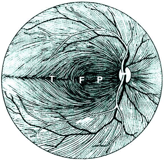
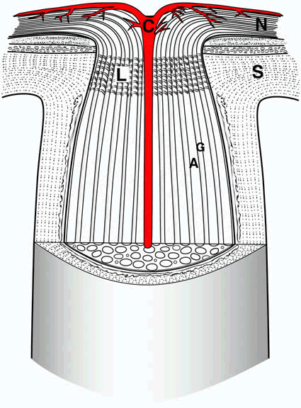
RETINAL GANGLION CELL AXONS WITHIN THE NERVE FIBER LAYER The optic nerve begins with the retinal ganglion cell, which is located in the innermost layer of the retina, within in the ganglion cell layer. Retinal ganglion cells receive input from bipolar cells and amacrine cells and project their axon toward the vitreous; then the axon makes an approximately 90° turn and projects toward the optic nerve head in the nerve fiber layer (Fig. 1). The fibers in the temporal part of the retina (corresponding to the nasal visual field) course away from the fovea, and then once in the nasal retina the fibers turn back toward the optic disc, entering in the superior and inferior portions of the disc. The retinal ganglion cell axons arising from retinal ganglion cells in the nasal retina project more directly to the disc, as follows: The fibers from the nasal half of the macula, forming the papillomacular bundle (or more properly, the maculopapillary bundle), enter at the temporal disc, whereas the fibers arising from ganglion cells nasal to the disc enter at the nasal part of the disc. In addition, there is strict segregation of those fibers arising from retinal ganglion cells located superior to the temporal horizontal meridian (temporal raphe) and those fibers arising from retinal ganglion cells located inferior to the temporal raphe. Because of this segregation, visual field defects corresponding to the injury to the retinal ganglion cell axons typically have stereotyped patterns (e.g., superior or inferior nasal steps, temporal wedges, or the arcuate scotomas). These are called nerve fiber bundle defects and are covered at length in the chapters on glaucoma. Once at the optic disc, the ganglion cell axons take a 90° turn away from the vitreous toward the brain (Fig. 2). This marks the beginning of the optic nerve, which is depicted in Figure 3. The neuroretinal rim of the disc is the part of the disc that corresponds to the projection of the nerve fibers into the optic nerve. The cup is the part of the disc that does not contain retinal ganglion cell axons. Depending on the size of the disc and whether there is any developmental or acquired injury, the cup-to-disc ratio may range from 0 to 1.0; the former represents a congenitally full disc, and the latter represents a disc that is completely “cupped out,” most commonly as a result of glaucomatous optic neuropathy. INTRAORBITAL OPTIC NERVE Although the retinal ganglion cell axon begins in the inner retina and continues along the nerve fiber layer of the retina, the optic nerve is considered to begin at the optic nerve head. There is an approximately 1-mm component of optic nerve within the intrascleral part of the globe and approximately a 30-mm length of optic nerve from the globe to the optic canal.1 The straight-line distance from the back of the globe to the optic canal is much less (the exact amount depending on individual orbital depth); the relative excess optic nerve is necessary for free movement of the globe during eye movements. In some cases excessive proptosis can exhaust the surplus length of optic nerve, resulting in tethering of the globe by the nerve. The retinotopic (topographic correspondence of optic nerve fibers to retinal location) segregation of retinal ganglion cell axons, particularly the segregation between axons arising from the superior retina and inferior retina, is gradually lost as the axons enter the nerve. There is only moderate retinotopy in the initial segment of the optic nerve.2 The retinotopy decreases distally3,4 and then becomes ordered for eventual nasal decussation near the chiasm.5 Within the orbit, the optic nerve travels within the muscle cone formed by the superior rectus, lateral rectus, inferior rectus, and medial rectus muscles. Tumors within the cone are common sources of compression of the optic nerve, or compressive optic neuropathy. Examples of these tumors include cavernous hemangioma, hemangiopericytoma, fibrous histiocytoma, lymphoma, and schwannoma. In addition, enlargement of the muscles themselves, particularly the inferior rectus and/or the medial rectus in Grave's (dysthyroid) ophthalmopathy, may also compress the optic nerve. THE OPTIC CANAL The optic nerve enters the cranium via the optic canal, a 5- to 12-mm passage that lies immediately superonasal to the superior orbital fissure. The optic canal contains some axons of sympathetic neurons destined for the orbit, as well as the ophthalmic artery. The latter lies immediately inferolateral to the optic nerve and is covered in dura. At the distal end of the canal, there is a half-moon-shaped segment of dura that overhangs the optic nerve superiorly and, thereby, lengthens the canal by a few millimeters. Within the canal, and immediately posterior to the optic canal, meningeal tissue is adjacent to the optic nerve. Benign tumors of the meninges, or meningiomas, are frequent causes of compressive optic neuropathies in these locations. Small tumors within the canal itself, where there is very little free space, may lead to compressive optic neuropathy without a radiographically visible tumor. INTRACRANIAL OPTIC NERVE AND THE CHIASM Once the nerve has entered the cranium, there is a highly variable (3 to 18 mm) length of nerve until the chiasm is reached. The length of the chiasm is approximately 8 mm. The intracranial optic nerve and chiasm are immediately above the planum sphenoidale and sella turcica (which contains the pituitary gland). There is approximately 10 mm between the inferior part of the nerve and the superior part of the pituitary. Tumors of the pituitary that are large enough to compress the chiasm may, therefore, cause compressive optic neuropathy. The nature of the retinotopic segregation changes as the fibers approach the chiasm; the temporal fibers are destined to remain ipsilateral, whereas nasal fibers cross. The ratio of fibers that cross to those that do not cross is approximately 52:48.6 This small difference between the number of crossing and noncrossing fibers is probably not responsible for the relative afferent pupillary defect seen in disorders of the optic tract, in which an afferent pupillary defect is seen contralateral to the injured tract (see also Chapter 34). Instead, some fibers from specialized cells within the retina that are responsible for the pupillary reflex may cross from the temporal retina into the contralateral optic tract.6 THE OPTIC TRACT AND LATERAL GENICULATE NUCLEUS Although the optic nerve anatomically ends at the chiasm, the retinal ganglion cell axons continue within the optic tract until the lateral geniculate nucleus or other targets (see later discussion and Chapter 34). |
OPTIC NERVE AXON COUNTS AND DIMENSIONS |
There are approximately 1,000,000 retinal ganglion cells in each retina, with
each cell sending a single axon down the optic nerve. Therefore, there
are approximately 1,000,000 axons within the optic nerve. Studies
in animals suggest that during development approximately 50% to 100% excess
retinal ganglion cells are produced, and during development
the number of retinal ganglion cells decreases by programmed cell death, in
many cases because not all of the axons reach their target regions
within the brain. Likewise in human development, excess retinal ganglion
cells die, presumably by programmed cell death during development. There
is also a large variation between individuals in the number of
axons within the optic nerve. Many factors affect the number of axons within the optic nerve, including inherited differences; damage to axons from disease; and the gradual loss of retinal ganglion cells during normal human aging, which averages approximately 5000 axons per year of life.7,8 |
MICROSCOPIC ANATOMY AND CYTOLOGY |
AXONS The most important components of the optic nerve are the axons of the retinal ganglion cells. Although the retinal ganglion cell bodies are situated within the retina, the axons are all located within the optic nerve. There are no other neuronal cell bodies within the optic nerve, thus, it is a pure white matter tract. Although the optic nerve may contain other small nerves, particularly tiny peripheral nerves (branches of the trigeminal system) that carry pain sensation or control vascular tone, most of the optic nerve is composed of the approximately 1,000,000 retinal ganglion cell axons. Optic nerve axons are collected in fascicles, which are separated by pia-derived septa. The mean axon diameter is slightly less than 1 μm. OLIGODENDROCYTES AND MYELIN Axonal conduction in the optic nerve depends on the presence of myelin, a fatty multilaminated structure that insulates each axon and greatly increases the speed and efficiency of conduction. This is discussed at length in the section on axonal conduction. The retrolaminar optic nerve is completely myelinated under normal circumstances. Oligodendrocytes are responsible for axonal myelination in the CNS, and there are approximately 20 patches of myelin contributed by each oligodendrocyte. Each oligodendrocyte may share in myelinating several axons, and each axon is myelinated by several oligodendrocytes. Each axon is myelinated with several lamellae of myelin bilayers, with the number of lamellae varying from axon to axon.9 Although oligodendrogliomas occur in other parts of the CNS, neoplasms of oligodendrocytes within the optic nerve are very rare. Intraretinal ganglion cell axons are not normally myelinated in most species because oligodendrocytes are absent in the retina. It has been hypothesized that the lamina cribrosa prevents migration of those cells, or their precursors, in development. Occasionally, myelination of part of the nerve fiber layer is seen; it is assumed to be secondary to ectopic oligodendrocytes or myelination of intraretinal axons by Schwann cells (as seen in normal cat10 and rat).11 ASTROCYTES The astrocyte is a major supporting cell of the optic nerve. Like oligodendrocytes, astrocytes are cells of glial lineage. Astrocytes have several functions, the most important are ionic homeostasis and serving as an energy source. Astrocytes are highly efficient at transporting potassium, and increases in the level of extracellular potassium as a result of repolarization are buffered by the presence of astrocytes. They also may serve as a glycogen storehouse, take up and inactivate neurotransmitters, aid in immune responses, and developmentally regulate axonal extension from the retina to the lateral geniculate nucleus. Astrocytes and their processes not only make up large portions of the optic nerve parenchyma in positions adjacent to neurons and vessels but also are present wherever neuroectodermal structures are adjacent to connective tissues, such as the pial septa, the adventitia of the central retinal artery and vein, and the pia in a layer named the glial mantle of Fuchs.12 Astrocytes, whether from optic nerve or from other CNS structures, may be immunohistochemically identified by the presence of intracellular glial fibrillary acidic protein (GFAP), an intermediate filament protein akin to the cytokeratins of epithelial cells. Two types of astrocytes are seen in the retina, the Müller cell and the retinal astrocyte. Müller cells are oriented perpendicular to the retinal plane, span it radially, and originate from neuroepithelium during retinal development.13 Retinal astrocytes are found in the nerve fiber layer, codistributed with retinal blood vessels.14 The retinal astrocyte, like that of the optic nerve, is probably a supporting cell for the ganglion cell axon.15 Astrocyte processes are concentrated at the nodes of Ranvier and have end feet in contact with nearby capillaries, presumably to allow transportation of substances between the local circulation and the axons. The most common intrinsic tumor of the optic nerve is an astrocytoma, or optic nerve glioma. This is usually a low-grade tumor of well-differentiated astrocytic cells that appear hair-like, or “pilocytic.” Pilocytic astrocytomas are usually seen in childhood and have a favorable prognosis. They are also commonly seen in association with neurofibromatosis. Rarely, more malignant neoplasms of astrocytic origin develop in adults and resemble the higher grade astrocytic neoplasms found elsewhere in the CNS. They are usually fatal. MICROGLIA Microglia are tissue macrophages found in the CNS. Although their origin was debated for decades, they have been shown experimentally to be of bone marrow origin16 and to share several markers with macrophages.17 They are associated with axon bundles but not necessarily with blood vessels. Microglia share several characteristics of the immune capacities of macrophages. By phagocytosing extracellular material, degradation within intracellular compartments can occur. This may be followed by antigen presentation on the cell surface and subsequent immune system activation. MENINGES The optic nerve is covered with three layers of meninges, dura, arachnoid, and pia. The meninges can also be divided up into pachymeninges (dura) and leptomeninges (arachnoid and pia). The outermost meningeal layer is the dura. This is a thick fibrovascular tissue, which is a direct extension of the sclera and which is in immediate contiguity with the periorbita and the dural layer of the lining of the cranial contents. The dura also continues within the optic canal. The arachnoid is the middle meningeal layer. It is a fairly loose, thin, fibrovascular tissue. The innermost meningeal layer is the pia, a tightly adherent layer that is extremely thin. Extensions of the pia continue within the nerve as the pial septa and provide the blood supply to the intraorbital and intracranial optic nerve, as discussed in the section on vasculature. The fibroblastic cells of the pial septa also form a matrix through which the fascicles of ganglion cell axons course.18 In the optic canal, there are numerous trabeculae connecting the dura through the arachnoid to the pia, which reduce the free space of the nerve sheath in this area.19 The pia extends collagenous septa into the optic nerve parenchyma, containing blood vessels that nourish the nerve. The space between the dura and arachnoid is the subdural space, and the space between the arachnoid and pia is the subarachnoid space. The subdural space around the optic nerve is small and is not in communication with the intracranial subdural space. The subarachnoid space, on the other hand, is in communication with the intracranial subarachnoid space. The optic nerve subarachnoid space ends anteriorly within a blind pouch just before the disc (see Fig. 3). As discussed in the section on increased intracranial pressure, the extension of the subarachnoid space from the brain into the orbit implies that elevated intracranial pressure has the potential to cause compression of the optic nerve via elevation of the hydrostatic pressure. Although all three layers of meninges are present in the intraorbital and intracanalicular optic nerve, only the pia continues along the intracranial optic nerve. The clinical relevance of this is that a tumor arising from the optic nerve meninges (i.e., an optic nerve sheath meningioma) will normally continue through the optic canal but then extend along the sphenoid bone and not along the course of the intracranial optic nerve. This means that it is extremely rare for an optic nerve sheath tumor to extend toward the chiasm and, thereby, affect the other side. The more common way for optic nerve sheath meningiomas to become bilateral is by directly extending along the sphenoid bone meninges. Mast Cells Mast cells, the tissue equivalent of circulating basophils, are responsible for immediate hypersensitivity (allergic) responses. They contain cytoplasmic granules of histamine, serotonin, and other inflammatory and vasoactive compounds. These granules are released when molecules of immunoglobulin E that are bound to high-affinity receptors on the mast cell surface contact particular target antigens. The products of degranulation are responsible for a local acute inflammatory response, with vasodilation and increased permeability of capillaries causing consequent edema. Cytokines released by mast cells may activate local inflammatory cells or may act as chemoattractants to recruit cells from the circulation. Mast cells have been histochemically identified in the meninges of human optic nerve,20 whereas histamine of mast cell origin has been described in rabbit and bovine optic nerve.21 OTHER CELLS Blood vessels within the optic nerve contain a variety of cell types. These are discussed further in the section on vascular supply to the optic nerve. Fibroblastic cells within the optic nerve are usually extensions of the meninges, and these are discussed in that section. The lamina cribrosa contains specialized fibroblastic cells; this important structure is discussed in Chapter 25. |
TARGETS OF RETINAL GANGLION CELL AXONS |
There are four main targets of retinal ganglion cells axons contained within
the optic nerve: (1) lateral geniculate nucleus, (2) pretectal nucleus, (3) superior
colliculus, and (4) suprachiasmatic nucleus. Most
axons are unbranched, but studies in many animals,22 including primates23 have demonstrated the existence of axon collaterals, suggesting that the
same may be true for human optic nerves. This section discusses each
of the synaptic targets of retinal ganglion cells axons. LATERAL GENICULATE NUCLEUS The main function of the optic nerve is for vision, and this is primarily subsumed by projections to the lateral geniculate nucleus (discussed in Chapter 34). The primate lateral geniculate nucleus is a six-layered structure, with layers 1 and 2 consisting of large (magnocellular) cells and layers 3, 4, 5, and 6 consisting of small (parvocellular) cells. The magnocellular layers receive input from the parasol retinal ganglion cells, whereas the parvocellular layers receive input from the midget retinal ganglion cells. Layers 2, 3, and 5 receive input from the ipsilateral retina, whereas layers 1, 4, and 6 receive input from the contralateral retina. In addition, there are interlaminar cells forming the koniocellular layers. These cells are smaller than those of the parvocellular layers and receive synaptic input from blue-yellow retinal ganglion cells.24 The first (and only) synapse of most axons within the optic nerve takes place at the lateral geniculate nucleus. These postsynaptic neurons then project to area V1 of the occipital cortex (i.e., the striate cortex). Details of this projection and the functional anatomy of the part of the cerebral cortex that is devoted to vision is discussed in Chapter 34. PRETECTAL NUCLEI A second function of the optic nerve is for the afferent portion of the pupillary response.25,26 These fibers project to the pretectal nuclei within the midbrain. Although the exact number is not known in humans, retrograde labeling studies in pigeons27 and rats28 indicate that only a small percentage of retinal ganglion cell axons are responsible for the pupillary reflex. The pretectal projection is bilateral, and, in addition, fibers from each pretectal nucleus project both ipsilaterally and contralaterally to the Edinger-Westphal subnuclei of the third nerve nucleus. The latter then projects parasympathetic axons along the course of the third nerve to the ciliary ganglion within the orbit, where a synapse is made. Postsynaptic parasympathetic fibers continue to the pupilloconstrictor muscle, constriction of which is visible as the pupillary light reflex. Because of the combined ipsilateral and contralateral projections from the retina to the pretectal nuclei and from the pretectal nuclei to the Edinger-Westphal subnuclei, a light stimulus that causes impulses along either optic nerve will cause equal constriction of both pupils. This is the physiologic basis for detecting an afferent pupillary defect (also called the swinging flashlight test or Marcus-Gunn pupil). If there is a functional difference in the number of optic nerve axons carrying impulses from each retina, then light shined into the eye with fewer axons will cause a lesser amount of (bilaterally symmetric) pupillary constriction than light shined into the other eye. By alternating the light between the two eyes and observing the difference in pupil size dependent on which eye the light is shined into, one can detect which optic nerve is functionally conducting less than the other. This measure is highly correlated with differences in the number of surviving retinal ganglion cells.29 SUPERIOR COLLICULUS A third set of optic nerve axons project to the superior colliculus, a paired structure of the dorsal midbrain. Although in lower animals (e.g., rodents) most fibers of the optic nerve project to the superior colliculus, only a minority do so in primates, including humans. The function of the superior colliculus is to coordinate retinal and cortical control of saccades and fixation.30 HYPOTHALAMUS A fourth projection of optic nerve axons is via the retinohypothalamic tract to the suprachiasmatic nucleus and the paraventricular nucleus31–33 within the hypothalamus. These fibers are responsible for circadian control of the sleep-wake cycle, temperature, and other systemic functions. |
OPTIC NERVE ELECTROPHYSIOLOGY |
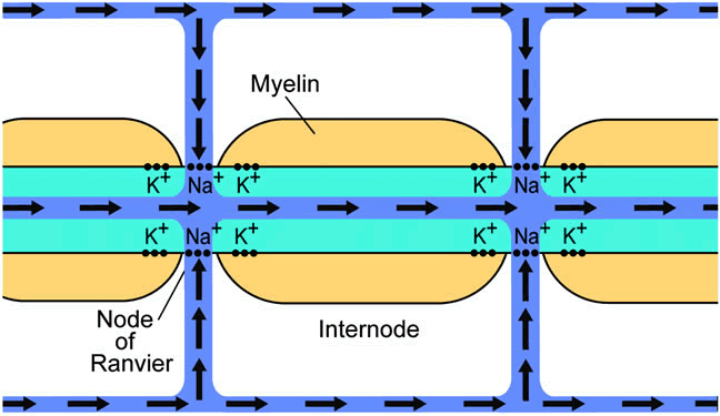
SYNAPTIC TRANSMISSION Retinal ganglion cells receive synaptic input from bipolar and amacrine cells. The bipolar inputs are the results of graded potentials; only retinal ganglion cells exclusively use action potentials (amacrine cells have features of both).34 The synapses from bipolar and amacrine cells primarily use glutamate as the major excitatory neurotransmitter. The retinal ganglion cell synapse on its targets, primarily the lateral geniculate nucleus in higher animals, is glutamate. Within the retina the levels of glutamate are controlled by Müller cells, which have glutamate transporters and also contain the enzyme glutamine synthetase, which converts glutamate to the amino acid glutamine. EXCITOTOXICITY Retinal ganglion cells are particularly susceptible to high levels of glutamate in the extracellular space. This causes cell death mediated via overexcitation, or excitotoxicity. It has been proposed that excitotoxic retinal ganglion cell death may explain pathologic conditions, such as glaucoma or retinal ischemia, in which an excess of retina ganglion cells die, akin to what occurs with cerebral infarct or other diseases. AXONAL CONDUCTION Action Potentials The primary function of the optic nerve is to carry information from the retina to target areas within the brain. Individual retinal ganglion cell axons transmit information via action potentials, which are all-or-nothing spikes of electrical activity. This is in contrast with intercellular communication within the retina, where graded potentials are used to transmit information. The retinal ganglion cells and their axons are the first neurons in the transmission of visual information from the eye to use action potentials as the mechanism for transmission. With action potentials, the actual amount of voltage change (i.e., depolarization) is the same, whereas the number of impulses per second and the distribution of impulses within various axons is the mechanism by which visual information is carried down the optic nerve. Conduction down individual axons occurs via the same biophysical mechanisms that occur in any myelinated axon (see later section on myelin). The mechanism of axonal conduction is straightforward. At rest, the inside of an axon is at a negative voltage with respect to the outside of the axon. This resting potential results from the higher concentration of potassium within the axon than outside the axon. As a few potassium ions flow down their concentration gradient from the inside of the axon to the outside, the movement of positive charge out of the axon results in a negative potential within the axon. Outward flux of potassium continues until the charge separation becomes too great and can no longer be driven by the concentration difference between inside and outside the axon. The point at which the gradient for potassium concentration is balanced by the gradient for separation of charge results in an equilibrium potential for potassium. Although the resting potential is primarily determined by potassium, there is also a smaller contribution from sodium flowing down its concentration gradient. In the case of sodium, the concentration in the extracellular space is higher than within the axon. This concentration gradient would induce sodium to enter the axon, and movement of a few sodium ions results in a positive sodium equilibrium potential. However, in a resting (nondepolarizing) axon the conductance for sodium is much smaller than that for potassium, and, therefore, the final resting potential is weighted much more by the equilibrium potential for potassium than for sodium. During axonal conduction, depolarization of a section of membrane induces opening of voltage-sensitive sodium channels located within adjacent membrane. These allow much greater amounts of sodium to enter the axon, and the positive sodium ions cause the axon to become more positive (i.e., depolarized). In this case, the potential across the membrane is now weighted far more by the sodium equilibrium potential than by the potassium equilibrium potential. The sodium channels open because they are voltage sensitive, and a relatively small depolarization of the membrane will cause them to open transiently. Increased potential within the axon rapidly affects adjacent sections of the axon and causes sodium channels in these adjacent areas of axonal membrane to likewise become partially depolarized. These then completely open and cause a compete depolarization. This chain of events continues down the axon, resulting in a transmission of action potential. Repolarization is the return of the axonal membrane potential to the original (negative resting potential) state and is necessary for more than a single action potential to be transmitted down the axon. Repolarization is due to the closure of the voltage-sensitive sodium channels and a transient opening of voltage-sensitive potassium channels. Once the latter occurs, the membrane resting potential is weighted more by the potassium equilibrium potential than by the sodium equilibrium potential. This results in a restoration of membrane potential to the resting state. In the process of transmission of an action potential there is movement of sodium and potassium ions along their concentration gradients. If action potentials continued indefinitely, the sodium and potassium concentrations would reach equilibrium across the axonal membrane, resulting in loss of their concentration gradients and blocking conduction. Therefore, to re-establish their concentration gradients, there is a relatively slow redistribution process of these ions via the Na+ = K+ = ATPase takes place. This is a highly energy-dependent process and will not happen if axonal metabolism is pathologically disturbed. Role of Myelin Myelin adds an additional complexity to axonal conduction. Myelin has two physiologic properties. It increases the resistance and decreases the capacitance of the axon. The decreased capacitance means less sodium ions (positive charge) are needed to enter the axon to depolarize the membrane to a given voltage. The increased resistance means that there is less leakage of charge across the membrane. Together, these properties decrease the amount of ionic flux needed to achieve changes in voltage across the membrane. If conduction can be achieved with less ionic flux, then less energy is needed to maintain ionic homeostasis after conduction, and, therefore, less Na, K-ATPase activity is necessary. Therefore, myelinated conduction is more efficient. Ionic channels are not distributed uniformly along a myelinated axon. Instead, the channels are segregated into patches located within the small areas where the axon is unmyelinated. These are called nodes of Ranvier (Fig. 4). Conduction in a myelinated axon, therefore, becomes much faster, because the depolarization of the axon at one node leads to depolarization at the next node by the intrinsic conduction within the axon (the “cable properties” of the axon), instead of by sequential opening of channels along the axon. The depolarization, thus, “jumps” from one node to another; this is called saltatory conduction (see Fig. 4). In cases of acquired loss of myelin, for example in idiopathic optic neuritis, a common inflammatory optic neuropathy, there is abnormal conduction because of the changes in resistive and capacitive properties of the membrane brought about by demyelination. Axonal conduction becomes slowed and in some cases may be blocked (“conduction block”), both of which result in decreased visual function. Even in a completely demyelinated axon, a low density of internodal sodium channels in demyelinated optic nerve may still allow conduction.35 Another phenomenon peculiar to demyelination is worsening of vision with heat or exercise, called Uhthoff's phenomenon. Increased temperature and exercise are thought to decrease the sodium channel open-time during depolarization, resulting in less charge entering the axon and a decreased likelihood that an adjacent section of demyelinated axon will be able to depolarize enough to cause opening of its own voltage-sensitive sodium channels. This leads to temperature-sensitive conduction block. |
AXONAL TRANSPORT |
The entire length of the retinal ganglion cell axon must be maintained
by transporting proteins and other subcellular constituents several centimeters
from the cell body, where the nucleus and virtually all of the
protein synthetic machinery resides. These axons are several centimeters
long, and, thus, the health of the nerve relies on axonal transport
of a multitude of substances over a distance many times the size of
the ganglion cell body. The processes underlying axonal transport have
been well studied, in the optic nerve and in other nerves, and several
important features can be identified. Axonal transport occurs in two directions, orthograde (away from the cell body and toward the brain) and retrograde (toward the cell body and away from the brain). By injecting radioactive, fluorescent, or enzymatically active macromolecular tracers into either the eye or the terminal fields of the axons, the course and timing of orthograde and retrograde transport can be determined. By analysis of the individual labeled intra-axonal substances, transport can be differentiated into several classes. Depending on the fineness of the separation, there appear to be two to five groups of substances that are axonally transported, each with their own characteristic velocity. Fast axonal transport carries several subcellular organelles, such as the vesicles of neurotransmitter used in synaptic transmission, as well as other particulate matter, toward the axon terminal.36 Proteins carried by this process are usually membrane-associated.37 This process has been measured at 90 to 350 mm/day in the rat and chick optic nerve.38,39 Fast axonal transport is sensitive to metabolic inhibitors and to agents such as colchicine, suggesting an energy- and microtubule-dependent mechanism for transport, respectively.40 This class of transport continues despite transection of the axon distal to the cell body, suggesting that intra-axonal components are sufficient for the process to occur. Slow axonal transport carries several types of proteins, many of which are as yet unidentified. Most components carried by slow axonal transport end up within the axon itself. There are two classes of slow transport. The slower class of slow axonal transport (SCa) occurs at 0.2 to 0.5 mm/day and carries cytoskeletal proteins (primarily tubulins), which make up microtubules, and neurofilament proteins, a member of the intermediate filament group. These are the major skeleton proteins for the neuron.41 The more rapid class of slow axonal transport (SCb) occurs at 1 to 6 mm/day and carries proteins such as actin and myosin, as well as the enzymes of intermediate metabolism and myelin-associated proteins.41–43 Mitochondria are transported by this process. Slow axonal transport does not continue after transection of the axon distal to the cell body, unlike fast axonal transport.44 Similarly, interruption of circulation to the proximal axon, for example by cutting the short posterior ciliary arteries, causes interruption of slow transport.45 Retrograde transport occurs at about half the velocity of fast orthograde transport and is the means by which endocytosed substances from the synapse, such as released neurotransmitter, may be returned to the cell body. Likewise, wear and breakdown products from the axon and its terminal are similarly returned to the cell soma. Retrograde transport of neurotrophic molecules during development may signal the cell body regarding the presence of a growth target. Axonal transport may be affected in disease, possibly potentiating nerve injury, such as in experimental allergic encephalomyelitis or glaucoma.46 Axonal transport within the optic nerve may have important clinical implications. For example, pressure can perturb axonal transport at the lamina cribrosa, experimentally demonstrated by focal accumulation of labeled proteins after injection of radiolabeled precursors into the vitreous.46,47 This suggests a role for interruption of axonal transport in the pathophysiology of axonal loss in glaucoma. Accumulation of labeled proteins also occurs at physiologic intraocular pressures,48 implying that a qualitatively greater disturbance of axonal transport must be necessary for damage to occur via this mechanism. Other important clinical correlations of altered axonal transport are in disc edema (in which blockage of axonal transport has been observed to occur, for example, with ocular hypotony or increased intracranial pressure)49,50 and in optic neuritis.51 |
BLOOD SUPPLY |
The blood supply to the optic nerve is complex and changes qualitatively
along its course. Because of the varying nature of the vascular supply
to the optic nerve, a wide variety of clinical syndromes may result
from ischemia or infarction at each location. This section discusses
the vascular supply to the retinal ganglion cell axons as a function of
location, starting at the retina, then the optic nerve head, the intraorbital
optic nerve, the optic canal, the intracranial optic nerve, and
the chiasm and optic tract. This material is covered in greater depth
in Chapter 25 and in recent reviews.52,53 RETINA Although the retina has two circulations, the retinal vascular circulation (derived from the central retinal artery) and the choroidal circulation (derived from the short posterior ciliary arteries), only the former is relevant to perfusion of the retinal ganglion cells and their axons within the nerve fiber layer. The retinal ganglion cells are located within the inner retina, in the ganglion cell layer, whereas the nerve fiber layer is vitread to the ganglion cell layer and is the innermost neuronal layer of the retina. Both derive their blood supply from capillary branches of the retinal circulation, although there presumably is some small amount of oxygen exchange with the vitreous. The clinical relevance of the retinal circulation perfusing the retinal ganglion cells and their axons is that an inner retinal infarction (e.g., from a central retinal artery occlusion) will result in an optic neuropathy, with a pale disc and loss of axons within the optic nerve. OPTIC NERVE HEAD The circulation of the optic nerve head is complex and varies depending on which particular layer is studied. Although the nerve head circulation is derived from two sources, the central retinal artery and the posterior ciliary arteries, the latter provides the most important contribution. The central retinal artery is a branch of the intraorbital ophthalmic artery, and enters the optic nerve during its intraorbital course, approximately 12 mm behind the globe. As it heads toward the disc, it provides minimal perfusion of the nerve through which it courses.54 As it branches on the disc into the retinal arterioles, it provides partial perfusion of the superficial disc via small capillaries. In contrast, branches of the short posterior ciliary arteries provide the major blood supply to the optic nerve head. There are usually two posterior ciliary arteries, a medial and a lateral, which originate from the ophthalmic artery and form several branches. One set of branches becomes the major perfusion of the choroid, forming the choriocapillaris. Another set of branches perfuses the optic nerve, both anteriorly via direct branches and posteriorly via a retrograde arteriolar investiture of the optic nerve (see later). Anastomoses of posterior ciliary artery branches55,56 form the circle of Zinn-Haller, which contributes significant perfusion to the optic nerve head. In addition, there are contributions from recurrent choroidal arterioles to the prelaminar and laminar optic nerve head and contributions from recurrent pial arterioles to the laminar and retrolaminar optic nerve head. Because of the common posterior ciliary arterial source of the choroid and deep optic nerve head, they will fluoresce simultaneously during the earliest phase of fluorescein angiography, before the retinal arterioles transit dye. Although both the choroid and the optic nerve head receive blood supply from the posterior ciliary circulation, there is a tremendous difference in the histologic characteristics of their capillaries. Choroidal vessels have fenestrated endothelial cells, whereas optic nerve vessels have nonfenestrated endothelial cells with tight junctions, surrounded by pericytes. Optic nerve vessels, therefore, share the same blood-nerve barrier characteristics as the blood-brain barrier. Thus, only a restricted number of molecules can cross the blood-nerve barrier. For example, clinically, gadolinium enhancement on magnetic resonance imaging is not seen unless there is some pathologic process within the optic nerve that would disrupt the blood-nerve barrier (e.g., inflammation).57,58 Another major difference between choroidal and optic nerve head vessels is that only the latter can autoregulate (i.e., maintain approximately constant blood flow despite most changes in intraocular pressure).59–61 The clinical implication is that there is compensation of perfusion for a wide range of intraocular pressures in normal individuals. INTRAORBITAL OPTIC NERVE AND OPTIC CANAL The pial circulation perfuses the intraorbital optic nerve. Branches of the ophthalmic artery either directly perfuse the pia or indirectly perfuse it anteriorly via recurrent branches of the short posterior ciliary arteries or a branch of the central retinal artery. The pia then sends penetrating (centripetal) vessels into the intraorbital optic nerve along the fibrovascular pial septa, from which a capillary network extends into neural tissue (axons and glia). Similarly, the intracanalicular optic nerve is perfused by one to three branches of the ophthalmic artery, which also perfuse the pial surface and then penetrate the nerve.62 An important clinical implication of the pia supplying the intraorbital and intracanalicular optic nerve relates to optic nerve sheath meningiomas. If a surgeon strips the meningioma away from the nerve, then the pia will be removed as well, resulting in loss of the blood supply to the nerve, infarction, and blindness. INTRACRANIAL OPTIC NERVE, CHIASM, AND OPTIC TRACT The intracranial optic nerve and chiasm are perfused by the internal carotid artery and its branches, primarily the anterior cerebral, anterior communicating, and superior hypophyseal artery. Branches of the posterior communicating artery may also perfuse the posterior chiasm. The optic tract is predominantly perfused by branches of the posterior communicating and anterior choroidal arteries. Similar to the intraorbital and intracanalicular optic nerve, the blood supply occurs via small pial penetrating vessels. However, there is no dura or arachnoid surrounding the optic nerve posterior to the optic canal, and, thus, the risk of infarction is not from stripping a tumor off the nerve but from inadvertent detachment of the fine vessels during surgical manipulation. |
AXONAL INJURY |
CLINICAL IMPLICATIONS The optic nerve is commonly involved in disease, resulting in optic neuropathy. Optic neuropathies are major causes of visual loss. The most common optic neuropathy is that associated with glaucoma (i.e., glaucomatous optic neuropathy). A wide variety of other optic neuropathies also cause visual loss. The inflammatory optic neuropathies are most commonly seen in young or middle-aged adults, and the most common is optic neuritis associated with the demyelinating disease multiple sclerosis. Ischemic optic neuropathy usually affects older adults. The most common type is nonarteritic anterior ischemic optic neuropathy, a disorder of unknown cause that results in sudden visual loss associated with disc edema; arteritic anterior ischemic optic neuropathy has a similar clinical presentation but is due to a vasculitic process, usually from giant cell arteritis. Compressive optic neuropathy, in which the optic nerve is compressed by a mass lesion, commonly a tumor or aneurysm, results in slowly progressive loss of visual function as the mass increases in size. Although not proven, some researchers believe that Leber's hereditary optic neuropathy and some of the toxic optic neuropathies may reflect damage to the retinal ganglion cell body within the retina. Except for these entities, most optic neuropathies are due to damage directly to the optic nerve. Therefore, most optic neuropathies involve axonal injury. In this section, how axonal damage results in death of the retinal ganglion cell is discussed. For example, glaucomatous optic neuropathy likely results from axonal injury at the optic nerve head, particularly at the lamina cribrosa. Nonarteritic anterior ischemic optic neuropathy also results from damage at the optic nerve head, in many cases just posterior to the lamina cribrosa. The locus of injury for compressive optic nerve injury is usually obvious and may range from within the orbit (e.g., with Grave's ophthalmopathy) to the chiasm (e.g., from a pituitary adenoma or craniopharyngioma). RETINAL GANGLION CELL DEATH AFTER AXONAL INJURY Death of retinal ganglion cells is the final common pathway underlying virtually all diseases of the optic nerve, including glaucomatous optic neuropathy, anterior ischemic optic neuropathy, optic neuritis, and compressive optic neuropathy. These disorders all cause injury to the axon of the retinal ganglion cell within the bulbar, orbital, intracanalicular, or intracranial optic nerve, yet the pathologic condition at the level of the retina is primarily loss of retinal ganglion cells. In many diseases affecting the retinal ganglion cell axons (e.g., glaucoma or arteritic ischemia), the visual loss is permanent, because retinal ganglion cell loss is irreversible. In some cases (e.g., chronic compressive optic neuropathy, acute optic neuritis, or papilledema), the visual loss can be reversed when the axonal damage is relieved, presumably because retinal ganglion cell death has not yet occurred. However, if allowed to continue, even these disorders eventually can result in retinal ganglion cell death. Most research on optic neuropathies has focused on the pathophysiology specific to the particular disorder (e.g., elevated intraocular pressure for glaucoma, inflammation for optic neuritis, and ischemia for anterior ischemic optic neuropathy). However, optic neuropathies all share in common retinal ganglion cell axonal injury, except for the few disorders in which the locus of injury is unknown (such as Leber's hereditary optic neuropathy). Therefore, an understanding of the molecular response of the retinal ganglion cell to axonal injury is applicable to a wide variety of diseases of the optic nerve, independent of the mechanism by which the nerve is injured. Time Course When axons contained within the optic nerve are transected or crushed, the associated retinal ganglion cells usually die. However, depending on the age and species of the animal, the cell body size, and the distance from the site of injury to the retinal ganglion cell, up to 70% may survive. In lower animals (e.g., goldfish and frogs) ganglion cells do not die but hypertrophy and regenerate axons within 1 to 2 months.63–65 Ganglion cells from squirrel and owl monkeys die approximately 4 to 6 weeks after axotomy.66,67 In humans with chiasmal compression for at least 6 months, there is extensive loss of ganglion cells from the nasal hemiretinas; yet even 35 days after transection of the ipsilateral optic tract there is sparing of some temporal ganglion cells.68 Apoptosis The mode of death of retinal ganglion cells after axonal injury is complex and appears to involve a programmed cell death process called apoptosis. Apoptosis is a form of programmed cell death, characterized by condensation of the nucleus and cytoplasm, budding of the cytoplasmic membrane, cleavage of DNA into 180 to 200 bp fragments, fragmentation of the cell into membrane-bound bodies, and heterophagy of these bodies.69,70 A variety of techniques have been used to show that the death of retinal ganglion cells after axotomy occurs by apoptosis.71–75 It is likely that neurotrophin deprivation or some other signal may induce a death program in these cells (see later discussion). Signaling of Axonal Injury There are several theories proposed to explain the mechanism by which axonal injury results in induction of apoptosis. Retinal ganglion cell death after optic nerve injury or target removal appears to take place through several mechanisms, including lack of neurotrophic factors from the target tissue, excitotoxicity from physiologic or pathologic levels of glutamate, free radical formation, leakage of cellular constituents out the end of the axon, proliferation of macroglia, activation of microglia, buildup of excess retrogradely transported macromolecules, and breakdown of the blood-brain barrier.76–83 Of greatest interest is neurotrophic factor deprivation from blocked retrograde transport of neurotrophic factors (neurotrophins) or decreased levels of endogenous ocular neurotrophins,84 which is analogous to the loss of a target during development. Retinal ganglion cells are known to be highly dependent on neurotrophic factors during development. The normal developmental loss of retinal ganglion cells is partly due to the competition for target-derived molecules, such as neurotrophins, while the cells attempt to extend their axons and form connections in target areas of the CNS. In the adult human CNS, the predominant targets of the axons contained within the optic nerve are the lateral geniculate nucleus, the superior colliculus, the pretectal nuclei, and the suprachiasmatic nucleus of the hypothalamus. It is thought that adult retinal ganglion cells, like other neurons, are partly dependent on neurotrophic agents for their survival. Support for the role of neurotrophin dependence comes from experiments using identified neurotrophic factors to rescue axotomized neurons, including retinal ganglion cells.81,85–89 TYPES OF OPTIC NERVE INJURY As discussed previously, injury to the axons contained within the optic nerve results in retinal ganglion cell death. However, axonal injuries differ substantially, especially with respect to clinical disease. This section discusses some of the major types of axonal injuries in the context of local pathology. Transection Transection of optic nerve axons results in a discontinuity of the axonal membrane and its contents. It is relatively rare for the human optic nerve to undergo an acute partial or complete transection; the most common examples are direct traumatic optic neuropathy (e.g., from a bullet or knife) and iatrogenic transection during surgical resection of an adjacent or intrinsic tumor. Although many animal models that are used to study the effects of optic nerve injury involve transection of axons, this is not reflected by clinical disease. This disparity means that many experimental studies of the pathways of retinal ganglion cell death after axonal injury do not necessarily reflect most diseases of the optic nerve, because of the great difference in time course of the initial insults. Ischemia The evidence backing ischemia as a cause of human optic neuropathies varies, depending on the disease. For example there is little doubt that arteritic anterior ischemic optic neuropathy resulting from giant cell arteritis is associated with histopathologically verifiable vasculitic occlusion of posterior ciliary arteries. On the other hand, there is a tremendous amount of controversy as the whether the effects of intraocular pressure causing axonal damage in glaucomatous optic neuropathy are primarily an ischemic or compressive process. Even nonarteritic anterior ischemic optic neuropathy has features that make it unlike many other ischemic diseases.90 Perhaps the least dispute relates to ischemia from hypotension or severe blood loss, although even in these cases the pathophysiology may vary.91,92 Inflammation Inflammation of the optic nerve, or optic neuritis, is the most common form of acute optic neuropathy in young and middle-aged adults and is a frequent harbinger of multiple sclerosis.93,94 Demyelination is the most common pathologic accompaniment of optic nerve inflammation, and conduction block resulting from this or other effects of inflammation is responsible for the usually temporary loss of visual function seen in patients with optic neuritis. This is discussed in a previous section on axonal conduction. Demyelination would not necessarily cause loss of retinal ganglion cells, but CNS inflammation in multiple sclerosis is associated with axonal loss.95–97 Axonal loss in optic neuritis has been long appreciated clinically as optic atrophy and loss of the nerve fiber layer,98 and experimental models of optic nerve inflammation can be used to elucidate how this axonal damage occurs.99–101 Although not completely clear, it is likely that one major effect of inflammation is on the axonal cytoskeleton, as witnessed by changes in microtubule and neurofilament organization in experimentally induced optic neuritis.101 This would also explain changes in axonal transport in experimental optic nerve inflammation.102–104 In addition, changes in the blood-nerve barrier consequent to the inflammatory process and generation of reactive oxygen species may also affect axonal function.57 Compression Compression of the optic nerve is a common clinical correlate of many optic neuropathies. Besides the obvious causes such as neoplasms and aneurysms, the optic nerve can also be compressed by enlarged extraocular muscles (as in Grave's ophthalmopathy), edema (as seen with indirect traumatic optic neuropathy, in which the nerve is contused within the optic canal), or optic disc drusen. Compression intrinsic to the nerve can occur in some forms of glaucomatous optic neuropathy, in which increased intraocular pressure causes bowing out and shifting of the lamina cribrosa, which constricts the bundles of optic nerve axons within the cribrosal pores.105 The effects of chronic experimental compression of the intraorbital optic nerve were delineated at the microscopic and ultrastructural level in a classic series of experiments by Clifford-Jones and colleagues.106,107 They found demyelination initially, followed by remyelination, even while the axons were still compressed. There were relatively minor findings of direct axonal loss. Demyelinated axons or the direct effects of pressure might be expected to lead to conduction block, which would be reversible. This could, therefore, explain the remarkable return of visual function after removal of tumors compressing the optic nerve.108 Glaucoma The most common optic neuropathy is glaucomatous optic neuropathy, distinguished by a distinct morphology of progressive excavation of the nerve head without significant pallor of the remaining neuroretinal rim. Clinical and histologic evidence from patients with glaucoma show changes in several locations along the course of the retinal ganglion cell axon and its targets. There are decreased numbers of retinal ganglion cell bodies in glaucoma,109–111 and this likely reflects death by apoptosis.74,112–114 The number of retinal ganglion cells lost correlates with the visual field deficit.115 In addition to the retinal ganglion cell body loss, there is loss of the ganglion cell axons, manifested by segmental loss of the nerve fiber layer,116–120 increased cup-to-disc ratio, thinning of the optic nerve121 and chiasm,122 and changes in postsynaptic cell counts within the lateral geniculate nucleus,123–126 the main target of retinal ganglion cell axons in higher animals. Although every point along the axon is eventually involved in the disc cupping of glaucomatous optic neuropathy, the site of injury is the optic disc; this is consistent with a wide variety of evidence demonstrating pathology at the level of the disc,111,127 particularly the lamina cribrosa,105,128–139 as well as the occurrence of splinter hemorrhages140–142 and focal notching143 at the disc. Studies of tissue from human patients with glaucoma and nonhuman primates with experimental glaucoma confirm changes at the optic nerve head, such as bowing out of the lamina cribrosa, intra-axonal accumulation of organelles (consistent with blocked axonal transport), and wallerian degeneration distal to the lamina cribrosa.105,144,145 Studies in experimental animals have shown that injury to the optic nerve, for example from increased intraocular pressure in experimental glaucoma, blocks the retrograde transport of the neurotrophic factor, brain-derived neurotrophic factor (BDNF), along with its associated receptor, trkB.146 Whether resulting from mechanical trauma of axons,71,147–149 ischemia,150 generation of nitric oxide,151,152 or other causes, axonal injury is known to cause changes in retinal ganglion cells, which eventually result in death. |
ACKNOWLEDGMENTS |
Supported by the Retina Research Foundation, the Glaucoma Foundation, NIH
EY12492, and an unrestricted departmental grant from Research to Prevent
Blindness, Inc. The author is a Research to Prevent Blindness Dolly
Green scholar. Portions of this chapter were adapted from: Levin LA. Biology of the optic nerve. In Albert DM, Jakobiec FA (eds). Principles and Practice of Ophthalmology. Philadelphia: WB Saunders, 2000; and Levin LA. Optic nerve. In Kaufman P, Alm A (eds). Adler's Physiology of the Eye. New York: Harcourt, 2001. |