Chapter 43 Epidemiology of Ocular Infections KIRK R. WILHELMUS Table Of Contents |
Epidemiology is “a peculiarly thoughtful approach to [the conduct
and interpretation of] studies amounting to applied common
sense.”1 The goals of epidemiologic analysis in ocular infectious diseases are
to validate operational definitions of ocular disorders, to ascertain
the frequency and the distribution of specific conditions, and to determine
the factors associated with their occurrence and prevention2 (Table 1). Although epidemiology is limited by variations and uncertainty in the
frequency and the spectrum of ocular diseases among different populations, in
diverse locations, and at various times, epidemiology is the
cornerstone of clinical research in ophthalmology and is particularly
useful in the study of ocular infections.
TABLE 43-1. Goals of Epidemiology in Infectious Eye Diseases Describe patterns of occurrence of eye infections
|
GLOBAL IMPACT OF INFECTIOUS EYE DISEASE | ||||||||||||||||||||||||||||||||||||||||||||||||||||||||||||||||||||||||||||||||||||||||||||||||||||||||||||||||||||||||||||||||||||||||||||||||
BLINDNESS AND INFECTION Public health ophthalmology seeks to identify the magnitude and the distribution of blinding eye diseases, to define their methods of transmission, to recognize vulnerable or high-risk groups, and to evaluate management and prevention programs that aim to improve eye health.3 National and international programs rely on surveillance statistics and on causal links.4 Infection is a major cause of eye disease that requires care. An estimated 38 million people worldwide are blind, and an additional 110 million have low vision.5 Extrapolations from data summarized by the World Health Organization (WHO) and other sources suggest that nearly 10 million people are visually disabled as a result of infectious diseases (Table 2), which account for about one fourth of the world's blind.6
TABLE 43-2. Ocular Infections of Global Importance*
*Compiled from multiple sources using data from 1975 to 1995. Estimates, particularly for numbers of visually disabled individuals, are hampered by incomplete sampling, underreporting, variable diagnostic criteria, and other uncertainties, and are not a reliable guide to resource allocation. Incidence is expressed as the number of new detectable ocular infections per year. Prevalence is the estimated current number of people with active ocular infection or residual scar.
Cross-sectional surveys in the United States show that socioeconomic status is one of the most important determinants of ocular health.7,8 The relationship between income and infectious eye disease has been presumed but not proven. However, international comparisons suggest that economic development accounts for a substantial portion of infectious blindness. In developed nations, infections are the cause of approximately 1% of partially sighted people; in less developed regions, the burden of visual loss resulting from infectious diseases is much greater. Populations at particular risk of infectious visual disability are the poor, who live and work in unhygienic conditions. EMERGENCE OF OPHTHALMIC EPIDEMIOLOGY The discipline of epidemiology began with the study of epidemics, the increased frequency of a disease in a specified population within a given time. Epidemics of eye disease have been recognized since antiquity, often in association with the natural environment or war.9 A recognition of contagion, an understanding of ocular inflammation, and advances in mathematics were prerequisite to the development of ophthalmic epidemiology. Abrupt outbreaks, such as those typical of viral conjunctivitis, are easily noticed, but knowledge of endemic-level disease frequency is needed to recognize epidemics of less common or of slow-onset infections. Census statistics collected by many nations during the 19th century included blindness prevalence and, by the end of that century, of the causes of blinding ocular disease. For example, infectious diseases accounted for 72% and 85% of all causes of blindness in northern Europe in 1883 and 1902, respectively. By 1926, population-based data indicated that the percentage had decreased to 47%,10 partly because the proportion of blindness due to ophthalmia neonatorum dropped from 18% of all blindness in 1902 to 5% in 1926, a reduction largely attributed to Credé prophylaxis. Although the reasons for a decline in the relative importance of other infections are less obvious, they probably relate to improved sanitation after the Industrial Revolution. Infection remained a major contributor to blindness until the 1930s; after the end of World War II, the percentage of blindness due to infectious diseases declined dramatically in many parts of the world11 (Fig. 1). The first major effect of epidemiology on ocular infections occurred during the early bacteriologic era. Recognition that trachoma and other external eye infections were among the world's leading causes of blindness led to enormous public health efforts at their containment and eradication. In the late 19th and early 20th centuries, immigration services routinely screened for trachoma (Fig. 2), and it was a common reason for deportation. Throughout the 20th century, much effort was aimed at stopping trachoma's spread and limiting its complications. Increased prosperity reduced the incidence of trachoma in Europe and the United States, but chlamydial and other infections are still major contributors to blindness in Africa and Asia (Table 3).
TABLE 43-3. Global Distribution of Blindness Due to Ocular Infection, 1990
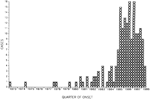
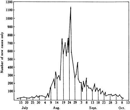
Ocular infections remain a global concern, even in developed nations, and are an individual as well as a community problem. For example, Americans contract an average of three ocular infectious diseases per year, with annual costs of more than $17 billion and of nearly $2 billion in lost work or school days.12 The worldwide frequency of ocular infections is uncertain. One surrogate measure is the number of topical ophthalmic anti-infective preparations prescribed, which runs into the hundreds of thousands each year (see http://ims-america.com, http://www.druginfonet.com, and http://www.fda.gov). Improved surveillance of ocular disease could better determine its frequency and distribution worldwide. EPIDEMIC OCULAR INFECTION An epidemic curve is a histogram showing the number of cases plotted over time. Sharp increases in disease frequency often reflect the exposure of several people to a point source of contamination. The downward slope may be skewed by waves of secondary cases from person-to-person transmission of a communicable infection. For example, the epidemic curve of an outbreak of adenovirus conjunctivitis often begins like that of a commonvehicle, single-exposure epidemic in which there is a sharp, initial rise in the number of cases with a subsequent, gradual fall. There are often one or more additional peaks and troughs as the virus continues to spread by serial and interpersonal propagation, superimposing in two or more epidemic curves over endemic, sporadic cases (Fig. 3). Each infectious disease is characterized by an incubation period (the interval between exposure to the infective agent and the onset of illness). During incubation, the pathogen multiplies to the threshold needed to produce symptoms in the host. The incubation period for an ocular infection such as adenovirus conjunctivitis varies among patients and produces a skewed curve along the time axis (Fig. 4). The downward slope is less steep than the upward slope because the incubation period is affected by factors such as inoculum size and host immunity. The date of symptom onset is usually obtained from the history of the patient taken at a subsequent date of diagnosis. Graphing the incubation periods of a common-vehicle epidemic approximates a lognormal distribution and represents the epidemic curve.13 The incubation period can also be estimated by the reciprocal of the incidence rate. Some ocular infections are so rare, such as conjunctivitis related to trichinosis or uveitis due to brucellosis, that even one case can be a sentinel of an emerging problem. Such outbreaks due to a common reservoir can seem to erupt unexpectedly within a population (Fig. 5). However, most epidemics occurring from a shared source or practice develop from a background of sporadic infections. Knowing the baseline rate of disease is necessary to identify an outbreak. Surveillance information on a disease's endemic level allows the epidemiologist to calculate the threshold for recognizing a significant increase. An example of threshold testing is the periodic monitoring of the number of cases of nosocomial conjunctivitis (Fig. 6). TIME, PLACE, AND PERSON An epidemic occurs when a disease clusters at the same time and in the same place. In addition to diseases spread by interpersonal contact, outbreaks of infection can occur after ocular surgery. For example, an outbreak of endophthalmitis can begin from a bottle of reused intraocular irrigant, a contaminated lot of intraocular lenses, or a contaminated batch of corneal preservation media. Increased use of a surgical procedure can raise the number of infected cases, but epidemicity is obscured when the incidence is low and cases are widely distributed across the eye care profession. The rise in cataract surgery, the wearing of contact lens, and other widespread exposures probably contribute to hidden epidemics of microbial endophthalmitis and bacterial keratitis. Epidemics are easiest to recognize when abrupt changes in disease frequency affect many individuals in a population within a short time. The recognition of secular trends during long periods is more challenging.14 Epidemiologic transitions from one era to another can be broadly assessed (Table 4), but accurate determination of the public health importance of specific ocular infections can be difficult.
TABLE 43-4. Epidemiologic Transitions in the Evolution of Infectious Blindness
Identification of an epidemic requires recognition of new cases of a disease. An outbreak is often depicted as an epidemic curve, the histogram display of cases occurring during a period. However, the epidemiology of many infections is both endemic and epidemic. For example, before widespread immunization, rubella in the United States occurred by interpersonal spread among schoolchildren and older individuals. Superimposed on this endemic-level disease was a cycle of epidemics at 7-year intervals (Fig. 7). Conjunctivitis is very common during rubella, and epithelial keratitis occasionally occurs. The recognition, first made by an ophthalmologist,15 that rubella virus was a potent teratogen, further showed the epidemiologic importance of this common disease. The 1964-1965 epidemic produced a substantial human and economic burden, involving 12.5 million rubella cases; 20,000 infants were born with congenital rubella syndrome, including 3580 who were blind.16 It is mainly because of the success of the rubella vaccine that no more than 50 cases per year of congenital rubella syndrome have occurred in the United States since 1966. Implementation of a national immunization policy suppresses endemic infection and blunts epidemics. In the prevaccine era, mumps occurred in epidemic cycles of 3-to-5 years, and a large pool of cases resulted in occasional cases of dacryoadenitis, mumps keratitis (Fig. 8), and encephalitis. Because of the widespread use of live mumps vaccine after 1967, the occurrence of mumps has become rare, and mumps keratitis has essentially disappeared.
Changes in disease frequency occur for many contagious diseases that have ocular manifestations. Many childhood viral infections are characterized by an incidence rate that increases with increased exposure. The incidence then gradually declines among older age groups because the number of susceptible people decreases. Herd immunity of a population may also partially explain the emergence of one viral strain as another wanes (Fig. 9), although route of transmission and mobility of individuals probably play major roles. Knowing the endemic disease frequency and the distribution of a condition is often a prerequisite for recognizing an emerging problem. |
OPHTHALMIC EPIDEMIOLOGY AND INFECTIOUS DISEASE | ||||||||||||||||||||||||||||||||||||||||||||||||||||||||||||||||||||||||||||||||||||||||||||||||||||||||||||||||||||||||||||||||||||||||||||||||||||||||||||||||||||||||||||||||||||||||||||||||||||||||||||||||||||||||||||||||||||||||||||||||||||||||||||
Ocular epidemiology interrelates the study of the frequency, causes, and
outcomes of microbial diseases of the eye and its adnexa. Defining disease
is the first step in determining its impact on society. Clinical, laboratory, and
epidemiologic criteria are used to classify cases as
suspected, probable, or confirmed.17 Ocular infections have unique epidemiologic characteristics that must
be recognized to practice therapeutic and preventive ophthalmology. CASE DEFINITION The term ophthalmia is one of the various and nonspecific descriptions used for centuries to designate infectious and inflammatory conditions of the eye. First divided by Beer in 1792 into ophthalmia externa and ophthalmia interna and later categorized by Rosas in 1830 with multiple -itises, the term ophthalmia (or the more contemporary red eye) is still widely used. In the study of epidemics of ocular infections, the grouping of diseases into a single category such as ophthalmia decreases specificity but increases disease detection by nonophthalmologists. One of the first large, population-based prevalence studies, performed on March 30, 1851, recorded the numbers of men and women with acute ophthalmia in Ireland and suggested regional differences in this widespread public health problem.18 The study of the epidemiologic patterns of ocular infections relies on how ophthalmic disease is subclassified. Ocular infections can be categorized by infectious agent, associated systemic syndrome, and form of ocular involvement. Most researchers now use an international coding scheme that incorporates etiologic agents, systemic diseases, and affected tissues of the eye. Laboratory Criteria Bacteriology emerged as a burgeoning science in the last quarter of the 19th century. Important human and veterinary diseases (e.g., cholera, typhoid fever, anthrax, plague, diphtheria, and tuberculosis) were shown to be bacterial infections. The ability to isolate bacteria from clinical specimens was first successfully applied to infectious conjunctivitis by Koch in 1883 and confirmed by Weeks in 1886. Laboratory quality assurance and proficiency testing are necessary to minimize false-positive, erroneous results that lead to misdiagnosis (pseudoinfection). Similarly, increased surveillance or development of new diagnostic techniques may produce an apparent but inaccurate increase in disease incidence (pseudo-outbreak). Diagnostic laboratories in ocular microbiology and virology are a useful resource for confirming outbreaks of ocular infection but they must, of course, be aware of the special requirements of handling ocular specimens.19 Accurate disease identification and laboratory confirmation enable dependable analysis of disease determinants and better planning of public health initiatives. For public health programs, it is important to distinguish between an unrelated group of community-acquired infections caused by various serotypes and that of a cluster of patients infected from a single strain acquired from a common source, such as a contaminated instrument in an ophthalmologist's office.20 The microbiology laboratory plays a key role in showing epidemiologic patterns by defining biomarkers and other parameters of molecular epidemiology.21,22 Identifying specific strains can also aid in the epidemiologic investigation of disease pathogenesis. One example is the use of phage typing to demonstrate that Staphylococcus aureus isolates from patients with postsurgical endophthalmitis were the same strains that colonized the eyelids and ocular surface preoperatively.23 Another example of molecular epidemiology is the use of nucleotide sequencing to monitor the international spread of coxsackievirus conjunctivitis (Fig. 10). Changing technology brings new risk factors, and the widespread use of anti-infective agents contributes to the emergence of resistance. New infectious agents continue to be recognized as causes of ocular disease (Table 5).
TABLE 43-5. Newly Identified Infectious Agents That Affect the Eye
*May differ from diagnosis of first case.
Screening Sensitivity and specificity are often used to describe laboratory test performance but are also applicable to the clinical description of ocular disease. For example, dendritic epithelial keratitis is a highly specific finding for herpes simplex virus (HSV) eye disease, although the diagnosis is not sensitive because most latently infected people do not develop overt ocular recurrences. The specificity of this clinical finding contributes to the ability to perform clinical trials of antiviral agents for ocular herpes. The clinical worth of a test is described by its predictive value. Unlike sensitivity and specificity, predictive value depends on disease prevalence. A test's reproducibility must also be considered. Dependent on interobserver and intraobserver variability, the reliability of a test can be measured by kappa. This test statistic has most often been used in ophthalmology to evaluate photographs of the optic nerve, the retina, or the cornea but is also applicable to visual acuity measurements, laboratory tests, and other diagnostic measurements. Possible correlation between right and left eye observations in binocular data need to be taken into account.24 Positive values approaching 1.0 indicate an increasing degree of observer agreement, whereas values less than 0.2 usually imply poor test repeatability. BURDEN OF DISEASE Accurate determination of the number of cases of most ocular infections is difficult. Several sources may provide information on ocular disease in the community (Table 6), but case definitions and population composition depend on the administrative purposes for screening.25 Linking files containing ophthalmic information with other databases (i.e., birth, death, employment, and school records) can be of help in epidemiologic studies. Several epidemiologic resources on infectious and other diseases are publicly available (see http://www.epibiostat.ucsf.edu/epidem/epidem.html).
TABLE 43-6. Information Sources for Ophthalmic Epidemiology Public Agencies Tax roles Private Health Agencies Managed care plans Disease Control Activities National registries Research Studies Cohort studies
The burden of ocular disease in a population can be estimated by the healthy life-years lost attributable to incident cases per year. Incorporating the case-disability ratio, duration of poor vision, and severity of visual loss, the burden of blindness for a prevalent ocular infection can be calculated. For example, trachoma accounts for about 25 healthy life-years lost per 100,000 per year in the developing world.26 Population Sampling Once the case definition has been set, the population from which the cases are to be drawn must be defined. Case reports typically describe selected patients with a given ocular infection. A broader study might include individuals whose ocular specimens have been sent to a microbiology laboratory, outpatients at a particular eye institute, or even the general community. However, a shortcoming of case reports and case series is the lack of information about how representative the cases are. Several methods for population sampling are available, each with its advantages and drawbacks. If an entire population of interest can be listed, one straightforward approach is to pick a fraction systematically. To ensure that the sample includes certain subgroups, a stratified sample can be taken. If a complete population list is not available but can be grouped (e.g., geographically), clusters can be randomly selected. Random digit telephone dialing is one way to perform population-based cluster sampling and has been used to determine the prevalence of infectious complications in the wearing of contact lens in the community. Results of a sample are often presented as a point estimate with a confidence interval that gives the range within which the estimate lies with a certain level of assurance. Laboratory sampling, such as testing for the presence or the absence of specific antibodies can show a population's level of susceptibility. A low prevalence of humoral immunity to enterovirus 70, for example, suggests that a population might be susceptible to an epidemic spread of acute hemorrhagic conjunctivitis when herd immunity falls below a certain level.27 A high degree of seroprevalence, on the other hand, can decrease the risk of spread of an infectious agent in a community. Although dependent on the specific disease, the population size, the infectivity duration, and other factors, herd immunity partially explains the periodicity of some infections. Ongoing surveillance shows how ocular infections change over time. For example, in the United States, the bacterial causes of acute suppurative keratitis have changed during the past 50 years from Streptococcus pneumoniae and other gram-positive cocci to Pseudomonas aeruginosa and other gram-negative rods.28 Sampling of other populations shows that the spectrum of bacteria causing ulcerative keratitis in developing nations resembles more closely the experience in the United States during the 1940s than the 1990s.29 Disease Frequency Two measures of disease occurrence are prevalence and incidence (Fig. 11). Prevalence is the proportion of existing cases per total population at a given time. Incidence is the number of new cases per population per given time and is often expressed using person-years when there are varying durations of observation. To compare incidence rates of different populations, rate standardization is used to reduce the distortion caused by age or other factors that affect disease occurrence. The relationship between prevalence and incidence usually depends on the duration of infection and its sequelae. Thus, the prevalence of epidemic viral keratoconjunctivitis at a particular time will be less than its incidence rate because most cases have a short duration. On the other hand, the prevalence of trachoma or of human immunodeficiency virus (HIV) infection is currently greater than their respective incidence densities. The duration of some ocular infections, such as HSV eye disease, can be difficult to assess accurately; thus, the continued presence of residual effects, such as conjunctival scarring and corneal opacification, is often included in HSV prevalence statistics (i.e., if all cases led to scarring, the prevalence would approach the total of its past incidence). TRANSMISSION OF DISEASE Defining pathogens and infections of the eye requires knowledge of the relationships that exist between microorganisms and their hosts. Pathogens are microbes capable of replicating in humans and of causing disease.30 Ocular pathogens fall into three broad categories: primary pathogens regularly cause disease; opportunistic pathogens cause disease in immunocompromised individuals; and incidental pathogens are commensal and other micro-organisms that can replicate in the eye when the normal, innate defenses are overcome. Primary human ocular pathogens depend on their ability to survive in people and to be transmitted; these traits may be encoded by deoxyribonucleic acid (DNA) segments called pathogenicity islands. Opportunistic and incidental pathogens reside in environmental niches and cause occasional infection in susceptible individuals or situations. Almost any organism can cause an eye infection, but relatively few substantially impact the public's ocular health. Microbial pathogens are able to adhere and to enter their hosts. Subsequent survival involves avoidance, subversion, or circumvention of the eye's clearance mechanisms. The goal of any bacterium or fungus is to become bacteria and fungi, and the successful pathogen replicates as a clone. An outbreak of an ocular infection is often recognizable by the molecular similarities of isolates from different individuals. Pathogenicity Pathogenicity is measured by the ratio of the number of clinically infected eyes divided by the number of eyes with the microorganism. An ocular example is the number of eyes that become overtly infected after cataract extraction compared with the number that harbor bacteria in the anterior chamber at the end of cataract surgery.31 One problem in assessing pathogenicity is the range of intraocular inflammation that can occur from intraocular inoculation of microorganisms. Signs may vary from mild, self-limited uveitis to purulent intraocular inflammation and destructive panophthalmitis. Thus, another measure of a pathogen's virulence is the rate of blindness or of severe visual disability, which is given by the ratio of the number of cases of blinding infections among the total number of infected eyes. Standardized classification of visual loss aids in comparing epidemiologic studies (Table 7).
TABLE 43-7. WHO Classification of Visual Impairment*
*Assign patient by better eye, select a provisional category based on visual field or visual acuity criteria, then make final determination by choosing an equivalent or worse category based on converse criteria. (Adapted from International Classification of Diseases, Manual of the International Statistical Classification of Diseases, Injuries and Causes of Death. Vol 1. Geneva, World Health Organization, 1977.)
Infectivity The techniques of epidemiology are useful in determining mechanisms of disease transmission to the eye32 (Table 8). Distinguishing epidemiologic patterns led to the first reports that ocular infections could be transmitted by purulent secretions in neonates33 or adults,34 by expired water droplets,35 or by vectors.36 Epidemics of ocular infections arise by transfer of an infectious agent between people or by contact with a single contaminated object. Ocular infections that are propagated by serial transfer among people often occur by means of respiratory and nasopharyngeal secretions that are coinfected with the same agent as the tear film. Commonvehicle epidemics in ophthalmology result from one or more exposures to a contaminated vehicle that is transmitted by inoculation or through the air. Some eye diseases, such as adenovirus keratoconjunctivitis, occur through a combination of interpersonal spread within the community and common-vehicle transfer in the eye clinic (Fig. 12).
TABLE 43-8. Routes of Transmission of Ocular Infections
The epidemiologic features of a group of patients with a given ocular infection may identify a source that would aid in developing preventive strategies. For example, identifying the features of postoperative infections by an infection control office can help ensure that devices used for ophthalmic surgery meet adequate sterility requirements. In the outpatient clinic, ongoing surveillance can lead to the rapid recognition of office outbreaks, although community-acquired cases continue to occur (Fig. 13). Some less common infectious eye diseases are caused by parasites with complex cycles; recognizing the role of an intermediate host is needed to describe the epidemiology of onchocerciasis, cysticercosis, and other conditions. Fatal infections transmitted by ocular contact include the acquired immunodeficiency syndrome (from contaminated blood), rabies (from corneal transplantation), and herpes B infection (from infected monkey). Zoonoses affecting the eye include cat-scratch diseases, toxoplasmosis, histoplasmosis, and psittacosis. Transmission of an infection can be expressed mathematically as R = B × C × D, where R = basic reproductive rate of an infection, B = average probability that an infected individual will infect a susceptible contact, C = average number of new contacts per unit time, and D = average duration of infectiousness.37 The number of new infections produced by an infected individual is thus determined by the transmissibility of the infective agent, the rate of contact, and the duration of infectivity. Infection control aims to reduce the rate of infection and to bring the reproductive rate below 1.0 by altering one or more these variables. Incidence can be used to measure infectivity. For example, one epidemiologic study reported that Candida parapsilosis endophthalmitis developed in 1.8% of patients exposed to a particular balanced salt solution during cataract surgery.38 The attack rate is the number of new cases of a disease occurring within the infection's incubation period divided by the number of people exposed to the index case. Table 9 shows the secondary attack rates in two day care centers and the tertiary attack rates among the children's families and within the community during an epidemic of adenovirus keratoconjunctivitis.39 In this example, differing secondary attack rates in two day care centers may reflect different susceptibilities between the two groups or the efficacy in the second center of a preventive strategy, such as handwashing or segregation of infected cases. Other epidemiologic studies have found adenovirus conjunctivitis in approximately 25% to 35% of close contacts. For comparison, household attack rates for other common viral diseases are 76% for measles, 61% for varicella, and 31% for mumps.40
TABLE 43-9. Incidence of Adenovirus Keratoconjunctivitis in an Acute Outbreak, Alice
Springs, Australia, 1989
CAUSALITY Epidemiology seeks to identify causes of effects. The findings of epidemiologic studies are typically presented as point estimates of the magnitude of an association. The rate ratio is comparison of the incidence of infection among the exposed with the incidence among the unexposed. The odds ratio is the comparison of the odds of exposure among the infected cases with the odds among the noninfected group. A relative risk whose 95% confidence interval does not include 1.0 suggests a significant association. The strength of an association requires judgments about validity, but a relative risk of more than 3.0 can indicate a relatively strong causal effect, and one of less than 0.3, a strongly protective one. More than the strength and specificity of an association between an exposure and a disease are needed to infer causation. For example, laboratory confirmation aids in recognizing disease outbreaks and in establishing causal links. In 1882 Koch recognized that causality could not be inferred merely by finding a microbe to be associated with a condition. Unknown components may lie in the causal pathway, and guilt by association cannot be reliably imputed. The Henle-Koch postulate used to be accepted as a definitive guideline for determining whether a microorganism was the causal agent of a disease:
Microbes producing many eye infections do not fulfill all these criteria. For example, Staphylococcus epidermidis is commonly found in the normal flora of the ocular surface yet is an important cause of corneal and intraocular infection. Also, nearly half of all infective agents causing ocular disease cannot be easily isolated in cell-free culture. Thus, a specific microorganism is neither necessary nor sufficient for most ocular infections. Other microbes can cause similar disease, and additional abnormalities must be present for infection to inevitably occur. Koch's bacteriologic criteria41 have been expanded to include virologic,42 serologic, histopathologic, epidemiologic,43 and molecular44 considerations. These criteria include consistency among different studies and concordance with biologic and other evidence. Guidelines applicable to ocular infections require judgments based on accumulated scientific evidence45 (Table 10). Issues to consider in determining causation include consistency among several studies, strength of the association, specificity of the apparent cause, and coherence with laboratory research and biologic mechanisms.46,47
TABLE 43-10. Microbiologic and Epidemiologic Criteria for Establishing
a Microbial Cause of Ocular Infection
Confirming an epidemiologic risk factor for a disease can be difficult, and myriad antecedents may lie beyond current knowledge. The interrelationships among heredity, lifestyle, and other components of ocular infectious disease are often complex and extends beyond exposure to an infectious agent. Most eye diseases are multifactorial, requiring various categories of risk factors (Table 11). For example, contact lens-related bacterial keratitis has many possible risk factors that may be behavioral (e.g., regular performance of the multiple steps of contact lens hygiene), physiologic (e.g., corneal epithelial edema or microerosions associated with prolonged contact lens wear), or even nonmodifiable (e.g., an ocular surface disorder caused by age or heredity). The determination of prognostic factors that affect the outcome of disease is also not known for many ocular disorders. A web of causation among risk and prognostic factors is often present.
TABLE 43-11. Model for Classifying Risk Factors Using Bacterial Keratitis
as an Example
Developing preventive strategies and deciding public health policy relies on knowing the degree of uncertainty of each association as much as on understanding causal mechanisms. A framework for understanding causation relies on balancing the advantages and relevancy of epidemiologic studies and on assessing their precision and validity. Precision Chance is evaluated during hypothesis testing, in which statistical significance is considered an unlikely event when it happens less than once every 20 times (p < 0.05) or less than 1 in a 100 (p < 0.01). The p value is the probability of obtaining a result at least as extreme as that observed, given that there is truly no relationship. When an association or effect is sufficiently large, clinical experience may be sufficient in lieu of probability testing. For example, the observation that topical sulfadiazine substantially reduced the blinding outcome of bacterial keratitis when compared with historical controls did not require statistics to show its importance. An adequate sample size is necessary to detect reliably a small but clinically important difference between groups. Small studies are often not informative when a null result is found. Calculating an expected sample size depends on the predetermined confidence level (given by 1 - α), and the power of the study (equivalent to 1 - β). Commonly set at 0.05 and 0.20, α and β are interrelated (Table 12).
TABLE 43-12. Hypothesis Testing in a Clinical Trial Showing Rejection and
Acceptance Errors
Validity Bias and confounding can affect a study's validity. Bias is a systematic error in selection or observation introduced by the examiner, study participants, or other sources.48 Confounding is a distortion by a factor independently associated with the exposure being assessed and the disease under investigation.49 The report of an epidemiologic study should discuss the measures taken to secure study validity (Table 13) and should identify the direction and magnitude of potential biases and confounders.
TABLE 43-13. Validity Issues of an Epidemiologic Study
Several epidemiologic studies of ocular infections are hampered by questions of selection or observation bias. For example, collections of cases of an ocular infection may be distorted by the inclusion of noninfective patients or, at least, those with negative culture results. This could potentially introduce an effect-cause bias when case comparisons are done because it is conceivable that an inflamed but noninfected eye could predispose to microbial adherence and colonization that, in turn, could produce a false-positive culture result. Ensuring a study's internal validity must precede coming to decisions about its generalizability to an at-risk population or its applicability to a given patient. The interpretation of a study's results must also avoid undue influence by an investigator's emotions and beliefs. Objective inference based on epidemiologic evidence is the foundation for rational decision making in ophthalmology. Interaction The choice of a model for assessing effect-measure modification depends on the expected relationship of the variables under investigation (Table 14). Deviations from additivity of attributable risks are important in allocating health delivery. Deviations from multiplicativity of the relative risks are helpful in understanding disease etiology.
TABLE 43-14. Interaction of Risk Factors
|
STRATEGIES IN OPHTHALMIC EPIDEMIOLOGY | |||||||||||||||||||||||||||||||||||||||||||||||||||||||||||||||||||||||||||||||||||||||||||||||||||||||||||||||||||||||||||||||||||||||||||||||||||||||||||||||||||||||||||||||||||||||||||||||||||||||||||||||||||||||||||||||||||||||||||||||||||||||||||||||||||||||||||||||||||||||||||
Knowledge of ocular infectious diseases continues to expand. Laboratory
research has contributed to a better understanding of disease pathogenesis
but clinical research is needed to apply biologic knowledge to daily
life. Epidemiology uses several types of investigational approaches50 (Table 15).
TABLE 43-15. Methods in Epidemiologic Research
Like other areas of clinical medicine,51 the ophthalmologic literature of ocular infections is largely based on descriptive studies in which ophthalmologists report the characteristics of specific cases (Fig. 14). The study of ocular infections and their therapy is still primarily based on anecdotal experience rather than on epidemiologic studies. Standards of care may even rely on the degree of agreement as measured by surveys of practicing ophthalmologists.52,53 Understanding the advantages of modern epidemiology can help extend knowledge of the causes, the cures, and the outcomes of eye infections. Analytic studies can clarify associations between exposures, risk factors, diseases, interventions, and outcomes. Population-based studies aid in determining a condition's public health importance (Table 16). Specific study designs include case-comparison studies that enroll individuals on the basis of disease, cohort studies that recruit individuals on the basis of exposure, and randomized clinical trials in which investigators allocate a study treatment or other intervention. Each of these approaches has advantages and limitations, and its use depends on the goals and feasibility of investigation. Table 17 outlines how these clinical epidemiologic studies contributed to knowledge of a specific ocular infection.54–59
TABLE 43-16. Population-based Prevalence and Incidence Studies in Ophthalmology
TABLE 43-17. Example of How Different Types of Epidemiologic Studies Contributed
to Knowledge of Acanthamoeba Keratitis
*Acanthamoeba keratitis cases not mentioned; also, this incidence study is not a complete cohort study since there was no comparison group of contact lens nonwearers.
DESCRIPTIVE EPIDEMIOLOGIC STUDIES Surveillance Population surveillance is the systematic, ongoing collection and dissemination of information and is often supervised by public health officials and infection control specialists. Infectious disease surveillance can be an active effort that is undertaken by trained individuals to collect information. For example, most hospitals have an infection control program that monitors nosocomial and postoperative infections associated with ophthalmic and other surgery. Community-based surveillance is largely focused on sexually transmitted diseases and other infections of public health importance, some of which may involve the eye (Table 18).
TABLE 43-18. Infectious Diseases of Ophthalmic Importance Under National
Surveillance in the United States, 1995
*Cytomegalovirus retinitis. (Adapted from Centers for Disease Control and Prevention: Summary of Notifiable Diseases, United States, 1995. MMWR 44:53, 1995.)
Passive surveillance relies on the recognition and reporting by practicing ophthalmologists.60 Sources of information on disease frequency can be sought from governmental statistics and surveillance programs, national and international disease registries, private health agencies, and professional organizations.61 A provisional registry can be established when a common-source outbreak of ocular infections occurs or when the emergence of a new infectious disease is suspected (Fig. 15). Goals of a registry are to identify at-risk patients, to detect cases, and to disseminate information. Surveillance data help determine secular trends, seasonal variations, and other clustering in time or place that can flag the emergence of an epidemic. For example, spatial and temporal clustering of cases of postsurgical endophthalmitis or infected scleral buckles is a tip-off to the hospital infection control office that a contaminated source in the operating suite should be sought.62 Changes in medical and ophthalmologic practices can also be monitored by surveillance systems, such as the Professional Services Review Organization, the Commission of Professional and Hospital Activities, and the Health Care Financing Association. Surveys A population survey assesses the prevalence of a disease or exposure. For example, comparing the number of patients treated for contact lens-related ulcerative keratitis to the total number of contact lens wearers yielded one of the first estimates of the proportion of microbial keratitis among people who used soft contact lenses.63 Surveys are also used to monitor the prevalence of trachoma in endemic parts of the world (Fig. 16). A geographic information system can map potential risk factors with disease occurrence. Procedures have been developed for population-based sampling surveys.64 National (see http://www.lib.umich.edu/hw/public.health/health.stats. html) and other cross-sectional statistics broadly suggest the importance of infectious diseases (Table 19). Economic development lowers the impact of infection to less than 1% of blindness. However, ocular infectious and inflammatory diseases still account for 5% of visual disability in some developed countries65 and from 10% to 50% in several developing nations.66–68 At the end of the 20th century, infections accounted for 26 million blind people and 42 million individuals with seriously impaired sight.
TABLE 43-19. Role of Ocular Infection on Global Blindness*
*Adapted from WHO statistics and other sources, where infection includes trachoma, onchocerciasis, and other corneal scarring.
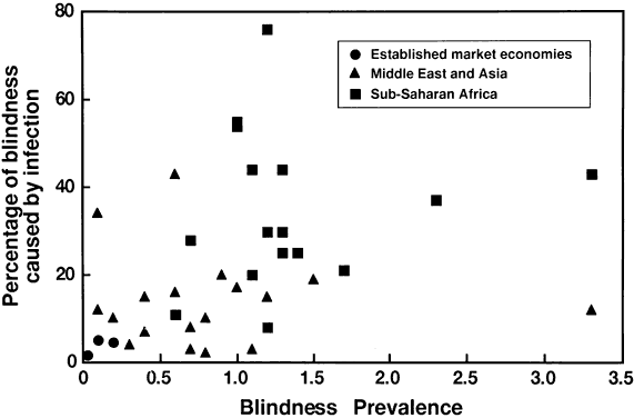
The impact of infections on childhood blindness is also substantial. There are an estimated 1.5 million blind children in the world; about one third are blind as a result of infection,69 mainly complications of bacterial conjunctivitis of neonates70 and childhood trachoma. The effect of blindness prevention programs would be substantial if eye infections could be reduced or eliminated among children. Correlational Studies Geographic ophthalmology, a term originating in the first half of the 20th century, is the comparison of different population groups to study associations between potential causes and effects. An ecologic or correlational study compares disease frequency in a group having a specific characteristic with those lacking it. An example of a correlational study in ophthalmology is the apparent correlation between a country's gross national product and the proportion of blindness caused by infection (Fig. 17). However, because these aggregate data may not represent the association at an individual level, it would be a potential fallacy to infer that an individual's income influences the individual's chance of getting or being blinded by an ocular infection. ANECDOTAL EPIDEMIOLOGIC STUDIES Case Reports Case reports have been used since ancient times to describe eye diseases and their treatments. For example, Galen used a case example to illustrate the benefit of bloodletting for acute ulcerative eye disease.71 The use of bloodletting was widely practiced in the early 19th century for treating various ophthalmias. It subsequently quickly declined, not so much because of epidemiologic studies but as a result of a general consensus that was based on clinical experience. Case reports make up the bulk of published information on ocular infections. At least one half of published reports of human ocular infections are reports of individual cases. These descriptive studies are the most frequent types of clinical research reports in the ophthalmologic literature and can give clues to the determinants of disease, disease severity, and outcome. History-taking is the most common method for identifying retrospectively exposures that may cause disease. Evaluating potential associations requires analytic studies. Case Series Groups of cases can highlight aspects of an exposure or a disease. For example, reported cases of P. aeruginosa keratitis caused by contaminated fluorescein eyedrops led to manufacturing guidelines for preparing sterile ophthalmic solutions.72 Descriptive studies of patients cannot reliably prove disease determinants. For example, a mid-19th century series of 1659 hospital cases of conjunctivitis found that 70% of the patients had gray or blue irides compared with 30% who had brown or hazel eyes.18 Studying an association between eye color and conjunctivitis would require an analytic epidemiologic study. Any risk factor suggested by a chart review must be questioned. A common example is a trend associating ocular disease with age. Case series are based on patients that an ophthalmologist sees. However, age distribution depends on the number of living persons in whom a disease can occur. Rather than giving numbers of cases in each age group, age-risk curves are better reported as population-based rates. Developments in analytic epidemiology have evolved to distinguish spurious correlations from true causal associations. ANALYTIC EPIDEMIOLOGIC STUDIES Case-Comparison Studies The use of epidemiologic methods in ophthalmology is relatively recent. The case-comparison approach, developed during the mid-20th century, compares cases selected by diagnostic criteria to controls who are randomly or systematically sampled from the same population. Case-control studies were first used in ophthalmology during the 1950s to link maternal rubella with congenital ocular malformations. Patients with a specific infection are thus compared with noninfected individuals to determine the potential effect of microorganisms and risk factors. By carefully defining case criteria and selecting suitable controls, associations between exposure and infection can be assessed even for relatively uncommon conditions.73 For example, a case-control study showed that overnight wear of soft contact lenses was a major risk factor for ulcerative keratitis.74 The case-comparison method has the advantage of being able to evaluate multiple etiologic factors for relatively uncommon conditions. Because incidence is not measured by this study design, the odds ratio is used to estimate the relative risk, assuming subjects are representative of diseased and control populations, incident cases are included, and the disease is rare. Case-control studies are limited by potential biases in selection and observation. One way to reduce confounding is to select controls by matching age, gender, or other variables that are not being studied in disease pathogenesis. Matching must be considered in the analysis. Cohort Studies A cohort study differs from a case-comparison study in that people without the disease are initially classified into an exposed and an unexposed group before they are followed up to ascertain the occurrence of disease (Table 20). Individuals with a suspected risk factor or exposure can be compared with others who are not affected or exposed to determine any association with the subsequent development of disease. In addition to being exposure-based, cohort studies are actually or conceptually longitudinal.
TABLE 43-20. Observational Studies in Epidemiology
*a, number exposed and diseased; b, number exposed and nondiseased; c, number unexposed and diseased; d, number unexposed and nondiseased.
The use of hospital or clinic records, in which the investigator selects individuals on the basis of a possible risk factor or other exposure, formed the basis for one of the first analytic epidemiology studies in ophthalmology (Table 21). In a prospective cohort study the investigator concurrently follows up persons into the future for a given period. For example, a prospective cohort study demonstrated how CD4 levels affect the risk of cytomegalovirus retinitis among HIV-infected persons.75
TABLE 43-21. Retrospective Cohort Study of the Risk of Neonatal Conjunctivitis, Stockholm, 1832
Data from Cederschjöld PG: On ophthalmia neonatorum. London Medical Gazette 27:382, 1840
A case-control study can form the basis for a cohort study. For example, after evaluating the risk factors for presumed ocular histoplasmosis syndrome (POHS),76 patients were followed for loss of visual acuity.77 POHS was considered as the disease in the case-control study and as the exposure in the cohort study. Cohort studies may be based on a population sample or can be selected from a particular group; certain eye diseases have been studied in the United States in Framingham, MA,78 Beaver Dam, WI,79 and Olmstead County, MN;80 and in Göthenburg, Sweden, as well as elsewhere. Although validity issues are of concern, private insurance claims databases and publicly available large databases, such as those maintained by the U.S. Health Care Finance Administration for Medicare inpatients (Medical Provider Analysis and Review) and outpatients (Part B Hospital Outpatient Facility), provide opportunities for follow-up studies in ophthalmology.81 EXPERIMENTAL EPIDEMIOLOGIC STUDIES A clinical trial is a controlled human experiment in which the investigator assigns the study treatment to persons having a specific condition. A clinical trial involves two or more treatment groups, as opposed to single-group, noncomparative “try-outs” to assess preliminary efficacy and safety. Most therapeutic trials in ocular infections have evaluated the efficacy of an anti-infective or anti-inflammatory agent for viral and bacterial diseases. Preventive trials, on the other hand, aim to reduce the risk of developing disease through the use of a prophylactic medication or procedure. Therapeutic trials thus study individuals with a given disease, and preventive trials study people at risk for a disease. A therapeutic trial can be classified as phase I if it primarily studies safety and pharmacokinetics among volunteers; as phase II or III if it assesses efficacy with limited or extensive numbers of subjects, respectively; and as phase IV if the study evaluates postmarketing safety or pharmacologic profile. Clinical trials begin with an expectation of benefit tempered by a reasonable doubt regarding efficacy that is called clinical equipoise. This ethical background to a trial, along with what is to be expected from patients, is part of informed consent.82 Use of randomization and masking (a term preferred to blinding83) reduce bias and confounding and help make the clinical trial a definitive tool for testing a scientific hypothesis in clinical practice. Key features of well-designed clinical trials that seek to minimize selection and observation bias are prospective observation, standardization of procedures and personnel, periodic monitoring, and ascertainment of one or more well-defined end points.84,85 An adequate sample size of compliant patients is needed. Other issues to consider in developing a clinical trial are its public health importance, feasibility, and cost. Although an ordeal, trials are indispensable in resolving uncertainty for major ophthalmologic dilemmas.86 When multiple studies are performed, systematic synthesis of several studies is possible. This may take the form of a literature review or a metaanalysis. Developed in 1976, this clinical research technique combines results from different clinical trials to achieve a comprehensive measure of a treatment's efficacy and safety. The Cochrane Collaboration has created guidelines for systematic reviews in ophthalmology (see http://www.cochrane.org).87 Decision modeling, economic analysis, and other methods can quantify the effectiveness of medical interventions.88 |
EPIDEMIOLOGY OF COMMON EYE INFECTIONS | ||||||||||||||||||||||||||||||||||||||||||||||||||||||||||||||||||||||||||||||||||||||||||||||||||||||||||||||||||||||||||||||||||||||||||||||||||||||||||||||||||||||||||||||||||||||||||||||||||||||||||||||||||||||||||||||||||||||||||||||||||||||||||||||||||||||||||||||||||||||||||||||||||||||||||||||||||||||||||||||||||||||||||||||||||||||||||||||||||||||||||||||||||||||||||||||||||||||||||||||||||||||||||||||||||||||||||||||||||||||||||||||||||||||||||||||||||||||||||||||||||||||||||||||||||||||||||||||||||||||||||||||||||||||||||||||||||
Details of the world's leading ocular infectious diseases are presented
in the CD-ROM version of this chapter. Table 22 summarizes some of the key determinants of these infections. Additional
information on prevalence and incidence, with references to literature
reports and Internet links, are also included in the computerized edition.
TABLE 43-22. Epidemiologic Features and Determinants of Ocular Infections
|
EPIDEMICS AND OUTBREAKS OF COMMON EYE INFECTIONS | ||||||||||||||||||||||||||||||||||||||||||||||||||||||||||||||||||||||||||||||||||||||||||||||||||||||||||||||||||||||||||||||||||||||||||||||||||||||||||||||||||||||||||||||||||||||||||||||||||||||||||||||||||||||||||||||||||||||||||||||||||||||||||||||||||||||||||||||||||||||||||||||||||||||||||||||||||||||
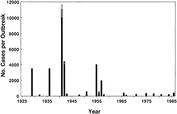
ADENOVIRUS CONJUNCTIVITIS Outbreaks of epidemic conjunctivitis were reported in Europe since the early 1700s.18 Around 1800, an acute ophthalmia spread among European militia and sailors. Mistakenly called Egyptian ophthalmia because it was presumed to have come from the Middle East, this contagious disease was largely confined to closed communities such as the military and students in boarding schools (Fig. 18). Once suspected to be trachoma, the rapid spread and communicability of this epidemic keratoconjunctivitis is more compatible with infection due to adenovirus or enterovirus, perhaps complicated by secondary bacterial infection caused by traditional treatment practices. For example, a gripping account of a highly communicable keratoconjunctivitis aboard a French slave ship in April, 1819, was probably, in retrospect, a viral rather than chlamydial infection, apparently made worse by traditional eye medicines.89 If adenovirus was a biologic cause of epidemic eye disease in the 19th century, crowding and international sea travel were its social determinants. Trading communities, with congested housing and poor sanitation, were susceptible to the spread of many communicable diseases. Historical epidemiology is needed to deduce when and where adenoviral conjunctivitis began. The clinical features of epidemic keratoconjunctivitis were first delineated during an epidemic in central Europe, occurring in 1889. As distances shrank in the 20th century, several sizable outbreaks occurred (Fig. 19). Coastal cities were affected during World War II. More than 100 major outbreaks of epidemic keratoconjunctivitis were reported during the 20th century in Europe, Asia, North America, and Australia.90,91 Most reports from 1900 to 1950 described large community epidemics; more recent reports evaluated the risk factors associated with outbreaks in occupational and medical settings. Epidemic outbreaks have occurred in prisons, military posts, refugee camps, children's camps, unchlorinated swimming pools and ponds, schools, factories, nursing homes, and hospitals. The incidence of the secondary spread of adenovirus during outbreaks varies widely, depending on the setting. The incidence of adenovirus conjunctivitis after exposure is generally low in the general population but much higher in households or institutions (Table 23), presumably because the likelihood of direct or indirect contact is greater. The secondary attack rate generally ranges from 5% to 20% for close contacts of infected individuals but can be 25%, 50%, or higher when a common source is involved. Most large outbreaks are caused by a single serotype, but two or more types have occasionally been found.
TABLE 43-23. Reported Attack Rates of Adenovirus Conjunctivitis
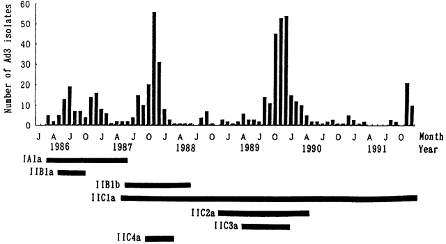
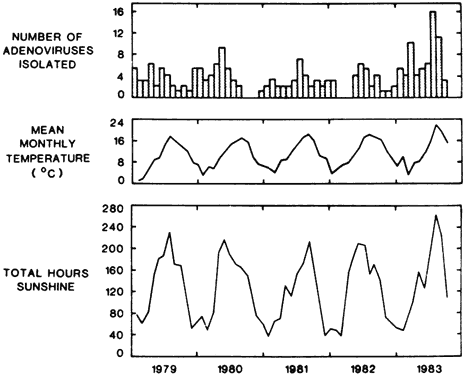
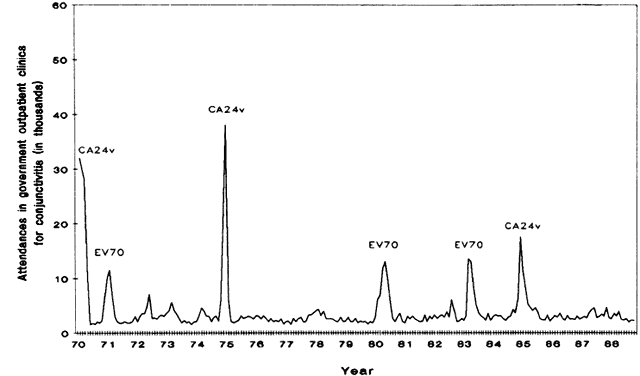
Types 3, 4, and 7 are prevalent causes of pharyngoconjunctival fever; types 8, 19, and 37 are most frequently associated with epidemic keratoconjunctivitis. Adenovirus serotypes occur sporadically over several years in many areas. Specific serotypes may recur in a cyclic pattern (Fig. 20). Genotype changes may account for the emergence of intermediate strains and may play a role in epidemic outbreaks in a susceptible population. Frequency can rise in warm months.92 The relationship of climate to the frequency of adenovirus conjunctivitis (Fig. 21) may be related to methods of transmission. ENTEROVIRUS CONJUNCTIVITIS The first recognized epidemic attributed to enterovirus 70 began in Ghana during 1969 (Fig. 22) and subsequently spread across Africa and the Middle Eastern crescent (Fig. 23), possibly associated with pilgrimages to Mecca. A parallel epidemic caused by enterovirus 70 also erupted in Indonesia in 1969. Another picornavirus, coxsackievirus A24, was first recognized as a cause of acute conjunctivitis causing an epidemic in Singapore in 1970. Both viruses have caused recurring epidemics in Asia (Fig. 24) and spread to Europe and the Americas by travelers and refugees. Several large outbreaks of enterovirus or coxsackievirus conjunctivitis have occurred in the late 20th century, predominantly in the coastal tropics of Africa, Asia, and Central America (Table 24). Small outbreaks and sporadic cases have been recognized in several cities of Europe, the Middle East, Asia, Australia, North America, and South America, and in laboratory accidents. Mixed epidemics of enterovirus 70 and coxsackievirus A24 and of enterovirus 70 and adenovirus have occurred.
TABLE 43-24. Countries Experiencing One or More Epidemics of Enterovirus 70 or
Coxsackievirus A24 Conjunctivitis Africa and Middle East Algeria Asia and Pacific Islands American Samoa Americas and Caribbean Antigua-Barbuda
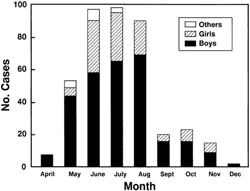
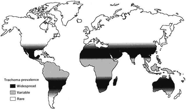
During an epidemic, reported attack rates range from 19% to 68.5%. In an outbreak in American Samoa, 48% of the population was affected. The population-based incidence rate during an epidemic in Morocco was 53 cases per 1000 people younger than 20 years. Household attack rates increase according to family size because of crowding and the sharing of beds, and can exceed 70%. Crowding also probably explains different attack rates among various socioeconomic groups. Epidemics do not have a clear-cut seasonal pattern (Fig. 25), although outbreaks in Latin America tend to occur in the hot, rainy season. TRACHOMA Chlamydial eye disease has probably been endemic in Egypt and other ancient civilizations for centuries. Once believed to be the cause of epidemics in the late 18th and early 19th centuries affecting soldiers and sailors,93 viral conjunctivitis and secondary bacterial infection is a more reasonable explanation for “military ophthalmia.”94 Trachoma does not usually spread in an acute fashion. Rather, recognizing an “epidemic” of Chlamydia trachomatis conjunctivitis requires a public health viewpoint. The disease occurs on all continents other than Antarctica and, until the mid-20th century, was prevalent in Europe and North America. Trachoma belts extend around the world (Fig. 26). Besides climate, prevalence is closely linked with socioeconomic development. In endemic areas, there is often familial clustering of active trachoma. In addition to sharing risk factors, genetic susceptibility may predispose to chlamydial infection and to trachomatous scarring.95 The blinding sequelae of corneal scarring and other complications of trachoma develop in a minority of individuals. Rates of hyperendemic trachoma were prospectively assessed during vaccine trials of the 1960s96–106 (Table 25). In the placebo groups, clinical signs of trachoma appeared in 10% to 30% of children during the first year of follow up. Within the next few years, trachoma will develop in about one half of all uninfected children in an endemic area. Neither prophylactic nor therapeutic vaccines have yet proven clinically effective. Childhood trachoma remains prevalent in endemic, underdeveloped regions of the world (Table 26).
TABLE 43-25. Prospective Cohort Studies of Tachoma in Hyperendemic Areas
*Development of trachoma in previously uninfected, placebo-treated children. †Vaccine efficacy assessed after 1 year.
TABLE 43-26. Prevalence of Active Childhood Trachoma in Rural Populations 1981-1991*
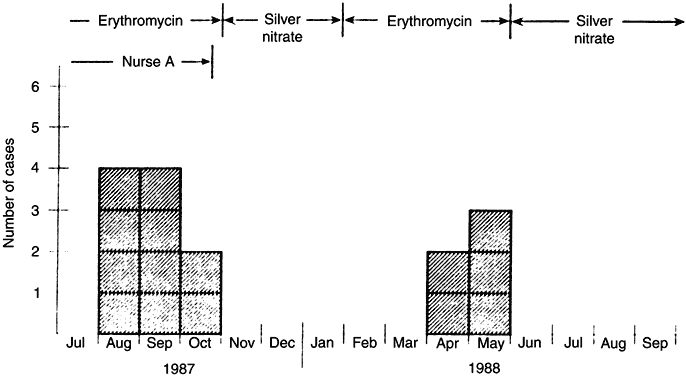
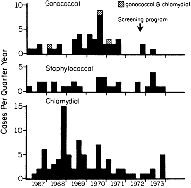
BACTERIAL CONJUNCTIVITIS Outbreaks of bacterial conjunctivitis are unusual but have occurred in day care centers,107 boarding schools,108 universities,109 dormitories, military camps, and nursing homes.110–112 Hospital outbreaks have occurred in nurseries, inpatient wards,113 intensive care units,114 and long-term nursing facilities.115 Young children are at greatest risk when epidemic bacterial infections spread within crowded households and day care centers. Determining that a bacterial species is responsible for a cluster of conjunctivitis cases can be difficult.116 Bacteria causing outbreaks of nonsexually transmitted conjunctivitis include several strains of Haemophilus influenzae,107,117,118 S. pneumoniae,109,119 Moraxella species,108,116 and Neisseria gonorrhoeae.120,121 School outbreaks of phlyctenular blepharokeratoconjunctivitis, presumably due to S. aureus, have also occurred.122 Contaminated traditional remedies can spread bacterial infection.123 Besides a common-source outbreak, an increased frequency of cases can result from an increase in oculogenital disease, such as the epidemics of gonococcal keratoconjunctivitis that occurred in Africa in the 1980s.124,125 Gonococcal conjunctivitis in a child raises the question of neglect or abuse.107 No cyclic variation has been established, although the frequency may increase in colder months.90 H. influenzae conjunctivitis and preseptal cellulitis are more common during summer and early autumn and, in warm climates, may occur in seasonal epidemics. CONJUNCTIVITIS IN NEONATES Before Credé, neonatal conjunctivitis was very common. The frequency of infective conjunctivitis in neonates changed with the use of anti-infective ocular prophylaxis at birth and with better prenatal care of the expectant mother. However, resurgence of chlamydial or gonococcal conjunctivitis of neonates still occurs. Each case of neonatal conjunctivitis represents a cluster of family members and associates with sexually transmitted disease.126 Nursery outbreaks have been reported in which infections have stemmed from a common source127–139 (Table 27). In one outbreak, 7% to 14% of infants acquired S. aureus conjunctivitis (Fig. 27). In two others, P. aeruginosa conjunctivitis developed in as many as 8%.
TABLE 43-27. Reported Outbreaks of Bacterial Conjunctivitis Among Neonates
After 1970
HNN, hospital newborn nursery; NICU, neonatal intensive care unit.
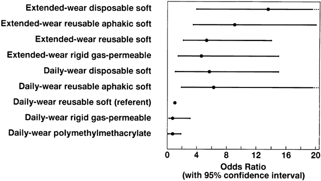
Gonococcal conjunctivitis develops in from 30% to 42% of newborns who are exposed to N. gonorrhoeae during delivery. Between 25% and 50% of neonates exposed to C. trachomatis during delivery acquire chlamydial conjunctivitis. The attack rate during a common-source outbreak in a newborn unit can exceed 10%. Seasonal variations in neonatal conjunctivitis correspond to fluctuations in maternal gonorrhea and chlamydiosis. Because the rate of adult gonorrhea peaks during late summer and fall in the United States, so does gonococcal neonatal conjunctivitis (Fig. 28). VARICELLA ZOSTER VIRUS DERMATOBLEPHARITIS Varicella zoster virus (VZV) outbreaks are common in closed communities such as schools, camps, and hospitals, where many nonimmunized children are exposed to an infected individual. VZV does not occur in an epidemic pattern, although a rising incidence is following the AIDS epidemic. Rare outbreaks of shingles, with an attack rate of 5% to 10%, have followed re-exposure to VZV. For chickenpox, secondary attack rates among susceptible household contacts is 70% to 90%. Chickenpox peaks in winter and early spring in the northern hemisphere; VZV has no seasonal variation. HERPES SIMPLEX VIRUS KERATITIS Outbreaks of primary HSV infection can occur among nonimmune individuals who are intimately exposed to another infected person. Most epidemics have occurred at schools, camps, and hospitals but infrequently involve the eye.140 In an outbreak among wrestlers attending a training camp, 34% were infected and 8% had primary herpetic blepharoconjunctivitis.141 Outbreaks have also occurred among rugby players and other athletes. MICROBIAL KERATITIS When proprietary eyedrops came into widespread use in the mid-20th century, some solutions such as fluorescein became contaminated and led to cases of pseudomonal keratitis.72 In-use contamination still occurs, but common-source outbreaks of keratitis in developed countries are rare. In many parts of the world, however, traditional medications used for treating the eye remain an important cause of corneal ulceration.142 A similar sequence of events occurred with the development of contact lenses. During the early years of production, some soft lenses were apparently contaminated at the manufacturer by P. aeruginosa or filamentous fungi and led to clusters of microbial keratitis when the lenses were fitted. Improper handling by an eye care practitioner is still a potential source for an outbreak of corneal infection.143 The widespread use of contact lenses and the associated increased frequency of microbial keratitis during the 1980s was described as an epidemic144 and led to a change in the spectrum of microorganisms causing corneal infection in developed countries. P. aeruginosa, Acanthamoeba species, and other waterborne organisms that could survive in contact lens fluids emerged as important infections during this time. The public health impact of contact lens-associated microbial keratitis is substantial, resulting in more than a 400% increase in corneal infections in the United States from 1950 to 1990.145 Several case-comparison studies have compared the relative odds of microbial keratitis for different contact lens types and wearing schedules (Fig. 29). Clusters of corneal infection have also occurred in obtunded, hospitalized patients related to tracheal suctioning and a poor blink reflex.114,146 OCULAR LEPROSY The distribution of leprosy is nonhomogeneous even in an endemic locale. Outbreaks of leprosy in families are often linked with the social and cultural context. In a family with an untreated lepromatous individual, skin lesions develop in up to 50% of that person's children, and approximately 5% to 10% of family members acquire leprosy. OCULAR ONCHOCERCIASIS Epidemic spread of onchocerciasis is measured in decades. In equatorial West Africa, a sequence of infection and blindness has led to a repeated pattern of migration of the population. Tribes settle in fertile river valleys and remain until the level of river blindness reaches an unsustainable level. They then abandon their villages and move to the hills, but move back to the rivers after several generations. This cycle may have gone on for centuries.147 Environmental and climatic changes play a role in the spread of onchocerciasis.148 It is possible that onchocerciasis spread to the Americas with the slave trade, appearing in regions where infected Africans were sent and blackfly vectors already existed. TOXOPLASMIC RETINOCHOROIDITIS Outbreaks of acquired toxoplasmosis are uncommon but have been associated with dust mixed with cat excrement, fecally contaminated water, infected meat or milk, and laboratory accidents (Fig. 30). The ophthalmologist may be the first to recognize a common-source epidemic.149 Epizootics have occurred at animal farms, in captive colonies, and in zoos. In a common-source outbreak of acquired toxoplasmosis, 18% to 42% of exposed people seroconvert. The cumulative risk of retinitis among those infected during a common-source outbreak is about 0.5%,149 although in one outbreak, 4% developed ocular toxoplasmosis.150 Pregnant women who acquire toxoplasmosis can transmit the infection to their fetus's retina.151 CYTOMEGALOVIRUS RETINITIS Cytomegalovirus retinitis does not occur in common-source outbreaks. A rising incidence followed the onset of the AIDS epidemic in the early 1980s then peaked at the end of the 20th century. MICROBIAL ENDOPHTHALMITIS Before 1865, endophthalmitis and other üblen Zufällen [unfortunate coincidences] occurred in 500 to 1100 cases per 10,000 cataract operations. The risk of endophthalmitis after cataract surgery fell with the introduction of antisepsis and improved surgical techniques in the late 19th century and has continued to decline before and during the antibiotic era (Fig. 31). The epidemiology of cataracts and cataract surgery has been studied,150 but large, prospective studies are needed. The postoperative rate of endophthalmitis varies slightly by intraocular procedure151 (Table 28). Compared with cataract surgery, the risk is probably greater for secondary intraocular lens implantation, pars plana vitrectomy, penetrating keratoplasty, and trabeculectomy. The median visual acuity after treatment for postsurgical endophthalmitis is 20/200.
TABLE 43-28. Risk of Microbial Endophthalmitis After Intraocular Surgery
After 1975
*(From Aaberg TM Jr, Flynn HW Jr, Schiffman J, Newton, J: Nosocomial acute-onset postoperative endophthalmitis survey. Ophthalmology 105:1004, 1998.) †Includes delayed-onset infections.
Most common-source outbreaks of endophthalmitis are related to contamination introduced during intraocular surgery62,154–167 (Table 29). Clusters of endogenous infection have also occurred. For example, endophthalmitis and other infections among neutropenic patients was traced to a contaminated skin lotion,168 and another cluster of fungal endophthalmitis followed exposure to a contaminated intravenous anesthetic agent.169 Sharing syringes among drug addicts can also produce outbreaks of fungal endophthalmitis.170 Determining whether an outbreak is171 or is not172 due to a common source is aided by techniques of molecular epidemiology.
TABLE 43-29. Reported Common-Source Outbreaks of Postsurgical Exogenous
Endophthalmitis after 1975
The frequency of intraocular infection after exposure to a contaminated irrigating solution or intraocular lens is not known. In a widespread outbreak of fungal endophthalmitis, infection developed in 4% of those exposed to a contaminated intraocular lens.157 However, defining an outbreak by time and place can be difficult. The paucity of reports of common-source epidemics of postsurgical endophthalmitis173 may belie its frequency. There is no seasonal fluctuation, although posttraumatic infection may be greater in warmer months.174 |
PREVENTION OF OCULAR INFECTIONS | |||||||||||||||||||||||||||||||||||||||||||||||||||||||||||||||||||||||||||||||||||||||||||||||||||||||||||||||||||||||||||||||||||||||
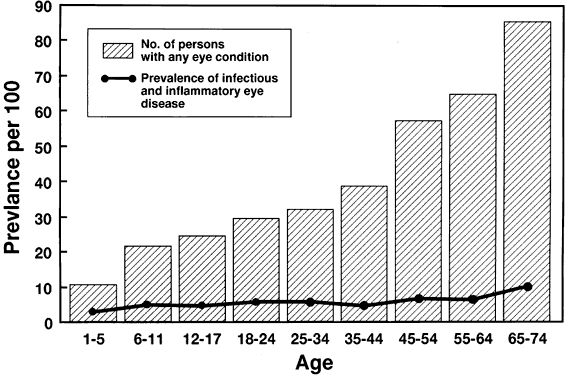
Infections account for much of the world's avoidable blindness.175 Most occur in less-developed areas. In an economically advantaged society, the
prevalence of infectious and inflammatory eye disease is about 5%, of
which 1 in 500 results in visual loss.176 Sanitation and other preventive strategies can help reduce the burden
of ocular infections, although chronic disorders associated with aging
then become relatively more important (Fig. 32). The goal of epidemiology is to discover causal associations that offer opportunities for disease prevention. To aid in decision making, ophthalmologists categorize infections by their clinical manifestations. Epidemiologists may group people by causal criteria, such as contact lens wear or foreign body injury, that might be targeted in a prevention program. Ideally, individuals could be identified before ocular infection occurs, and their risk classified as high, moderate, or negligible. Knowledge of disease pathogenesis can then allow preventive strategies to be tailored to specific situations and circumstances. PRIMARY PREVENTION Prevention of ocular infections can occur at multiple levels (Table 30). Many cases of blinding ocular infection are avoidable by primary and secondary prevention (Fig. 33). Recommendations for public health ophthalmology must judge the relative advantages and shortcomings of each alternative (Table 31).
TABLE 43-30. Levels of Prevention in Ophthalmic Epidemiology
TABLE 43-31. Strategies in Preventive Ophthalmology
Practices that are in widespread use by ophthalmologists are universal precautions,177 aseptic and no-touch techniques with barrier protection during surgical procedures,178 and prophylactic anti-infective agents for patients at increased risk.179 Examples of institutional programs that prevent the spread of pathogens are good manufacturing practices by ophthalmic pharmaceutical companies and serological donor screening programs by eye banks. Community-wide programs in preventive ophthalmology that involve public health agencies include the Onchocerciasis Control Program, which aims to control the blackfly vector in endemic areas of Africa180 and trachoma control programs (Fig. 34) that involve both primary and secondary prevention.181,182 Behavior Ways to reduce the risk of acquiring or transmitting infection should be promoted among patients exposed to a known or suspected risk factor for ocular infection. For example, adherence to recommended hygiene practices among contact lens wearers reduces corneal complications. Similarly, improvements in facial cleanliness and avoidance of shared towels reduces trachoma in an endemic area. Education begins by making the public aware of prevalent ocular infections and the corresponding risk factors. Some ocular infections may even be a sentinel for unsafe practices, such as folk remedies, that need to be addressed in educational efforts. At a community level, educators concerned with health maintenance should discuss eye safety recommendations to prevent recreational and sports trauma. Segregation Identification of infected patients permits reducing their direct and indirect contact with susceptible persons. Levels of preventive strategies can be recommended to limit microbial spread in the family and at the workplace. In the eye care setting, segregation of patients with symptomatic conjunctivitis to a so-called red eye room, deferring nonessential procedures such as tonometry, and avoiding unnecessary follow-up can reduce the chance of initiating an office outbreak. Segregation and other control measures can reduce the potential for spread whenever there is concern of exposing a large number of people to a highly infectious condition.* Counseling the patient with a communicable ocular infection can avert risky behavior and reduce the number of contacts. Based on the National Quarantine Act of 1878 that excluded any “person suffering from a loathsome or dangerous contagious disease”, the United States federal government assumed responsibility in 1891 for the medical inspection of immigrants. Screenings were performed by physicians of the US Public Health Service at immigration stations such as Ellis Island. A button-hook, used to fasten high-top shoes, helped to evert eyelids to check for trachoma and was cleaned with a Lysol-soaked towel between exams. Prospective immigrants seen to have a possible defect would be marked with chalk on their clothing: “C” for conjunctivitis, “CT” for trachoma, and “E” for eye disease. Some cases of ocular infection were hospitalized, at their own expense or assisted by an immigrant aid fund, until the condition resolved; others were sent home on a return ship. More restrictive immigration laws were enacted in 1924. Emigrants are now usually examined before embarkation by local physicians approved by the US Immigration and Naturalization Service. Sanitation Hygiene is among the most important elements of preventive ophthalmology. Hand washing using liquid soap dispensers and either disposable paper towels or hot air blowers are probably the most easily used preventive measures in the eye clinic. Door handles, telephones, and many other shared devices offer opportunities for improved design. The use of disposable gloves or fingerlings or a no-touch technique with disposable applicator sticks to manipulate the eyelids is recommended for examining infected eyes. All contact instruments, especially tonometer tips, need to be cleaned and sterilized between patients. Diagnostic instruments that come into contact with ocular secretions should also be disinfected after each use. Eyedropper tips should not come in contact with the patient's ocular surface or tear film. Multiuse ophthalmic solutions can be periodically discarded or replaced with unit doses. Special precautions are needed during office outbreaks of communicable ocular infection.20,183 On a global scale, most eye infections occur in less developed regions. Haphazard urbanization without a sanitary infrastructure breeds infectious outbreaks. Improving crowded and dirty living conditions among the economically disadvantaged can reduce the incidence of infectious eye diseases. International agencies that accelerate economic development make a large impact on a society's ocular health. ANTI-INFECTIVE PROPHYLAXIS The relative benefits and disadvantages of using topical ophthalmic antibiotics for a noninfected patient need to be carefully weighed, especially if used routinely and often. Special-exposure groups need to be identified for cases in which anti-infective agents might prevent initial or recurrent infection. In addition, although antimicrobial therapy is generally considered secondary prevention, antiinfective treatment of patients with a communicable infection can reduce transmission. Neonatal Prophylaxis The first demonstration of the use of topical antisepsis for preventing purulent conjunctivitis in neonates was by Credé (Table 32). Public health policy is still evolving on the role of instilling a prophylactic agent into the eyes of all newborn infants because maternal screening is also an effective strategy for preventing ophthalmia neonatorum. In a locale lacking universal prenatal care, an antiinfective preparation should be instilled onto the eyes of every neonate as soon as possible after delivery.184
TABLE 43-32. Comparison of the Annual Incidence of Acute Conjunctivitis
in Neonates, Leipzig, 1874-82
Perioperative Prophylaxis The first demonstration of the efficacy of an antiseptic agent for reducing the risk of postoperative endophthalmitis was by von Graefe.185 From 1877 to 1884, the frequency of endophthalmitis after cataract surgery ranged from 4.7% to 6.25% per year; subsequent use of a 1:5000 sublimate solution reduced the proportion infected to 1% to 2%. In spite of the lack of a prospective cohort study or randomized clinical trial, multiple topical, subconjunctival, systemic, and intraocular antimicrobial agents have been widely used ever since. Antiviral Prophylaxis An antiviral agent is used during corticosteroid therapy of HSV keratitis to prevent recurrent epithelial infection.186 Chronic suppression with an antiviral agent can also prevent various forms of HSV recurrence in patients with a previous episode. The development of a safe and an effective prophylactic agent for adenovirus and other viruses would offer a way to limit spread of communicable viral conjunctivitis in a susceptible population. IMMUNIZATION AND OCULAR INFECTION Immunization programs against viral infections contribute to the prevention of several forms of ocular infection (Table 33). Identifying the proportion of the population that needs to be immune to prevent the development of an epidemic of a specific viral disease is a basis for setting vaccination policy. To prevent congenital rubella syndrome by vaccination during early childhood, for example, approximately 85% of the susceptible population must be immunized.
TABLE 43-33. Vaccines for Preventing Diseases That Have Ocular Manifestations
The effect of systemic immunization against bacterial ocular infection is less clear. Immunization against H. influenzae, S. pneumoniae, and other ocular pathogens may or may not beneficially alter the conjunctival colonization of these microbes or affect the risk of ulcerative keratitis, bleb-associated endophthalmitis, and other eye infections caused by these organisms in susceptible individuals. A better understanding of mucosal immunity offers opportunities for developing improved ophthalmic vaccines for common ocular diseases. Vaccination can potentially be used for the following:
Topical ocular immunization may have an advantage over systemic administration (Table 34). Development toward a vaccine for Moraxella bovis conjunctivitis in cattle, experimental work on immunizing against P. aeruginosa and other bacterial keratitis, and success with DNA vaccines in various animal models offer emerging opportunities for preventing infectious diseases of the eye.
TABLE 43-34. Differential Effects of Immunization by Route
|