Chapter 3
Nongranulomatous Inflammation: Uveitis, Endophthalmitis, Panophthalmitis, and Sequelae
RUSSELL W. READ, EHUD ZAMIR and NARSING A. RAO
Main Menu   Table Of Contents

Search

CELLULAR COMPONENTS OF INFLAMMATION
SOLUBLE COMPONENTS OF THE IMMUNE SYSTEM
IMMUNE PRIVILEGE IN THE OCULAR ENVIRONMENT
CLASSIFICATION OF INFLAMMATION
UVEITIS
ENDOPHTHALMITIS AND PANOPHTHALMITIS
SEQUELAE
REFERENCES

Inflammation can be characterized as a process or series of processes mounted by an organism in response to a stimulus interpreted to be hazardous or undesirable. The stimulus may be extrinsic to the organism, such as an invading microorganism, or may be a misdirected attack against an intrinsic component of the organism itself, such as in autoimmunity. The resultant inflammatory processes function to contain and destroy the stimulus, then effect repair of any tissue that may have been damaged by the stimulus itself or the organism's response to the stimulus. Organisms have multiple options in accomplishing this response, and together these options make up what is known as the immune system.

Many different components make up the immune system, including cellular elements such as neutrophils, lymphocytes, and macrophages, and soluble elements such as cytokines, complement, and antibodies. Each of these components has unique but interdependent functions within the immune system. For example, cytokines (i.e., small proteins that serve as intercellular signals) such as interleukin-2 (IL-2) are produced by leukocytes to stimulate and attract other cellular elements to the area of an insult. These recruited cells then produce additional cytokines, antibodies, and so forth, which act to contain and destroy the stimulus. These components are required for normal functioning of the immune system.

Back to Top
CELLULAR COMPONENTS OF INFLAMMATION
The inflammatory response is dependent on the actions of many different cell types. These cells express varied surface markers that serve to identify them and allow them to interact with other cells to perform their various functions.

MAJOR HISTOCOMPATIBILITY COMPLEX

The major histocompatibility complex (MHC) is a region of genes that encodes for the production of small proteins found on the surface of cells. These proteins serve to present antigen in a recognizable form to other effector cells of the immune system, such as T lymphocytes. The term major histocompatibility complex is derived from the original description of these molecules in relation to their function in the rejection of transplanted tissues. The MHC system is made up of three subclasses: I, II, and III. All are located on chromosome 6. MHC class I molecules are found on all nucleated cells. This molecule class functions in the regulation of the actions of CD8+ cytotoxic T cells, especially in the defense against viral infection. Viral antigen present within a cell is processed and presented on the cell surface in association with an MHC class I molecule. This presentation serves to activate CD8+ T cells (see description below) to destroy the infected cell, thus preventing further viral production. MHC class II molecules are present only on certain cells of the immune system, such as macrophages and other antigen-presenting cells. Processed antigen is presented in association with MHC class II molecules to CD4+ helper T cells. MHC class III molecules consist of complement and are not true histocompatibility markers. These molecules play a role in the recruitment of neutrophils and other inflammatory cells and in the amplification of the inflammatory process.

The MHC system in humans is termed the human leukocyte antigen (HLA) system, because these molecules were first discovered on the surface of leukocytes. In the human, MHC class I antigens consist of HLA types A, B, and C. MHC class II molecules consist of HLA type D. It is now known that HLA molecules play an important role in an organism's susceptibility to certain inflammatory diseases. The exact mechanism by which HLA molecules predispose an individual to disease is not known, but several theories exist that are summarized in Table 1.


TABLE 1. Proposed Mechanisms of Human Leukocyte Antigen Disease Association

  Unique presentation of disease-inducing peptide
  Errors in development of the T-cell receptor repertoire
  Infectious agents that carry a molecular mimic of a self peptide
  Unmasking sequestered antigens that bind upregulated class II

(Davey MP, Rosenbaum JT: The human leukocyte antigen complex and chronic ocular inflammatory disorders. Am J Ophthalmol 129:235---243, 2000.)


INFLAMMATORY CELL TYPES

The hallmark of acute inflammation is the polymorphonuclear leukocyte, or neutrophil. The most numerous of the circulating leukocytes, neutrophils live an estimated 2 to 3 days in circulation. Their primary function is in phagocytosis of foreign material. Neutrophils are attracted by C5a (an activated complement component), IL-8, fibrin breakdown products, and neutrophil chemotactic factor, as well as other substances.1

Lymphocytes are the hallmark of chronic inflammatory infiltrates, although they may be present from the onset of inflammation. Lymphocytes are divided into B and T types as well as natural killer cells. After their production in the bone marrow from pluripotent stem cells, B and T lymphocytes undergo differentiation in the fetal liver/adult bone marrow and thymus, respectively. It is here that these cells gain the ability to recognize antigen. T cells are characterized by the presence of T-cell receptors (TCRs) on their surface, of which two main types exist. TCR-1 cells (5% to 10% of circulating T cells) function primarily as cytotoxic cells. TCR-2 cells (90% to 95% of circulating T cells) can be further divided into those that carry the CD4 surface marker and recognize antigen in the context of MHC class II molecules and those that carry the CD8 marker and recognize antigen in association with MHC class I molecules. The CD4 positive subset of T cell, also know as the helper T cell, can again be further subdivided into Th1 and Th2 type cells. Th1 cells produce IL-2 and interferon-γ and promote the functions of cellular immunity. Th2 cells produce IL-4, IL-5, IL-6, and IL-10, which help regulate the functions of macrophages and stimulate B cells to produce antibodies (humoral immunity).1,2

After stimulation by helper T cells in association with binding to their target antigen, B cells produce specific antibody directed to the antigen recognized. Further stimulation leads to their differentiation into plasma cells. As plasma cells produce antibodies, they may accumulate excess amounts within their cytoplasm, producing amorphous eosinophilic structures, referred to as Russell bodies.

Mononuclear cells provide important functions in the areas of antigen presentation and phagocytosis. Mononuclear cells are widely distributed in the body, both in circulating form as well as resident tissue monocytes/macrophages such as Kupffer cells in the liver, alveolar macrophages in the lung, serosal macrophages, and microglia in the brain and retina.1

In addition to the above cell types, eosinophils and basophils/mast cells participate in immune reactions as well, usually in relation to allergic stimuli or parasitic invasion (Fig. 1A).

Fig. 1. A. Photomicrograph of conjunctival biopsy specimen from patient with parasitic infection. Eosinophils are visible in the upper right corner of the image. Red-pink material at left of image represents the Splendore-Hoeppli phenomenon seen in such infections (hematoxylin and eosin; × 500). B. HLA-B27-associated acute anterior uveitis. Intense conjunctival injection, posterior synechiae, and hypopyon with admixed hemorrhage can be seen.

Back to Top
SOLUBLE COMPONENTS OF THE IMMUNE SYSTEM
In addition to the cellular elements described above, the immune system achieves its goal via production of numerous soluble factors that serve to carry out its functions. Compounds such as cytokines serve to signal between cells, some stimulating the system and others inhibiting it. Others such as complement serve both to attract the cellular elements as well as control invading organisms. Table 2 lists the sources and functions of some of the better-studied cytokines.


TABLE 2. Features of Selected Cytokines


CytokineCellular SourcesMain Cell Targets<st>Main Actions<st>
IFN-<gg>*T cells, NK cellsLymphocytes, monocytes, tissue cellsImmunoregulation, B cell differentiation, some antiviral action
TNF<ga>, TNF<gb>*Macrophages, lymphocytesFibroblasts, endotheliumInflammation, catabolism, fibrosis; production of other cytokines (IL-1, IL-6, GM-CSF) and adhesion molecules
IL-1<ga>, IL-1<gb>*Monocytes, dendritic cells, some B cells, fibroblasts, epithelial cells, endothelium, astrocutes, macrophagesThymocytes, neutrophils, T and B cells, tissue cellsImmunoregulation, inflammation, fever
IL-2*T cells, NK cellsT cells, B cells, monocytesProliferation, activation
IL-3*T cellsStem cells, progenitorsPan-specific colony-stimulating factor
IL-4*T cellsB cells, T cellsDivision and differentiation
IL-5*T cellsB cells, eosinophilsDifferentiation
IL-6*Macrophages, T cells, fibroblasts, some B cellsT cells, B cells, thymocytes, hepatocytesDifferentiation, acute phase protein synthesis
IL-8* (family)Macrophages, skin cellsGranulocytes, T cellsChemotaxis
IL-10*B cells, macrophages/microgliaB cells, macrophagesInhibits IFN-<gg> production, inhibits antigen presentation and IL-1, IL-6, and TNF-<ga> production
IL-12<sd>T cellsMacrophages, NK cellsInhibits Th2 responses and T-cell proliferation, decreases granuloma formation
IL-18<dd>T cellsT cellsActivates Th1 cells and induces IFN-<gg> production

INF, interferon; IL, interleukin; TNF, tumor necrosis factor; NK, natural killer; GM-CSF, granulocyte-monocyte colony stimulating factor.
*Information on IFN-<gg>, TNF, IL-1, -2, -3, -4, -5, -6, -8, and -10 modified from Roitt I, Brostoff J, Male D: Immunology. 4th ed. London: Mosby, 1996.
<sd>Information on IL-12 from Yang JQ, Tasaka K, Chuang CK et al: Dynamic analysis of T-lymphocyte function in relation to hepatopathologic changes and effect of interleukin-12 treatment in mice infected with Schistosoma japonicum. J Parasitol 85:257---262, 1999; and Taha RA, Minshall EM, Olivenstein R et al: Increased expression of IL-12 receptor mRNA in active pulmonary tuberculosis and sarcoidosis. Am J Respir Crit Care Med 160:1119---1123, 1999.
<dd>Information on IL-18 from Sugawara I, Yamada H, Kaneko H et al: Role of interleukin-18 (IL-18) in mycobacterial infection in IL-18-gene---disrupted mice. Infect Immun 67:2585---2589, 1999.
<st>Every function and target of each cytokine is not shown.


Back to Top
IMMUNE PRIVILEGE IN THE OCULAR ENVIRONMENT
To maintain its role as a functioning organ of sense, the eye has an absolute need for a clear visual axis, normal functioning of the delicate neural networks of the retina and optic nerve, and production of aqueous humor by the ciliary body. Therefore, whereas the ocular environment needs the protection of the immune system as detailed above, the eye cannot tolerate unchecked inflammation with its attendant destruction of tissue and repair by fibrosis. Evolutionary processes have thus driven the development of unique modifications to the immune system's standard repertoire as they relate to the eye.3 Briefly, tight junctions between the cells of the ciliary epithelium, retinal pigment epithelium, and retinal vascular endothelium inhibit penetration into the eye of molecules and cells from the circulation, thus creating a physical blood-ocular barrier.4–8 Lack of a lymphatic drainage system from the intraocular space results in the initial presentation within the spleen of antigen derived from the eye. In addition, the unique distribution of antigen-presenting cells within the eye,9 the presence of immunosuppressive factors within the aqueous humor,10 and the presence of cell surface molecules within the eye that modify the function of the immune system11 all contribute to a modified state of immunity. Initially described in relation to the anterior chamber, this modified immune state is known as anterior chamber-associated immune deviation (ACAID). It subsequently has been found to be present in the vitreous cavity and subretinal space as well.3

A more complete discussion of the various components of the immune system, the inflammatory cascade as it relates to the ocular environment, and its unique modifications is contained elsewhere in this series.

Back to Top
CLASSIFICATION OF INFLAMMATION
Multiple systems of nomenclature have been developed to describe the various causes, patterns, and spectra of ocular inflammation. These classification schemes are variably based on the relationship to the organism's environment (e.g., exogenous versus endogenous), anatomic area of involvement (e.g., anterior, posterior; cornea, retina, iris), temporal course (e.g., acute, delayed onset, chronic), etiologic agent (e.g., bacteria, viral, autoimmune), or specific histopathologic pattern (e.g., granulomatous or nongranulomatous; suppurative or nonsuppurative). In actual practice, a combination of these classifications typically is used.

RELATION TO ORGANISM

Stimuli can be classified as exogenous (i.e., coming from outside the organism) or endogenous (i.e., coming from within the organism). Under this scheme, a septic emboli from an infected heart valve that lodges in the eye and produces a bacterial endophthalmitis would be termed endogenous, whereas an acute bacterial endophthalmitis occurring after cataract surgery would be termed exogenous, even though both might be caused by the same bacterial species. This scheme is useful in helping to emphasize whether the source is within the organism, prompting an evaluation to identify this source.

ANATOMIC

Here the portion of the eye that is primarily involved in the inflammatory process is used with the modifying suffix -itis to characterize the disease process. Thus, involvement of the cornea with an inflammatory process secondary to herpes simplex is a keratitis, whereas the same viral organism, affecting the retina, becomes a herpetic retinitis (also known as acute retinal necrosis). (Conditions may have multiple names; some developed before the actual etiology of the condition were fully elucidated.) Similarly, one can have an iridocyclitis, vasculitis, papillitis, choroiditis, scleritis, or myositis, for example. When more than one anatomic structure is involved, anatomic terms can be combined, resulting in entities such as keratouveitis, sclerouveitis, and retinochoroiditis. The structure primarily involved is listed first, followed by those with secondary involvement. Further modifying terms such as multifocal, focal, and diffuse also can be used. When an inflammatory process involves an ocular cavity and its adjacent structures, the term endophthalmitis is used. When the inflammatory process extends to involve the outer coats of the eye, Tenon's capsule, and orbit, the term panophthalmitis is used.12 Although not necessarily infectious processes, panophthalmitis and endophthalmitis are used most often in this context. Exceptions such as phacoanaphylactic endophthalmitis or “sterile” endophthalmitis exist.

ETIOLOGIC AGENT

Inflammation within the eye may be induced by any number of sources, exogenous or endogenous, as detailed above. When caused by an endogenous noninfectious condition, the actual process at work may not be known or may be associated with a systemic condition or marker, such as HLA-B27-associated anterior uveitis. When caused by an infectious condition, any microbial agent can serve as the initiating organism, including bacteria, fungi, viruses, protozoa, and helminths. It obviously is important to differentiate infectious from noninfectious causes, as the former causes are potentially curable with appropriate antimicrobial therapy.

TEMPORAL COURSE AND HISTOPATHOLOGIC PATTERN

When faced with a condition of uncertain etiology, samples of intraocular fluids; tissues such as iris, retina, and choroid; or both can be obtained.13,14 It is in these situations that the ophthalmic pathologist can be of maximal benefit by helping to establish the pattern of inflammation present, as well as the presence of any infectious agents. The advent of modern molecular biologic techniques such as in situ hybridization, polymerase chain reaction, and in situ polymerase chain reaction now allows detection of specific DNA sequences from infectious organisms within the tissue or fluid15,16 and will no doubt be of increasing importance in ocular diagnosis.

In these situations, the inflammatory condition may be characterized by its time course and characteristic cellular makeup into acute inflammation and chronic inflammation. In acute inflammation, the onset of disease typically is sudden with rapid onset of pain, photophobia, and decreased visual acuity. Based on the histopathologic pattern, acute inflammation can be further divided into acute suppurative inflammation, such as in an infectious endophthalmitis due to virulent bacteria after cataract surgery; or acute nonsuppurative inflammation, such as in an HLA-B27-associated anterior uveitis (see Fig. 1) or Behçet's disease (BD). Suppurative inflammation is characterized predominately by the presence of neutrophils and their byproducts, collectively known as pus. Suppurative inflammation is characteristic of acute bacterial infections, such as the case mentioned of acute postoperative bacterial endophthalmitis. Certain bacterial species such as staphylococci commonly produce this type of inflammation and are referred to as pyogenic (pus-producing) bacteria. Conversely, acute nonsuppurative inflammation is that which does not produce a purulent material. The predominant cell type in acute nonsuppurative inflammation remains the neutrophil, but there is no formation of purulent material.

In contrast to acute inflammation, with chronic inflammation, the onset of disease typically is slower, such as in a Propionibacterium acnes endophthalmitis, in which signs of persist inflammation may not appear until several weeks after surgery. Whatever the etiology of the inflammation, when it persists for an extended period, typically longer than 6 weeks in cases of uveitis, it is termed chronic. Based again on histopathologic pattern, chronic inflammatory processes can be divided into the two broad categories of chronic granulomatous inflammation and chronic nongranulomatous inflammation. Granulomatous inflammation is typified by the formation of granulomas, with the presence of epithelioid histiocytes against a background of lymphocytes and plasma cells. Multinucleated giant cells may or may not be present. This tissue pattern is produced in response to certain bacterial or fungal infections such as Mycobacterium tuberculosis and Aspergillus as well as noninfectious processes such as retained foreign bodies, lipid extrusion from a ruptured chalazion, and sarcoidosis. Chronic nongranulomatous inflammation thus is defined by the lack of these findings, with the predominate feature being an infiltration of lymphocytes and plasma cells.

Back to Top
UVEITIS
Uvea is derived from the Latin uva, meaning grape, because of the resemblance of removed uveal tissue to that of its Latin namesake. Thus, strictly speaking, uveitis refers only to inflammation of the uveal tract, composed of the iris, ciliary body, and choroid. In actual practice, however, uveitis frequently is used to refer to any form of intraocular inflammatory disease. Uveitis may take many forms, from the sudden, explosive onset of an HLA-B27-associated anterior uveitis with hypopyon to the more smoldering course that can be seen in entities such as sarcoidosis. With anterior involvement, patients typically report a red, photophobic eye associated with a decrease in visual acuity and discomfort around the involved eye. Examination in the acute stage shows conjunctival injection and edema with the classically described sign of a ciliary flush, indicating increased blood flow through the ciliary body. The cornea may show edema and keratic precipitates. The anterior chamber shows the presence of leukocytes and possibly a hypopyon (see Fig. 1). The outpouring of inflammatory mediators and fibrin may result in the formation of adhesions between the iris and the anterior lens capsule, termed posterior synechiae. These adhesions may progress to involve 360 degrees of the pupil, producing a secluded pupil and subsequent iris bombé. The narrowing of the anterior chamber angle thus produced, in association with the fibrin, may result in peripheral anterior synechiae formation, worsening the degree of angle closure. Because of inflammation of the ciliary body, intraocular pressure typically is low in acute iridocyclitis. However, with iris bombé formation and peripheral anterior synechiae, elevated intraocular pressure may result, leading to glaucomatous damage to the optic nerve. When involvement is primarily in the posterior segment, the most common complaint may be of blurred vision and “floaters.” Vague discomfort may be felt around the involved eye.

Acute nongranulomatous uveitis typically does not result in the need for enucleation or even biopsy; therefore, histopathologic examination of human eyes in the acute stages of the disease is rare. When enucleation is needed, it usually is at the end stage of the disease, when all attempts to preserve vision or comfort or both have failed. Thus, most histopathologic descriptions of human uveitis are from chronically inflamed eyes and show mostly an infiltration with chronic inflammatory cells (Fig. 2). Most of the information regarding events in acute uveitis has been gained from the use of various animal models of uveitis. Several models exist, but all are based on a similar principle: that of bypassing the normally existing state of self-tolerance and stimulating an organism to attack its own ocular tissues. This is accomplished by the subcutaneous inoculation of various substances (including retinal S antigen, interphotoreceptor retinoid-binding protein [IRBP], and melanin-associated proteins) into laboratory animals. A variable period later, depending on the specific model, uveitis develops, which can then be studied. In experimental autoimmune anterior uveitis, inoculation of Lewis rats with melanin-associated proteins produces primarily anterior segment disease,17–20 with minimal retinal involvement. Perhaps the most widely studied animal model of endogenous posterior uveitis is experimental autoimmune uveoretinitis (EAU), induced by the inoculation of either the retinal S antigen or IRBP into susceptible strains of animals. Other models of uveitis include bacterial product-induced intraocular inflammation. In endotoxin-induced uveitis, injection of bacterial endotoxins directly into the vitreous cavity or systemically produces inflammation localized primarily to the anterior segment in Lewis rats.21

Fig. 2. A. Photomicrograph of iris root and anterior chamber angle. There is apposition of iris to trabecular meshwork with closure of the angle by peripheral anterior synechiae (arrows) (hematoxylin and eosin; × 200). B. Chronic inflammatory cells including plasma cells (arrow) and lymphocytes (arrowhead) can be seen within the iris stroma (hematoxylin and eosin; × 500).

Early events in EAU include expression of adhesion molecules on the surface of retinal vascular endothelium, allowing the adherence of circulating leukocytes and subsequent infiltration into the retina.22,23 In Lewis rats, the acute cellular infiltrate is composed of neutrophils and primarily affects the retina (Fig. 3) and anterior segment.24 CD4+ T cells have been found necessary for the development of EAU25 and are stimulated by presentation of antigen in the setting of MHC class II-expressing cells.26 There is likely a genetic susceptibility to the development of EAU,27 which may help explain occurrence of uveitis in only certain individuals. Once initiated, much of the damage resulting from EAU, and most likely in human uveitis, is caused by the production of numerous oxygen-derived radicals (e.g., superoxide, hydrogen peroxide, hydroxyl radicals, peroxynitrite), which directly attack the photoreceptor layer, producing oxidative damage to these sensitive light-sensing cells.28–30

Fig. 3. Photomicrograph of retina of Lewis rat 13 days after injection of S antigen in complete Freund's adjuvant to hind footpad. Inflammatory cell infiltrate exists within the retina as well as in the subretinal space. Photoreceptor layer is destroyed (hematoxylin and eosin; × 200).

REPRESENTATIVE NONGRANULOMATOUS HUMAN UVEITIC ENTITIES IN WHICH PATHOLOGY IS KNOWN

Behçet's Disease

Behçet's disease is a systemic inflammatory disease predominantly involving the skin, genitalia, and eyes, where it may manifest as a panuveitis. The disease is most commonly encountered in Japan and in the Mediterranean basin area, along the historic “silk route,” an ancient trade route that connected China and imperial Rome, initially developing between 100 BC and the fifth century AC. Disease characteristics and severity vary among patients from different geographic locations, specifically inside or outside the silk route. At least five different sets of diagnostic criteria have been proposed; therefore, an international consensus classification was developed and published in 1991 (Table 3).31


TABLE 3. Behçet's Disease Diagnostic Criteria*

  Diagnosis requires the presence of:

  Recurrent oral ulceration

  Minor aphthous, major aphthous, or herpetiform ulceration observed by physician or patient, which recurred at least 3 times in one 12-month period and at least two of the following:


  Recurrent genital ulceration

  Aphthous ulceration or scarring, observed by physician or patient


  Eye lesions

  Anterior uveitis, posterior uveitis, or cells in vitreous on slit-lamp examination; or retinal vasculitis observed by ophthalmologist


  Skin lesions

  Erythema nodosum observed by physician or patient, pseudofolliculitis, or papulopustular lesions; or acneiform nodules observed by physician in postadolescent patients not receiving corticosteroid treatment


  Positive athergy test results

  Read by physician at 24---24 hours


*Findings applicable only in absence of other clinical explanations.
(Modified from International Study Group for Behçet's Disease: Criteria for diagnosis of Behçet's disease. Lancet 335:1078---1080, 1990.)


According to some reports, an association with HLA-B51 is present in 81% of Asian patients versus 13% of white patients.32 The pathogenic significance of HLA-B51 is unknown. It has been shown by polymerase chain reaction methods that the pathogenic gene may exist near the HLA-B region, probably between it and the tumor necrosis factor (TNF) region.33 Selected human heat shock protein (HSP)-derived peptides (specifically from a 60-kd HSP) show some homology to microbial HSPs and may act as epitopes for T-cell activation in BD.32,34–37 These human epitopes are uveitogenic when injected in Lewis rats with an adjuvant.37 One suggested mechanism for BD is that repeated exposure to an antigen may result in positive selection of antigen-specific T cells. Through molecular mimicry mechanisms, these T cells may recognize human HSPs, among other targets, as their autoantigens and therefore initiate an inflammatory response against them.32 The HLA-B51 phenotype itself seems to promote neutrophil hyperfunction, as evident by hyperactive chemotaxis of neutrophils from HLA-B51-positive patients.38 Similarly, the ability of neutrophils to generate superoxide seems to correlate with the presence of the B51 phenotype, regardless of the presence of BD.38 T lymphocytes of patients with BD have a lower threshold for production of interferon gamma than do those of control subjects when exposed to low concentrations of staphylococcal enterotoxins. Thus, these patients have an antigen-inspecific T-cell hyperreactivity, with abnormalities in signal transduction triggered by perturbation of TCRs, which may play a role in the pathogenesis of BD.39

A Japanese national survey of BD has shown that ocular symptoms occur in 86% of patients with BD.40 Common clinical presentations include anterior uveitis with hypopyon (Fig. 4), vitritis, retinal vasculitis (Figs. 5 and 6), and chronic ischemic optic neuropathy. The hypopyon in BD has been shown to be composed of neutrophils41 and, in its later stages, lymphocytes as well.42 The disease tends to have a remitting-relapsing clinical course and carries a high potential for visually devastating complications.

Fig. 4. Hypopyon in patient with Behçet's disease. (Photograph provided courtesy of Professor Masahiko Usui, Tokyo Medical University, Japan.)

Fig. 5. A. Left fundus showing focal area of retinitis with hemorrhage in a patient with Behçet's disease. B. Resolution of retinitis after treatment with prednisone.

Fig. 6. Occlusive retinal vasculitis in a patient with Behçet's disease.

Histopathologic data about Behçet's uveitis come from a few pathologic reports on enucleated, blind, painful eyes with chronic disease.43 Light microscopy usually shows a panuveitis with infiltrating polymorphonuclear leukocytes, lymphocytes, plasma cells, and macrophages (Fig. 7). Immunohistochemical studies have shown a perivascular choroidal infiltration of activated CD4+ , but not CD-8+ , T cells,44,45 as well as focal aggregates of B cells and plasma cells, with increased expression of cell adhesion molecules on vascular endothelial cells.43 These findings imply a mixture of cellular and humoral components in the pathogenesis of Behçet's ocular vasculitis, in which CD4+ cells activate B cells, resulting in enhanced local production of antibodies and immune complex deposition.43 However, the nature of the antigen in the complex is not known.

Fig. 7. Photomicrograph from eye of patient with Behçet's disease showing infiltration of retina with acute and chronic inflammatory cells, especially in the perivascular region (hematoxylin and eosin; × 400). (Photomicrograph courtesy of Professor Hajime Inomata, Kyushu University, Fukuoka, Japan.)

Intermediate Uveitis

Intermediate uveitis, as manifested in the classic pars planitis subtype, is characterized by a chronic vitritis with fibrosis over the inferior pars plana and peripheral retina. The inferior vitreous typically shows white aggregates of inflammatory cells or “snowballs.” Patients usually have minimal external inflammatory signs but report symptoms of floaters and reduced visual acuity, which result from both vitreous opacification and the often-accompanying cystoid macular edema (CME). Other complications may include cataract, glaucoma, cyclitic membrane formation, peripheral retinal neovascularization, macular epiretinal membrane, and retinal detachment. Phthisis may follow in rare, severe cases. This idiopathic condition should be differentiated from other disease entities that may present similarly, such as peripheral toxocariasis, toxoplasmosis, sarcoidosis, and Lyme disease. There is a well-documented association with multiple sclerosis,46–51 as well as evidence for the association of both conditions with the HLA-DR15 allele, coding for one of the two HLA-DR2 subtypes.47,49,51

Pars planitis tends to occur in young adults and generally runs a benign course. Most histopathologic data, however, are derived from severely complicated cases that required enucleation and therefore represent the most severe consequences of this entity. The iris and ciliary body usually are reported to be minimally, if at all, inflamed.52–55 The anterior part of the choroid may reveal a lymphoid cell infiltration in some patients. In other patients with chronic, uncomplicated pars planitis, eyes obtained at autopsy showed no uveal inflammation.53

Much attention has been given to the histopathologic nature of the “snowbanks,” which has been studied by both light and electron microscopy.52,54,56–58 These fibrovascular-glial proliferations are found over the inferior pars plana and peripheral retina. They are composed of dense connective tissue derived from condensed, collapsed vitreous and often are vascularized. These vessels may reveal features of high endothelial venules (Fig. 8). Such endothelia are known to play a role in the homing of activated lymphocytes.54 The cellular elements of these membranes include fibroglial cells that are believed, based on their electron microscopic characteristics, to represent fibrous astrocytes, as well as nonpigmented ciliary epithelial cells, fibroblasts, and lymphocytes (Fig. 9).52–55,59,60 The vitreous is infiltrated by chronic inflammatory cells, and despite the nongranulomatous nature of the inflammatory component in the snowbanks, vitreous snowballs have been reported to consist of epithelioid cell granulomas.52

Fig. 8. Electron micrograph from eye with pars planitis (see also Fig. 9) showing collagen deposition and high endothelial venules (arrow).

Fig. 9. A. Photomicrograph of “snowbank” in patient with pars planitis showing a fibroglial mass with proliferated epithelial elements (hematoxylin and eosin; × 31). B. Higher magnification showing epithelial elements and glial proliferation (hematoxylin and eosin; × 80). C. Chronic inflammatory cell infiltrate in pars planitis (hematoxylin and eosin; × 200).

The retina often shows a perivasculitis, CME, and epiretinal membrane formation. Inflammation-induced contraction of the vitreous body may cause CME and, through breaks in the inner limiting membrane, predispose to epiretinal fibrous proliferation.53 However, prostaglandins and cytokines released from inflammatory cells also may play a role in the development of CME as well.

Fuchs' Heterochromic Iridocyclitis

Fuchs' heterochromic iridocyclitis (FHI) is a chronic, typically unilateral, nongranulomatous anterior uveitis. There usually are no acute inflammatory symptoms or signs and no synechiae formation. Characteristic clinical signs include iris hypochromia and atrophy with diffuse, stellate keratic precipitates, a chronic low-grade iridocyclitis, fine pathologic anterior chamber angle vessels, and cataract formation. The pupil may be dilated and sluggishly reactive. Secondary glaucoma occurs in 15% to 27%.61–66

FHI was considered unusual in the past, accounting for only 1.8% of cases of anterior uveitis in a series of 600 patients.67 However, this clinical entity probably has been underdiagnosed because of its subtle findings. In a series of 77 patients from Fearnley and Rosenthal,63 only about half of patients with FHI were diagnosed correctly. The prevalence of FHI in later studies has increased to as high as 8%.68

Several theories have been raised regarding the pathophysiology of FHI. None has been proved systematically, and the etiology remains largely unknown. An ischemic mechanism has been suggested based on perfusion defects seen on anterior segment fluorescein angiography69,70 and the iris neovascularization. This hypothesis also is supported by histopathologic findings of hyalinized iris blood vessels in patients with FHI.71 However, these changes may be secondary to inflammation or to other primary insults. A possible association exists between FHI and ocular sympathetic denervation. FHI has been reported in eyes with Horner's syndrome72 and, as a part of hemifacial atrophy (Parry-Romberg syndrome), an idiopathic inflammatory disorder characterized by sympathetic denervation and chronic neurotrophic changes of the ipsilateral facial and intracranial structures.73–75 Electron microscopic studies on irides of patients with FHI indeed have shown membranous degeneration of nerve fibers. This may account for the defective melanin production in the irides of patients with FHI.76 This finding, however, was not reported in an earlier, larger series by Wobmann.77 Infectious agents, particularly Toxoplasma gondii, have been associated with the development of FHI, although it is controversial whether a true association exists.78–82 An association with retinitis pigmentosa also has been reported.83–86 It is therefore possible that FHI represents a syndrome, rather than a single entity, and is the result of different pathogenic events. Further discussion can be found in two comprehensive reviews by Loewenfeld and Thompson61 and by Jones.82

Beginning with Fuchs' original article in 1906,87 several histopathologic reports have been published on FHI. Although there are no pathognomonic findings, typical features include a chronic, nongranulomatous inflammatory infiltration of the iris, ciliary body and trabecular meshwork with plasma cells, lymphocytes, and monocytes.61,71,76,82,87,88

Despite the nongranulomatous nature of FHI, there is the occasional formation of iris nodules. Other findings include iris stromal atrophy and depigmentation, iris blood vessel wall hyalinization, and iris sphincter atrophy.61 An interesting clinicopathologic sign is the occasional presence of minute, glistening iris crystals (Fig. 10) representing Russell bodies. Although not specific, this finding seems to be typical of FHI and probably is more common than realized previously.89–93 It probably reflects chronic local production of immunoglobulins in the inflamed iris. This assumption also may be supported by the high fraction of immunoglobulin G1 (IgG1) subclass found in the aqueous of patients with FHI.94 Electron microscopic studies by Melamed and associates76 show an abundance of plasma cells and the presence of immature melanin granules within melanocytes.

Fig. 10. Iris crystal in patient with longstanding Fuchs' heterochromic iridocyclitis (arrow).

Ocular Inflammatory Disease Related to the Acquired Immunodeficiency Syndrome

Infection with the human immunodeficiency virus (HIV) produces a progressive loss of CD4+ T cells, resulting in an increased susceptibility to certain opportunistic infections, many of which may involve the eye primarily or as part of a generalized somatic infection. In fact, approximately 95% of patients with the acquired immunodeficiency syndrome (AIDS) have some ocular manifestation of the disease, including noninfectious ones, whereas 75% have infectious manifestations.95 Because CD4+ T cells are required for the formation of granulomas,96 infectious entities that might usually result in granulomatous inflammation do not necessarily do so in individuals with AIDS. The viruses cytomegalovirus (CMV) and herpes zoster or simplex, as well as the protozoan Toxoplasma gondii may produce necrotizing retinitises, which are difficult to distinguish clinically. In addition, infectious choroiditises from Pneumocystis carinii, Cryptococcus neoformans, and mycobacterial species may occur, which are likewise difficult to distinguish based on clinical findings alone.97,98 The profound immunosuppression produced by HIV infection also has resulted in an increase in the incidence of well-known infectious entities that had been in decline, such as tuberculosis.99

Discussed below are examples of the more common infectious ocular diseases in patients with AIDS. A more complete discussion can be found in the review by Kuo and Rao.95

CYTOMEGALOVIRUS RETINITIS. CMV retinitis is the most common opportunistic ocular infection in patients with HIV. Before the AIDS era, it was a rare disease, seen mostly in immunosuppressed patients, such as after renal transplantation. It typically manifests as a hemorrhagic, necrotizing retinitis with minimal vitreous inflammation. A sharp demarcation may exist between atrophic, affected areas and healthy retina. Because of the thin, fragile nature of the damaged retina, CMV retinitis often is complicated by rhegmatogenous retinal detachment. Risk for development of CMV retinitis is greatest when the CD4+ cell count is below 50/mm3.

Pathologic findings in a series of 35 autopsy eyes reported by Pepose and associates100 revealed CMV retinitis in 34%, with findings of retinal hemorrhages, exudative or rhegmatogenous retinal detachment, and neutrophilic infiltration of the retina and choroid. Immunohistochemical evidence for viral antigens was found throughout the neurosensory retinal layers, the retinal pigment epithelium, and rarely in the choroid, but not in retinal vascular endothelial cells. In contrast, Read and colleagues101 showed by dual immunohistochemical staining the presence of CMV antigen within retinal vascular endothelial cells. It was theorized that the endothelium may in fact be the initial site of infection in the retina and that dislodged endothelial cells may be a mechanism of spread of the lesions.

Other histologic findings include full-thickness retinal necrosis and inclusion bodies within large cells that probably are neurons. Two types of inclusion bodies may be seen: eosinophilic bodies measuring 0.5 to 1 μm and larger and basophilic bodies measuring 5 to 10 μm with an owl's eye appearance (Fig. 11). Multinucleated giant cells also may be seen, which probably represents syncytium formation resulting from cytopathic viral effects. Electron microscopy shows viral particles wrapped in a double-walled envelope within the nucleus and cytoplasm of infected cells.102–105 These envelopes may appear empty after antiviral therapy with ganciclovir.100,105,106 Retinal necrosis may be followed by calcification.104,107

Fig. 11. A and B. Photomicrographs of retina from a patient with cytomegalovirus retinitis showing retinal atrophy and characteristic large (megalo-) cells (arrows) with intracytoplasmic and intranuclear inclusion bodies (hematoxylin and eosin; A × 200, B × 500). C. Immunoperoxidase stain for cytomegalovirus antigen shows presence of virus (indicated by rust color) within cells in the retina, including the retinal pigment epithelium (immunoperoxidase; × 500).

PROGRESSIVE OUTER RETINAL NECROSIS. Descriptively named, progressive outer retinal necrosis is a progressive, multifocal, necrotizing inflammation of the outer retina. Unlike CMV retinitis, it is not associated with significant retinal vasculitis. Caused by the varicella zoster virus, it often follows an episode of cutaneous zoster.108 Rutzen and associates109 reported the histopathology from two cases of progressive outer retinal necrosis requiring retinal biopsy. Retinal necrosis was present in one, with the other showing only retinal atrophy (Fig. 12). Both revealed herpes group viral DNA. Unlike the typical occlusive vasculitis seen in CMV retinitis, retinal vessels were patent in the case with retinal necrosis.

Fig. 12. Photomicrograph of retina from a patient with progressive outer retinal necrosis showing loss of normal retina architecture but preservation of retinal vessels (arrow) (hematoxylin and eosin; × 200).

TOXOPLASMOSIS RETINOCHOROIDITIS. Toxoplasmosis accounts for 1% to 5% of AIDS-related retinal infections and usually is acquired as opposed to being congenital.110 Toxoplasmosis in the immunocompromised host differs in its clinical manifestations to that of the immunocompetent host. There tends to be less intraocular inflammation, lack of a preexisting pigmented chorioretinal lesion, and larger, more hemorrhagic areas of involvement, which may be bilateral.110–112 Histopathologic analysis of cases of toxoplasmosis in patients with AIDS shows the presence of Toxoplasma cysts, necrosis of the retina, and a minimal lymphocytic infiltrate within all layers of the retina (Fig. 13).110 In addition, the choroid shows the presence of histiocytes, plasma cells, and eosinophils.113

Fig. 13 A. Right fundus from patient with the acquired immunodeficiency syndrome showing a large area of necrotizing retinitis (pigmentation within area of necrosis, possibly representing previous site of activity) with hemorrhage, which, on histopathologic analysis (B and C), revealed retinal necrosis and the presence of cysts consistent with Toxoplasma gondii (hematoxylin and eosin; B × 200, C × 500). D. Electron microscopy shows tachyzoites within cysts.

Back to Top
ENDOPHTHALMITIS AND PANOPHTHALMITIS
As defined previously, endophthalmitis refers to inflammation involving an ocular cavity with adjacent inflammation of the ocular coats, although without full-thickness involvement. It is not necessarily infectious in nature, such as in cases of lens-induced endophthalmitis or autoimmune-mediated sclerouveitis. In common practice, however, endophthalmitis usually is used in relation to an infectious process. The source of the infection may be either endogenous or exogenous, and the inflammation may be either suppurative or nonsuppurative. Etiologic categories of endophthalmitis are detailed in Table 4. When the inflammatory process extends to involve not only an ocular cavity but also all coats of the eye as well as Tenon's capsule and the surrounding orbital tissue, it is termed a panophthalmitis. In this setting, patients typically present with pain, extreme orbital congestion, external ophthalmoplegia, and proptosis (Fig. 14).


TABLE 4. Classification of Endophthalmitis

  Infectious endophthalmitis

  Postoperative

  Acute postoperative endophthalmitis
  Delayed-onset endophthalmitis (onset >6 weeks postoperatively)
  Conjunctival filtering bleb associated


  Endogenous
  Posttraumatic


  Noninfectious endophthalmitis

  Sterile uveitis
  Phacoanaphylactic endophthalmitis
  Sympathetic ophthalmia


(Kresloff MS, Castellarin AA, Zarbin MA: Endophthalmitis. Surv Ophthalmol 43:193---224, 1998.)


Fig. 14. Endophthalmitis and panophthalmitis. A. Endophthalmitis developed in the patient following surgery for retinal detachment. Hypopyon is seen. B. Panophthalmitis following perforating surgery. Massive chemosis can be seen. C. Endophthalmitis shown by a vitreous adscess and iridocyclitis. The sclera contains no inflammatory reaction. D. Panophthalmitis shown by involvement of the vitreous cavity and all coats of the eye including the sclera. The cornea has perforated. (A and B, clinical [SEI 79-5 and 79-6]; C and D, hematoxylin and eosin, × 3 [SEI 79-7 and 79-8].

ACUTE INFECTIOUS POSTOPERATIVE ENDOPHTHALMITIS

Acute postoperative endophthalmitis has been reported to occur after cataract extraction in between 0.07% and 0.13% of cases.114–117 Advanced age, male sex, intracapsular cataract extraction, and anterior vitrectomy have been associated with an increased risk of postoperative endophthalmitis.118 The most common cause of acute postoperative bacterial endophthalmitis after cataract extraction is coagulase-negative Staphylococcus species,119,120 such as S. epidermidis. The likely source of organisms is from the normal flora of the conjunctiva, with inoculation into the eye at the time of surgery.121–126 It has been reported that in up to 29% of eyes undergoing cataract extraction, bacteria can be cultured from the aqueous humor at the time of surgery.125 Because the incidence of postoperative endophthalmitis is more than two orders of magnitude lower than 29%, factors other than the mere presence of bacteria must play a role in the development of endophthalmitis. Experimental studies in rabbits have shown that a certain “threshold” inoculum size must be reached before sustainable infection can be achieved; otherwise, the ocular inflammatory reaction results in “autosterilization” of the intraocular environment.127 Similarly, in a rabbit model of S. epidermidis endophthalmitis, a more severe disease resulted when a larger inoculum of bacteria was injected, which persisted for a longer period than did the disease produced when a smaller number of bacteria were injected.128 It is possible as well that a genetic susceptibility to infectious endophthalmitis exists.129

Patients with acute postoperative bacterial endophthalmitis may present with decreased visual acuity, pain, redness, and intense anterior chamber inflammatory reaction with hypopyon, corneal edema, vitritis, and retinitis. Suspicion must be high and treatment rapid and broad in scope to cover the most likely organisms. Typically, multiple intravitreous antibiotics to cover both gram-positive and gram-negative organisms are injected in the office setting after sampling of the aqueous and vitreous humors for culture. Neither systemic antibiotics nor immediate vitrectomy has been shown to be of benefit, except vitrectomy when visual acuity is light perception or worse at presentation.130

DELAYED-ONSET ENDOPHTHALMITIS

Differentiation of delayed-onset infectious endophthalmitis from endogenous autoimmune-mediated uveitis occurring in a patient who happens to be postcataract extraction is difficult at best.131 Both may respond initially to corticosteroid use, but similarly, both may become resistant to such therapy. It has been shown that bacteria can adhere to the surface of intraocular lenses, and thus their implantation may increase the risk of endophthalmitis.132 Certain clinical clues, such as the presence of a white plaque within the capsular bag, may be helpful. Suspicion must be high for infection, and definitive diagnosis usually requires sampling of the ocular contents, either through aqueous humor tap or vitreous sampling (by tap or biopsy) for culture or detection of bacterial DNA by the polymerase chain reaction or both.15 The most common organisms resulting in this type of infection are the coagulase-negative Staphylococcus species,133P. acnes, as well as fungi.134 When conservative therapy with antibiotics alone fails to eradicate the infectious agent, surgical removal of the intraocular lens may be required.135

CONJUNCTIVAL FILTERING BLEB-ASSOCIATED ENDOPHTHALMITIS

Infection in eyes with previous glaucoma-filtering procedures may present as a localized blebitis or a frank endophthalmitis with vitreous involvement.136 Similar to other cases of acute postoperative endophthalmitis, Staphylococcus species are the most common isolates in the acute setting.136 When bleb-associated infection occurs later, however, other organisms in addition to Staphylococcus species may be responsible, including Streptococcus species and Haemophilus influenzae.137,138

POSTTRAUMATIC ENDOPHTHALMITIS

Gram-positive cocci remain the most common cause of posttraumatic endophthalmitis. However, while one series from southern India reported the most common isolate was Streptococcus species,139 others have reported a higher incidence of S. epidermidis140 or Bacillus species, perhaps the most feared etiologic agent in posttraumatic endophthalmitis,141 because of its generally poor visual outcome.

ENDOGENOUS INFECTIOUS ENDOPHTHALMITIS

Less common than exogenous endophthalmitis, endogenous endophthalmitis results from the hematogenous spread of organisms from a site distant to the eye. The finding of an endogenous endophthalmitis should prompt an immediate and thorough search for the source. In one series, endocarditis and the gastrointestinal tract were the most common primary sources of organisms, with the most common causes being streptococcal species and Staphylococcus aureus. In most patients, visual outcome was poor.142 Other organisms, some uncommon pathogens to the eye, also can cause endogenous endophthalmitis, among them Neisseria meningitidis,143Listeria monocytogenes,144Serratia marcescens,145Rickettsia conorii,146Nocardia,147 and Ochrobactrum anthropi.148 When fungi are the responsible endogenous agents, Candida species are most common, followed by Aspergillus species.149

HISTOLOGY OF INFECTIOUS ENDOPHTHALMITIS

Histopathologic examination of eyes with acute bacterial endophthalmitis typically shows an infiltration of polymorphonuclear leukocytes with the formation of suppurative inflammation and destruction of ocular structures (Fig. 15). Depending on the duration of the inflammation, chronic inflammatory cells can be seen as well.

Fig. 15. A. Photomicrograph of an eye enucleated for acute purulent bacterial endophthalmitis. Cells at right of figure are polymorphonuclear leukocytes with admixed hemorrhage. There is destruction of the retina (arrow) (hematoxylin and eosin; × 31). B. Higher power showing purulent material composed of viable and degenerating polymorphonuclear leukocytes and exudative material (hematoxylin and eosin; × 500).

Back to Top
SEQUELAE
Despite the modifications of the immune system in the eye that are designed to eliminate noxious stimuli from the ocular environment while maintaining ocular function, if a severe threat occurs or the stimulus is such that these regulatory mechanisms are circumvented, the inflammatory process can lead to ocular tissue destruction with a resultant decrease in function. This destructive process can affect any part of the eye.

CORNEA

Any process that decreases the clarity of the cornea results in a decrement in acuity. This may occur secondary to corneal edema from endothelial cell dysfunction caused by inflammation, opacification due to scarring with ingrowth of fibrovascular pannus, or opacification due to a deposition of material on or within the cornea, such as in band keratopathy.

The corneal endothelium functions to maintain a state of relative dehydration of the corneal stroma, which permits the orderly arrangement of the stromal lamellae and, thus, transparency.150 Because the corneal endothelium does not regenerate, loss of endothelial cells results in the enlargement of adjacent endothelial cells to compensate. With continued damage and loss, these enlarged cells are unable to maintain the required degree of fluid transport and corneal edema results, eventually involving the entire stroma and finally the epithelium (Fig. 16).150 Edema of the corneal epithelium results in the failure of the hemidesmosomes between the basal epithelium and Bowman's membrane, with subsequent bullae formation. Bullae may rupture, and eventually a fibrovascular tissue grows to cover the defect, resulting in corneal scarring (Fig. 17).

Fig. 16. Corneal epithelium showing edema of the basal epithelial layer (hematoxylin and eosin; × 200).

Fig. 17. A. Limbal region of an eye enucleated after endophthalmitis showing angle closure due to peripheral anterior synechiae as well as a chronic inflammatory cell infiltrate within the iris (hematoxylin and eosin; × 31). B. Fibrovascular pannus ingrowth along Bowman's layer of the cornea (hematoxylin and eosin; × 200).

Prolonged inflammation may result in the deposition of calcium along Bowman's membrane, which typically occurs within the intrapalpebral space, producing a bandlike appearance (Fig. 18). Pathologically, the calcium usually is limited to Bowman's membrane and the anteriormost aspect of the corneal stroma (Fig. 19).151 This form of corneal degeneration is more common in children, such as in patients with juvenile rheumatoid arthritis-related uveitis, but can be seen in any chronic inflammatory condition.

Fig. 18. Band keratopathy in a patient with chronic idiopathic uveitis.

Fig. 19. Photomicrograph showing basophilic stippling along Bowman's membrane, characteristic of calcific band keratopathy (arrows). A fibrous membrane is present between the epithelium and Bowman's membrane as well (arrowhead) (hematoxylin and eosin; × 200).

IRIS, ANGLE, AND ANTERIOR CHAMBER

An integral portion of inflammation is the release of fibrin and other inflammatory mediators into the intraocular environment. The presence of these factors in the aqueous humor may result in the formation of adhesions between the iris and underlying lens capsule, termed posterior synechiae (Fig. 20); or iris and peripheral cornea, termed peripheral anterior synechiae. Development of posterior synechiae for 360 degrees of the pupillary margin results in a secluded pupil and prevents the normal flow of aqueous from the posterior chamber to the anterior chamber. Assuming aqueous production continues, the resultant increased pressure in the posterior chamber results in the forward bowing of the mid and peripheral iris, producing iris bombé. Development of iris bombé may result in elevated intraocular pressure as the peripheral iris occludes the trabecular meshwork. If apposition between the peripheral iris and trabecular meshwork continues, then the previously described peripheral anterior synechiae may form, making the angle closure permanent (Fig. 21). This is hastened if active inflammation is present at the time of apposition. Timely creation of an alternate bypass channel for aqueous flow from the posterior to anterior chambers of the eye (before the development of peripheral anterior synechiae) allows equalization of the pressure and resolution of iris bombé. This bypass channel may be created using the argon or neodymium:YAG (Nd:YAG) lasers (and is then called a peripheral iridotomy) or surgically, by removing a piece of peripheral iris through a limbal incision (termed a peripheral iridectomy). With active inflammation, the generally larger opening of the surgical iridectomy may be preferred to decrease the chance of the opening from closing because of fibrosis. If fibrosis continues, then growth of a membrane may occur across the pupillary space as well. When the pupil is covered, it is said to be occluded (Fig. 22). Because of the common mechanism of formation, seclusion and occlusion of the pupil often are seen, at least somewhat, in tandem.

Fig. 20. Right eye of patient with uveitis showing localized area of posterior synechiae.

Fig. 21. Photomicrograph of eye enucleated after endophthalmitis. Broad peripheral anterior synechiae are present, as well as a fibrous membrane on the surface of the iris (arrow) (periodic acid-Schiff; × 31).

Fig. 22. A. Photomicrograph showing fibrous membrane stretching across the pupil (arrow). Lens visible at lower portion of figure shows fibrotic anterior subcapsular cataract (arrowhead) (hematoxylin and eosin; × 31). B. Higher magnification highlights the fibrous cataract present beneath the lens capsule (periodic acid-Schiff; × 80).

Neovascularization of the iris may occur due to ischemia because of inflammation or from nonischemically induced upregulation of vascular endothelial growth factor production by Müller cells during uveitis.152 Regardless of the underlying mechanism, growth of new vessels on the iris surface may extend to involve the angle. This fibrovascular membrane may then contract, resulting in angle closure glaucoma as well as the turning out of the pupillary margin, or ectropion uvea.

Atrophy may occur in the iris after an inflammatory insult. Ischemia due to a localized occlusive vasculitis, such as in herpes zoster-related uveitis, may result in characteristic atrophy of the iris stroma with clinically evident transillumination defects. Damage may occur to any portion of the iris, including the stroma, dilator or constrictor muscles or both, and pigment epithelium.

LENS

The lens, in the face of an intact capsule and lack of maturity of cataract, is rarely if ever primarily involved in an inflammatory process. Formation of a cataract as a result of inflammation, steroid use, or the combination, however, is common. The lens epithelium, which normally is located only anteriorly, may migrate posteriorly to form a posterior subcapsular cataract. Damage or insult to the lens may result in fibrous metaplasia of the lens epithelium and formation of anterior subcapsular cataracts as well (see Fig. 22). Localized cataract may be induced by posterior synechiae formation. In addition, the presence of posterior synechiae makes cataract extraction difficult because of poor pupillary dilation and localized fibrosis, making the capsulorhexis more challenging. In advanced cases, calcification of the lens may occur.

CILIARY BODY

Production of aqueous humor is vital for the continued health of the ocular structures. Aqueous provides the necessary intraocular pressure that allows the eye to maintain its shape and provides nutrition for the nonvascular corneal endothelium and lens epithelium. Inflammation of the ciliary body, as in acute or chronic iridocyclitis, may result in a decrease in aqueous production and thus the characteristic finding of reduced intraocular pressure. In vivo studies of the human ciliary body during acute anterior uveitis show an enlarged cross-sectional area and occasional epithelial cyst formation.153 Pathology of the ciliary body in cases of chronic feline uveitis shows infiltration with chronic inflammatory cells,154 findings that are typical for human cases as well. Continued inflammation in the eye may result in the formation of a fibrous membrane stretching from ciliary process to ciliary process, across the retropupillary space (Fig. 23). Contraction of this fibrotic cyclitic membrane may result in ciliary body detachment with decreased production of aqueous humor, eventually leading to chronic hypotony if the process is not reversed.

Fig. 23. Ciliary body region of eye showing a fibrous membrane emanating from the ciliary processes (arrows). This membrane reached across to the ciliary processes on the opposite side of the eye. Bone formation can be seen at the lower portion of the figure (arrowhead) (hematoxylin and eosin; × 31).

VITREOUS

It is unknown whether the vitreous body itself can be the primary target of an inflammatory attack or whether it is always merely the repository for cells and inflammatory mediators as a result of inflammation of adjacent structures. Regardless, similar to the processes that lead to pupillary membrane formation, the inflammatory debris found in the vitreous can undergo organization and fibrosis to produce traction on the retina and resultant retinal detachment or simply a hazy or sometimes opaque media.

In addition, inflammation may lead to the formation of neovascularization of the optic nerve155 or retina,156 possibly secondary to ischemia of the retina. These new vessels are fragile and grow into the vitreous cavity. With development of a posterior vitreous detachment, these vessels may tear, producing a vitreous hemorrhage.156 Blood within the vitreous cavity, at the least, results in temporary opacification of the optical media but also may lead to fibrous organization with membrane formation, similar to that described above and with the anterior chamber and ciliary body.

RETINA

Inflammation of the intraocular environment may lead to alterations in normal retinal function that are transient or permanent, even if the retina is not directly involved in the inflammatory process. Perhaps the most common type of retinal involvement is CME, in which fluid collects within the outer plexiform layer of the retina due to leakage from perifoveal capillaries. The characteristic petaloid pattern seen on fluorescein angiography (Fig. 24) is from the vertical anatomic arrangement of the fibers in Henle's layer, which confines the fluid to several large spaces with surrounding smaller spaces.157 The pathophysiology of CME is not completely understood but most likely relates to breakdown of the blood-ocular barrier in the perifoveal capillaries, possibly because of the effects of prostaglandins. A perivasculitis may develop, such as that seen early in the course of EAU in the Lewis rat.158

Fig. 24. Fluorescein angiogram of the left eye of a patient with birdshot choroidopathy showing the characteristic petaloid pattern of cystoid macular edema.

Inflammation may result in the formation of a thin, fibrous epiretinal membrane on the surface of the internal limiting membrane. Similar to other fibrous membranes, there is a contractile component to these membranes, which may result in distortion of the retina and a significant decrease in visual acuity.159,160

Long-term loss of retinal function is mediated primarily by free radical-mediated damage to the retina.161 The tissue damage is mediated mainly by phagocytic inflammatory cells, such as macrophages, by the release of various proteolytic enzymes, arachidonic acid metabolites, cytokines, and free radicals. Tissue damage is caused by peroxidation of lipid cell membranes by peroxynitrite, which is formed from nitric oxide and superoxide.161

Breakdown of the outer blood-ocular barrier (i.e., the tight junctions between the retinal pigment epithelial cells) allows exudation of fluid beneath the retina, producing detachment. Because of the high protein content of this fluid, the retinal detachment is characterized by shifting subretinal fluid. Subretinal fibrosis may occur in this setting. The retinal pigment epithelial cells may undergo atrophy or conversely undergo hyperplasia or hypertrophy in response to an inflammatory insult.

CHOROID

Diseases such as Vogt-Koyanagi-Harada syndrome and sympathetic ophthalmia (both granulomatous inflammatory processes) primarily affect the choroid, causing diffuse choroidal thickening, which is evident on ultrasonography162 and histopathologically (Fig. 25). The inflammation in these conditions, which is presumed to be directed against a component associated with the melanocyte, results in the destruction of choroidal melanocytes and their absence in chronic cases.163

Fig. 25. A. Retina and choroid of eye with sympathetic ophthalmia showing marked thickening of the choroid due to infiltration with chronic inflammatory cells (hematoxylin and eosin; × 200). B. Granuloma formation in the choroid. There is a loss of melanocytes in this region (hematoxylin and eosin; × 500).

Disruption of Bruch's membrane in inflammatory processes may predispose to the development of choroidal neovascular membranes. Inflammatory entities in which choroidal neovascular membranes have been reported to occur include Vogt-Koyanagi-Harada syndrome,164 serpiginous choroiditis,165 acquired syphilis,166 sarcoidosis,167Toxoplasma retinochoroiditis,168 multifocal choroiditis,169 toxocariasis,170 and after Candida chorioretinitis.171 These lesions may be managed similarly to other choroidal neovascular membranes with laser photocoagulation or subretinal surgery.172 Histopathologic analysis shows a fibrovascular membrane.

OPTIC NERVE

Inflammatory processes such as sarcoid can directly involve the optic nerve,173 but more common is the situation in which damage to the optic nerve occurs secondary to glaucoma. Many of the mechanisms in which glaucoma may be induced in the inflamed or previously inflamed eye already have been described, including angle closure due to growth of a neovascular membrane across the trabecular meshwork with contracture, peripheral anterior synechiae induced by apposition of the iris to the peripheral cornea (which may be the result of iris bombé), and anterior rotation of the lens or iris diaphragm due to ciliary body edema.174–176 Retinal detachment also has been reported to result in angle closure because of this mechanism.177 Intraocular pressure may be elevated transiently by inflammatory cells and debris blocking aqueous outflow through the trabecular meshwork. The trabecular meshwork itself may be inflamed, producing a “trabeculitis” and decreased aqueous outflow. This may be seen most often in cases of herpesvirus-related uveitis.178

Regardless of the underlying mechanism, persistent elevation of the intraocular pressure may result in progressive optic nerve damage, manifesting histopathologically as loss of nerve fibers and ganglion cells with “cupping” of the optic nerve head (Fig. 26).

Fig. 26. Optic nerve head shows loss of axons (“cupping”) due to chronic glaucoma (hematoxylin and eosin; × 31).

END STAGE OF INFLAMMATORY OCULAR DISEASES

When failure to adequately control intraocular inflammation occurs, the eye inevitably undergoes a progressive loss of function and normal organization. Components of the eye undergo atrophy as detailed above, including the iris and retina. Uncontrolled glaucoma results in optic atrophy with nerve fiber layer loss and dropout of ganglion cells. In the pediatric patient, elevated intraocular pressure may result in the development of buphthalmos. Conversely, eyes may undergo atrophy with shrinkage, termed atrophia bulbi. Here the globe retains its normal anatomic structures but becomes smaller and hypotonus. With hypotony, the eye becomes soft and the tension created by the extraocular muscles may produce a squared-off appearance to the globe.12 When the internal contents of the eye become disorganized, in association with shrinkage of the overall globe, then phthisis bulbi is said to exist.12 Metaplasia of cellular elements, especially the retinal pigment epithelium, may occur and result in the formation of cancellous bone within the phthisical eye (Fig. 27).179,180

Fig. 27. Markedly thickened sclera (arrow) and choroid with bone formation (arrowhead), characteristic of phthisis bulbi (hematoxylin and eosin; × 31).

Back to Top
REFERENCES

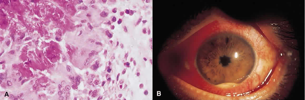
1. Roitt I, Brostoff J, Male D: Immunology. 4th ed. London: Mosby, 1996

2. Morel PA, Oriss TB: Cross-regulation between Th1 and Th2 cells. Crit Rev Immunol 18:275–303, 1998

3. Streilein JW: Immunoregulatory mechanisms of the eye. Progr Retinal Eye Res 18:357–370, 1999

4. Castenholtz A: Studies on the “vascular labeling” phenomenon in the iris and the ciliary body of the rat after intravenous injection of colloidal carbon. Graefes Arch Klin Exp Ophthalmol 202:27, 1977

5. Streilein JW: Immune privilege as the result of local tissue barriers and immunopressive microenvironments. Curr Opin Immunol 5:482–492, 1993

6. Tonjum AM, Pederson OO: The permeability of the human ciliary and iridic epithelium to horseradish peroxidase: An in vitro study. Acta Ophthalmol 55:781, 1977

7. Raviola G: The structural basis of the blood-ocular barriers. Exp Eye Res 25:27, 1977

8. Cunha-Vaz J: The blood-ocular barriers. Surv Ophthalmol 23:279, 1979

9. Streilein JW, Toews G, Bergstresser P: Corneal allografts fail to express Ia antigens. Nature 282:325–327, 1979

10. Kaiser C, Ksander B, Streilein JW: Inhibition of lymphocyte proliferation by aqueous humor. Reg Immunol 2:42–49, 1989

11. Griffith T, Brunner T, Fletcher S: Fas ligand-induced apoptosis as a mechanism of immune privilege. Science 270:1189–1192, 1995

12. Hogan MJ, Zimmerman LE (eds): Ophthalmic Pathology: An Atlas and Textbook. 2nd ed. Philadelphia: WB Saunders, 1962

13. Rutzen AR, Ortega-Larrocea G, Dugel PU et al: Retinal and choroidal biopsy in intraocular inflammation: Clinicopathologic study. Trans Am Ophthalmol Soc 92:431–455, 1994

14. Freeman WR, Wiley CA, Gross JG et al: Endoretinal biopsy in immunosuppressed and healthy patients with retinitis. Indications, utility, and techniques. Ophthalmology 96: 1559–1565, 1989

15. Therese KL, Anand AR, Madhavan HN: Polymerase chain reaction in the diagnosis of bacterial endophthalmitis. Br J Ophthalmol 82:1078–1082, 1998

16. Lohmann CP, Heeb M, Linde HJ et al: Diagnosis of infectious endophthalmitis after cataract surgery by polymerase chain reaction. J Cataract Refract Surg 24:821–826, 1998

17. Kim MC, Kabeer NH, Tandhasetti MT et al: Immunohistochemical studies on melanin associated antigen (MAA) induced experimental autoimmune anterior uveitis (EAAU). Curr Eye Res 14:703–710, 1995

18. Broekhuyse RM, Kuhlmann ED, Winkens HJ: Experimental autoimmune anterior uveitis (EAAU). III. Induction by immunization with purified uveal and skin melanins. Exp Eye Res 56:575–583, 1993

19. Broekhuyse RM, Kuhlmann ED: Experimental autoimmune anterior uveitis. The preparation of uveitogenic ocular melanin. Invest Ophthalmol Vis Sci 34:698–700, 1993

20. Broekhuyse RM, Kuhlmann ED, Winkens HJ: Experimental autoimmune anterior uveitis (EAAU): Induction by melanin antigen and suppression by various treatments. Pigment Cell Res 6:1–6, 1993

21. Rosenbaum JT, McDevitt HO, Guss RB et al: Endotoxin-induced uveitis in rats as a model for human disease. Nature 286:611–613, 1980

22. Parnaby-Price A, Stanford MR, Biggerstaff J et al: Leukocyte trafficking in experimental autoimmune uveitis in vivo. J Leukoc Biol 64:434–440, 1998

23. Hamada M, Ogura Y, Miyamoto K et al: Retinal leukocyte behavior in experimental autoimmune uveoretinitis of rats. Exp Eye Res 65:445–450, 1997

24. Gery I, Wiggert B, Redmond TM et al: Uveoretinitis and pinealitis induced by immunization with interphotoreceptor retinoid-binding protein. Invest Ophthalmol Vis Sci 27: 1296–1300, 1986

25. Atalla L, Linker-Israeli M, Steinman L et al: Inhibition of autoimmune uveitis by anti-CD4 antibody. Invest Ophthalmol Vis Sci 31:1264–1270, 1990

26. Forrester JV, Liversidge J, Dua HS: Regulation of the local immune response by retinal cells. Curr Eye Res 9:183–191, 1990

27. Sun SH, Silver PB, Caspi RR et al: Identification of genomic regions controlling experimental autoimmune uveoretinitis in rats. Int Immunol 11:529–534, 1999

28. Ishimoto S, Wu GS, Hayashi S et al: Free radical tissue damages in the anterior segment of the eye in experimental autoimmune uveitis. Invest Ophthalmol Vis Sci 37:630–636, 1996

29. Wu GS, Zhang J, Rao NA: Peroxynitrite and oxidative damage in experimental autoimmune uveitis. Invest Ophthalmol Vis Sci 38:1333–1339, 1997

30. Goto H, Wu GS, Chen F et al: Lipid peroxidation in experimental uveitis: Sequential studies. Curr Eye Res 11:489–499, 1992

31. Criteria for diagnosis of Behçet's disease: International Study Group for Behçet's Disease. Lancet 335:1078–1080, 1990

32. Sakane T: New perspective on Behçet's disease. Int Rev Immunol 14:89–96, 1997

33. Mizuki N, Inoko H, Ohno S: Pathogenic gene responsible for the predisposition of Behçet's disease. Int Rev Immunol 14:33–48, 1997

34. Lehner T, Lavery E, Smith R et al: Association between 65-kilodalton heat shock protein Streptococcus sanguis and the corresponding antibodies in Behçet's syndrome. Infect Immunol 59:1434, 1991

35. Lehner T: The role of heat shock protein, microbial and autoimmune agents in the aetiology of Behçet's disease. Int Rev Immunol 14:21–32, 1997

36. Yamashita N: Hyperreactivity of neutrophils and abnormal T cell homeostasis: A new insight for pathogenesis of Behçet's disease. Int Rev Immunol 14:11–19, 1997

37. Stanford MR, Kasp E, Whiston R et al: Heat shock protein peptides reactive in patients with Behçet's disease are uveitogenic in Lewis rats. Clin Exp Immunol 97:226–231, 1994

38. Chajek-Shaul T, Pisanty S, Knobler H et al: HLA-B51 may serve as an immunogenetic marker for a subgroup of patients with Behçet's syndrome. Am J Med 83:666–72, 1987

39. Hirohata S, Hashimoto T: Abnormal T cell responses to bacterial superantigens in Behçet's disease (BD). Clin Exp Immunol 112:317–324, 1998

40. Mishima S, Masuda K, Izawa Y et al: The Eighth Frederick H. Verhoeff Lecture. Presented by Saiichi Mishima, MD, Behçet's disease in Japan: Ophthalmologic aspects. Trans Am Ophthalmol Soc 77:225–279, 1979

41. Shimada K, Yaoita H, Shikano S: Chemotactic activity for polymorphonuclear leucocytes of the aqueous humor in patients with Behçet's disease. Nippon Ganka Gakkai Zasshi 75:2100–2105, 1971

42. Nussenblatt RB. Uveitis in Behçet's disease. Int Rev Immunol 14:67–79, 1997

43. George RK, Chan CC, Whitcup SM et al: Ocular immunopathology of Behçet's disease. Surv Ophthalmol 42:157–162, 1997

44. Charteris DG CC, Rosenthal AR et al: Behçet's disease: Activated T-lymphocytes in retinal perivasculitis. Br J Ophthalmol 76:499–501, 1992

45. Charteris DG BK, McCartney ACF et al: CD4+ lymphocyte involvement in ocular Behçet's disease. Autoimmunity 12:201–206, 1992

46. Aracil P, Moyenin P, Ryckewaert M et al: Pars planitis and multiple sclerosis. Bull Soc Ophtalmol Fr 86:485–487, 1986

47. Malinowski SM, Pulido JS, Goeken NE et al: The association of HLA-B8, B51, DR2, and multiple sclerosis in pars planitis. Ophthalmology 100:1199–1205, 1993

48. Palimeris G, Marcomichelakis N, Konstantinidou V et al: Intermediate uveitis: What is the natural course of the disease and its relationship with other systemic diseases? Eur J Ophthalmol 4:223–227, 1994

49. Tang WM, Pulido JS, Eckels DD et al: The association of HLA-DR15 and intermediate uveitis. Am J Ophthalmol 123:70–75, 1997

50. Biousse V, Trichet C, Bloch-Michel E et al: Multiple sclerosis associated with uveitis in two large clinic-based series. Neurology 52:179–181, 1999

51. Raja SC, Jabs DA, Dunn JP et al: Pars planitis: Clinical features and class II HLA associations. Ophthalmology 106:594–5999, 1999

52. Green WR, Kincaid MC, Michels RG et al: Pars planitis. Trans Ophthalmol Soc UK 101:361–367, 1981

53. Pederson JE, Kenyon KR, Green WR et al: Pathology of pars planitis. Am J Ophthalmol 86:762–774, 1978

54. Yoser SL, Forster DJ, Rao NA: Pathology of intermediate uveitis. Dev Ophthalmol 23:60–70, 1992

55. Eichenbaum JW, Friedman AH, Mamelok AE: A clinical and histopathological review of intermediate uveitis (“pars planitis”). Bull NY Acad Med 64:164–174, 1988

56. Brockhurst RJ, Schepens CL, Okamura ID: Uveitis. II. Peripheral uveitis: Clinical description, complications and differential diagnosis. Am J Ophthalmol 49:1257–1266, 1960

57. Kimura SJ, Hogan MJ: Chronic cyclitis. Arch Ophthalmol 71:193–201, 1964

58. Wetzig RP, Chan CC, Nussenblatt RB et al: Clinical and immunopathological studies of pars planitis in a family. Br J Ophthalmol 72:5–10, 1988

59. Gartner J: The fine structure of the vitreous base of the human eye and pathogenesis of pars planitis. Am J Ophthalmol 71:1317–1327, 1971

60. Gartner J: The vitreous base of the human eye and “pars planitis.”Mod Probl Ophthalmol 10:250–255, 1972

61. Loewenfeld IE, Thompson HS: Fuchs's heterochromic cyclitis: A critical review of the literature. I. Clinical characteristics of the syndrome. Surv Ophthalmol 17:394–457, 1973

62. Merayo-Lloves J, Power WJ, Rodriguez A et al: Secondary glaucoma in patients with uveitis. Ophthalmologica 213: 300–304, 1999

63. Fearnley IR, Rosenthal AR: Fuchs' heterochromic iridocyclitis revisited. Acta Ophthalmol Scand 73:166–170, 1995

64. La Hey E, de Vries J, Langerhorst CT: Treatment and prognosis of secondary glaucoma in Fuchs' heterochromic iridocyclitis. Am J Ophthalmol 116:327–340, 1993

65. Jones NP: Glaucoma in Fuchs' heterochromic uveitis: Aetiology, management and outcome. Eye 5:662–667, 1991

66. La Hey E, Baarsma GS, De Vries J et al: Clinical analysis of Fuchs' heterochromic cyclitis. Doc Ophthalmol 78:225–235, 1991

67. Henderly DE, Genstler AJ, Smith RE et al: Changing patterns of uveitis. Am J Ophthalmol 103:131–136, 1987

68. Pivetti-Pezzi P, Accorinti M, La Cava M et al: Endogenous uveitis: An analysis of 1,417 cases. Ophthalmologica 210: 234–238, 1996

69. Saari M, Vuorre I, Tiilikainen A et al: Genetic background of Fuchs' heterochromic cyclitis. Can J Ophthalmol 13:240–246, 1978

70. Berger BB, Tessler HH, Kottow MH: Anterior segment ischemia in Fuchs' heterochromic cyclitis. Arch Ophthalmol 98:499–501, 1980

71. Goldberg MF, Erozan YS, Duke JR et al: Cytopathologic and histopathologic aspects of Fuchs' heterochromic iridocyclitis. Arch Ophthalmol 74:604–609, 1965

72. Regenbogen LS, Naveh-Floman N: Glaucoma in Fuchs' heterochromic cyclitis associated with congenital Horner's syndrome. Br J Ophthalmol 71:844–849, 1987

73. Collier M: Progressive facial hemiatrophy, heterochromic cyclitis, Fuchs type, and micropapilla. Bull Soc Ophtalmol Fr 67:595–603, 1967

74. Fulmek R: Hemiatrophia progressiva faciei (Rombergsyndrome) associated with heterochromia complicata (Fuchs-syndrome) (author's translation). Klin Monatsbl Augenheilkd 164:615–628, 1974

75. La Hey E, Baarsma GS: Fuchs' heterochromic cyclitis and retinal vascular abnormalities in progressive hemifacial atrophy. Eye 7:426–428, 1993

76. Melamed S, Lahav M, Sandbank U et al: Fuch's heterochromic iridocyclitis: An electron microscopic study of the iris. Invest Ophthalmol Vis Sci 17:1193–1199, 1978

77. Wobmann P: Fuchs's heterochromic cyclitis. Electronmicroscopic study of nine iris biopsies (author's translation). Albrecht Von Graefes Arch Klin Exp Ophthalmol 199:167–178, 1976

78. La Hey E, Rothova A, Baarsma GS et al: Fuchs' heterochromic iridocyclitis is not associated with ocular toxoplasmosis. Arch Ophthalmol 110:806–811, 1992

79. La Heij E, Rothova A: Fuchs's heterochromic cyclitis in congenital ocular toxoplasmosis. Br J Ophthalmol 75:372–373, 1991

80. La Hey E, de Jong PT, Kijlstra A: Fuchs' heterochromic cyclitis: Review of the literature on the pathogenetic mechanisms. Br J Ophthalmol 78:307–312, 1994

81. Schwab IR: The epidemiologic association of Fuchs' heterochromic iridocyclitis and ocular toxoplasmosis. Am J Ophthalmol 111:356–362, 1991

82. Jones NP: Fuchs' heterochromic uveitis: An update. Surv Ophthalmol 37:253–272, 1993

83. van den Born LI, van Schooneveld MJ, de Jong PT et al: Fuchs' heterochromic uveitis associated with retinitis pigmentosa in a father and son. Br J Ophthalmol 78:504–505, 1994

84. Vuorre I, Saari M, Tiilikainen A et al: Fuchs' heterochromic cyclitis associated with retinitis pigmentosa: A family study. Can J Ophthalmol 14:10–16, 1979

85. Yalvac IS, Altintas AK, Gokdere A et al: Fuchs' heterochromic uveitis associated with retinitis pigmentosa. Acta Ophthalmol Scand 76:243–244, 1998

86. Chowers I, Zamir E, Banin E et al: Retinitis pigmentosa associated with Fuchs' heterochronic uveitis. Arch Ophthalmol 118:800–802, 2000

87. Fuchs E: Uber Komplicationen der heterochromie. Z Augenheilkd 15:191–212, 1906

88. Dithmar S, Tetz MR, Volcker HE: Fuchs' heterochromic cyclitis. Clinico-histopathologic findings of nodular iritis.Klin Monatsbl Augenheilkd 209:158–162, 1996

89. Bialasiewicz A, Gierth K, Naumann GO: Heterochromia complicata Fuchs, crystalline iridopathy and increased immunoglobulin G in aqueous humor: A case report. Klin Monatsbl Augenheilkd 198:205–206, 1991

90. Lam S, Tessler HH, Winchester K et al: Iris crystals in chronic iridocyclitis. Br J Ophthalmol 77:181–182, 1993

91. Zamir E, Margalit E, Chowers I: Iris crystals in Fuchs' heterochromic iridocyclitis. Arch Ophthalmol 116:1394, 1998

92. Callear AB, Reynolds A, Harry J et al: Iris crystals in chronic uveitis. Br J Ophthalmol 83:703–706, 1999

93. Goldstein DA, Edward DP, Tessler HH: Iris crystals in Fuch heterochromic iridocyclitis. Arch Ophthalmol 116: 1692–1693, 1998

94. Bloch-Michel E, Lambin P, Debbia M et al: Local production of IgG and IgG subclasses in the aqueous humor of patients with Fuchs heterochromic cyclitis, herpetic uveitis and toxoplasmic chorioretinitis. Int Ophthalmol 21:187–194, 1997

95. Kuo I, Rao NA: Ocular disease in AIDS. Springer Semin Immunopathol 21:161–177, 1999

96. Hogan LH, Weinstock JV, Sandor M: TCR specificity in infection induced granulomas. Immunol Lett 68:115–120, 1999

97. Morinelli EN, Dugel PU, Riffenburgh R et al: Infectious multifocal choroiditis in patients with acquired immune deficiency syndrome. Ophthalmology 100:1014–1021, 1993

98. Morinelli EN, Dugel PU, Lee M et al: Opportunistic intraocular infections in AIDS. Trans Am Ophthalmol Soc 90:97–108, 1992

99. Perlman DC, El-Helou P, Salomon N: Tuberculosis in patients with human immunodeficiency virus infection. Semin Respir Infect 14:344–352, 1999

100. Pepose JS, Holland GN, Nestor MS et al: Acquired immune deficiency syndrome. Pathogenic mechanisms of ocular disease. Ophthalmology 92:472–484, 1985

101. Read RW, Zhang JA, Ishimoto SI et al: Evaluation of the role of human retinal vascular endothelial cells in the pathogenesis of CMV retinitis. Ocul Immunol Inflamm 7:139–146, 1999

102. Neuwirth J, Gutman I, Hofeldt AJ et al: Cytomegalovirus retinitis in a young homosexual male with acquired immunodeficiency. Ophthalmology 89:805–808, 1982

103. Yoser SL, Forster DJ, Rao NA: Systemic viral infections and their retinal and choroidal manifestations. Surv Ophthalmol 37:313–352, 1993

104. Palestine AG, Rodrigues MM, Macher AM et al: Ophthalmic involvement in acquired immunodeficiency syndrome. Ophthalmology 91:1092–1099, 1984

105. Anand R, Font RL, Fish RH et al: Pathology of cytomegalovirus retinitis treated with sustained release intravitreal ganciclovir. Ophthalmology 100:1032–1039, 1993

106. Rodrigues MM, Palestine A, Nussenblatt R et al: Unilateral cytomegalovirus retinochoroiditis and bilateral cytoid bodies in a bisexual man with the acquired immunodeficiency syndrome. Ophthalmology 90:1577–1582, 1983

107. Faber DW, Crapotta JA, Wiley CA et al: Retinal calcifications in cytomegalovirus retinitis. Retina 13:46–49, 1993

108. Engstrom RE, Jr., Holland GN, Margolis TP et al: The progressive outer retinal necrosis syndrome; A variant of necrotizing herpetic retinopathy in patients with AIDS. Ophthalmology 101:1488–1502, 1994

109. Rutzen AR, Ortega-Larrocea G, Dugel PU et al: Clinicopathologic study of retinal and choroidal biopsies in intraocular inflammation. Am J Ophthalmol 119:597–611, 1995

110. Moorthy RS, Smith RE, Rao NA: Progressive ocular toxoplasmosis in patients with acquired immunodeficiency syndrome. Am J Ophthalmol 115:742–747, 1993

111. Smith RE: Toxoplasmic retinochoroiditis as an emerging problem in AIDS patients [editorial]. Am J Ophthalmol 106:738–739, 1988

112. Holland GN, Engstrom RE Jr, Glasgow BJ et al: Ocular toxoplasmosis in patients with the acquired immunodeficiency syndrome. Am J Ophthalmol 106:653–667, 1988

113. Park KL, Rao NA: Ocular pathology of opportunistic infections and neoplasms associated with AIDS. Ophthalmol Clin North Am 10:73, 1997

114. Aaberg TM Jr, Flynn HW Jr, Schiffman J et al: Nosocomial acute-onset postoperative endophthalmitis survey: A 10-year review of incidence and outcomes. Ophthalmology 105:1004–1010, 1998

115. Powe NR, Schein OD, Gieser SC et al: Synthesis of the literature on visual acuity and complications after cataract extraction with intraocular lens implantation. Arch Ophthalmol 112:239–252, 1994

116. Kattan HM, Flynn HW, Pfugfelder SC et al: Nosocomial endophthalmitis survey: Current incidence of infection after intraocular surgery. Ophthalmology 98:227–238, 1991

117. Javitt JC, Vitale S, Canner JK et al: National outcomes of cataract extraction: Endophthalmitis after inpatient surgery. Arch Ophthalmol 109:1085–1089, 1991

118. Norregaard JC, Thoning H, Bernth-Petersen P et al: Risk of endophthalmitis after cataract extraction: Results from the International Cataract Surgery Outcomes study. Br J Ophthalmol 81:102–106, 1997

119. Han DP, Wisniewski SR, Wilson LA et al: Spectrum and susceptibilities of microbiologic isolates in the Endophthalmitis Vitrectomy Study. Am J Ophthalmol 122:1–17, 1996; published erratum appears in Am J Ophthalmol 122:920, 1996

120. Kunimoto DY, Das T, Sharma S et al: Microbiologic spectrum and susceptibility of isolates: Part I. Postoperative endophthalmitis. Endophthalmitis Research Group. Am J Ophthalmol 128:240–242, 1999

121. Mistlberger A, Ruckhofer J, Raithel E et al: Anterior chamber contamination during cataract surgery with intraocular lens implantation. J Cataract Refract Surg 23:1064–1069, 1997

122. Egger SF, Huber-Spitzy V, Skorpik C et al: Different techniques of extracapsular cataract extraction: Bacterial contamination during surgery: Prospective study on 230 consecutive patients. Graefes Arch Clin Exp Ophthalmol 232:308–311, 1994

123. Egger SF, Huber-Spitzy V, Scholda C et al: Bacterial contamination during extracapsular cataract extraction. Prospective study on 200 consecutive patients. Ophthalmologica 208:77–81, 1994

124. Beigi B, Westlake W, Chang B et al: The effect of intracameral, per-operative antibiotics on microbial contamination of anterior chamber aspirates during phacoemulsification. Eye 12:390–394, 1998

125. Beigi B, Westlake W, Mangelschots E et al: Peroperative microbial contamination of anterior chamber aspirates during extracapsular cataract extraction and phacoemulsification. Br J Ophthalmol 81:953–955, 1997

126. Ariyasu RG, Nakamura T, Trousdale MD et al: Intraoperative bacterial contamination of the aqueous humor. Ophthalmic Surg 24:367–373, 1993

127. Maxwell DP, Brent BD, Orillac R et al: A natural history study of experimental Staphylococcus epidermidis endophthalmitis. Curr Eye Res 12:907–912, 1993

128. Pleyer U, Mondino BJ, Adamu SA et al: Immune response to Staphylococcus epidermidis-induced endophthalmitis in a rabbit model. Invest Ophthalmol Vis Sci 33:2650–2663, 1992

129. Davis JL, Winward KR, Lonardo EC et al: Association of Propionibacterium acnes endophthalmitis with HLA-DQw5. Ocular Immunol Inflam 4:25–32, 1996

130. Endophthalmitis Vitrectomy Study Group: Results of the endophthalmitis vitrectomy study: A randomized trial of immediate vitrectomy and of intravenous antibiotics for the treatment of postoperative bacterial endophthalmitis. Arch Ophthalmol 113:1479–1496, 1995

131. Ormerod LD, Puklin JE, Giles CL: Chronic Propionibacterium acnes endophthalmitis as a cause of intermediate uveitis. Ocul Immunol Inflamm 5:67–68, 1997

132. Doyle A, Beigi B, Early A et al: Adherence of bacteria to intraocular lenses: A prospective study. Br J Ophthalmol 79:347–349, 1995

133. Menikoff JA, Speaker MG, Marmor M et al: A case-control study of risk factors for postoperative endophthalmitis. Ophthalmology 98:1761–1768, 1991

134. Rao NA, Nerenberg AV, Forster DJ: Torulopsis Candida (Candida famata) endophthalmitis simulating Propionibacterium acnes syndrome. Arch Ophthalmol 109:1718–1721, 1991

135. Busin M, Cusumano A, Spitznas M: Intraocular lens removal from eyes with chronic low-grade endophthalmitis. J Cataract Refract Surg 21:679–684, 1995

136. Ciulla TA, Beck AD, Topping TM et al: Blebitis, early endophthalmitis, and late endophthalmitis after glaucoma-filtering surgery. Ophthalmology 104:986–995, 1997

137. Waheed S, Ritterband DC, Greenfield DS et al: New patterns of infecting organisms in late bleb-related endophthalmitis: A ten year review. Eye 12:910–915, 1998

138. Kangas TA, Greenfield DS, Flynn HW, Jr. et al: Delayed-onset endophthalmitis associated with conjunctival filtering blebs. Ophthalmology 104:746–52, 1997

139. Kunimoto DY, Das T, Sharma S et al: Microbiologic spectrum and susceptibility of isolates: Part II. Posttraumatic endophthalmitis. Endophthalmitis Research Group. Am J Ophthalmol 128:242–244, 1999

140. Abu el-Asrar AM, al-Amro SA, al-Mosallam AA et al: Post-traumatic endophthalmitis: Causative organisms and visual outcome. Eur J Ophthalmol 9:21–31, 1999

141. Duch-Samper AM, Chaques-Alepuz V, Menezo JL et al: Endophthalmitis following open-globe injuries. Curr Opin Ophthalmol 9:59–65, 1998

142. Okada AA, Johnson RP, Liles WC et al: Endogenous bacterial endophthalmitis: Report of a ten-year retrospective study. Ophthalmology 101:832–838, 1994

143. Wong JS, Balakrishnan V: Neisseria meningitidis endogenous endophthalmitis: Case report and literature review. J Pediatr Ophthalmol Strabismus 36:145–152, 1999

144. Lohmann CP, Gabel VP, Heep M et al: Listeria monocytogenes-induced endogenous endophthalmitis in an otherwise healthy individual: Rapid PCR-diagnosis as the basis for effective treatment. Eur J Ophthalmol 9:53–57, 1999

145. Marinella MA, Warwar R: Endogenous endophthalmitis due to Serratia marcescens. South Med J 91:388–391, 1998

146. Mendivil A, Cuartero V: Endogenous endophthalmitis caused by Rickettsia conorii. Acta Ophthalmol Scand 76: 121–122, 1998

147. Davitt B, Gehrs K, Bowers T: Endogenous Nocardia endophthalmitis. Retina 18:71–73, 1998

148. Berman AJ, Del Priore LV, Fischer CK: Endogenous Ochrobactrum anthropi endophthalmitis. Am J Ophthalmol 123:560–562, 1997

149. Essman TF, Flynn HW Jr, Smiddy WE et al: Treatment outcomes in a 10-year study of endogenous fungal endophthalmitis. Ophthalmic Surg Lasers 28:185–194, 1997

150. Tuft SJ, Coster DJ: The corneal endothelium. Eye 4:389–424, 1990

151. O'Connor GR: Calcific band keratopathy. Trans Am Ophthalmol Soc 70:58–81, 1972

152. Vinores SA, Youssri AI, Luna JD et al: Upregulation of vascular endothelial growth factor in ischemic and non-ischemic human and experimental retinal disease. Histol Histopathol 12:99–109, 1997

153. Gentile RC, Liebmann JM, Tello C et al: Ciliary body enlargement and cyst formation in uveitis. Br J Ophthalmol 80:895–899, 1996

154. Peiffer RL, Jr., Wilcock BP: Histopathologic study of uveitis in cats: 139 cases (1978-1988). J Am Vet Med Assoc 198:135–138, 1991

155. Kalina PH, Pach JM, Buettner H et al: Neovascularization of the disc in pars planitis. Retina 10:269–273, 1990

156. Phillips WBD, Bergren RL, McNamara JA: Pars planitis presenting with vitreous hemorrhage. Ophthalmic Surg 24:630–631, 1993

157. Gass J: Stereoscopic Atlas of Macular Diseases: Diagnosis and treatment. Vol 42, 4th ed. St Louis: Mosby-Year Book, 1997

158. McMenamin PG, Forrester JV, Steptoe R et al: Ultrastructural pathology of experimental autoimmune uveitis in the rat. Autoimmunity 16:83–93, 1993

159. Dev S, Mieler WF, Pulido JS et al: Visual outcomes after pars plana vitrectomy for epiretinal membranes associated with pars planitis. Ophthalmology 106:1086–1090, 1999

160. Gilbert C, Hiscott P, Unger W et al: Inflammation and the formation of epiretinal membranes. Eye 2:S140–S156, 1988

161. Rao NA, Wu GS: Free radical mediated photoreceptor damage in uveitis. Prog Retin Eye Res 19:41–68, 2000

162. Forster DJ, Cano MR, Green RL et al: Echographic features of the Vogt-Koyanagi-Harada syndrome. Arch Ophthalmol 108:1421–1426, 1990

163. Inomata H, Sakamoto T: Immunohistochemical studies of Vogt-Koyanagi-Harada disease with sunset sky fundus. Curr Eye Res 9:35–40, 1990

164. Moorthy RS, Chong LP, Smith RE et al: Subretinal neovascular membranes in Vogt-Koyanagi-Harada syndrome. Am J Ophthalmol 116:164–170, 1993

165. Jampol LM, Orth D, Daily MJ et al: Subretinal neovascularization with geographic (serpiginous) choroiditis. Am J Ophthalmol 88:683–689, 1979

166. Halperin LS, Lewis H, Blumenkranz MS et al: Choroidal neovascular membrane and other chorioretinal complications of acquired syphilis. Am J Ophthalmol 108:554–562, 1989

167. Frank KW, Weiss H: Unusual clinical and histopathological findings in ocular sarcoidosis. Br J Ophthalmol 67:8–16, 1983

168. Fine SL, Owens SL, Haller JA et al: Choroidal neovascularization as a late complication of ocular toxoplasmosis. Am J Ophthalmol 91:318–322, 1981

169. Korobelnik JF, Hannouche D, Marin F et al: Surgical treatment of retrofoveal choroid neovascularization in multifocal choroiditis. J Fr Ophtalmol 21:146–151, 1998

170. Lampariello DA, Primo SA: Ocular toxocariasis: A rare presentation of a posterior pole granuloma with an associated choroidal neovascular membrane. J Am Optom Assoc 70:245–252, 1999

171. Jampol LM, Sung J, Walker JD et al: Choroidal neovascularization secondary to Candida albicans chorioretinitis. Am J Ophthalmol 121:643–649, 1996

172. Maas S, Deutman AF, Bandhoe F et al: Surgical removal of subretinal neovascular membranes. Eur J Ophthalmol 5:48–55, 1995

173. Kosmorsky GS, Prayson R: Primary optic pathway sarcoidosis in a 38-year-old white man. J Neuroophthalmol 16:188–190, 1996

174. Kishi A, Nao-i N, Sawada A: Ultrasound biomicroscopic findings of acute angle-closure glaucoma in Vogt-Koyanagi-Harada syndrome. Am J Ophthalmol 122:735–737, 1996

175. Gohdo T, Tsukahara S: Ultrasound biomicroscopy of shallow anterior chamber in Vogt-Koyanagi-Harada syndrome. Am J Ophthalmol 122:112–114, 1996

176. Kawano Y, Tawara A, Nishioka Y et al: Ultrasound biomicroscopic analysis of transient shallow anterior chamber in Vogt-Koyanagi-Harada syndrome. Am J Ophthalmol 121:720–723, 1996

177. Krohn J, Seland JH: Exudative retinal detachment in nanophthalmos. Acta Ophthalmol Scand 76:499–502, 1998

178. Falcon MG, Williams HP: Herpes simplex kerato-uveitis and glaucoma. Trans Ophthalmol Soc UK 98:101–104, 1978

179. Allen CB, Ulshafer RJ, Ellis EA et al: Scanning electron microscopic analysis of intraocular ossification in advanced retinal disease. Scanning Microsc 1:233–239, 1987

180. Loewenstein JI, Hogan RN, Jakobiec FA: Osseous metaplasia in a preretinal membrane. Arch Ophthalmol 115:117–9, 1997; published erratum appears in Arch Ophthalmol 115:589, 1997

Back to Top