Chapter 11
Pathology of the Uvea
P. KUMAR RAO
Main Menu   Table Of Contents

Search

EMBRYOLOGY
CONGENITAL AND DEVELOPMENTAL ABNORMALITIES
IRIS
CILIARY BODY
CHOROID
NEOPLASTIC CONDITIONS
REFERENCES

The uvea is the pigmented vascular middle layer of the eye, lying between the sclera and neuroepithelium. It consists of three parts: the iris, which is the anterior part of the uvea; the ciliary body, forming the middle; and the choroid, which is the posterior section. Common pathologic changes involving the uvea include inflammatory and neoplastic diseases. Inflammatory changes are clinically recognized as various forms of uveitis. Among the neoplasms, both primary and metastatic tumors are found in all parts of the uvea.
Back to Top
EMBRYOLOGY
Neural crest cells are responsible for the development of many structures of the eye and orbit.1,2 Anterior segment development results from three waves of neural crest cell migration between the surface ectoderm and lens vesicle.3 The first wave of migrating neural crest cells gives rise to trabecular meshwork and endothelium. The second wave becomes keratocytes, and the third wave differentiates into iris stroma.3 The muscular elements of the iris and its pigment epithelium derive from the neural epithelium. The neural epithelium is also responsible for the development of the neural (sensory) retina and the retinal pigment epithelium. Other derivatives of neural crest are pigmented and nonpigmented portions of iris and choroidal stroma, scleral fibroblasts, and many adnexal structures.4–6 Various anterior segment disorders can be explained by abnormal neural crest development (Table 1)


Table 1. Classification of Anterior Segment Disorders Based on Neural Crest Origin


Cell AbnormalitySecondary Disorders
Deficient neural crest formation(brain-eye-face malformations) 
Abnormal crest cell migrationCongenital glaucoma
 Posterior embryotoxin
 Axenfeld's anomaly and syndrome
 Rieger's anomaly and syndrome
 Peters' anomaly
 Sclerocornea
Abnormal crest cell proliferationIridocorneal endothelial (ICE) syndrome (iris nevus syndrome, Chandler's syndrome, essential iris atrophy)
Abnormal crest cell terminal induction (final differentiation)Congenital hereditary endothelial dystrophy
 Posterior polymorphous dystrophy
 Fuchs' endothelial dystrophy
Acquired abnormalitiesMetaplasia
 Abiotrophy
 Proliferation

(Modified from Bahn CF, Falls HF, Varley GA et al: Classification of corneal endothelial disorders based on neural crest origin. Ophthalmology 91:558, 1984.)


Reciprocal cellular interactions are particularly important in the differentiation of the ciliary body and iris.7 Lens epithelium must interact with the potential ciliary epithelium and iris in order to differentiate properly.7 Without the developing lens, the neural plate epithelium that would have become ciliary epithelium differentiates into retina.8 Ciliary epithelium in turn induces underlying neural crest tissue to differentiate into ciliary muscle and stroma.7

The choriocapillaris begins to differentiate at the fourth or fifth week of gestation at approximately the same time that the retinal pigment epithelium begins its differentiation. In this way the developing human eye is completely enveloped in a capillary layer by the sixth gestational week. The external, large Haller's vessels develop during the fourth month of gestation. They consist of branches of short posterior ciliary arteries and veins draining the choriocapillaris. Sattler's vessels develop during the fifth month between the choriocapillaris and Haller's layer of vessels. By week 23, all choroidal vascular layers are present, and arteries, as distinct from arterioles, are present.9,10

Back to Top
CONGENITAL AND DEVELOPMENTAL ABNORMALITIES

COLOBOMA

Coloboma, a localized defect within tissue, may involve any part of the uveal tract. Typical colobomas occur inferonasally, in the region of the embryonic fissure. They may be complete or incomplete (iris stromal hypoplasia) or, when in the region of the choroid, may appear cystic. Atypical colobomas occur in regions other than the inferonasal area. After initial formation of the optic vesicles from neuroectoderm, invagination of the optic vesicle occurs and forms the optic cup. The embryonic fissure forms secondary to progressive invagination along the inferior edge of the optic cup and stalk. Prior to closure of this fissure, it receives the ingrowth of the hyaloid artery and permits retinal axons to form the optic nerve. Closure of the embryonic fissure begins in the middle and progresses proximally and distally. Improper fusion of the inner optic cup layers leads to failure of the outer layers to become confluent, resulting in a coloboma. The embryonic fissure is located in the inferonasal region of the eye, and failure of fusion leads to typical colobomas in that area. The embryonic fissure closes by 33 to 40 days of embryonic life.11,12

Histologically, a simple chorioretinal coloboma consists of bare sclera that is not covered by choroid or normal retina. The sclera may be covered by a membrane of undifferentiated retina that may contain blood vessels.11

Ocular coloboma usually is sporadic or inherited as an autosomal dominant disorder that is not associated with extraocular abnormalities. Recessive inheritance also has been reported.(11) Coloboma may be a component of many syndromes. Only a few coloboma-associated syndromes of particular interest to ophthalmologists are discussed here.

CHARGE syndrome consists of coloboma, heart defects, atresia of choanae, retarded growth and development, genital hypoplasia, and ear abnormalities or hearing loss.14 Chestler and France found that in 54 cases of ocular coloboma, six patients (11%) met the diagnostic criteria for CHARGE syndrome.14 Patients had vision of 20/200 or less in at least one eye, microphthalmos, iris and chorioretinal defects, and optic nerve colobomas.

Patients with Aicardi's syndrome also suffer from colobomas. Font and associates have documented the ocular abnormalities in Aicardi's syndrome.15 Findings included bilateral microphthalmos, bilateral optic nerve hypoplasia, bilateral colobomas of the juxtapapillary choroid and optic disc, bilateral total retinal detachment with dysplastic rosettes and chorioretinal lacunae characterized by focal thinning, and atrophy of the retinal pigment epithelium and choroid.15

Back to Top
IRIS

NORMAL ANATOMY

The iris is made up of three layers: an anterior layer composed of fibroblasts, melanocytes and collagen, a middle layer of stroma, and a posterior layer composed of the dilator muscle and pigment epithelium. The anterior layer terminates at the iris root with the exception of some spoke-like extensions that continue into Schwalbe's line, and its density determines iris color. The stroma makes up the bulk of the iris and consists of pigmented and nonpigmented cells in a loose extracellular matrix of collagen and mucopolysaccharides. There is a rich blood supply to this layer, in addition to nerves. In the pupillary zone, the iris sphincter muscle is present. Unlike the anterior layer, the stroma appears to have a similar structure regardless of its iris color. In the posterior layers, the dilator muscle extends from the pupillary zone to the periphery. The pigment epithelium is composed of two layers of apposed epithelia which are arranged apex to apex. The anterior cuboidal border is continuous with the pigmented epithelium of the ciliary body. The posterior epithelial cells are columnar in nature.16

ANIRIDIA

Aniridia describes a clinical appearance of complete iris hypoplasia. In actuality, a small stump of iris is present on gonioscopic examination as well as histologically. The majority of cases are familial and are usually inherited in an autosomal dominant manner.17 The condition may be accompanied by glaucoma, attributable to a progressive synechiae of the trabecular meshwork and iris remnant.18 An association of Wilms' tumor, aniridia, genitourinary abnormalities, and mental retardation is known as the WAGR syndrome.19,20 A deletion in the 11P13 region may account for the this syndrome.21

AXENFELD-REIGER ANOMALY

In the Axenfeld-Reiger anomaly there is thickening and anterior displacement of Schwalbe's line. Iris stromal processes may be adherent to this abnormally thickened Schwalbe's line.

IRIDOCORNEAL ENDOTHELIAL (ICE) SYNDROME

The ICE syndrome refers to a set of acquired abnormalities affecting the cornea, angle, and iris. This syndrome may be subdivided into iris nevus syndrome, Chandler's syndrome, and essential iris atrophy. Abnormal proliferation of the corneal endothelium is a feature of this syndrome, as well asproduction of a basement membrane.22,23 Adhesion of the iris stroma to the endothelium result in peripheral anterior synechiae with progression to angle closure glaucoma. Each condition demonstrates endothelial attenuation and extension with production of a basement membrane. If the basement membrane grows over the trabecular meshwork, endothelialization and descemetization of the chamber angle may be seen. As the endothelium extends over the trabecular meshwork, changes in the iris result. Specular microscopy has demonstrated pleomorphism of the endothelium early in the disease.24,25 Histologically the endothelial cells may show pleomorphism, anisocytosis, and occasional paired oval nuclei.22

IRIS CYSTS

Iris cysts can be classified based on their location.26 In general, iris cysts are of the pigment epithelium or of the iris stroma. Most iris epithelial cysts remain stable and do not progress. If the cyst is located in an area of iris pigment epithelium, the cyst will fail to transilluminate. In contrast, if the iris cyst is more peripheral and located near the ciliary body nonpigmented epithelium, the cyst may allow transillumination. As its name implies, a cyst of the pigment epithelium is lined by the iris pigment epithelial cells. On the other hand, cysts of the iris stroma are lined by stratified squamous epithelium with goblet cells and may resemble conjunctival epithelium (Fig. 1). These findings and the fact that stromal cysts are usually found in infants support the idea of a congenital rest of ectopic surface epithelium as the source of these lesions.

Fig. 1. Iris cyst. Iris stromal cyst lined by stratified epithelium that resembles conjunctival epithelium. (Hematoxylin-eosin ×25.)

Acquired cysts of the iris may be due to epithelial down-growth in postsurgical or traumatic cases or may be due to intraocular tumors such as medulloepithelioma. Additionally, some parasitic infections such as cysticercosis may present with cystic changes.

IRIS NODULES

Iris nodules may appear in many conditions and have different appearances based on their etiology. Etiologies include congenital diseases as well as inflammatory, neoplastic, and infectious causes. Congenital accumulations of nevus cells may be variable in their pigmentation. These nevi may be seen alone or in association with neurofibromatosis.27 An iris freckle is seen as a darkly pigmented flat area on the anterior iris surface and is composed of melanocytes containing an increased amount of pigmentation without an increase in the number of cells. Brushfield spots seen in Down's syndrome appear as elevated white-to-yellow lesions in the periphery of the iris and histopathologically appear as areas of relatively normal iris stroma surrounded by a ring of mild iris hypoplasia28 In the iris nevus (Cogan-Reese) syndrome, diffuse nevi of the iris associated with unilateral glaucoma and peripheral anterior synechia may be seen (ICE syndrome).

Inflammatory conditions may also lead to iris nodules. Patients suffering from fungal endophthalmitis may demonstrate an irregular yellow-white mass on the iris. Histologically, these appear as necrotizing granulomas containing mycotic agents (Fig. 2). In juvenile xanthogranuloma, a yellowish-gray iris lesion may be associated with spontaneous hyphema, and histopathologically the nodules demonstrate diffuse histiocytic infiltrate (Fig. 3). Multinucleated giant cells displaying peripheral foamy cytoplasm are also noted; these cells are known as Touton giant cells.29 The giant cells and the histiocytes contain lipid that can be demonstrated by oil red O stain.

Fig. 2. Coccidioidomycosis. Iris stroma shows necrotizing granuloma containing mycotic organisms. The organisms show features of Coccidioides immitis. (Hemotoxylin-eosin ×60.)

Fig. 3. Juvenile xanthogranuloma. The iris is infiltrated by histiocytes, which form nodular aggregates on the anterior surface of the iris. (Hemotoxylin-eosin ×25.) Inset (×200) shows oil red O-positive histiocytes.

Patients suffering from classic granulomatous anterior uveitis may demonstrate Koeppe's nodules that occur at the pupillary border, or Busacca's nodules at the anterior iris surface. Such nodules are seen in patients with sarcoidosis, tuberculosis, Vogt-Koyanagi-Harada' disease, and others Histologically, they consist of aggregates of epithelioid cells mixed with other mononuclear cells. However in tuberculosis, the granulomas show necrosis, and acid-fast stains may reveal the mycobacteria.

Neoplastic causes of iris nodules include melanoma, leiomyoma, leukemia, metastatic carcinoma, and retinoblastoma. Melanoma may occur as a nodular (Fig. 4A) or flat growth or as tapioca-like nodules. All these lesions show spindle shape tumor cells (Fig. 4B), or such cells may be mixed with epithelioid melanoma cells. Both spindle A and spindle B cells may be present.30 These tumors may display occasional mitotic figures, foci of necrosis, and melanophages. Iris melanomas usually are small, mainly low-grade spindle tumors and carry a relatively good prognosis when compared to ciliary body and choroidal melanomas.31–39

Fig. 4. Iris melanoma. A pigmented mass involves the iris (A), which histologically reveals spindle cells (B). (Hemotoxylin-eosin ×60.)

Histologically, leiomyoma may appear similar to amelanotic spindle cell melanoma; immunohistochemical analysis with anti-muscle specific actin and electron microscopy may be required for clear differentiation.40,41

In leukemia, rarely nodular or milky lesions may be present. Histologically, the nodules reveal infiltration of abnormal lymphoid (Fig. 5) or myeloid cells and iris architecture is lost. As the lesion becomes thickened, a pseudohypopyon is common.42 In cases of retinoblastoma, white foci on the anterior iris surface may appear and a pseudohypopyon may also be present.43 Histologically, these foci show abnormal, hyperchromatic, round cells with scant cytoplasm as well as frequent abnormal mitotic figures. Adenomas of the pigmented iris epithelium and adenomas of nonpigmented or pigmented ciliary epithelium may present with nodular lesions pushing the iris stroma. These benign tumors rarely enlarge, seldom undergo malignant transformation, and consist of proliferations of the iris or ciliary body epithelium. Other rare tumors include neonatal hemangiomatosis, and this entity shows multiple vascular spaces that contain erythrocytes and are lined by endothelium (Fig. 6).

Fig. 5. Lymphoma of the iris. Iris stroma is infiltrated by monomorphic-appearing lymphoid cells. The tumor cells were positive for T-cell marker CD3. (Hemotoxylin-eosin ×60.)

Fig. 6. Neonatal hemangiomatosis. Multiple vascular spaces lined by endothelial cells; some of the spaces contain erythrocytes. (Hemotoxylin-eosin ×25.)

DIABETES

Iris changes resulting from diabetes can be seen histopathologically in proliferative diabetic retinopathy. Neovascularization may be seen on the anterior surface of the iris and may contract and pull the pigmented iris epithelium around the pupillary edge, creating ectropion uvea. Additionally, the neovascular membrane may grow over the trabecular meshwork, leading to neovascular glaucoma (Fig. 7A). Lacy vacuolization of the iris pigment epithelium may be seenhistologically if enucleation is performed during a hyperglycemic state. These intraepithelial vacuoles contain glycogen and are periodic acid–Schiff (PAS) positive. In addition, the basement membrane of the ciliary pigment epithelium may show diffuse thickening (Fig. 7B).44

Fig. 7. A. Neovascular glaucoma. The anterior chamber angle is blocked by peripheral anterior synechia. Neovascularization of the iris is present. (Hemotoxylin-eosin ×25.) B. Diabetes. The ciliary epithelium shows diffuse thickening of the basement membrane. (Periodic acid–Schiff ×35).

Back to Top
CILIARY BODY

NORMAL ANATOMY

The ciliary body consists of an anterior portion called the pars plicata and a posterior portion called the pars plana. The stroma of these areas are filled with melanocytes, rich vascular networks, fibrous connective tissue, and bundles of smooth muscle. The smooth muscle comprises the majority of the ciliary body and can be divided into three portions: an outer longitudinal portion, a middle oblique portion, and the inner circular component. The ciliary muscles connect to the scleral spur just posterior to the trabecular meshwork. The ciliary body is covered by a bilayered epithelium; the outer layer of this epithelium is pigmented, the inner layer is not. The pars plicata is denoted by multiple finger-like extensions of ciliary body stroma covered by the ciliary body epithelium. The pars plana does not have these extensions and ends at the ora serrata. In children, the processes of the ciliary body are thin and finger-like. With age, these ciliary processes become thickened and show eosinophilic acellular material.

Pathologic changes involving the ciliary body can be divided into those related to congenital abnormalities and others related to acquired changes. The latter includes pathologic alterations resulting from trauma, inflammatory and infectious changes, and neoplastic and degenerative changes.

CONGENITAL ABNORMALITIES

Iris and Ciliary Body Cysts

In a review of patients with primary iris cysts, Shields and associates studied 45 patients with cysts located near the junction of the iris and ciliary body.45 These cysts tended to be unilateral, affected women, and were usually located in the temporal quadrants. Most cysts remained stable and were not associated with anterior segment complications such as glaucoma, cataract or corneal edema. Histologically these cysts were located where the ciliary body's nonpigmented epithelium joined the iris pigment epithelium, and the presence of some nonpigmented epithelium in the cyst wall explained why these cysts would transilluminate. Some of the cysts are lined by pigment epithelium (Fig. 8).

Fig. 8. Ciliary epithelial cyst. The cyst arising from the ciliary epithelium is lined by pigment epithelial cells. (Hemotoxylin-eosin ×25.)

Stratified squamous epithelium with goblet cells was found in primary stromal cysts and may indicate a congenital rest of ectopic surface epithelium as the source of these lesions. In a recent study of 232 presumed normal eyes, 54% had ciliary body cysts when examined with ultrasound techniques. There was no gender predominance in this study.46 Congenital cysts of the vitreous have also been reported.47,48 Their origin is unknown. Cysts in the anterior vitreous may lie behind the lens or ciliary body and may be connected by thread-like extensions. It is possible that these anterior cysts may be derived from ciliary body epithelium.

Persistant Hyperplastic Primary Vitreous, Trisomy 13-15

Persistent hyperplastic primary itreous (PHPV) is usually a unilateral condition that leads to fibrovascular connections in the retrolental area. These connections may reach the posterior retina and cause a severe tractional detachment. Adipose tissue, cartilage, and smooth muscle may be seen in the retrolental mass. Ciliary processes are stretched or elongated as well.49–54 A group of disorders characterized by trisomy 13-15 may also demonstrate the elongated ciliary processes and retrolental findings seen in PHPV.55–57 This condition (also termed Patau's syndrome) demonstrates retinal dysplasia and rosette-like configurations of the retina.

ACQUIRED ABNORMALITIES

Cysts of the Pars Plana

Pars plana cysts appear to be acquired rather than congenital and may represent a separation of the two epithelial layers analogous to retinal detachments seen posteriorly. They are presumed to be a degenerative change. Histologically, they appear as intraepithelial cysts within the nonpigmented ciliary epithelium of the pars plana.58–60 They appear empty in routinely stained sections but have been shown to contain hyaluronidase-sensitive material, presumably hyaluronic acid.59

Cysts of the pars plana have been demonstrated in patients with multiple myeloma. In contrast to typical pars plana cysts, those associated with myeloma become opaque when fixed in formaldehyde and are eosinophilic on hematoxylin-eosin stain. Additionally, they are PAS positive.58,61,62 These cysts have been demonstrated to contain a gamma globulin and may be seen in patients with nonmyelomatous hypergammaglobulinemia.63

TRAUMA

Following blunt injury to the eye, a cleft between the circumferential and longitudinal ciliary body muscles may develop (Fig. 9). This is described clinically as a recessed angle. Histologically, one can determine angle recession by tracing an imaginary line through the scleral spur and parallel to a line passing between the pupil and optic nerve. If the ciliary body is posterior to this line, the angle can be termed recessed. Complete dislocation of the ciliary body from the scleral spur is known as cyclodialysis.64

Fig. 9. Ciliary body with acute angle recession. There is a cleft between the ciliary muscle and hemorrhage involving angle structures. (Hemotoxylin-eosin ×25.)

The uveitis/glaucoma/hyphema (UGH) syndrome represents episodes of recurrent hemorrhage associated with uveitis and elevated intraocular pressure following implantation of an intraocular lens. The condition is thought to result from repeated trauma to the iris and ciliary body structures from the implanted intraocular lens. Erosion of the posterior chamber intraocular lens haptics into the substance of the ciliary body can occur.65,66

INFLAMMATORY AND INFECTIOUS CHANGES

Intermediate Uveitis (Chronic Cyclitis, Pars Planitis)

The pars plana and peripheral choroid can be involved with nongranulomatous chronic inflammatory infiltrate. Clinically, such inflammation is seen as vitritis and whitish exudative changes at the pars plana. When it is idiopathic, such inflammatory infiltrate may be termed pars planitis. A moderate to dense vitritis may be present. The vitreous inflammation may clump together and form “snow balls.” Typically, dense white material is present at the pars plana and posterior ciliary body; this is called a “snow bank.” Histologically, enucleated eyes with pars planitis reveal a lymphocytic infiltration in the pars plana and peripheral choroids (Fig. 10). The snow bank is made of fibroglial proliferation containing elements of ciliary epithelium, and vascular channels surrounded by mononuclear cells67–69 Features similar to pars planitis are also observed in patients with multiple sclerosis. Intermediate uveitis without a clear snow bank is seen in patients with sarcoidosis, Lyme disease, and intraocular inflammation associated with human T-cell lymphotropic virus type 1 (HTLV-1) infection.70–73

Fig. 10. Pars planitis. A. Peripheral choroid is infiltrated by lymphocytes. (Hemotoxylin-eosin ×35.) B. The “Snow bank” is made up of fibroglial cells and vascular channels lined by prominent endothelial cells ((×2000).

The ciliary body is involved in other inflammatory disorders that affect the iris, choroid, and lens. These include sarcoidosis, which appears as noncaseating granuloma, phacoantigenic endophthalmitis showing granulomatous inflammation, and diffuse granulomas seen in association with sympathetic ophthalmia.74–78 Infectious changes can be seen with viral infections such as varicella zoster virus (VZV) and other herpesviruses, bacterial infections including acid-fast organisms,and various fungal and parasitic infections. VZV infection can cause necrosis of the ciliary body and may show granulomatous inflammation.79 The bacterial infections result in both suppurative and nonsuppurative inflammations as well as granulomatous infiltration. The latter is typically seen with tuberculosis.80

Children with juvenile rheumatoid arthritis (JRA) develop inflammation of the iris and ciliary body. The pauciarticular form of JRA (fewer than five joints involved) accounts for the majority of cases associated with iritis. The majority of patients with iridocyclitis and JRA are antinuclear nuclear antibody (ANA) positive. Rheumatoid factor assay is usually negative. Chronic inflammation may result in peripheral anterior synechia, posterior synechia, cystoid macular edema, vitreous debris and opacification, and chronic band keratopathy. Secondary glaucoma and cyclitic membranes may also be seen. Histologically, inflammation may be seen primarily in the iris (Fig. 11) and ciliary body and consists of nongranulomatous chronic inflammatory infiltration. The infiltrate often consists of lymphocytes and plasma cells. Russell bodies may be common in chronic cases.81,82 Other disorders with a similar nongranulomatous inflammation of the uveal tract include ankylosing spondylitis, Reiter's syndrome, ulcerative colitis, and Behçet's disease.

Fig. 11. Juvenile Rheumatoid Arthritis. Non-granulomatous chronic inflammation is present in the iris. The inflammatory infiltrate contains plasma cells. (Hemotoxylin-eosin ×140.)

NEOPLAMS

Ciliary body tumors arise from the epithelium or from the stroma. Epithelial tumors include medulloepithelioma, adenoma, and adenocarcinoma. Melanoma, melanocytoma, and leiomyoma arise from the ciliary body stroma.

Medulloepithelioma

Medulloepithelioma is classified as benign or malignant and may be teratic. Histologically, benign medulloepithelioma is usually composed of multi-layered sheets or cords of immature neuroepithelial cells (Fig. 12). The presence of poorly differentiated neuroblastic cells resembling neuroblastoma, significantly increased pleomorphism, mitotic activity, sarcomatous areas resembling chondrosarcoma or rhabdomyosarcoma (Fig. 13), and invasion of the uvea or cornea have been listed as criteria for malignancy.83,84 When elements such as hyaline cartilage or skeletal muscle are present, the tumor is designated teratic but may also be classified as benign or malignant based on the previously listed cytologic criteria.

Fig. 12. Medulloepithelioma. The tumor displays cords and tubular structures made up of medullary epithelium. (Hemotoxylin-eosin ×180.)

Fig. 13. Malignant teratoid medulloepithelioma. Clusters of eosinophilic myoblastic cells are seen in malignant teratoid medulloepithelioma. (Hemotoxylin-eosin ×180.)

Adenoma and Adenocarcinoma

Fuchs' adenoma (pseudoadenomatous hyperplasia) rarely becomes evident clinically but may appear as a glistening, white irregular tumor arising from the ciliary body.85,86 It consists of a benign proliferation of nonpigmented ciliary epithelial cells with accumulation of basement membrane-like material (Fig. 15).

Fig. 14. Teratoid medulloepithelioma. The tumor shows cartilage and foci of medullary epithelial cords at the periphery. (Hemotoxylin-eosin ×60.)

Fig. 15. Fuchs adenoma. Histologically, pseudoadenomatous hyperplasia of the nonpigmented ciliary epithelium is noted. (Hemotoxylin-eosin ×15.)

Ciliary epithelial adenomas are much larger than Fuchs' adenomas and may resemble a ciliary body melanoma clinically. These tumors rarely undergo malignant transformation. Histologically the adenomas have a tubular (Fig. 16), papillary, solid,or pleomorphic appearance. The neoplastic cells show small nuclei and inconspicuous nucleoli. Adenocarcinomas are composed of gland-like accumulations of cells with prominent nuclei, nucleoli and marked pleomorphism.87 Both adenomas and adenocarcinomas can be pigmented or nonpigmented.

Fig. 16. Adenoma of the ciliary body. The adenoma shows tubular proliferation of pigment epithelium of the ciliary body.

Melanoma, Melanocytoma, Leiomyoma

Primary stromal tumors of the ciliary body include melanocytoma, melanoma, and leiomyoma. Histologically, melanocytomas appear as heavily pigmented tumors with large cuboidal cells and small nuclei.88 Bleached sections reveal typical balloon-shaped cells containing small nuclei and inconspicuous nucleoli (Fig. 17). Melanomas of the ciliary body have a similar histologic appearance to melanomas in the choroid and demonstrate spindle- to epithelioid-type cells with variable pigmentation. Mitotic figures may be seen. The tumor can extend anteriorly to involve angle structures. Melanomas can also invade the sclera and can cause cataracts.

Fig. 17. Melanocytoma of the ciliary body. The bleached preparation of the ciliary body tumor shows several cells with benign cytology and pale cytoplasm. There are also pigment-containing cells. (Hemotoxylin-eosin ×60.)

Leiomyoma of the ciliary body appears as a dome-shaped mass. Histopathologically, two types are recognized: mesoectodermal and mesodermal. Mesodermal leiomyoma exhibits amelanotic spindle-shaped cells with eosinophillic cytoplasm and long oval nuclei. The neoplastic cells show immunoreactivity to smooth muscle actin (Fig. 18). Electron microscopic examination reveals fusiform densities and other smooth muscle characteristics.89–95 Mesoectodermal leiomyomas show interlacing bundles of spindle-shaped cells containing fibrillary eosinophillic cytoplasm and elongated oval nuclei. The tumor cells posses both myogenic and neurogenic features.

Fig. 18. Leiomyoma of the ciliary body. The tumor shows bundles of eosinophilic spindle cells. The neoplastic cells are immunoreactive with muscle-specific actin (inset ×35). (Hemotoxylin-eosin ×35.)

The ciliary body is also affected by metastatic tumors or by direct invasion by other ocular tumors, including retinoblastoma, metastatic carcinoma, leukemia, and lymphoma. Acute granulocytic and lymphocytic leukemias may be seen arising from the ciliary body uveal tract. These infiltrates may present as part of the generalized disease.96

Back to Top
CHOROID

NORMAL ANATOMY

The choroid is the principal vascular and pigmented tissue of the eye and forms the middle coat of the posterior section of the eye. It extends from the ora serrata to the optic nerve and is attached to the sclera by connective tissue strands and by the numerous blood vessels and nerves that enter the choroid from the sclera. Small amounts of choroidal tissue may extend into the scleral canals through which ciliary vessels and nerves enter the eye.

Histologically, the choroid reveals a thick inner membrane called Bruch's membrane. Bruch's membrane can be divided into five components: the basement membrane of the retinal pigment epithelium, an inner collagenous zone, an elastic layer, an outer collagenous zone, and the basement membrane of the endothelium of the choriocapillaris. The choriocapillaris is a capillary layer that provides nutrition to the retinal pigment epithelium and outer retinal layers. It is arranged in lobules with a central arteriolar feeder. The endothelial cells lining the choriocapillaris are fenestrated and joined by gap junctions.

Another component of the choroid, the stroma, contains larger arteries and veins. The arteries decrease in caliber as they approach the choriocapillaris. The stroma is surrounded by the lamina fusca, which is analogous to the suprachoroidal layer and consists of collagenous and elastic fibers and many fibrocytes and melanocytes.

Pathologic changes affecting the choroid can be classified as dystrophies and degenerations, including congenital diseases, inflammatory and infectious diseases, neoplastic conditions, and traumatic changes.

DYSTROPHIES, DEGENERATIONS, AND CONGENITAL ANOMALIES

Aging Changes

As the eye ages, an accumulation of drusen may be seen on Bruch's membrane. These drusen represent eosinophillic deposits of proteinaceous debris in Bruch's membrane. Bruch's membrane may become calcified and broken in areas. These breaks allow the development of choroidal neovascularization, which may enter the subretinal space. Grossniklaus and Gass have classified these membranes as type I and type II.97 Type I is typically found in patients with age-related macular degeneration and consists of neovascular tissue that lies beneath the retinal pigment epithelium (RPE). Type II membranes are those typically seen in patients who suffer from presumed ocular histoplasmosis syndrome and consists of a single break in Bruch's membrane, which allows the choroidal neovascular tissue to penetrate the membrane and enter the subretinal space.

Aicardi Syndrome

Aicardi syndrome consists of infantile spasms, absence of the corpus callosum, mental retardation, and chorioretinal abnormalities known as “lacunar defects.”98 This is believed to be an X-linked anomaly that is lethal to males during early embryo development. Coloboma of the optic disc may be seen in patients with this disorder. Other ocular abnormalities found in this syndrome include bilateral microphthalmos, bilateral optic nerve hypoplasia, total retinal detachment with dysplastic rosettes, and chorioretinal lacunae that appear as focal thinning and atrophy of the retinal pigment epithelium and choroid.15

Regional Choroidal Dystrophies

Choriocapillaris atrophy in the macula has been termed central progressive areolar choroidal dystrophy. It has been associated with dominant inheritance patterns.99,100 Fluorescein angiography demonstrates a small hyperfluorescent parafoveal area secondary to RPE loss. Later in the progression of the disease, a well-circumscribed loss of choriocapillaris within the macula may be seen. The lesions may progress slowly and are limited to the posterior pole, thereby causing a central scotoma but not night blindness. Histologically, areas of involvement show loss or degeneration of the choriocapillaris, retinal pigment epithelium, and outer retinal layers.101

Total diffuse choroidal vascular atrophy is also known as gyrate atrophy of the choroid.102 This is an autosomal recessive condition that becomes more progressive in the second and third decades of life. Declining vision and night blindness are prominent symptoms. Clinically, atrophic chorioretinal areas develop in the periphery and progress to the central retina. In the late stages of the disease, the majority of the posterior pole may be involved. Patients suffer from deficient activity of the enzyme ornithine aminotransferase. Chronic reduction of ornithine with an arginine-restricted diet may slow the progression of this disorder.103–105 Histologically, there may be focal areas of photoreceptor atrophy with adjacent retinal pigment epithelial hyperplasia. Zones of almost total atrophy of the retina, retinal pigment epithelium, and choroid are present in the fundus midperiphery. Electron microscopic examination discloses abnormalities of the mitochondria of the corneal endothelium and the nonpigmented ciliary epithelium.106

Choroideremia is an X-linked disorder characterized by complete degeneration of the retina and choroid that becomes manifest in childhood and is slowly progressive. The fundus examination of female carriers may resemble the early stages in affected men in that peripheral retinal pigment epithelium changes have a salt-and-pepper appearance. Histologic findings include extensive chorioretinal atrophy and disruption of Bruch's membrane. Hypoproduction of basement membrane by vascular endothelial cells and their pericytes may be seen in the uveal tract; this change could be associated with loss of retinal pigment epithelial cells.107

Sorsby Fundus Dystrophy

Sorsby fundus dystrophy is dominantly inherited and characterized by yellowish fundus lesions that may be accompanied by disciform scarring or result in atrophic macular degeneration.108 Atrophy of the choroid, retinal pigment epithelium, and retina may progress slowly into the periphery. Lipid-rich deposits may be found on Bruch's membrane, and there may be loss of the outer retina and RPE and atrophy of the choriocapillaris.109

ISCHEMIC LESIONS

Cobblestone degeneration and Elschnig spots represent focal areas of choroidal ischemia. In cobblestone degeneration, individual lobules or confluent lobules of the choriocapillaris are involved. Larger infarctions involving multiple lobules of the choriocapillaris are termed Elschnig spots.110,111 Occlusion of the choriocapillary circulation results in atrophy of the tissues that it supplies, such as the retinal pigment epithelium and outer retina. In some instances, the RPE may remain and demonstrate hypopigmentation or hyperplastic changes. A variety of clinical situations may lead to choroidal occlusive disease such as malignant hypertension,110,111 temporal arteritis,112,113 sickle cell disease,114,115 toxemia of pregnancy,110,112,116,117 and disseminated intravascular coagulopathy.118,119

INFLAMMATORY AND INFECTIOUS DISEASES

Non-granulomatous Inflammations

Diffuse or focal lymphocytic infiltration is seen as part of inflammatory processes extending from the ciliary body. These conditions are described in detail in the section on ciliary body inflammatory and infectious changes in this chapter. Histologically, most cases classified as idiopathic choroiditis show non-granulomatous inflammation displaying infiltrates of lymphocytes, plasma cells and histiocytes. Similar choroidal infiltrates are seen in Behçet's disease.

Granulomatous Inflammation

Granulomatous inflammation is defined by the presence of epithelioid histiocytes with or without the presence of inflammatory giant cells. Clinically, one may note the presence of large mutton-fat keratic precipitates (KPs) on the endothelium of the cornea. These mutton-fat KPs are composed of aggregates of epithelioid histiocytes. Granulomatous inflammation may be a reaction to acid-fast organisms, fungi, spirochetes, or foreign material. Autoimmune diseases such as sarcoidosis or a reaction to lens material or lipid may also result in granulomatous inflammation.

Three histologic patterns of granulomatous inflammation may be seen: diffuse, discrete, and zonal. In diffuse granulomatous inflammation, the epithelioid histiocytes are scattered throughout the involved uveal tissue (Fig. 19). There may be an accompanying background of lymphocytes and plasma cells. Discrete granulomatous inflammation reveals well-circumscribed areas of epithelioid histiocytes (Fig. 20). Zonal granulomatous inflammation consists of a central zone of necrosis and/or polymorphonuclear leukocytes surrounded by epithelioid histiocytes, which is in turn surrounded by a zone of non-granulomatous inflammation consisting of granulation tissue, lymphocytes and plasma cells (Fig. 21).

Fig. 19. Sympathetic ophthalmia. Diffuse granulomatous inflammation involving the choroid. (Hemotoxylin-eosin ×65.)

Fig. 20. Sarcoidosis. Discrete granulomatous inflammation is seen in sarcoidosis. (Hemotoxylin-eosin ×100.)

Fig. 21. Tuberculous choroiditis. A zonal granulomatous inflammation is present. Central area shows necrosis. (Hemotoxylin-eosin ×65.)

Diffuse granulomatous inflammation is typically seen in sympathetic ophthalmia and Vogt-Koyanagi-Harada (VKH) disease. Sympathetic ophthalmia has classically been described as an intraocular inflammatory response in the non-traumatized fellow eye following penetrating injury to the contralateral eye. The disorder is characterized by waxing and waning episodes of chronic inflammation; untreated, it may lead to blindness in both eyes. The patient may present with mild pain, photophobia, and vision loss. The exciting eye (previously traumatized) may demonstrate decreased vision and increased photophobia. Both eyes may reveal ciliary injection and vitritis.The exciting eye may develop keratic precipitates. The sympathizing eye may demonstrate mild uveitis and KPs on the endothelium. Posterior segment findings include papillitis, Dalen-Fuchs nodules (yellow-white lesions beneath the RPE), choroidal granulomas, and exudative retinal detachments. Histologically, both the traumatized and sympathizing eyes show diffuse granulomatous inflammation made up of nests of epithelioid cells and giant cells mixed with lymphocytes. This inflammation does not extend to involve the choriocapillaris. Dalen-Fuchs nodules are composed of nodular clusters of epithelioid cells lying between the RPE and Bruch's membrane (Fig. 22). Sympathetic ophthalmia represents an autoimmune process that may be related to an altered T-cell response to uveal antigens or other intraocular proteins.120–124

Fig. 22. Sympathetic ophthalmia. Dalen-Fuchs nodule is composed of clusters of epithelioid cells lying between Bruch's membrane and the retinal pigment epithelium. (Hemotoxylin-eosin ×140.)

Similar diffuse granulomatous choroiditis is seen in VKH disease. VKH disease typically presents with clinical features of meningeal irritation, patches of cutaneous depigmentation, poliosis (Fig. 23), and dysacusia. Lumbar puncture reveals pleocytosis of the cerebrospinal fluid.125 Although the choriocapillaris is spared from the infiltration of the inflammatory cells, chronic cases of VKH disease with recurrences may show involvement of these vascular structures.

Fig. 23. Vogt-Koyangi-Harada (VKH) disease. Note vitiligo and depigmentation in this patient with VKH disease.

The discrete form of granulomatous inflammation is seen in sarcoidosis, and ocular involvement is a common feature of this disease. Systemic manifestations of sarcoidosis include granulomatous inflammations of the lungs, liver, lymph nodes, skin, and even the central nervous system. Clumps of epithelioid histiocytes are sharply demarcated from a surrounding infiltrate of lymphocytes. These discrete granulomas do not carry any caseating centers and therefore lack the cheesy central necrosis that is characteristic of tuberculosis. Although the histologic appearance may differ from that of tuberculosis, there is enough similarity such that special stains for acid-fast bacilli are recommended. A careful examination for foreign material should also be undertaken. This may be aided by polarization microscopy techniques.

Zonal granulomatous inflammation may be seen in cases of phaco-antigenic uveitis in which injury of the natural lens leads to this severe type of intraocular inflammation. The etiology is described in detail in the chapter on the lens. Zonal granulomatous inflammations can also be observed adjacent to the necrotic sclera in patients with rheumatoid arthritis and in cases of infectious choroiditis or retinochoroiditis.

Infectious endophthalmitis refers to inflammation due to an infectious organism, usually bacterial, but it may also be caused by yeast or filamentous fungi. The vitreous is typically involved and supports the growth of the infectious intraocular organism. The reaction to the organism is usually severe and is characterized by abundant polymorphonuclear leukocyte infiltration. The infection is usually accompanied by tissue destruction due to the release of proteolytic enzymes in the severe inflammatory reaction. Uveal and retinal necrosis may occur during these infections. Endogenous endophthalmitis refers to hematogenous dissemination of organisms to the eye and can be seen in cases of septicemia from bacterial or fungal causes. The latter can be caused by Candida or Aspergillus species. Aspergillosis usually causes destructive choroidal inflammation (Fig. 24) with vascular occlusions. The infective organisms are found in the choriocapillaris or along the sub-RPE and sub-retinal space.126

Fig. 24. Aspergillus chorioretinitis. A. Hemorrhagic necrotizing retinitis and choroiditis are noted in a patient with disseminated aspergillosis (hemotoxylin-eosin ×60). B. Note the branching hypae of the organisms involving the retina and choroid (periodic acid–Schiff ×240).

Retinochoroiditis

Necrotizing retinitis with secondary choroiditis is seen in protozoal infections such as toxoplasmosis or in herpetic infections. Infection with Toxoplasma gondii leads to retinitis and secondary choroiditis (Fig. 25), usually granulomatous.127 Congenital infection can be acquired in utero by transplacental transmission of the parasite from the infected mother to the fetus.128 Acquired disease occurs after ingestion of oocysts or tissue cysts.129–132 The congenital form of infection leads to atypical macular colobomas. Reactivation of the infection is caused by release of organisms that have remained dormant in the margins of old congenital retinal scars.133 The slowly proliferating form of the organism, termed the bradyzoite, can be seen in cysts. The rapidly multiplying form, termed the tachyzoite, may be difficult to identify in an infected retina or in immunocompetent individuals, but they are frequently seen in the retinitis of immunocompromised hosts.134 Active infection usually causes focal retinal opacification and an intense vitritis. These findings may give the appearance of a “headlight in a fog” in an immunocompetent person. In contrast, this clinical presentation is rare in patients with AIDS, in whom diffuse retinitis is observed with non-granulomatous choroiditis.134

Fig. 25. Toxoplasma retinochoroiditis. Necrotic retina shows cysts of toxoplasma gondii, and the choroid reveals granulomatous inflammation. (Hemotoxylin-eosin ×65.) Inset (Gomori methenamine silver ×160) shows cysts of the organisms.

VZV, human Hherpes simplex virus type 1 (HSV-1) and type 2 (HSV-2) can cause necrotizing retinitis and secondary choroiditis.135 Such uveal inflammation occurs in immunocompetent individuals. In contrast, VZV and HSV retinitis in patients with AIDS shows minimal or no choroidal inflammation136

Back to Top
NEOPLASTIC CONDITIONS

BENIGN TUMORS

Choroidal Nevi

Most uveal nevi are asymptomatic and are congenital in nature. Choroidal nevi may be detected during a routine ophthalmologic evaluation. The typical choroidal nevus appears as a small, brown tumor with a bland surface. Drusen or retinal pigment epithelium clumping may be present. Most nevi are less than 2-mm thick and less than 5 mm in diameter. In some cases, choroidal neovascularization may be associated with the nevus. Nevi change very little over the course of several years. Should enlargement be seen during periodic evaluation of the patient's lesion, nevus should be reclassified as a malignant melanoma. Histologically, a nevus contains spindle-shaped cells with small nuclei and inconspicuous nucleoli and without mitotic figures.137

Melanocytoma

Melanocytoma, also known as magnocellular nevus, is usually located on the inferotemporal portion of the optic nerve head. It appears as a deeply pigmented lesion. Similar lesions occur in the choroid. Melanocytomasrarely enlarge over long periods of time. Histologically, it is composed of deeply pigmented polyhedral cells with a small nuclei, and bleached preparations reveal benign cytologic features.137,138

Hemangioma

Choroidal hemangioma appears in one of two patterns: a circumscribed form, which is usually isolated, and a diffuse form, which is usually part of the Sturge-Weber syndrome. Histologically, the isolated choroidal hemangioma is composed of cavernous spaces filled with red blood cells and is usually demarcated from normal choroid. Clinically, it appears reddish-orange and is usually in the posterior half of the fundus. Usuallyy the lesion is 1- to 3-mm thick. Irregular depigmentation of the retinal pigment epithelium may be seen. The neurosensory retina may appear thickened or cystic. The diffuse choroidal hemangioma appears as a saturated red choroid and has been termed “tomato ketchup fundus.” The choroid tends to be diffusely thickened.139,140

Choroidal Osteoma

Choroidal osteoma is an acquired bony tumor of the choroid usually seen in young women. It is usually a circum-papillary, yellowish-to-orange choroidal lesion with well-defined margins. The tumor may appear relatively flat, but the surface may appear uneven. If the lesion affects the macula, severe vision loss may occur in combination with degeneration of the overlying RPE. Choroidal neovascularization has been noted to develop in chronic cases.141 Histologically, normal-appearing compact bone may be seen within the choroid. The bony material may contain osteocytes, and intratrabecular spaces are filled with connective tissue containing large and small blood vessels.142

Leiomyoma

Leiomyoma is a rare tumor of the choroid composed of smooth muscle cells and vascular channels. Clinically and histologically these tumors resemble amelanotic melanomas. Histologic diagnosis may require immunohistochemistry (immunoreactivity to muscle-specific actin) and in some cases electron microscopic examination. The latter reveals spindle-shaped cells containing fusiform densities in the cytoplasm and cells surrounded by basement membranes.143

Neurofibroma and Schwannoma

Neurofibromas of the choroid are composed of Schwann's cells, fibroblasts, and fibrous connective tissue. They are usually not encapsulated and may contain nerve axons.144 In contrast, schwannomas are encapsulated and represent a proliferation of Schwann's cells in one of two patterns, Antoni types A and B. In the Antoni-A pattern, spindle-shaped cells are densely packed and arranged in fascicles or bundles. In the Antoni-B pattern, the cells are loosely packed together and are separated by a mucoid substance.145

MALIGNANT TUMORS

Malignant Melanoma of the Uveal Tract

Virchow recognized that melanomas of the uveal tract arise from melanocytes and that these tumors are not always pigmented.146 Several risk factors for the development of uveal melanomas have been identified; these include age, race, male gender, and predisposing lesions.147 The incidence of uveal melanoma increases with age. In one study, the median age at diagnosis was 53 years.148 The tumor rarely occurs in African-Americans; in a large series of malignant melanoma of the uveal tract, only 1% of patients were African-American.149 There was an 8.5 times greater incidence of choroidal melanoma in Caucasians.150,151 Uveal malignant melanoma is more common in men than in women,147,148 and congenital melanosis and nevi may predispose to the development of uveal malignant melanoma.150,152–155 Congenital oculodermal melanocytosis (nevus of Ota) is more common in African-Americans and Asians than in Caucasians, but progression to uveal melanoma is most often observed in Caucasians.156 Only rarely have there been familial cases of uveal melanoma.157,158 Chromosomal abnormalities associated with melanoma include changes on chromosomes 3 and 8, as well as the tumor suppressor gene P53.159–163

Clinically, uveal melanomas may arise in the iris, ciliary body, or choroid arising in the iris are visible and easily noticed by the patient when relatively small. Choroidal and ciliary body melanomas may be divided into four stages based on their clinical presentation. In the first stage, the patient is often asymptomatic and the tumor is discovered on routine ophthalmoscopic examination. In the second stage, there is either a field defect or vision loss that is usually due to an associated retinal detachment. In the third stage, ocular pain develops from glaucoma or inflammation. In the fourth stage, extraocular extension occurs, leading to proptosis or visible subconjunctival masses.164

Choroidal tumors are more common than ciliary body melanomas. The former tumors can be divided into three groups: small (largest dimension is less than 10 mm), medium (the largest dimension is 11 to 15 mm), and large (the largest dimension is greater than 15 mm). As these tumors grow, they produce a mass in the choroid that distends Bruch's membrane, which eventually leads to rupture of that structure. The tumor herniates through the rupture, growing into the subretinal space, and at this point may develop a typical “collar button” or mushroom-like appearance. The collar button or mushroom configuration is seen in many medium-sized tumors. The retina overlying the tumor may undergo atrophy or cystoid degeneration, and there may be a serous detachment of the retina adjacent to the tumor. Larger tumors invade and may destroy other ocular tissues. Some tumors may follow the course of nerves and vessels through the scleral wall into the orbit.

A diffuse, infiltrating type of pattern of choroidal melanoma is occasionally seen. These tumors grow within the choroid without causing much thickening. They are more likely to invade through the sclera and may produce an orbital mass.165 Occasionally, neoplastic cells invade the lumen of vortex veins, but vascular invasion within the tumor is a more common source of hematogenous metastasis. Malignant melanomas arising from the peripapillary choroid may invade the optic nerve head but rarely extends retrolaminarly within the optic nerve.

In 1931, Callender classified uveal melanomas based on cytologic features.166 He described six groups, four of which were based on cytology and two on other histologic features. The groups based on cytology were tumors composed of (1) spindle-A cells, (2) spindle-B cells (Fig. 26), (3) epithelioid cells (Fig. 27), (4) a mixture of epithelioid and spindle cells (Fig. 28). The fifth group consisted of tumors with a fascicular pattern, and the the sixth group was composed of tumors that could not be classified in the other groups because of extensive necrosis (Fig. 29). Spindle cells are described as fusiform and arranged in tightly cohesive bundles. The plasma membranes of the cells are indistinct and have a syncytial appearance. Spindle-A cells have a slender nucleus with fine chromatin and a longitudinal fold in the nuclear envelope that gives the appearance of a line. Spindle-B cells have a slightly plumper nucleus, coarser chromatin, and a more prominent and eosinophillic nucleolus. Epithelioid cells are larger and more pleomorphic. They have an abundant glassy cytoplasm, a polyhedral shape, and a distinct cell border and are less cohesive. Epothelioid cells tend to have a larger and rounder nucleus than the other types, with a more angular nuclear envelope and irregular indentations. The chromatin is coarse and marginated, and large eosinophillic nucleoli are present.

Fig. 26. Choroidal melanoma. Note spindle-B melanoma cells with nucleoli. (Hemotoxylin-eosin ×160.)

Fig. 27. Choroidal melanoma. The epithelioid melanoma cells show large nuclei and prominent nucleoli. (Hemotoxylin-eosin ×160.)

Fig. 28. Choroidal melanoma. The tumor shows a mixture of spindle cells and epithelioid cells. Both spindle A and spindle B cells are present. (Hemotoxylin-eosin ×160.)

Fig. 29. Choroidal melanoma. Necrotic tumor cells are mixed with melanophages. (Hemotoxylin-eosin ×200.)

Immunohistochemical studies may aid in the diagnosis of amelanotic melanomas or those tumors with atypical features. HMB-45 is a monoclonal antibody against a protein obtained from cutaneous malignant melanoma and is a marker for cells derived from melanocytes. Choroidal melanomas tend to be strongly positive when stained with HMB-45 and may show positivity when stained with S-100 protein or melan-A.167,168

Tumors composed of spindle-A or spindle-B cells have a better prognosis than other types. Patients with these tumors have a 22% death rate due to metastasis when followed for 5 years; patients with tumors composed of necrotic, epithelioid, or mixed cell types had a 62% death rate due to metastasis when followed for 5 years.169 Other factors that may affect prognosis include tumor size, invasion into the sclera, mitotic activity, necrosis, and the presence of closed vascular loops.170–175 Most fatal cases with extraocular extension are due to hematogenous metastasis to the liver.

Lymphomatous and Leukemic Infiltrations

Patients with acute or chronic leukemia may develop infiltrations of the choroid, retina, or optic disc. As in intraocular lymphoma, dispersed intravitreal cells may be present. The choroid is the primary intraocular site of involvement in leukemia as well as in systemic lymphomas. Uveal lymphoid infiltration can occur without systemic lymphoma, and these localized cases show massive thickening of choroid from benign reactive lymphoid cells (Fig. 30) or malignant cells. Usually this entity shows episcleral lymphoid infiltration.

Fig. 30. Massive reactive lymphoid hyperplasia of the uvea. The choroid shows diffuse lymphoid infiltration. (Hemotoxylin-eosin ×100.)

Metastatic Tumors

Ocular metastasis can be seen in individuals of all ages but are most common in those aged 40 to 70 years. Presenting signs usually depend on the size, location, and secondary effects of the tumors, but blurred vision and pain are most common. There tends to be a slight female predominance in most series because breast carcinoma tends to metastasize most frequently. Other primary sites include lung, kidney, gastrointestinal tract, prostrate, and thyroid.176,177 Multiple choroidal tumors are more apt to occur with metastatic disease. Metastatic tumors tend to be flat and diffusely infiltrating in the choroid. Histopathologic features generally reflect those of the primary tumors, but the metastatic lesion may be composed of undifferentiated cells and may not allow easy identification of the primary site.

Metastatic breast carcinomas usually are moderately or well differentiated. They often maintain the features of adenocarcinoma with cells arranged in ducts. Some cells may produce mucin. Metastatic bronchogenic carcinomas are frequently undifferentiated and may rarely have a squamous appearance. Oat cell carcinoma of the lung usually consists of small hyperchromatic cells that are often undifferentiated. Metastatic tumors from the gastrointestinal tract may demonstrate mucin production. Such mucin production can be demonstrated histologically by alcian blue or mucicarmine stains. Metastatic prostate carcinoma tends to maintain an adenoid or glandular pattern. Metastatic thyroid carcinoma is usually of the follicular type. In cases of poorly differentiated metastatic carcinoma to the uvea, a clear clinical history of the primary site is needed to make the definitive diagnosis, and in some cases immunohistochemical preparations are helpful.

Back to Top
REFERENCES

1. Johnston MC, Bhakdinaronk A, Reid YC: An expanded role of the neural crest in oral and pharyngeal development. In Bosma JD (ed): Fourth Symposium on Oral Sensation and Perception: Development in the Fetus and Infant, pp 37–52. Washington, DC, U.S. Government Printing Office, 1974:37–52.

2. Johnston MC, Noden DM, Hazelton RD et al: Origins of avian ocular and periocular tissues. Exp Eye Res 29:27, 1979.

3. Bahn CF, Falls HF, Varley GA et al: Classification of corneal endothelial disorders based on neural crest origin. Ophthalmology 91:558, 1984.

4. Ozanics V, Jakobiec FA: Prenatal development of the eye and its adnexa. In Duane TD, Jaeger EA (eds): Biomedical Foundations of Ophthalmology, Vol 1, pp 1–23. Philadelphia: Harper & Row, 1987.

5. Tripathi BJ, Tripathi RC: Neural crest origin of human trabecular meshwork and its implications for the pathogenesis of glaucoma. Am J Ophthalmol 107:671, 1989.

6. Kaiser-Kupfer MI: Neural crest origin of trabecular meshwork cells and other structures of the anterior chamber. Am J Ophthalmol 107:671, 1989.

7. Beebe DC: Development of the ciliary body: A brief review. Trans Ophthalmol Soc UK 105:123, 1986.

8. Stroeva OG: The role of the lens epithelium in the induction of iris and ciliary body tissue. Dokl Akad Nauk SSSR 151:464, 1963.

9. Sellheyer K: Spitznas M. Morphology of the developing choroidal vasculature in the human fetus. Graefes Arch Clin Exp Ophthalmol 226:461, 1988.

10. Sellheyer K: Development of the choroid and related structures. Eye 4:255, 1990.

11. Pagon RA: Ocular coloboma. Surv Ophthalmol 25:223, 1981.

12. Mann I: The Development of the Human Eye, pp 1–29. New York, Grune and Stratton, 1928.

13. Pagon RA, Graham JM Jr, Zonana J et al:. Coloboma, congenital heart disease, and choanal atresia with multiple anomalies: CHARGE association. J Pediatr 99:223, 1981

14. Chestler RJ, France TD: Ocular findings in CHARGE syndrome: Six case reports and a review. Ophthalmology 95:1613, 1988.

15. Font RL, Marines HM, Cartwright J Jr et al: Aicardi syndrome: A clinicopathologic case report including electron microscopic observations. Ophthalmology 98:1727, 1991.

16. Fine BS, Yanoff M: Ocular Histology: A Text and Atlas, pp 167–212. New York, Harper & Row, 1972.

17. Nelson LB, Spaeth GL, Nowinski TS et al: Aniridia: A review. Surv Ophthalmol 28:621, 1984.

18. Grant WM, Walton DS: Progressive changes in the angle in congenital aniridia with development of glaucoma. Am J Ophthalmol 78:842, 1974.

19. Miller RW, Fraumeni JF Jr, Manning MD: Association of Wilms' tumor with aniridia, hemihypertrophy and other congenital malformations. N Engl J Med 270:922, 1964.

20. Shannon RS, Mann JR, Harper E et al: Wilms's tumour and aniridia: Clinical and cytogenetic features. Arch Dis Child 57:685, 1982.

21. Rose EA, Glaser T, Jones C et al: Complete physical map of the WAGR region of 1lp13 localizes a candidate Wilms' tumor gene. Cell 60:495, 1990.

22. Eagle RC Jr, Shields J: Iridocorneal endothelial syndrome with contralateral guttata endothelial dystrophy. Ophthalmology 94:862, 1987.

23. Alvarado JA, Murphy CG, Juster RP et al: Pathogenesis of Chandler's syndrome, essential iris atrophy and the Cogan-Reese syndrome, II: Estimated age at disease onset. Invest Ophthalmol Vis Sci 27:873, 1986.

24. Hirst LW, Quigley HA, Stark WJ et al: Specular microscopy of iridocorneal endothelial syndrome. Am J Ophthalmol 89:11, 1980.

25. Hetherington J Jr: The spectrum of Chandler's syndrome. Trans Am Acad Ophthalmol Otolaryngol 85:240, 1978.

26. Shields JA: Primary cysts of the iris. Trans Am Ophthalmol Soc 79:771, 1981.

27. Spencer WH: Ophthalmic Pathology: An Atlas and Textbook, p 1474. 4th ed. Philadelphia, WB Saunders, 1990.

28. Donaldson DD: The significance of spotting of the iris in Mongoloids. Brushfield spots. Arch Ophthalmol 4:26, 1961.

29. Spencer WH: Ophthalmic Pathology: An Atlas and Textbook, p 1544. 4th ed. Philadelphia, WB Saunders, 1990.

30. Spencer WH: Ophthalmic Pathology: An Atlas and Textbook, p 1612. 4th ed. Philadelphia, WB Saunders, 1990.

31. Duke JR, Dunn SN: Primary tumors of the iris. Arch Ophthalmol 59:204, 1958.

32. Ashton N: Primary tumours of the iris. Br J Ophthalmol 48:650, 1964.

33. Arentsen JJ, Green WR: Melanoma of the iris: Report of 72 cases treated surgically. Ophthalmic Surg 6:23, 1975.

34. Ashton N, Wybar K: Primary tumor of the iris. Ophthalmologica 151:97, 1966.

35. Callender GR, Wilder HC, Ash JE: Five hundred melanomas of the choroid and ciliary body: Followed five years or longer. Am J Ophthalmol 25:562, 1942.

36. Jakobiec FA, Silbert G: Are most iris “melanomas” really nevi? Arch Ophthalmol 99:2117, 1981.

37. Makley TA Jr: Management of melanomas of the anterior segment. Surv Ophthalmol 19:135, 1974.

38. Rones B, Zimmerman LE: The prognosis of primary tumors of the iris treated by iridectomy. Arch Ophthalmol 60:193, 1958.

39. Stallard HB: Surgery of malignant melanoma of iris. Br J Ophthalmol 35: 774, 1951.

40. Yanoff M, Fine BS: Ocular Pathology. 2nd ed. Philadelphia, Lippincott, 1982.

41. Eoss AJ, Pecorella I, Alexander RA et al: Are most intraocular “leiomyomas” really melanocytic lesions? Ophthalmology 101:919, 1994.

42. Spencer WH: Ophthalmic Pathology: An Atlas and Textbook, p 1672. 4th ed. Philadelphia, WB Saunders, 1990.

43. Spencer WH: Ophthalmic Pathology: An Atlas and Textbook, p 1350. 4th ed. Philadelphia, WB Saunders, 1990.

44. Spencer WH: Ophthalmic Pathology: An Atlas and Textbook, p 32. 4th ed. Philadelphia, WB Saunders, 1990.

45. Shields JA, Kline MW, Augsburger JJ: Primary iris cysts: A review of the literature and report of 62 cases. Br J Ophthalmol 68:152, 1984.

46. Kunimatsu S, Araie M, Ohara K, Hamada C: Ultrasound biomicroscopy of ciliary body cysts. Am J Ophthalmol 127:48, 1999.

47. Bullock JD: Developmental vitreous cysts. Arch Ophthalmol 91:83, 1974.

48. Feman SS, Straatsma BR: Cyst of the posterior vitreous. Arch Ophthalmol 91:328, 1974.

49. Reese AB: Persistence and hyperplasia of primary vitreous: Retrolental fibroplasia—Two entities. Arch Ophthalmol 41:527, 1949.

50. Reese AB: Persistent hyperplastic primary vitreous. Am J Ophthalmol 40:317, 1955.

51. Manschot WA: Persistent hyperplastic primary vitreous. Arch Ophthalmol 59:188, 1958.

52. Haddad R, Font RL, Reeser F: Persistent hyperplastic primary vitreous. A clinicopathologic study of 62 cases and review of the literature. Surv Ophthalmol 23:123, 1978.

53. Reese AB, Payne F: Persistence and hyperplasia of the primary vitreous. Am J Ophthalmol 29:1, 1964.

54. Font RL, Yanoff M, Zimmerman LE: Intraocular adipose tissue and persistent hyperplastic primary vitreous. Arch Ophthalmol 82:43, 1969.

55. Cogan DG, Kuwabara T: Ocular pathology of the 13-15 trisomy syndrome. Arch Ophthalmol 72:246, 1964.

56. Miller M, Robbins J, Fishman R et al: A chromosomal anomaly with multiple ocular defects, including retinal dysplasia. Am J Ophthalmol 55:901, 1963.

57. Sergovich F, Madronich JS, Barr ML et al: The D trisomy syndrome: A case report with a description of ocular pathology. Can Med Assoc J 89:151, 1963.

58. Alien RA, Miller DH, Straatsma BR: Cysts of posterior ciliary body (pars plana). Arch Ophthalmol 66:302, 1961.

59. Zimmerman LE, Fine BS: Production of hyaluronic acid by cysts and tumors of the ciliary body. Arch Ophthalmol 72:365, 1964.

60. Gartner J: Fine structure of pars plana cysts. Am J Ophthalmol 73:971, 1972.

61. Baker TR, Spencer WH: Ocular findings in multiple myeloma: A report of two cases. Arch Ophthalmol 91:110, 1974.

62. Johnson BL, Storey JD: Proteinaceous cyst of the ciliary epithelium: I. Their clear nature and immunoelectrophoretic analysis in case of multiple myeloma. Arch Ophthalmol 84:166, 1970.

63. Johnson BL: Proteinaceous cysts of the ciliary epithelium: II. Their occurrence in nonmyelomatous hypergammaglobulinemic conditions. Arch Ophthalmol 84:171, 1970.

64. Spencer WH: Ophthalmic Pathology: An Atlas and Textbook, pp 21–24. 4th ed. Philadelphia: WB Saunders, 1990.

65. McDonnell PJ, Green WR, Maumenee AE et al: Pathology of intraocular lenses in 33 eyes examined postmortem. Ophthalmology 90:386, 1983.

66. Crawford JB: A histopathologic study of the position of the Shearing intraocular lens in the posterior chamber. Am J Ophthalmol 91:458, 1981.

67. Kenyon KR, Pederson JE, Green WR et al: Fibroglial proliferation in pars planitis. Trans Ophthalmol Soc UK 1975;95:391–396

68. Pederson JE, Kenyon KR, Green WR et al: Pathology of pars planitis. Am J Ophthalmol 86:762, 1978.

69. Green WR, Kincaid MC, Michels RG et al: Pars planitis. Trans Ophthalmol Soc UK 101:361, 1981.

70. Prieto JF, Dios C, Gutierrez JM et al: Pars planitis: Epidemiology, treatment, and association with multiple sclerosis. J Ocul Immunol Inflamm 9.2:93, 2001.

71. Zierhut M, Foster CS: Multiple sclerosis, sarcoidosis and other diseases in patients with pars planitis. J Dev Ophthalmol 23:41, 1992.

72. Takahashi T, Takase H, Urano T et al: Clinical features of human T-lymphocytic virus type I uveitis: a long term follow-up. J Ocul Immunol Inflamm 8:234, 2000.

73. Breeveld J, Rothova A, Kupier H: Intermediate uveitis and Lyme borreliosis. Br J Ophthalmol 76.3:181, 1992.

74. Mizuno K, Watanabe T: Sarcoid granulomatous cyclitis. Am J Ophthalmol 81:82, 1976.

75. Mizuno K, Takahashi J: Sarcoid cyclitis. Ophthalmology 93:511, 1986.

76. Schachat AP (ed): Retina pp 1758–1759. 3rd ed. St. Louis, Mosby, 2001.

77. Albert DM, Diaz-Rohena R: A historical review of sympathetic ophthalmia and its epidemiology. Surv Ophthalmol 34:1, 1989.

78. Croxatto JO, Rao NA, McLean IW et al:. Atypical histopathologic features in sympathetic ophthalmia: A study of a hundred cases. Int Ophthalmol 4:129, 1982.

79. Hedges TR III, Albert DM: The progression of the ocular abnormalities of herpes zoster: Histopathologic observations of nine cases. Ophthalmology 89:165, 1982.

80. Seward DN: Tuberculoma of the ciliary body. Med J Aust 1:297, 1973.

81. Sabates R, Smith T, Apple D: Ocular histopathology in juvenile rheumatoid arthritis. Ann Ophthalmol 11:733, 1979.

82. Merriam JC, Chylack LT Jr, Albert DM: Early-onset pauciarticular juvenile rheumatoid arthritis. A histopathologic study. Arch Ophthalmol 101:1085, 1983.

83. Zimmerman LE: Verhoeff's “terato-neuroma”: A critical reappraisal in light of new observations and current concepts in embryonic tumors. Am J Ophthalmol 72:1039, 1971.

84. Broughton WL, Zimmerman LE: A Clinicopathologic study of 56 cases of intraocular medulloepitheliomas. Am J Ophthalmol 85:407, 1978.

85. Bateman JB, Foos RY: Coronal adenomas. Arch Ophthalmol 97:2379, 1979.

86. Brown HH, Glasgow BJ, Foos RY: Ultrastructural and immunohistochemical features of coronal adenomas. Am J Ophthalmol 112:34, 1991.

87. Dryja TP, Zakov ZN, Albert DM: Adenocarcinoma arising from the epithelium of this iris and ciliary body. Int Ophthalmol Clin 20:177, 1980.

88. Bowers JF: Melanocytoma of the ciliary body. Arch Ophthalmol 71:649, 1964.

89. Blodi FC: Leiomyoma of the ciliary body. Am J Ophthalmol 33:939, 1950.

90. Meyer SL, Fine BS, Font RL et al: Leiomyoma of the ciliary body. Electron microscopic verification. Am J Ophthalmol 66:1061, 1968.

91. Jakobiec FA, Font RL, Tso MO et al: Mesectodermal leiomyoma of the ciliary body. A tumor of presumed neural crest origin. Cancer 39:2102, 1977.

92. Shields JA, Shields CL, Eagle RC Jr et al: Observations on seven cases of intraocular leiomyoma. Arch Ophthalmol 112:521, 1994.

93. Weiss SW, Langloss JM, Enzinger FM: Value of S-100 protein in the diagnosis of soft tissue tumors with particular reference to benign and malignant Schwann cell tumors. Lab Invest 49:299, 1983.

94. Croxatto JO, Malbran ES: Unusual ciliary body tumor. Mesectodermal leiomyoma. Ophthalmology 89:1208, 1982.

95. Takagi T, Ueno Y, Matsuya N: Mesectodermal leiomyoma of the ciliary body. An ultrastructural study. Arch Ophthalmol 103:1711, 1985.

96. Barr CC, Green WR, Payne JW et al: Intraocular reticulum-cell sarcoma: Clinicopathologic study of four cases and review of the literature. Surv Ophthalmol 19:224, 1975.

97. Grossniklaus HE, Gass JD: Clinicopathologic correlations of surgically excised Type 1 and Type 2 submacular choroidal neovascular membranes. Am J Ophthalmol 126:59, 1998.

98. Aicardi J, LeFebvre J, Lerique-Koechlin A: A new syndrome: Spasm in flexion, callosal agenesis, ocular abnormalities. Electroencephalogr Clin Neurophysiol 19:609, 1965.

99. Hughes AE, Luterg AJ, Silvestri G: Five localizations of the gene for central areolar choroidal dystrophy on chromosome 17p. J Med Genet 135:770, 1998.

100. Hoyng CB, Heutin KP, Testers L et al: Autosomal dominant central areolar choroidal dystrophy caused by a mutation coder 142 in the periphery/RDS gene. Am J Ophthalmol 124:623, 1996.

101. Ferry AP, Llovera I, Shafer DM: Central areolar choroidal dystrophy. Arch Ophthalmol 88:39, 1972.

102. Krill AE, Archer D: Classification of the choroidal atrophies. Am J Ophthalmol 72:562, 1971.

103. Takki K: Gyrate atrophy of the choroid and retina associated with hyperornithinaemia. Br J Ophthalmol 58:3, 1974.

104. Kaiser-Kupfer MI, Valle D, Del Valle LA: A specific enzyme defect in gyrate atrophy. Am J Ophthalmol 85:200, 1978.

105. Kaiser-Kupfer MI, Caruso RC, Valle D: Gyrate atrophy of the choroid and retina: Long term reduction of orinthine slows retinal degeneration. Arch Ophthalmol 109:1539, 1991.

106. Wilson DJ, Weleber RG, Green WR: Ocular clinicopathologic studies of gyrate atrophy. Am J Ophthalmol 111:24, 1991.

107. Cameron JD, Fine BS, Shapiro I: Histopathologic observations in choroideremia with emphasis on vascular changes of the uveal tract. Ophthalmology 94:197, 1987.

108. Hamilton WK, Ewing CC, Ives EJ et al: Sorsby's fundus dystrophy. Ophthalmology 96:1755, 1989.

109. Capon MRC, Marshall J, Kraft JI et al: Sorsby's fundus dystrophy: A light and electrom microscopic study. Ophthalmology 96:1769, 1989.

110. Klein BA: Ischemic infarcts of the choroid (Elschnig spots). A cause of retinal separation in hypertensive disease with renal insufficiency. A clinical and histopathologic study. Am J Ophthalmol 66:1069, 1968.

111. Morse PH: Elschnig's spots and hypertensive choroidopathy. Am J Ophthalmol 66:844, 1968.

112. Amalric P: Acute choroidal ischemia. Trans Ophthalmol Soc UK 91:305, 1971.

113. Foulds WS, Lee WR, Taylor WO: Clinical and pathologic aspects of choroidal ischemia. Trans Ophthalmol Soc UK 91:323, 1971.

114. Condon PI, Serjeant GR, Ikeda M: Unusual chorioretinal degeneration in sickle cell disease. Possible sequelae of posterior ciliary occlusion. Br J Ophthalmol 57:81, 1973.

115. Dizon RV, Jampol LM, Goldberg MF et al: Choroidal occlusive disease in sickle cell hemoglobinopathies. Surv Ophthalmol 23:297, 1979.

116. Fasternberg DM, Fetkenhour CL, Choromokos E et al: Choroidal vascular changes in toxemia of pregnancy. Am J Ophthalmol 89:362, 1980.

117. Folk VC, Weingeist TA: Fundus changes in toxemia. Ophthalmology 88:1173, 1981.

118. Cogan DG: Ocular involvement in disseminated intravascular coagulopathy. Arch Ophthalmol 93:1, 1975.

119. Samples JR, Buettner H: Ocular involvement in disseminated intravascular coagulopathy. Ophthalmology 90:914, 1983.

120. Jakobiec FA, Marboe CC, Knowles DM et al: Human sympathetic ophthalmia: An analysis of the inflammatory infiltrate by hybridoma-monoclonal antibodies, immunochemistry, and correlative electron microscopy. Ophthalmology 90:76, 1983.

121. Marak GE Jr: Recent advances in sympathetic ophthalmia. Surv Ophthalmol 24:141, 1979.

122. Rao NA, Robin J, Hartmann D, Sweeney JA et al: The role of the penetrating wound in the development of sympathetic ophthalmia: experimental 
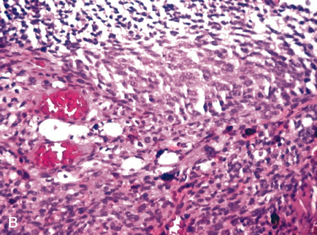
observations. Arch Ophthalmol 101:102, 1983.

123. Rao NA, Wacker WB, Marak GE Jr: Experimental allergic uveitis: Clinicopathologic features associated with varying doses of S antigen. Arch Ophthalmol 97:1954, 1979.

124. Sugita S, Sagawa K, Mochizuki M et al: Melanocyte lysis by cytotoxic T lymphocytes recognizing the MART-1 melanoma antigen in HLA-A2 patients with Vogt-Koyanagi-Harada disease. Int Immunol 8:799, 1996.

125. Read RW, Holland GN, Rao NA et al: Revised diagnostic criteria for Vogt-Koyangi-Harada disease: Report of an international committee on nomenclature. Am J Ophthalmol 131:647, 2001.

126. Rao NA, Hidayat AA: Endogenous mycotic endophthalmitis: Variations in clinical and histopathologic changes in candidiasis compared with aspergillosis. Am J Ophthalmol 132:244, 2001.

127. Wilder H: Toxoplasma chorioretinitis in adults. Arch Ophthalmol 48:127, 1952.

128. Desmontis G, Remington JS, Couvreur J: Congenital toxoplasmosis. In Stern L, Vert P (eds): Neonatal Medicine, pp 992–678. New York, Masson, 1987.

129. Stagno S, Dykes AC, Amos CS et al: An outbreak of toxoplasmosis linked to cats. Pediatrics 65:706, 1980.

130. Akstein RB, Wilson LA, Teutsch SM: Acquired toxoplasmosis. Ophthalmology 89:1299, 1982.

131. Michelson JB, Shields JA, McDonald R et al: Retinitis secondary to acquired systemic toxoplasmosis with isolation of parasite. Am J Ophthalmol 86:548, 1978.

132. Hausmann N, Richard G: Acquired ocular toxoplasmosis. Ophthalmology 98:1647, 1991.

133. Rothova A: Ocular involvement in toxoplasmosis. Br J Ophthalmol 77:371–377, 1993.

134. Holland GN, Engstrom RE Jr, Glasgow BJ et al: Ocular toxoplasmosis in patients with acquired immunodeficiency syndrome. Am J Ophthalmol 106:653, 1988.

135. Sternberg P Jr, Knox DL, Finkelstein D et al: Acute retinal necrosis syndrome. Retina 2:145, 1982.

136. Greven CM, Ford J, Stanton C et al: Progressive outer retinal necrosis secondary to varicella zoster virus in acquired immune deficiency syndrome. Retina 15:14, 1995.

137. Naumann G, Yanoff M, Zimmerman LE: Histogenesis of malignant melanomas of the uvea. Arch Ophthalmol 76:784, 1966.

138. Timmerman LE: Melanocystes, melanocytic nevi and melanocytomas. Invest Ophthalmol 4:11, 1965.

139. Witschel H, Font RL: Hemangioma of the choroid. A clinicopathologic study of 71 cases and a 
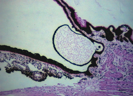
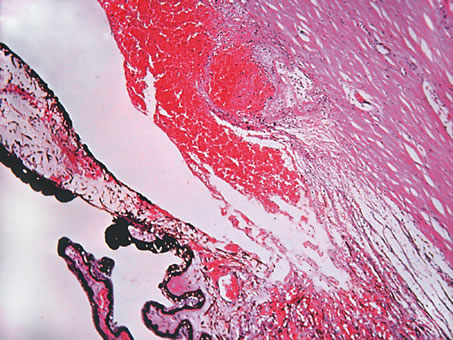
review of the literature. Surv Ophthalmol 20:415, 1976.

140. Anand R, Augsburger JJ, Shields JA: Circumscribed choroidal hemangiomas. Arch Ophthalmol 107:1338, 1989.

141. Aylword GW, Chang TS, Pautler SE et al: A long-term follow-up of choroidal osteoma. Arch Ophthalmol 116:1337, 1998.

142. Williams AT, Font RL, Van Dyk HJL et al: Osseous choristoma of the choroid simulating a choroidal melanoma. Arch Ophthalmol 96:1875, 1978.

143. Perri P, Pauduano B, Incorvaia C et al: Mesectodermal leiomyoma exclusively involving the posterior choroid. Am J Ophthalmol 134:451, 2002.

144. Harkin JC, Reed RJ: Tumors of the peripheral nervous system. In Armed Forces Institute of Pathology: Atlas of Tumor Pathology. 2nd Series, Fascicle 3. Washington, DC, 1969.

145. Freedman SF, Elner VM, Donev R et al: Intraocular neurilemmoma arising from the posterior ciliary nerve in neurofibromatosis. Ophthalmology 195:1559, 1988.

146. Zimmerman LE: Malignant melanoma. In Spencer WH (ed): Ophthalmic Pathology: An Atlas and Textbook, pp 2072–2141. 3rd ed. Philadelphia, WB Saunders, 1985.

147. Gallagher RP, Elwood JM, Rootman J: Epidemiologic aspects of intraocular malignant melanoma. Cancer Treat Res 43:73, 1988.

148. Cutler SJ, Young JL: Third National Cancer Survey. Incidence Data, pp 1–9. NCI Monograph. Vol 41. Bethesda, National Institutes of Health, 1975.

149. Margo CE, McLean IW: Malignant melanoma of the choroid and ciliary body in black patients. Arch Ophthalmol 102:77, 1984.

150. Scotto J, Fraumenti JF Jr, Lee JA: Melanomas of the eye and other noncutaneous sites: Epidemiologic aspects. J Natl Cancer Inst 56:489, 1976.

151. Gonder JR, Ezell PC, Sheilds, JA et al: Ocular melanocytosis. A study to determine the prevalence rate of ocular melanocytosis. Ophthalmology 89:950, 1982.

152. Shields JA, Albert DM: Malignant melanoma of the choroid associated with oculodermal melanocytosis. Ophthalmology 88:372, 1981.

153. Gass JD: Observation of suspected choroidal and ciliary body melanomas for evidence of growth prior to enucleation. Ophthalmology 187:523, 1980.

154. Naumann G, Yanoff M, Zimmerman LE: Histiogenesis of malignant melanomas of the uvea. I. Histopathologic characteristics of nevi of the choroid and ciliary body. Arch Ophthalmol 1966;76:784–96

155. Yanoff M, Zimmerman LE: Histogenesis of malignant melanomas of the uvea. II. Relationship of uveal nevi to malignant melanomas. Cancer 20:493, 1967.

156. Nik ND, Glew WB, Zimmerman LE: Malignant melanoma of the choroid in the nevus of Ota of a black patient. Arch Ophthalmol 76:784, 1966.

157. Lynch HT, Anderson DE, Krush AJ: Heredity and intraocular malignant melanoma. Cancer 21:119, 1968.

158. Walker JP, Weiter JJ, Albert DM et al: Uveal malignant melanoma in three generations of the same family. Am J Ophthalmol 88:723, 1979.

159. Prescher G, Bornfeld N, Becher R: Nonrandom chromosomal abnormalities in primary uveal melanoma. J Natl Cancer Inst 82:1765, 1990.

160. Sisley K, Cottam DW, Rennie IG et al: Non-random abnormalities of chromosomes 3, 6 and 8 associated with posterior uveal melanoma. Genes Chromosomes Cancer 5:197, 1992.

161. Wiltshire RN, Elner VM, Dennis T et al: Cytogenic analysis of posterior uveal melanoma. Cancer Genet Cytogenet 66:47, 1993.

162. Tobal K, Warren W, Cooper CS et al.: Increased expression and mutation of p53 in choroidal melanoma. Br J Cancer 66:900, 1992.

163. Jay M, McCartney AC: Familial malignant melanoma of the uvea and p53: A Victorian detective story. Surv Ophthalmol 37:457, 1993.

164. Zimmerman LE, McLean IW: Do growth and onset of symptoms of uveal melanomas indicate subclinical metastasis? Ophthalmology 91:685, 1984.

165. Font RL, Spaulding AG, Zimmerman LE: Diffuse malignant melanoma of the uveal tract. A clinicopathologic report of 54 cases. Trans Am Acad Ophthalmol Otolaryngol 72:877, 1968.

166. Callender GR: Malignant melanotic tumors of the eye: a study of histologic types in 111 cases. Trans Am Acad Ophthalmol Otolaryngol 36:131, 1931.

167. Burnier MN Jr, McLean IW, Carmel JW: Immunohistochemical evaluation of uveal melanocytic tumors. Expression of HMB-45, S-100 protein, and neuron-specific enolase. Cancer 68:809, 1991.

168. Iwamoto S, Burrows RC, Kalina RE et al: Immunophenotypic differences between uveal and cutaneous melanomas. Arch Ophthalmol 130:466, 2002.

169. Wilder HC, Paul EV: Malignant melanoma of the choroid and ciliary body: a study of 2,535 cases. Mil Surg 109:370, 1951.

170. McLean IW, Foster WD, Zimmerman, LE: Prognostic factors in small malignant melanomas of choroid and ciliary body. Arch Ophthalmol 95:48, 1977.

171. Gamel JW, McCurdy JB, McLean IW: A comparison of prognostic covariates for uveal melanoma. Invest Ophthalmol Vis Sci 33:1919, 1992.

172. Hayton S, Lafreniere R, Jerry LM et al: Ocular melanoma in Alberta: A 38 year review pointing to the importance of tumor size and tumor histology as predictors of survival. J Surg Oncol 42:215, 1989.

173. Shammas HF, Blodi FC: Prognostic factors in choroidal and ciliary body melanomas. Arch Ophthalmol 95:53, 1977.

174. Affeldt JC, Minckler DS, Azen SP et al.: Prognosis in uveal melanoma with extrascleral extension. Arch Ophthalmol 98:1975, 1980.

175. Folberg R, Pe'er J, Gruman LM et al: The morphologic characteristics of tumor blood vessels as a marker of tumor progression in primary human uveal melanoma: A matched case-control study. Hum Pathol 23:1298, 1992.

176. Ferry AP, Font RL: Carcinoma metastatic to the eye and orbit. I. A clinicopathologic study of 227 cases. Arch Ophthalmol 92:276, 1974.

177. Stephens RF, Shields JA: Diagnosis and management of cancer metastatic to the uvea: A study of 70 cases. Ophthalmology 86:1336, 1979.

Back to Top