Chapter 21
Retinoblastoma and Simulating Lesions
RALPH C. EAGLE JR.
Main Menu   Table Of Contents

Search

CLINICAL PRESENTATION
NATURAL HISTORY
GROSS PATHOLOGY
HISTOPATHOLOGY
ROSETTES AND PHOTORECEPTOR DIFFERENTIATION
MOLECULAR GENETICS AND THE RETINOBLASTOMA ONCOGENE
DIFFERENTIAL DIAGNOSIS OF RETINOBLASTOMA

Retinoblastoma is the most common intraocular tumor of childhood. In Asia, Africa, and South America, where uveal melanoma is relatively rare, retinoblastoma probably is the most common primary intraocular tumor. In Europe and the United States, it is second in overall prevalence, reflecting the propensity of lightly pigmented adult Europeans to develop uveal melanoma. The incidence of retinoblastoma has been estimated to be 1 in 15,000 to 1 in 34,000 births. Retinoblastoma is cosmopolitan and affects all races. Both sexes are affected equally, and the tumor has no predilection for the right or left eye. The average age at diagnosis is 18 months. Although cases have been reported in adults, retinoblastoma is rare after age 4 years.

Retinoblastoma has many unusual and interesting clinical characteristics. Foremost, it is a hereditary cancer: 5% to 10% of the tumors are inherited. In affected families, retinoblastoma is transmitted as an autosomal dominant trait, and the tumor frequently affects both eyes. Bilaterality is a clinical marker for potentially transmissible disease. About one third of all retinoblastomas are bilateral; the additional bilateral cases are caused by new germline mutations. The great majority of retinoblastomas arise sporadically in patients with a negative family history. About 75% of the sporadic tumors are caused by somatic mutations and are not transmissible. Sporadic tumors caused by somatic mutations are always unilateral. Unfortunately, unilateral involvement does not exclude heritable disease: about 10% to 15% of infants with unilateral sporadic tumors have heritable germinal mutations. The retinoblastoma gene is discussed below.

Back to Top
CLINICAL PRESENTATION
The clinical presentation of retinoblastoma depends on the size, location, growth pattern, and stage of the lesion at diagnosis and the availability and sophistication of medical care in the community. In the United States and Europe, about 90% of patients present with leukocoria, an abnormal, typically white pupillary reflex (Figs. 14). Leukocoria is the classic clinical manifestation of retinoblastoma. In the older literature, this finding was fancifully likened to the illuminated tapetal light reflex of the cat (amaurotic cat's-eye reflex). The white pupillary reflex is caused by tumor in the vitreous cavity (endophytic tumors) or total retinal detachment (exophytic tumors).

Fig. 1. Leukocoria, unilateral sporadic retinoblastoma. A white pupillary reflex is the presenting manifestation of retinoblastoma in about 90% of patients in the United States. (Photo courtesy of Dr. Jerry A. Shields, Wills Eye Hospital)

Fig. 2. Bilateral leukocoria, familial retinoblastoma. The presence of bilateral tumors indicates that the affected patient is a carrier of familial retinoblastoma who can transmit the tumor to progeny. Bilateral tumors occur in about two thirds of patients with familial retinoblastoma. (Photo courtesy of Dr. Jerry A. Shields, Wills Eye Hospital)

Fig. 3. Leukocoria, endophytic retinoblastoma. Tumor growing in the vitreous cavity produces white pupillary reflex. (Photo courtesy of Dr. Jerry A. Shields, Wills Eye Hospital)

Fig. 4. Leukocoria, exophytic retinoblastoma. The tumor has arisen from the outer layers of the retina, producing a retinal detachment. Retinal vessels are seen behind the lens. (Photo courtesy of Dr. Jerry A. Shields, Wills Eye Hospital)

Strabismus or ocular misalignment occurs in about one third of cases and may herald a relatively small tumor that involves the fovea. For this reason, careful ophthalmoscopy should be performed in all children with strabismus to exclude retinoblastoma or some other significant retinal pathology. About half of the eyes enucleated for retinoblastoma have iris neovascularization, which may produce neovascular glaucoma, iris heterochromia, and even secondary buphthalmos. Pupillary block glaucoma caused by anterior displacement of the lens-iris diaphragm may be found in eyes with exophytic tumors and total retinal detachments.

Endophytic tumors and the rare diffuse infiltrating variant of the disease often seed the anterior chamber, forming a pseudohypopyon of tumor cells that may be initially misdiagnosed as inflammation. An orbital cellulitis-like picture is another type of pseudoinflammatory presentation. In such cases, histopathologic examination usually reveals severe neovascular glaucoma that has caused extensive infarction of the tumor and other intraocular structures.

In underdeveloped countries, patients may present in the late stages of the disease with a tumor that has extended extraocularly, causing exophthalmos and an orbital mass.

Rare tumors are manifest at birth. Congenital retinoblastoma may present with a massive hyphema and an enlarged ectatic cornea that spontaneously perforates (Fig. 5).

Fig. 5. Congenital retinoblastoma. Clinical photo shows affected infant several days after birth. The perforated cornea is markedly ectatic, and blood fills the anterior chamber.

Back to Top
NATURAL HISTORY
Retinoblastoma is a highly malignant neoplasm that grows relentlessly and almost always has a fatal outcome if untreated. The tumor arises from retinal cells and initially is confined to the retina. Clinically, there appears to be a direct correlation between patient age and retinal topography that follows a central to peripheral distribution. Macular tumors present earliest; anterior tumors present last. In Abramson's series of 565 eyes with bilateral retinoblastoma, all macular tumors and most retinoblastomas of the posterior pole were detected before age 24 months.

As it grows, the tumor invades the vitreous cavity and/or the subretinal space. Endophytic retinoblastomas that arise from the inner layers of the retina seed the inner surface of the retina, the vitreous cavity, and eventually the anterior chamber, where the tumor cells may infiltrate the iris stroma or trabecular meshwork or form a pseudohypopyon in the inferior angle (Fig. 6). Exophytic tumors arising from the outer retinal layers typically cause a serous retinal detachment. The retinal detachment is often high and bullous in configuration, and the retina typically is plastered against the posterior surface of the lens (Fig. 7). Such eyes frequently have secondary closed-angle glaucoma due to pupillary block. The subretinal fluid often contains seeds of tumor, and a sheet of tumor cells may blanket the inner surface of the retinal pigment epithelium (RPE). Retinoblastoma cells often invade the space between the RPE and Bruch's membrane, causing focal RPE detachments. Bruch's membrane initially serves as a mechanical barrier to invasion. Eventually, however, the retinoblastoma cells breach Bruch's membrane and invade the choroidal stroma (Fig. 8). The uveal tract's rich blood supply nurtures the proliferating tumor cells and can serve as a major route for distant hematogenous dissemination. Although multivariant analysis suggests that the presence of extrascleral extension is a much better indicator of poor prognosis, massive uveal invasion does carry a risk for distant metastasis. In one series, the mortality rate was about 25% with minimal choroidal invasion; this rose to 65% if the invasion was massive. Metastatic disease usually is evident within 1 or 2 years after therapy. Late metastasis is so rare in retinoblastoma that it should raise the possibility of a second independent primary tumor.

Fig. 6. Retinoblastoma, endophytic growth pattern. A white encephaloid mass arises from the inner retina and invades the vitreous cavity. Most of the retina remains attached.

Fig. 7. Exophytic retinoblastoma. Macrophoto of enucleated eye with exophytic retinoblastoma shows encephaloid tumor in subretinal space and total bullous retinal detachment that adheres to the back of the lens. The lens-iris diaphragm is displaced anteriorly, causing secondary closure of the angle.

Fig. 8. Choroidal invasion. A basophilic infiltrate of poorly differentiated, viable retinoblastoma diffusely thickens the choroid. No tumor necrosis is evident in the richly vascularized choroid. The RPE is variably pigmented and the retina is detached. (Hematoxylin and eosin, × 50)

After a retinoblastoma has filled the globe and destroyed its contents, it extends extraocularly. Anteriorly, extraocular extension occurs through the aqueous outflow pathways, pre-existing emissarial canals, or perforations in the cornea. Posterior segment tumors that have invaded the choroid can extend extraocularly along emissarial canals or can invade the orbit by directly infiltrating and destroying the sclera. Patients from underdeveloped countries often present with orbital masses of tumor tissue, which contain ocular remnants, mainly sclera. Secondary buphthalmos and staphylomas caused by secondary glaucoma can facilitate extrascleral extension. Secondary closed-angle glaucoma caused by pupillary block or iris neovascularization is relatively common in eyes with retinoblastoma. Angiogenic factors such as VEGF produced by the tumor cells or the ischemic retina are the probable cause of the iris neovascularization.

The optic nerve is the most common avenue for extraocular extension by retinoblastoma. Retinoblastoma has a marked proclivity to invade the optic nerve (Fig. 9). Some degree of optic nerve invasion was present in 29.5% of 814 retinoblastomas reported by Magramm and coworkers. In this respect, the behavior of retinoblastoma contrasts sharply with that of choroidal melanoma, which invades the optic nerve only 5% of the time. The tumor may travel to the brain along the parenchyma of the nerve or via the subarachnoid space, or both. Rare tumors can travel around the chiasm and reach the posterior aspect of the fellow eye. Retinoblastoma cells also can be disseminated widely through the cerebrospinal fluid in the subarachnoid space.

Fig. 9. Retinoblastoma, grade III optic nerve invasion. The retrolaminar parenchyma of the optic nerve contains a basophilic infiltrate of retinoblastoma cells. (Hematoxylin and eosin, × 10)

Optic nerve invasion is an extremely important prognostic factor in retinoblastoma. Mortality rates correlate directly with the depth of optic nerve invasion. In Magramm's series, the mortality rate was 10% if there was superficial invasion of the nerve head only (grade I) and 29% for involvement up to and including the lamina cribrosa (stage II). It rose to 42% when there was retrolaminar invasion (grade III) and 78% when the tumor extended to the surgical margin (grade IV).

Hence, the surgeon must obtain a long segment of optic nerve when enucleating an eye that is known or suspected to contain a retinoblastoma. If possible, enucleation should not be performed by an inexperienced surgeon. In addition, special care must be taken during gross dissection to avoid contaminating this important surgical margin with tumor. The optic nerve margin should be marked with indelible pencil or India ink before sectioning, and the optic nerve should always be removed before the globe is opened and submitted in a separate cassette. This is particularly important if fresh tumor is harvested in the operating room. In such cases, the marginal segment of nerve should be submitted in a separate container. Blood-staining, obliquity of sectioning, and slight crushing serve to identify the true surgical margin in the laboratory.

Retinoblastoma typically metastasizes hematogenously to lungs, bones, brain, and other organs after the tumor gains access to the blood vessels in the uvea or orbital tissues. Cervical and preauricular adenopathy occasionally develops when advanced tumors with extensive anterior segment involvement gain access to lymphatics in the conjunctival stroma. Lymphatics are not present in the orbit.

Retinoblastoma occasionally undergoes spontaneous regression. In the past, the incidence of spontaneous regression was estimated to be 1%, which is higher than for any other malignant tumor. Currently, however, it is believed that many lesions that once were classified as spontaneously regressed retinoblastomas are actually benign nonprogressive retinal tumors called retinocytomas or retinomas (some favor the term “spontaneously arrested retinoblastoma”).

Clinically, retinocytomas were thought to represent spontaneously regressed retinoblastomas because they resemble retinoblastomas that have regressed after radiation therapy (Fig. 10). They have a translucent “fish flesh” appearance, contain abundant calcification that has been likened to cottage cheese, and are surrounded by a ring of RPE depigmentation. Retinocytomas generally are small tumors that are found in eyes that retain useful vision. They may be incidental findings or are discovered in a parent or siblings when the detection of retinoblastoma in a child prompts examination of other family members. Rare cases of retinocytoma have been observed to undergo malignant transformation into retinoblastoma. Histopathologic examination in one such a case confirmed the presence of benign cytology and photoreceptor differentiation in the original basal part of the tumor. Retinocytomas are relatively resistant to radiation, as are other benign tumors. It, therefore, is not surprising that foci of photoreceptor differentiation are found more often in eyes that are enucleated after external-beam radiotherapy or chemoreduction.

Fig. 10. Retinocytoma. The tumor has a translucent “fish flesh” appearance and contains abundant calcification that has been likened to cottage cheese. Depigmented RPE surrounds the tumor. This lesion was stable for several years before undergoing malignant transformation. (From Eagle RC Jr, Shields JA, Donoso L, Milner RS: Malignant transformation of spontaneously regressed retinoblastoma, retinoma/retinocytoma variant. Ophthalmology 96:1389, 1989)

Bona fide cases of spontaneous regression of retinoblastoma do occur but are rare. They typically are found in blind, phthisical eyes. Both the regression and phthisis bulbi probably are caused by intraocular infarction in an eye with neovascular glaucoma and markedly elevated intraocular pressure (Fig. 11).

Fig. 11. Spontaneously regressed retinoblastoma, phthisical eye. Basophilic foci of necrotic, calcified tumor cells are the only remnants of retinoblastoma in this phthisical eye. The calcific foci are located in the posterior chamber behind the necrotic iris. (Hematoxylin and eosin, × 25)

Several staging systems for retinoblastoma are available. The Reese-Ellsworth classification for intraocular tumors has prognostic significance for retention of an eye and the control of local disease (Table 1). The St. Jude Children's Research Hospital Clinical Staging System (Table 2) attempts to relate the extent of the disease within and outside the eye to prognosis for sight as well as for freedom from systemic disease. This system is histologically based and requires enucleation.


TABLE ONE. Reese and Ellsworth Classification

  Group I: Very favorable for maintenance of sight

  1. Solitary tumor, smaller than 4 disc diameters in size, at or behind the equator
  2. Multiple tumors, none larger than 4 disc diameters in size, all at or behind the equator

  Group II: Favorable for maintenance of sight
  1. Solitary tumor, 4–10 disc diameters in size, at or behind the equator
  2. Multiple tumors, 4–10 disc diameters in size, behind the equator

  Group III: Possible for maintenance of sight
  1. Any lesion anterior to the equator
  2. Solitary tumor, larger than 10 disc diameters in size, behind the equator

  Group IV: Unfavorable for maintenance of sight
  1. Multiple tumors, some larger than 10 disc diameters in size
  2. Any lesion extending anteriorly to the ora serrata

  Group V: Very unfavorable for maintenance of sight
  1. Massive tumors involving more than one half the retina
  2. Vitreous seeding


Approximately 90% of patients present with one or both eyes categorized as group V.


TABLE TWO. St. Jude Children's Research Hospital Clinical Staging System

  Stage I: Tumor confined to the retina

  1. Occupying one quadrant or less
  2. Occupying two quadrants or less
  3. Occupying more than 50% of the retinal surface

  Stage II: Tumor confined to globe
  1. With vitreous seeding
  2. Extension to optic nerve head
  3. Extension to choroid
  4. Extension to choroid and optic nerve head
  5. Extension to emissaries

  Stage III: Regional extraocular extension of tumor
  1. Extension beyond cut ends of optic nerve
  2. Extension through sclera into orbital contents
  3. Extension to choroid beyond cut end of optic nerve (including subarachnoid extension)
  4. Extension through sclera into orbital contents and beyond cut end of optic nerve (including subarachnoid extension)

  Stage IV: Distant metastases
  1. Extension via optic nerve to brain (i.e., gross tumor in the CNS or tumor cells in the cerebrospinal fluid)
  2. Blood-borne metastases to soft tissue, bone, or viscera
  3. Bone marrow metastases


Approximately 80% of patients present with one or both eyes classified as stage I–II.


Back to Top
GROSS PATHOLOGY
The gross appearance of retinoblastoma is somewhat variable, reflecting the stage of the disease at enucleation. Because the retina can be considered to be a peripheral colony of brain cells, it is not surprising that the tumor characteristically has a white encephaloid, or brainlike, appearance grossly. The whitish tumor thickens, replaces and destroys part or all of the retina, and may totally fill the vitreous cavity. The mass typically contains lighter foci of necrotic tumor or calcification. Gritty crystals of calcium apatite or lemon-yellow pigmentation may be encountered in some advanced lesions.

Totally necrotic retinoblastomas that have undergone acute infarction occasionally are encountered. Such tumors typically have a blood-tinged, orange, or soupy, grayish necrotic appearance grossly. Infarction caused by severe neovascular glaucoma also affects other intraocular structures, including the iris, ciliary body, and retina, causing inflammatory signs and symptoms. Affected infants may be initially misdiagnosed as having orbital cellulitis.

Several tumor growth patterns are recognized macroscopically. Exophytic retinoblastomas arise from the outer retinal layers, proliferate in the subretinal space, and cause secondary retinal detachment (see Fig. 7). The detached retina is typically highly elevated, and its vessels are visible behind the lens on clinical examination. Eyes with exophytic retinoblastomas often develop secondary pupillary block glaucoma. Clinically, exophytic retinoblastomas can be confused with simulating lesions that cause retinal detachment, such as Coats' disease.

Endophytic retinoblastomas grow into the vitreous cavity, obscuring the retina, which remains attached (see Fig. 6). Tumor fills the vitreous cavity by direct extension and seeding. Retinal vessels are not evident in the white pupil on slit-lamp examination. A pseudohypopyon of tumor cells often develops if seeding involves the anterior chamber. Hence, endophytic tumors can be confused with primary inflammatory disorders such as toxocariasis, mycotic endophthalmitis, or granulomatous uveitis. As a rule, purely endophytic or exophytic retinoblastomas are uncommon. Most tumors exhibit foci of both growth patterns.

A diffuse infiltrative growth pattern occurs in less than 2% of cases. Such diffuse infiltrative tumors are often overlooked or misdiagnosed because they diffusely thicken the retina and do not form a discrete tumefaction (Fig. 12). Diffuse infiltrative retinoblastoma occurs in older children, whose mean age at diagnosis is about 6 years. Such tumors are invariably unilateral and sporadic. Like endophytic retinoblastoma, diffuse infiltrative retinoblastoma typically presents with anterior segment seeding and pseudohypopyon; it is often misdiagnosed as a primary inflammatory disorder or juvenile xanthogranuloma. Some have suggested that this variant of retinoblastoma may have a relatively good prognosis despite delays in diagnosis, but this issue remains controversial.

Fig. 12. Diffuse infiltrating retinoblastoma. The diffusely infiltrating tumor thickens and opacifies much of the retina but does not form a distinct mass. Tumor seeds blanket the pars plana. The unilateral sporadic tumor was found in a 7-year-old boy who presented with a pseudohypopyon of retinoblastoma cells.

Retinoblastoma is a friable, poorly cohesive neoplasm that can readily disperse or seed widely. Tumor seeds deposited on the inner or outer surfaces of the retina frequently give rise to secondary tumors. The latter may be impossible to differentiate grossly or histologically from multifocal primary lesions. This is unfortunate because true multifocal involvement of the retina signifies hereditary, potentially transmissible disease.

Back to Top
HISTOPATHOLOGY
Retinoblastoma has a characteristic appearance and staining pattern in routine sections stained with hematoxylin and eosin that instantly suggests the diagnosis under low magnification. The vitreous cavity is filled totally, or in part, by a basophilic mass that arises from and destroys the retina (Fig. 13). The basophilic tumor usually contains eosinophilic and reddish-purple foci (Fig. 14).

Fig. 13. Endophytic retinoblastoma. The basophilic neoplasm arises from and destroys the retina, which remains attached as the endophytic tumor invades the vitreous cavity. Rosettes are not seen. (Hematoxylin and eosin, × 25)

Fig. 14. Retinoblastoma. Photomicrograph shows basophilic sleeves of viable tumor surrounding vessels. Extensive tumor necrosis is present. The necrotic area is eosinophilic and contains a reddish-purple focus of dystrophic calcification. (Hematoxylin and eosin, × 10)

Viable parts of a retinoblastoma appear blue because they are composed of poorly differentiated neuroblastic cells that have scanty cytoplasm and intensely basophilic nuclei (Fig. 15). Many mitoses and fragments of apoptotic nuclear debris usually are present. Retinoblastoma grows rapidly and has a marked propensity to outgrow its blood supply and undergo spontaneous coagulative necrosis. This usually occurs when the proliferating cells have extended about 90 to 110 μm away from a blood vessel (see Fig. 15). The necrotic tumor cells lose their basophilic nuclear DNA and become pink or eosinophilic. The residual viable cells typically form cuffs or sleeves around vessels, imparting a multilobulated or papillary appearance to some tumors. These perivascular cuffs were called pseudorosettes by some in the past. Foci of dystrophic calcification develop in the necrotic parts of the tumor in many cases. Histopathologically, the calcific foci appear reddish-purple in hematoxylin-and-eosin sections, and the presence of calcium can be confirmed by the von Kossa or alizarin red stains. Electron microscopy suggests that calcification probably begins in the mitochondria of necrotic cells. Clinically, the demonstration of calcification by ultrasonography or computed tomography can help to differentiate retinoblastoma from other simulating lesions.

Fig. 15. Retinoblastoma, poorly differentiated neuroblastic cells. The malignant tumor is composed of cells with scanty cytoplasm and prominent basophilic nuclei. Mitotic figures and a few apoptotic cells are present. (Hematoxylin and eosin, × 250)

Intensely basophilic deposits of DNA released from necrotic tumor cells are another characteristic histopathologic feature of retinoblastoma. DNA deposition generally is found in eyes with extensively necrotic tumors. Typically, the DNA deposits surround retinal or iris vessels or are found in the trabecular meshwork or in basement membranes such as the lens capsule or the internal limiting membrane of the retina.

Back to Top
ROSETTES AND PHOTORECEPTOR DIFFERENTIATION
The most characteristic histopathologic feature of retinoblastoma is the rosettes of tumor cells described by Simon Flexner in 1881 and Hugo Wintersteiner in 1897 (Fig. 16). Flexner-Wintersteiner rosettes represent an early attempt at retinal differentiation. Histologically, these rosettes are composed of a ring of cuboidal cells surrounding a central lumen. The lumen, which corresponds to the subretinal space, contains hyaluronidase-resistant acid mucopolysaccharide (AMP) similar to photoreceptor matrix AMP. The cells surrounding the lumen are joined near their apices by intercellular connections (zonulae adherentes), analogous to the external limiting membrane of the retina. Cilia, which exhibit the 9 + 0 pattern of microtubular doublets found in the central nervous system, project into the lumen. Cilia are hypothesized to be the precursor of photoreceptor outer segments.

Fig. 16. Flexner-Wintersteiner rosettes. Flexner-Wintersteiner rosettes have a central lumen that corresponds to the subretinal space. The tumor cells surrounding the lumen are joined by cellular connections that are analogous to the retina's external limiting membrane. (Hematoxylin and eosin, × 250)

The presence of many Flexner-Wintersteiner rosettes in a tumor may have prognostic significance. In one series, patients who had moderately well-differentiated tumors that contained abundant Flexner-Wintersteiner rosettes had about a sixfold better prognosis than those whose tumors lacked rosettes. Flexner-Wintersteiner rosettes are more frequent in small, relatively early tumors and may be totally absent in large tumors, which usually are poorly differentiated. Flexner-Wintersteiner rosettes rarely are found in foci of metastatic retinoblastoma.

Another less-differentiated type of rosette found in retinoblastoma was described by James Homer Wright (Fig. 17). Wright rosettes are indicative of neuroblastic differentiation. They lack a central lumen, and their constituent cells encompass a central tangle of neural filaments. Wright rosettes are relatively nonspecific. They also occur in neuroblastoma and are a characteristic feature of cerebellar medulloblastoma.

Fig. 17. Wright rosettes. An irregular ring of nuclei encompasses a central tangle of neural processes. No lumen is present. (Hematoxylin and eosin, × 250)

Fleurettes, described by Ts'o, Zimmerman, and Fine in 1970, are aggregates of neoplastic photoreceptors that represent a greater degree of retinal differentiation (Fig. 18). Found in approximately 6% of cases, fleurettes typically occur in a viable area of tumor that appears less cellular and relatively more eosinophilic than adjacent undifferentiated retinoblastoma during low-magnification microscopy. The term “fleurette” denotes a bouquet-like arrangement of cytologically benign cells joined by a series of zonulae adherentes that may form a short segment of neoplastic external limiting membrane. Bulbous eosinophilic processes that represent abortive photoreceptor inner segments form the “flowers” of the bouquet. Electron microscopy occasionally discloses stacks of cellular membranes representing early outer segment differentiation. The demonstration and characterization of photoreceptor differentiation firmly established that retinoblastoma is not a glioma of the retina and affirmed that the adoption of that name by the American Ophthalmological Society in 1926 at Frederick Verhoeff's suggestion was indeed appropriate.

Fig. 18. Photoreceptor differentiation (fleurettes). Bulbous neoplastic photoreceptor inner segments form eosinophilic bouquet-like structures. Some cells are aligned along a segment of neoplastic external limiting membrane. The nuclei are bland and mitoses and necrosis are not evident. (Hematoxylin and eosin, × 250)

Foci of photoreceptor differentiation are found more frequently (40% of cases) in eyes that are enucleated after radiotherapy or chemoreduction. Presumably, the chemo- or radiotherapy kills the malignant part of the tumor and discloses the benign foci, which are relatively radio- or chemoresistant. Retinal tumors composed entirely of fleurettes are thought to be a benign variant of retinoblastoma that is incapable of metastasis. Such tumors are called retinocytomas (see above). Compared to retinoblastoma, the cells composing retinocytoma are quite bland, and mitotic activity is uncommon. In addition, calcification occurs in viable parts of retinocytomas.

Retinoblastoma is thought to be derived histogenetically from primitive retinal cells that are capable of differentiation into both neuronal and glial elements. Immunocytochemical studies of retinoblastomas have demonstrated cells that are immunoreactive for either photoreceptors or the giant glial cells of Mueller. Although the actual cell of origin is uncertain, there is evidence that retinoblastoma could be derived from photoreceptors.

Back to Top
MOLECULAR GENETICS AND THE RETINOBLASTOMA ONCOGENE
Retinoblastoma is a heritable cancer. Between 5% and 10% of cases are inherited as what appears to be a classic mendelian autosomal dominant trait with incomplete penetrance and expressivity. In other words, a carrier of familial retinoblastoma transmits the tumor to half of his or her offspring. Molecular genetic studies have shown that the gene responsible for retinoblastoma actually is recessive on the molecular level. In fact, the retinoblastoma gene is the paradigm of a human recessive oncogene or tumor suppressor gene.

This retinoblastoma or RB gene is located in the 14 band of the Q or long arm of chromosome 13. The RB gene is 180,388 base pairs in length, and its protein product pRB comprises 928 amino acids. pRB is abundant in the nucleus, where it is involved in control of the cell cycle. During the G1 or resting phase of the cell cycle, pRB is bound to transcription factors such as E2F. Phosphorylation of pRB causes release of E2F. Uncomplexed E2F, in turn, activates a variety of other genes and transcription factors that are important in the initiation of DNA synthesis (S phase). Absence of pRB causes continual cell division and lack of terminal differentiation. The RB protein is phosphorylated by cyclin D2 and its cyclin-dependent kinase (cdk) partner. Certain oncoviruses cause tumors by synthesizing proteins (e.g., adenoviral protein E1A and SV40 large T protein) that bind to pRB and inactivate it.

The RB gene causes cancer when its protein product is absent or dysfunctional. Healthy persons have two normal or wild-type RB genes. Both alleles of the RB gene are absent or inactivated in the cells of a retinoblastoma. Carriers of familial retinoblastoma are heterozygous for the RB gene. Although the pRB produced by a heterozygote's single functional gene is sufficient to inhibit tumorigenesis, heterozygotes are at substantial risk to develop retinoblastoma.

The genotype of a child with familial retinoblastoma includes one functional copy of the RB gene. The other copy is absent or is mutant and codes for dysfunctional RB protein. Retinoblastoma develops when the solitary remaining wild-type gene is lost or inactivated, by chance, in a cell in the developing retina. Cytogenetic mechanisms responsible for gene inactivation (and resultant homozygosity for the recessive allele) include chromosomal loss or deletion, somatic recombination, and point mutation. The spontaneous mutation rate of the normal wild-type gene RB is 1 in 10 million or greater. It is estimated that 100 million mitoses occur during the growth and development of each retina. Hence, it is highly probable that the second functional copy of the RB gene will be lost or inactivated in at least one retinal cell in an individual whose genotype is heterozygous. Further, it is equally probable that gene inactivation and tumorigenesis will occur in both eyes.

An analogous situation occurs in patients who have 13Q- or chromosomal deletion retinoblastoma, which is associated with a karyotypically apparent deletion that includes the Q14 band of chromosome 13. In an analogous fashion, retinoblastoma develops after the chance loss or inactivation of the solitary suppressor gene in a retinal cell. Patients with 13Q- syndrome have severe mental retardation and other congenital abnormalities that may include microcephaly, hypertelorism, ptosis, micrognathia, deformed low-set ears, a wide nasal bridge, cardiac anomalies, anal atresia, microphthalmia, colobomas, and cataracts.

There is no evidence to support the interesting speculation that a “triple dose” of suppressor substance could be responsible for the hypoplastic ocular anomalies, such as anophthalmia, microphthalmia, and cyclopia/synophthalmia, that occur in patients with trisomy 13.

Immune surveillance appears to arrest some tumors, despite the appropriate second mutation. This probably is responsible in part for the incomplete penetrance of the RB gene, which is estimated to be approximately 80%—in other words, there is an 80% chance that one tumor will develop in one eye. A few families with low-penetrance retinoblastoma have been reported. They have reduced levels of wild-type RB protein or mutant RB protein that retains partial activity.

Most retinoblastomas are sporadic tumors that occur in patients who have a negative family history and a normal karyotype. Three fourths of the sporadic tumors are caused by somatic mutations. Always unilateral and solitary, such somatic sporadic retinoblastomas presumably are caused by the chance inactivation of both RB genes in a single cell in the developing retina (somatic mutation). The remaining 25% of sporadic retinoblastomas are caused by germinal mutations. Such sporadic germinal tumors represent new heritable cases. The primary mutation or gene inactivation probably occurs early in embryogenesis in the fusing gametes or in the fertilized ovum and thus is present in all the patient's cells, including gametes. Hence, persons who have this variety of sporadic retinoblastoma can transmit the tumor to their offspring. Like familial tumors, retinoblastomas caused by sporadic germinal mutations typically are bilateral and multifocal and occur at an earlier age.

About one third of patients with retinoblastoma have bilateral tumors (see Fig. 2). The presence of bilateral tumors in a patient with sporadic retinoblastoma indicates that that patient has a germline mutation and is capable of transmitting the disease to one half of his or her offspring. Unfortunately, the opposite is not true. As a result of incomplete penetrance and expressivity, 10% to 15% of unilateral, sporadic retinoblastomas are caused by potentially transmissible, germline mutations. Statistics used for genetic counseling (Table 3) reflect both the effect of gene penetrance and the proportion of familial, chromosomal deletion, and sporadic somatic and germinal retinoblastomas in the population.


TABLE THREE. Genetic Counseling: Risk That Subsequent


Child Will Have Retinoblastoma 
Unilateral Retinoblastoma 
Affected parent with no affected children3%
Normal parents, one affected child3%
One affected parent, one affected child30%
Bilateral Retinoblastoma 
One affected parent, no affected child40%
Normal parents, one affected child10%
One affected parent, one affected child50%


Molecular genetics readily explains several hitherto puzzling clinical features of retinoblastoma. For example, retinoblastoma is predominantly a tumor of early childhood. Although rare adult cases have been reported, the mean age at diagnosis is 18 months and the tumor is extremely rare after age 4 years. Cytogenetic misadventures that cause gene inactivation generally occur during cellular division. Most mitotic activity in the retina actually ceases before birth, making neoplastic transformation in older persons highly unlikely. Congenital retinoblastoma has been observed in both premature and term infants.

A heritable or sporadic tumor caused by a germinal mutation typically develops at an earlier age than a unilateral sporadic somatic retinoblastoma (mean age 12 months versus 23 months), presumably because only a single RB gene allele, rather than two, must be inactivated. Knudson graphically compared the ages of patients who had unilateral and bilateral tumors with the logarithm of the proportion in each group as yet undiagnosed. His results led him to postulate that two separate events or “hits” are necessary for the development of sporadic retinoblastomas (“two-hit hypothesis”). The curve for bilateral, hereditary tumors is a simple exponential relation, consistent with a single gene inactivation or “hit” superimposed on the inherited genotypic defect.

Patients who are heterozygous carriers of familial retinoblastoma are predisposed to develop other malignant tumors. A survivor of bilateral retinoblastoma has a 20% to 50% chance of developing a second tumor within 20 years (Armed Forces Institute of Pathology series: 26% within 30 years). These secondary nonocular tumors include osteogenic and other soft tissue sarcomas, carcinomas of the upper respiratory passages, malignant melanomas, and carcinomas of the skin. Most second tumors occur within the field of irradiation many years after external-beam radiotherapy for intraocular retinoblastoma. However, a 500-fold increase in the incidence of osteogenic sarcoma in the nonirradiated femur has been reported.

Some of the most interesting secondary nonocular neoplasms that develop in heterozygous carriers of the RB gene are tumors of the pineal gland or parasellar region that resemble ectopic retinoblastomas. This association between pinealoma and bilateral hereditary retinoblastoma has been termed “trilateral retinoblastoma.” The pineal gland, which serves as a “third eye” in some primitive reptiles, shares antigenic determinants with the retina and exhibits transient photoreceptor differentiation in the neonatal rat. Photoreceptor differentiation has been identified in human pineal tumors.

The RB gene has been implicated in a variety of other systemic malignancies, including breast, lung, and bladder cancer. This is not surprising, considering the RB gene's fundamental role in control of the cell cycle.

Back to Top
DIFFERENTIAL DIAGNOSIS OF RETINOBLASTOMA
Although untreated retinoblastoma is almost invariably fatal, the prognosis is excellent if the tumor is treated in its early stages. Because patient survival depends on prompt, accurate diagnosis and expeditious therapy, the ophthalmologist must be thoroughly familiar with retinoblastoma's varied clinical presentations. In addition, he or she must be able to differentiate this pediatric malignancy from a variety of benign simulating lesions (Table 4). In Europe, such masquerading lesions occasionally are still termed pseudogliomas, reflecting Rudolph Virchow's erroneous dictum that retinoblastoma is a retinal glioma.


TABLE FOUR. Differential Diagnosis of Leukocoria: Retinoblastoma


LesionUnilateralBilateralAgeComments
RetinoblastomaXXMean 18 mo.Calcification on imaging studies; pseudoinflammatory presentations
ToxocariasisX6–11 yr.Contact with puppies, serial sectioning may disclose worm in eosinophilic abscess; negative ELISA excludes
PHPVXPresent at birthMicrophthalmic eye with retrolental fibrovascular plaque; inwardly drawn ciliary processes; iris shunt vessels
Coats' diseaseXrare18 mo. to 18 yr. (peak: end 1st decade)Two thirds male; abnormal retinal vessels (Leber's miliary aneurysms); bullous exudative RD with lipid-rich subretinal fluid; massive exudation
Retinopathy of prematurityXIn infancy, but not congenitalPrematurity, supplemental oxygen therapy
Incontinentia pigmenti (Bloch-Sulzberger)XInfancyPerinatal bullous skin eruptions with eosinophila; whorled skin pigmentation; retinal vascular abnormalities; secondary retinal detachment; X-linked dominant (lethal in males)
Norrie diseaseXCongenitalMales, X-linked recessive; bilateral “pseudogliomas” caused by detachment of dysplastic retina; deafness, mental retardation; norrin gene
MedulloepitheliomaX4 yrs“Diktyoma,” benign and malignant; teratoid and nonteratoid. Teratoid tumors contain cartilage, muscle, brain.
Retinal dysplasiaX CongenitalMicropthalmia; most have 13 trisomy.

Other lesions: Retinal astrocytomas and astrocytic hamartomas (tuberous sclerosis), colobomas, myelinated nerve fibers, congenital cataract, retinal detachment, vitreous hemorrhage, trauma.


In the past, the clinical differentiation of retinoblastoma from other benign simulating lesions was somewhat academic because both typically occurred in blind eyes and were treated by enucleation. Although enucleation is always psychologically abhorrent, it did not have major functional consequences in such cases. Diagnostic accuracy has become increasingly important in recent years, however, because chemotherapy is being used as primary therapy for some cases of retinoblastoma. In addition, some common simulating lesions, such as persistent hyperplastic primary vitreous (PHPV), are now treated surgically. Another reason to avoid unnecessary enucleation in infancy is the orbital hypoplasia and deforming facial asymmetry that often result from surgical anophthalmia. It must be strongly emphasized, however, that diagnostic enucleation remains the only prudent therapeutic choice if the diagnosis is at all uncertain.

Many lesions that simulate retinoblastoma produce leukocoria. The causes of the white pupillary reflex in such cases include opacification of the lens, retrolenticular fibrosis, retinal detachment, material in the vitreous cavity, or depigmented lesions in the posterior retina or choroid. The three most common simulating lesions are toxocariasis, PHPV, and Coats' disease.

TOXOCARIASIS

Inflammatory disorders can simulate retinoblastoma, and retinoblastoma occasionally is misdiagnosed initially as inflammation. Endophytic or diffuse infiltrative retinoblastomas typically seed the vitreous humor and occasionally the anterior chamber, forming a pseudohypopyon of tumor cells. The most common inflammatory disease that simulates retinoblastoma is ocular toxocariasis, particularly the nematode endophthalmitis form of the disorder (Fig. 19). Representing about one fourth of cases, presumed ocular toxocariasis was the most common simulating lesion in Shields' series of 76 cases of pseudoretinoblastomas. Leukocoria in toxocariasis results from the accumulation of inflammatory cells or debris in the vitreous and mimics endophytic retinoblastoma.

Fig. 19. Ocular toxocariasis. Eosinophil-rich vitreous abscess contains a fragment of a nematode larva. (Hematoxylin and eosin, × 100)

Ocular toxocariasis is an infestation by second-stage larvae of the canine ascarid Toxocara canis and is an ocular manifestation of visceral larval migrans. Ocular toxocariasis almost always occurs unilaterally in children ages 6 to 11 years. Clinically, ocular toxocariasis presents most often as a large retinal inflammatory mass accompanied by a marked inflammatory reaction in the vitreous, or as an isolated granuloma with scant vitritis. The typical lesion is a white chorioretinal mass in the periphery of the fundus. Such peripheral lesions typically exert extreme vitreoretinal traction and may produce falciform retinal folds or total retinal detachment. Although it generally is stated that the inflammatory response follows the death of the intraocular parasite, eosinophils do interact with living larvae under experimental conditions. Experimental studies also suggest that parasites potentially may escape immune surveillance by shedding material from the outer portion of their cuticles. This may explain why nematodes occasionally are not found when a globe that contains a presumptively diagnostic eosinophilic vitreous abscess is serially sectioned. Patients rarely have systemic eosinophilia. Clinically, a negative serum enzyme-linked immunosorbent assay (ELISA) for Toxocara antigen is evidence against toxocariasis, but of course a positive ELISA test does not exclude retinoblastoma: in some socioeconomic groups, 10% of the population tests positive for antibodies to Toxocara.

Other inflammatory diseases that occasionally are misdiagnosed as retinoblastoma include metastatic endophthalmitis, congenital toxoplasmosis, cytomegalovirus retinitis, herpes simplex virus retinitis, and peripheral uveoretinitis.

PERSISTENT HYPERPLASTIC PRIMARY VITREOUS

PHPV is an extremely common variety of pseudoretinoblastoma. Second only to presumed ocular toxocariasis, it constituted 20% of the cases in the series reported by Shields. PHPV is a congenital anomaly that is present at birth. Although exceptions have been reported, PHPV classically occurs in a microphthalmic eye that has a shallow anterior chamber and a clear lens. The disorder is unilateral. Abnormal shunt vessels often traverse the iridic surface. Histopathologically, a plaque of fibrovascular tissue that resembles primary vitreous adheres to the posterior lens capsule, and the hyaloid artery is often patent. The tips of the ciliary processes typically adhere to the margins of the retrolenticular plaque. As the eye enlarges, the processes are drawn centrally and elongated. Visible behind the clear lens through a dilated pupil, these inwardly drawn ciliary processes are a helpful diagnostic sign (Figs. 20 and 21).

Fig. 20. Persistent hyperplastic primary vitreous (PHPV). Pupillary mydriasis discloses stretched ciliary processes that adhere to the edge of a retrolental fibrovascular plaque. The findings were present at birth, and the affected eye was microphthalmic.

Fig. 21. Persistent hyperplastic primary vitreous (PHPV). An elongated ciliary process adheres to the margin of a fibrovascular plaque on the back of the lens. An anterior subcapsular cataract is also present. (Hematoxylin and eosin, × 10)

PHPV may occur in isolation or in association with other ocular abnormalities. It is a common finding in eyes with trisomy 13. Rare cases have been reported in which both PHPV and retinoblastoma have occurred together, either ipsilaterally or contralaterally. The posterior lens capsule is interrupted in some cases of PHPV. This occasionally leads to cataract formation and may allow mesenchymal tissue to invade the interior of the lens. Intraocular adipose tissue and even bone have been reported histopathologically. Approximately one fifth (10/47) of eyes with PHPV contain mature adipose tissue. Goldberg has suggested that persistent fetal vasculature may be a more appropriate term for this disorder.

Other congenital or developmental disorders occasionally confused with retinoblastoma include congenital retinoschisis, dominant exudative vitreoretinopathy, congenital retinal fold, the morning-glory disk anomaly, and idiopathic retinal vascular hypoplasia.

COATS' DISEASE

Simulating lesions that cause retinal detachment typically are confused with exophytic retinoblastoma. The exudative detachment of Coats' disease is a classic example (Figs. 2224). The exudative retinal detachment in Coats' disease is caused by leakage of fluid from abnormal telangiectatic retinal vessels. Often called Leber's miliary aneurysms, these fusiform or saccular “light bulb” venous dilatations tend to involve the temporal parafoveal quadrant of the retina and are especially common superotemporally. Intravenous fluorescein angiography often discloses an area of capillary nonperfusion adjacent to the abnormal vessels. Some cases may present with decreased vision due to macular exudation.

Fig. 22. Coats' disease. Thickened, totally detached retina adheres to the back of the lens and the pars plana. The angle is closed by anterior displacement of the lens and iris. The amber gelatinous subretinal exudate contains sparkling cholesterol crystals and clumps of lipid-laden histiocytes. The glaucomatous eye was enucleated because it was blind and painful and retinoblastoma could not be totally excluded.

Fig. 23. Coats' disease. The detached retina adheres to the back of the lens. Eosinophilic exudates massively thicken the outer retina. (Hematoxylin and eosin, × 50)

Fig. 24. Coats' disease. The eosinophilic, densely proteinaceous subretinal fluid contains slitlike cholesterol clefts and clumps of foamy lipid-laden histiocytes. (Hematoxylin and eosin, × 100)

Coats' disease usually is unilateral, and two thirds of cases occur in boys. Although most cases are diagnosed in the second half of the first decade (age 4 to 10 years), the disease can affect children between 18 months and 18 years.

Macroscopically, enucleated eyes with Coats' disease have a high bullous retinal detachment that typically abuts the back surface of the lens and ciliary body (see Fig. 22). The lens-iris diaphragm is displaced anteriorly, causing secondary closed-angle glaucoma via a pupillary block mechanism. The yellow, gelatinous subretinal fluid contains glistening crystals of cholesterol. Rarely, the lipid-rich subretinal fluid enters the anterior chamber through defects in the retina and anterior vitreous.

Histopathologically, parts of the inner retina contain an increased number of large telangiectatic vessels. The outer retinal layers are massively thickened by exudates, which appear as pools of eosinophilic and transudate positive for periodic acid-Schiff (see Fig. 23). The densely proteinaceous and lipid-rich subretinal fluid contains aggregates of foamy histiocytes and empty clefts that contained free, rhomboidal, crystals of cholesterol that were dissolved by lipid solvents during processing (see Fig. 24).

RETINOPATHY OF PREMATURITY

Organization or fibrosis of the vitreous (e.g., cyclitic membrane formation), which may lead to tractional retinal detachment, occurs in several conditions that simulate retinoblastoma, including retinopathy of prematurity (ROP). The term retrolental fibroplasia, originally applied to this largely iatrogenic disorder, emphasizes the causal role of vitreous organization in the tractional retinal detachment that complicates its final stages. ROP usually is bilateral and rarely is present at birth, two features it shares with retinoblastoma. Classically, ROP develops in premature infants who have received supplemental oxygen therapy. Infants weighing less than 1500 g at birth and those born at a gestational age of less than 32 weeks are at risk for developing ROP. However, occasional cases have been reported in full-term infants and in premature infants who did not receive oxygen therapy. Vascular proliferation and secondary vitreous fibrosis are thought to result from the effect of increased oxygen levels on the immature, incompletely vascularized retina. ROP typically arises in the temporal quadrant of the retina, because the temporal retina usually is not completely vascularized at term, especially in premature infants.

Experiments performed in newborn kittens suggest that excessive oxygen induces transient vasoconstriction, subsequent dilatation, and ultimately vaso-obliteration of immature retinal vessels. Electron microscopic studies suggest that retinal neovascularization in the human may be a response of the vanguard of primitive, vasoformative spindle cells in the nonvascularized peripheral retina to elevated oxygen levels or, perhaps, diminished levels of vitamin E. Increased numbers of intercellular gap junctions between vasoformative cells have been observed in vitamin E-deficient infants who subsequently developed ROP. The role of this natural antioxidant compound in the causation and therapy of ROP remains controversial.

In the early active stages of ROP, a band of glomeruloid capillaries proliferates at the junction between the peripheral nonperfused and the posterior perfused retina. The proliferating vessels break through the internal limiting membrane and invade the vitreous, inciting fibrosis and contraction. In the later cicatricial stages of ROP, the retina is folded on itself by the organized vitreous, forming a fibroneural mass that drags the macula and optic disc temporally (Fig. 25). The end stage of the disease is marked by total retinal detachment, leukocoria, blindness, and phthisis bulbi.

Fig. 25. Retinopathy of prematurity. A mass of folded retina is present in the temporal periphery behind the lens. This mass was caused by vitreoretinal neovascularization. The eye was obtained postmortem from a premature infant who had received supplemental oxygen.

RETINAL DETACHMENT

Retinal detachment in childhood can be confused with retinoblastoma, and vice versa. The possibility of an underlying retinoblastoma should always be considered when a child presents with retinal detachment and vitreous hemorrhage, even when a history of trauma is obtained. Appropriate preoperative studies (ultrasonography or computed tomography) are indicated; if vitrectomy is performed, the specimen should be submitted for cytologic examination. The clinician must recall that retinoblastoma occasionally presents in older children, who may have the diffuse infiltrative form of the tumor that typically does not cause a discrete tumefaction or leukocoria. In addition, retinoblastoma (or another intraocular malignancy such as medulloepithelioma) must be ruled out in any infant or child with neovascular glaucoma. Neovascular glaucoma in a child is retinoblastoma until proven otherwise. In addition, the ophthalmologist always should recall the association between retinal detachment and child abuse.

INCONTINENTIA PIGMENTI

Peripheral vascular abnormalities, which are believed to cause secondary retinal detachment and leukocoria, occur in females who have incontinentia pigmenti or the Bloch-Sulzberger syndrome. This rare multisystem disorder has an X-linked dominant inheritance; normal males die in utero, but the disorder has been reported in males with Klinefelter's syndrome (XXY). Affected female infants develop vesiculobullous skin lesions on the trunk and extremities at birth or shortly thereafter. The bullae contain many eosinophils, and systemic eosinophilia is often present. The term incontinentia pigmenti refers to the incontinence, or loss, of melanin from epidermal basal cells. The pigment collects in the dermis as free granules or as aggregates of melanophages. Clinically, the affected skin has a characteristic marbleized or whorllike pattern of pigmentation. Other manifestations of the syndrome include alopecia, dental anomalies (late dentition, absent or misshapen teeth), and central nervous system abnormalities (seizures, mental retardation).

Ocular findings occur in approximately one third of patients and include strabismus (18.2%) and pseudoglioma caused by a retrolental mass of organized, chronically detached retina (15.4%). Histopathologic examination, limited to “end-stage” eyes enucleated as possible retinoblastomas, has revealed relatively nonspecific findings. Although pigmentary disturbances have been reported clinically, the RPE abnormalities noted histopathologically, including papillary proliferation and fibrotic nodules, probably are a nonspecific reaction to chronic retinal detachment. Of pathogenetic significance are the retinal vascular abnormalities found in the temporal equatorial retina in 7 of 19 patients by Watzke and coworkers. These border the nonperfused, avascular peripheral retina and are composed of bizarre, arborizing arteriovenous anastomoses and frondlike clusters of new vessels that leak fluorescein. These retinal vascular changes have a striking similarity to the vascular lesions of early ROP, but none of the patients were premature. There is a single report of retinoblastoma arising in a patient with incontinentia pigmenti.

NORRIE DISEASE

Norrie disease, or the progressive oculoacousticocerebral degeneration of Norrie, is a rare, X-linked recessive heritable disorder characterized by bilateral leukocoria caused by retinal detachment. Affected boys classically have a triad of blindness, deafness, and mental retardation. Apparent at birth or in early infancy, the ocular findings usually progress to phthisis bulbi. An identical disorder in a Maltese kindred is called Episkopi blindness.

The Norrie disease gene is located on the short arm of the X chromosome. The function of norrin, the gene's protein product, is uncertain. Mutations in the norrin gene have been found in patients with other heritable retinal disorders, including X-linked exudative vitreoretinopathy, and may predispose some premature infants to develop severe ROP.

RETINAL DYSPLASIA AND TRISOMY 13

In the broadest sense, retinal dysplasia refers to an aberrant proliferation of the developing retina as branching tubules that communicate with the subretinal space. In routine histologic sections, such tubules appear as dysplastic rosettes. Variable in appearance, dysplastic rosettes generally are larger and more elliptical than the neoplastic Flexner-Wintersteiner rosettes of retinoblastoma and usually are composed of multiple retinal layers.

Retinal dysplasia and PHPV are characteristic ocular findings in trisomy 13; in fact, trisomy 13 was called retinal dysplasia before the chromosomal defect was identified. The multitude of systemic and ocular findings found in patients with trisomy 13 may include bilateral leukocoria. Rarely, retinal dysplasia occurs unilaterally in the congenitally malformed eyes of otherwise healthy persons.

MEDULLOEPITHELIOMA AND OTHER PEDIATRIC INTRAOCULAR TUMORS

Embryonal medulloepithelioma is the second most common intraocular tumor of childhood (Figs. 26 and 27). This rare intraocular neoplasm is thought to arise from congenital rests of the medullary epithelium, which normally lines the forebrain and optic vesicle during early embryonic life. Embryonal medulloepithelioma is also called diktyoma. Fuchs applied that term to an early case of the tumor that had an interlacing, netlike pattern of neuroepithelial cells, but the diktyomatous pattern is actually uncommon.

Fig. 26. Teratoid medulloepithelioma. Histopathology (see Fig. 27) shows that the white foci in this ciliary body tumor are hyaline cartilage. A delicate cyclitic membrane is present, and the hyaloid artery persists. (From Shields JA, Eagle RC Jr, Shields CL et al: Fluorescein angiography and ultrasonography of malignant intraocular medulloepithelioma. J Pediatr Ophthalmol Strabismus 33:193, 1996)

Fig. 27. Teratoid medulloepithelioma. This teratoid tumor contains large basophilic foci of hyaline cartilage. The myxoid stroma contains cords and ribbons of neuroepithelial cells. Teratoid medulloepitheliomas may contain other heteroplastic elements such as striated muscle, rhabdomyoblasts, and brain. (Hematoxylin and eosin, × 25)

Embryonal medulloepithelioma presents most often with poor vision or blindness (39%), pain (30%), an iris or ciliary body mass (18%), or leukocoria (18%). In 16% of cases, the tumor is an unexpected, incidental finding in an enucleated blind, painful eye. Unusual presentations include cataract, multiple translucent neoplastic cysts in the posterior or anterior chamber, and lens “colobomas” due to segmental loss of zonular fibers. In some cases, the tumor stroma forms a diaphanous cyclitic membrane behind the lens. Although the average age at presentation is about 4 years, treatment often is delayed; thus, the median age at surgery and histopathologic diagnosis is 5 years.

Histopathologically, medulloepithelioma is composed of tubules, cords, and bands of polarized neuroectodermal cells that resemble neoplastic medullary epithelium. Although Homer Wright and Flexner-Wintersteiner rosettes can be observed in retinoblastoma-like foci within malignant medulloepitheliomas, most rosettes in medulloepithelioma are larger and are composed of multiple layers of elongated neuroepithelial cells, which closely resemble the embryonic ciliary epithelium. The lumina of the rosettes and slitlike structures formed by the polarized epithelium correspond to the subretinal space. The outer side is lined by a basement membrane analogous to the retina's internal limiting membrane. Tumors often contain pools of loose, mesenchymal stroma rich in hyaluronic acid. This “neoplastic vitreous” typically adheres to the side of the neuroepithelium that is lined by basement membrane.

In approximately 37.5% of cases, which are called teratoid medulloepitheliomas, the tumor stroma contains heteroplastic elements such as brain, hyaline cartilage, and rhabdomyoblasts (see Fig. 27). Both benign and malignant teratoid and nonteratoid medulloepitheliomas occur. Two thirds of the medulloepitheliomas in Broughton and Zimmerman's series of 56 cases from the Armed Forces Institute of Pathology were malignant, and almost half (46%) of these were teratoid and contained a variety of heteroplastic elements. Only one fifth (21%) of the benign tumors were teratoid. Histologic criteria for malignancy include (1) areas of poorly differentiated neuroblastic cells resembling those of retinoblastoma, (2) pleomorphism and mitotic activity, (3) a sarcomatous appearance of the stroma, and (4) aggressive behavior evidenced by intraocular invasion of the uvea, cornea, sclera, or optic nerve, with or without extrascleral extension. Although most medulloepitheliomas are ciliary body tumors, the tumor occasionally arises from the retina or optic nerve. Direct intracranial invasion is responsible for most deaths and is seen in cases with extraocular extension and orbital spread. Enucleation is the treatment of choice because there is a high incidence of recurrence after local resection.

Other rare intraocular neoplasms that present in childhood may be confused with retinoblastoma clinically. These include glial neoplasms such as retinal astrocytomas and astrocytic hamartomas, and a reactive, nonneoplastic proliferation of retinal glial cells. The latter, termed massive gliosis, has a variety of causes, usually is unsuspected clinically, and is diagnosed histopathologically. Massive gliosis rarely is confused clinically with an intraocular neoplasm. It usually is found incidentally during the histopathologic examination of a blind and painful or phthisical eye. Frequently, massive gliosis is caused by old presumed toxocariasis.

Retinal astrocytomas or astrocytic hamartomas usually are found in patients with tuberous sclerosis or von Recklinghausen's neurofibromatosis. Astrocytic lesions occur in more than 50% of cases of tuberous sclerosis. Most are small, slow-growing astrocytic hamartomas. These vary from small, semitranslucent plaques of the nerve fiber layer to elevated, partially calcified nodules. The accumulation of calcospherites often imparts a mulberry-like appearance to the nodules. Large retinal tumors that resemble subependymal giant cell astrocytomas of the brain histopathologically occur rarely. These often affect the posterior part of the retina and are readily confused with retinoblastoma clinically. They are often extensively necrotic and may be associated with total retinal detachment and florid iris neovascularization. The term giant drusen of the optic nerve has been applied to astrocytomas located in the prelaminar part of the disc.

Glioneuroma is an extremely rare anterior segment tumor that arises from the anterior lip of the optic cup; it actually may be a choristomatous malformation. Histopathologically, glioneuroma contains both glial and neuronal-like cells and has a brainlike appearance. In contrast to retinoblastoma that has invaded the anterior chamber, glioneuroma is a more distinct, cohesive mass.

A primitive neuroectodermal tumor of the retina with melanocytic differentiation has been reported. This “retinal melanoma” was misdiagnosed clinically as retinoblastoma.

OTHER SIMULATING LESIONS

Cataract, congenital corneal opacities, colobomas, and myelinated nerve fibers also are listed in the differential diagnosis of retinoblastoma.

Although leukocoria caused by cataract should be obvious to the ophthalmologist, secondary cataracts may be misleading. The author has seen a fatal case of retinoblastoma in which the patient had undergone inappropriate cataract surgery elsewhere. Rarely, retinoblastoma and congenital cataract occur together. Secondary cataractogenesis is fortunately a late manifestation of retinoblastoma that usually occurs in eyes with extensive intraocular necrosis. Necrosis of the ciliary epithelium may also cause spontaneous dislocation of the lens in such eyes.

Corneal opacities or white pupillary reflexes caused by choroidal colobomas or myelinated nerve fibers understandably could confound the pediatrician or a general physician unskilled at ophthalmoscopy. Such lesions should be easily diagnosed by an ophthalmologist, however.

Back to Top
REFERENCES

EPIDEMIOLOGY
  Abramson DH: Retinoblastoma incidence in the United States [letter; comment]. Arch Ophthalmol 108:1514, 1990
  al-Idrissi I, al-Kaff AS, Senft SH: Cumulative incidence of retinoblastoma in Riyadh, Saudi Arabia. Ophthalmic Paediatr Genet 13:9, 1992
  BenEzra D, Chirambo MC: Incidence of retinoblastoma in Malawi. J Pediatr Ophthalmol 13:340, 1976
  Berkow RL, Fleshman JK: Retinoblastoma in Navajo Indian children. Am J Dis Child 137:137, 1983
  Devesa SS: The incidence of retinoblastoma. Am J Ophthalmol 80:263, 1975
  Fitzgerald PH, Stewart J, Suckling RD: Retinoblastoma mutation rate in New Zealand and support for the two-hit model. Hum Genet 64:128, 1983
  Freedman J, Goldberg L: Incidence of retinoblastoma in the Bantu of South Africa. Br J Ophthalmol 60:655, 1976
  Kock E, Naeser P: Retinoblastoma in Sweden 1958-1971. A clinical and histopathological study. Acta Ophthalmol (Copenh) 57:344, 1979
  Tarkkanen A, Tuovinen E: Retinoblastoma in Finland 1912—1964. Acta Ophthalmol 49:293, 1971

CLINICAL FEATURES
  Abramson DH, Frank CM, Susman M et al: Presenting signs of retinoblastoma. J Pediatr 132:505, 1998
  Abramson DH, Gombos DS: The topography of bilateral retinoblastoma lesions. Retina 16:232, 1996
  Bhatnagar R, Vine AK: Diffuse infiltrating retinoblastoma. Ophthalmology 98:1657, 1991
  Byrnes GA, Shields CL, Shields JA, De Potter P, Eagle RC Jr: Retinoblastoma presenting with spontaneous hyphema and dislocated lens. J Pediatr Ophthalmol Strabismus 30:334, 1993
  Fontanesi J, Pratt C, Meyer D et al: Asynchronous bilateral retinoblastoma: the St. Jude Children's Research Hospital experience. Ophthalmic Genet 16:109, 1995
  Mietz H, Hutton WL, Font RL: Unilateral retinoblastoma in an adult: report of a case and review of the literature [see comments]. Ophthalmology 104:43, 1997
  Morgan G: Diffuse infiltrating retinoblastoma. Br J Ophthalmol 55: 600, 1971
  Nicholson DH, Norton EW: Diffuse infiltrating retinoblastoma. Trans Am Ophthalmol Soc 78:265, 1980
  Plotsky D, Quinn G, Eagle R Jr, Shields J, Granowetter L: Congenital retinoblastoma: a case report. J Pediatr Ophthalmol Strabismus 24:120, 1987
  Rubenfeld M, Abramson DH, Ellsworth RM, Kitchin FD: Unilateral vs. bilateral retinoblastoma. Correlations between age at diagnosis and stage of ocular disease. Ophthalmology 93:1016, 1986
  Schofield PB: Diffuse infiltrating retinoblastoma. Br J Ophthalmol 44:35, 1960
  Shields CL, Shields JA, Shah P: Retinoblastoma in older children. Ophthalmology 98:395, 1991
  Shields JA, Michelson JB, Leonard BC et al: Retinoblastoma in an 18-year-old. J Pediatr Ophthalmol 13:275, 1976
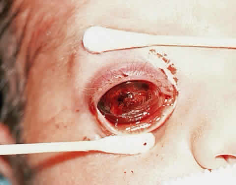
  Shields JA, Shields CL, Eagle RC, Blair CJ: Spontaneous pseudohypopyon secondary to diffuse infiltrating retinoblastoma. Arch Ophthalmol 106:1301, 1988
  Shields JA, Shields CL, Eagle RC, De Potter P, Douglas CH: Calcified intraocular abscess simulating retinoblastoma [letter]. Am J Ophthalmol 114:227, 1992
  Shields JA, Shields CL, Eagle RC Jr, Barrett J, De Potter P: Endogenous endophthalmitis simulating retinoblastoma. Retina 15:213, 1995
  Shields JA, Shields CL, Suvarnamani C, Schroeder RP, DePotter P: Retinoblastoma manifesting as orbital cellulitis. Am J Ophthalmol 112:442, 1991
  Spaulding AG: Rubeosis iridis in retinoblastoma and pseudoglioma. Trans Am Ophthalmol Soc 76:584, 1978
  Stafford WR, Yanoff M, Parnell B: Retinoblastoma initially misdiagnosed as primary ocular inflammation. Arch Ophthalmol 82:771, 1969
  Takahashi T, Tamura S, Inoue M et al: Retinoblastoma in a 26-year-old man. Ophthalmology 90:179, 1979
  Verhoeff FH: Retinoblastoma: Report of a case in a male age 48. Arch Ophthalmol 2:643, 1929
  Walton DS, Grant WM: Retinoblastoma and iris neovascularization. Am J Ophthalmol 65:598, 1968
  Yoshizumi MO, Thomas JV, Smith TR: Glaucoma-inducing mechanisms in eyes with retinoblastoma. Arch Ophthalmol 96:105, 1978
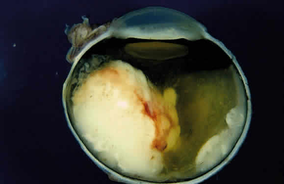
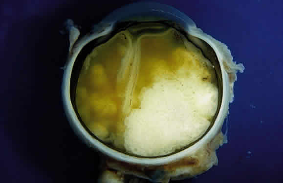
  Zimmerman LE: Retinoblastoma and retinocytoma. In Spencer WH (ed): Ophthalmic Pathology: An Atlas and Textbook, 3rd ed, vol 2, pp 1292–1351. Philadelphia, WB Saunders, 1985
  Zimmerman LE: Retinoblastoma, including a report of illustrative cases. Med Ann DC 38:366, 1969

PROGNOSTIC FACTORS
  Carbajal UM: Metastasis in retinoblastoma. Am J Ophthalmol 48:47, 1959
  DeBuen S: Retinoblastoma with spread by direct continuity to the contralateral optic nerve: report of a case. Am J Ophthalmol 49:815, 1960
  Dunkel IJ, Gerald WL, Rosenfield NS et al: Outcome of patients with a history of bilateral retinoblastoma treated for a second malignancy: the Memorial-Sloan Kettering experience. Med Pediatr Oncol 30:59, 1998
  Howarth C, Meyer D, Hustu HO et al: Stage-related combined modality treatment of retinoblastoma: results of a prospective study. Cancer 45:851, 1980
  Khelfaoui F, Validire P, Auperin A et al: Histopathologic risk factors in retinoblastoma: a retrospective study of 172 patients treated in a single institution. Cancer 77:1206, 1996
  Kopelman JE, McLean IW, Rosenberg SH: Multivariate analysis of risk factors for metastasis in retinoblastoma treated by enucleation. Ophthalmology 94:371, 1987
  Lennox EL, Draper GJ, Sanders BM: Retinoblastoma: a study of natural history and prognosis of 268 cases. Br Med J 3:731, 1975
  MacKay CJ, Abramson DH, Ellsworth RM: Metastatic patterns of retinoblastoma. Arch Ophthalmol 102:391, 1984
  Magramm I, Abramson DH, Ellsworth RM: Optic nerve involvement in retinoblastoma. Ophthalmology 96:217, 1989
  McLean IW, Rosenberg SH, Messmer EP et al: Prognostic factors in cases of retinoblastoma: analysis of 974 patients from Germany and the United States treated by enucleation. In Bornfeld N, Gragoudas ES, Lommatzsch PK (eds): Tumors of the Eye. Proceedings of the International Symposium on Tumors of the Eye, pp 69–72. Amsterdam, Kugler Publications, 1991
  Messmer EP, Heinrich T, Hopping W et al: Risk factors for metastases in patients with retinoblastoma. Ophthalmology 98:136, 1991
  Redler LD, Ellsworth RM: Prognostic importance of choroidal invasion in retinoblastoma. Arch Ophthalmol 90:294, 1973
  Rootman J, Ellsworth RM, Hofbauer J, Kitchen D: Orbital extension of retinoblastoma: a clinicopathological study. Can J Ophthalmol 13:72, 1978
  Rootman J, Hofbauer J, Ellsworth RM, Kitchen D: Invasion of the optic nerve by retinoblastoma: a clinicopathological study. Can J Ophthalmol 11:106, 1976
  Rubin CM, Robison LL, Cameron JD et al: Intraocular retinoblastoma group V: an analysis of prognostic factors. J Clin Oncol 3:680, 1985
  Shields CL, Shields JA, Baez K, Cater JR, De Potter P: Optic nerve invasion of retinoblastoma. Metastatic potential and clinical risk factors. Cancer 73:692, 1994
  Shields CL, Shields JA, Baez KA, Cater J, De Potter PV: Choroidal invasion of retinoblastoma: metastatic potential and clinical risk factors [see comments]. Br J Ophthalmol 77:544, 1993
  Taktikos A: Investigation of retinoblastoma with special reference to histology and prognosis. Br J Ophthalmol 50:225, 1966
  Wiggs J, Nordenskjold M, Yandell D et al: Prediction of the risk of hereditary retinoblastoma, using DNA polymorphisms within the retinoblastoma gene. N Engl J Med 318:151, 1988
  Wolter JR: Retinoblastoma extension into the choroid. Pathological study of the neoplastic process and thoughts about its prognostic significance. Ophthalmic Paediatr Genet 8:151, 1987

RETINOMA/RETINOCYTOMA/SPONTANEOUS REGRESSION
  Aaby AA, Price RL, Zakov ZN: Spontaneously regressing retinoblastomas, retinoma, or retinoblastoma group 0. Am J Ophthalmol 96:315, 1983
  Abramson DH: Retinoma, retinocytoma, and the retinoblastoma gene [editorial]. Arch Ophthalmol 101:1517, 1983
  Andersen SR, Jensen OA: Retinoblastoma with necrosis of central retinal artery and vein and partial spontaneous regression. Acta Ophthalmol 52:183, 1974
  Balmer A, Munier F, Gailloud C: Retinoma. Case studies. Ophthalmic Paediatr Genet 12:131, 1991
  Boniuk M, Zimmerman LE: Spontaneous regression of retinoblastoma. Int Ophthalmol Clin 2:525, 1962
  Eagle RC Jr, Shields JA, Donoso L, Milner RS: Malignant transformation of spontaneously regressed retinoblastoma, retinoma/retinocytoma variant. Ophthalmology 96:1389, 1989
  Gallie BL, Ellsworth RM, Abramson DH et al: Retinoma: spontaneous regression of retinoblastoma or benign manifestation of the mutation? Br J Cancer 45:513, 1982
  Gallie BL, Phillips RA, Ellsworth RM, Abramson DH: Significance of retinoma and phthisis bulbi for retinoblastoma. Ophthalmology 89:1393, 1982
  Gangwar DN, Jain IS, Gupta A et al: Bilateral spontaneous regression of retinoblastoma with dominant transmission. Ann Ophthalmol 14:479, 1982
  Margo C, Hidayat A, Kopelman J, Zimmerman LE: Retinocytoma. A benign variant of retinoblastoma. Arch Ophthalmol 101:1519, 1983
  Smith JLS: Histology and spontaneous regression of retinoblastoma. Trans Ophthalmol Soc UK 94:953, 1974

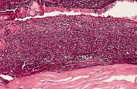
HISTOLOGY AND ULTRASTRUCTURE
  Albert DM, Craft J, Sang DN: Ultrastructure of retinoblastomas: transmission and scanning electron microscopy. In Jakobiec FA (ed): Ocular and Adnexal Tumors, pp 157–171. Birmingham, AL, Aesculapius, 1978
  Bunt AH, Tso MO: Feulgen-positive deposits in retinoblastoma. Incidence, composition, and ultrastructure. Arch Ophthalmol 99:144, 1981
  Burnier MN, McLean IW, Zimmerman LE, Rosenberg SH: Retinoblastoma. The relationship of proliferating cells to blood vessels. Invest Ophthalmol Vis Sci 31:2037, 1990
  Datta BN: DNA coating of blood vessels in retinoblastomas. Am J Clin Pathol 62:94, 1974
  Kyritsis AP, Tsokos M, Triche TJ, Chader GJ: Retinoblastoma: a primitive tumor with multipotential characteristics. Invest Ophthalmol Vis Sci 27:1760, 1986
  Loeffler KU, McMenamin PG: An ultrastructural study of DNA precipitation in the anterior segment of eyes with retinoblastoma. Ophthalmology 94:1160, 1987
  Mullaney J: Retinoblastomas with DNA precipitation. Arch Ophthalmol 82:454, 1969
  Nork TM, Millecchia LL, de Venecia GB, Myers FL, Vogel KA: Immunocytochemical features of retinoblastoma in an adult. Arch Ophthalmol 114:1402, 1996
  Nork TM, Schwartz TL, Doshi HM, Millecchia LL: Retinoblastoma. Cell of origin. Arch Ophthalmol 113:791, 1995
  Pe'er J, Neufeld M, Baras M et al: Rubeosis iridis in retinoblastoma. Histologic findings and the possible role of vascular endothelial growth factor in its induction. Ophthalmology 104:1251, 1997
  Popoff NA, Ellsworth RM: The fine structure of retinoblastoma: In vivo and in vitro observations. Lab Invest 25:389, 1971
  Shields JA, Shields CL, Ehya H, Eagle RC Jr, De Potter P: Fine-needle aspiration biopsy of suspected intraocular tumors. Ophthalmology 100:1677, 1993
  Stowe GC, Zakov ZN, Albert DM et al: Vascular basophilia in ocular and orbital tumors. Invest Ophthalmol Vis Sci 18:1068, 1979
  Ts'o MO, Fine BS, Zimmerman LE: The Flexner-Wintersteiner rosettes in retinoblastoma. Arch Pathol 88:664, 1969
  Ts'o MO, Fine BS, Zimmerman LE: The nature of retinoblastoma. II. Photoreceptor differentiation: an electron microscopic study. Am J Ophthalmol 69:350, 1970
  Ts'o MO, Zimmerman LE, Fine BS, Ellsworth RM: A cause of radioresistance in retinoblastoma: photoreceptor differentiation. Trans Am Acad Ophthalmol Otolaryngol 74:959, 1970
  Ts'o MO, Zimmerman LE, Fine BS: The nature of retinoblastoma. I. Photoreceptor differentiation: a clinical and histopathologic study. Am J Ophthalmol 69:339, 1970
  Tsokos M, Kyritsis AP, Chader GJ, Triche TJ: Differentiation of human retinoblastoma in vitro into cell types with characteristics observed in embryonal or mature retina. Am J Pathol 123:542, 1986
  Verhoeff FH, Jackson E: Minutes of Proceedings, 62nd Annual Meeting. Trans Am Ophthalmol Soc 24:38, 1926
  Vrabec T, Arbizo V, Adamus G et al: Rod cell-specific antigens in retinoblastoma. Arch Ophthalmol 107:1061, 1989

THE RETINOBLASTOMA GENE AND THE GENETICS OF RETINOBLASTOMA
  Balaban G, Gilbert F, Nichols W et al: Abnormalities of chromosome 13 in retinoblastoma from individuals with normal constitutional karyotypes. Cancer Genet Cytogenet 6:213, 1982
  Benedict WF, Murphree AL, Banerjee A et al: Patient with chromosome deletion: evidence that the retinoblastoma gene is a recessive cancer gene. Science 219:973, 1983
  Bookstein R, Lee WH: Molecular genetics of the retinoblastoma suppressor gene. Crit Rev Oncog 2:211, 1991
  Cavenee WK, Dryja TP, Phillips RA et al: Expression of recessive alleles by chromosomal mechanisms in retinoblastoma. Nature (Lond) 305:779, 1983
  Cavenee WK, Hansen MF, Norenskjold M et al: Genetic origin of mutations predisposing to retinoblastoma. Science 228:501, 1985
  Cavenee WK, Murphree AL, Shull MM et al: Prediction of familial predisposition to retinoblastoma. N Engl J Med 314:1201, 1986
  Dryja TP, Bruns GAP, Orkin SH et al: Isolation of DNA fragments from chromosome 13. Retina 3:121, 1983
  Dryja TP, Cavenee W, White R et al: Homozygosity of chromosome 13 in retinoblastoma. N Engl J Med 310:550, 1984
  Dryja TP, Rapaport J, McGee TL, Nork TM, Schwartz TL: Molecular etiology of low-penetrance retinoblastoma in two pedigrees. Am J Hum Genet 52:1122, 1993
  Dryja TP, Rapaport JM, Weichselbaum R, Bruns GAP: Chromosome 13 restriction fragment length polymorphisms. Hum Genet 65:320, 1984
  Gallie BL, Dunn JM, Chan HS et al: The genetics of retinoblastoma: relevance to the patient. Pediatr Clin North Am 38:299, 1991
  Gallie BL, Phillips RA: Retinoblastoma: a model of oncogenesis. Ophthalmology 91:666, 1984
  Godbout R, Dryja TP, Squire J et al: Somatic inactivation of genes on chromosome 13 is a common event in retinoblastoma. Nature 304:451, 1983
  Gordon H: Family studies in retinoblastoma. Birth Defects 10:185, 1974
  Harbour JW: Overview of RB gene mutations in patients with retinoblastoma. Implications for clinical genetic screening. Ophthalmology 105:1442, 1998
  Knudson AG Jr, Hethcote HW, Brown BW: Mutation and childhood cancer: a probabilistic model for the incidence of retinoblastoma. Proc Natl Acad Sci USA 71:5116, 1975
  Knudson AG Jr: Mutation and cancer: a statistical study of retinoblastoma. Proc Natl Acad Sci USA 68:820, 1971
  Knudson AG Jr: Retinoblastoma: a prototypic hereditary neoplasm. Semin Oncol 5:57, 1978
  Kratzke RA, Otterson GA, Hogg A et al: Partial inactivation of the RB product in a family with incomplete penetrance of familial retinoblastoma and benign retinal tumors. Oncogene 9:1321, 1994
  McGee TL, Yandell DW, Dryja TP: Structure and partial genomic sequence of the human retinoblastoma susceptibility gene. Gene 80:119, 1989
  Murphree AL, Benedict WF: Retinoblastoma: clues to human oncogenesis. Science 223:1028, 1984
  Murphree AL: Molecular genetics of retinoblastoma. Ophthalmol Clin North Am 8:155, 1995
  Nielson M, Goldschmidt E: Retinoblastoma among offspring of adult survivors in Denmark. Acta Ophthalmol 46:736, 1968
  Otterson GA, Chen W, Coxon AB, Khleif SN, Kaye FJ: Incomplete penetrance of familial retinoblastoma linked to germ-line mutations that result in partial loss of RB function. Proc Natl Acad Sci USA 94:12036, 1997
  Shields CL, Shields JA, Donoso LA: Clinical genetics of retinoblastoma. Int Ophthalmol Clin 33:67, 1993
  Vogel F: Genetics of retinoblastoma. Hum Genet 52:1, 1979
  Weichselbaum RR, Zakov ZN, Albert DM et al: New findings in the chromosome 13 long-arm deletion syndrome and retinoblastoma. Ophthalmology 86:1191, 1979
  Weinberg RA: The retinoblastoma gene and gene product. Cancer Surv 12:43, 1992
  Yunis JJ, Ramsay N: Retinoblastoma and subband deletion of chromosome 13. Am J Dis Child 132:161, 1978

SECONDARY NONOCULAR TUMORS
  Abramson DH, Ellsworth RM, Zimmerman LE: Nonocular cancer in retinoblastoma survivors. Trans Am Acad Ophthalmol Otolaryngol 81:454, 1976
  Bader JL, Meadows AT, Zimmerman LE et al: Bilateral retinoblastoma with ectopic intracranial retinoblastoma: trilateral retinoblastoma. Cancer Genet Cytogenet 5:203, 1982
  Holladay DA, Holladay A, Montebello JF, Redmond KP: Clinical presentation, treatment, and outcome of trilateral retinoblastoma. Cancer 67:710, 1991
  Jakobiec FA, Tso MOM, Zimmerman LE et al: Retinoblastoma and intracranial malignancy. Cancer 39:2048, 1977
  Moll AC, Imhof SM, Bouter LM et al: Second primary tumors in patients with retinoblastoma. A review of the literature. Ophthalm Genet 18:27, 1997
  Pesin SR, Shields JA: Seven cases of trilateral retinoblastoma. Am J Ophthalmol 107:121, 1989
  Roarty JD, McLean IW, Zimmerman LE: Incidence of second neoplasms in patients with bilateral retinoblastoma. Ophthalmology 95:1583, 1988
  Wong FL, Boice JD, Abramson DH et al: Cancer incidence after retinoblastoma: radiation dose and sarcoma risk. JAMA 278:1262, 1997

LESIONS THAT SIMULATE RETINOBLASTOMA
  
General References
  Apple DJ, Hamming NA, Gieser DK: Differential diagnosis of leukocoria. In Peyman GA et al (eds): Intraocular Tumors, pp 235–283. New York, Appleton/Century/Crofts, 1977
  Francois J: Differential diagnosis of leukokoria in children. Ann Ophthalmol 10:1375, 1978
  Francois J: Neonatal or juvenile leucocoria. Ophthalmologica 179:129, 1979
  Howard GM, Ellsworth RM: Differential diagnosis of retinoblastoma: a statistical survey of 500 children: 1. Relative frequency of the lesions which simulate retinoblastoma. Am J Ophthalmol 60:610, 1965
  Kogan L, Boniuk M: Causes for enucleation in children with special reference to pseudoglioma and unsuspected retinoblastoma. Int Ophthalmol Clin 2:507, 1962
  Margo CE, Zimmerman LE: Retinoblastoma: the accuracy of clinical diagnosis in children treated by enucleation. J Pediatr Ophthalmol Strabismus 20:227, 1983
  Robertson DM, Campbell RJ: Analysis of misdiagnosed retinoblastoma in a series of 726 enucleated eyes. Mod Probl Ophthalmol 18:156, 1977
  Shields JA, Parsons HM, Shields CL, Shah P: Lesions simulating retinoblastoma. J Pediatr Ophthalmol Strabismus 28:338, 1991
  Shields JA, Shields CL: Differential diagnosis of retinoblastoma. In: Intraocular Tumors: A Text and Atlas, pp 341–362. Philadelphia, WB Saunders, 1992
  Shields JA, Shields CL, Parsons HM: Differential diagnosis of retinoblastoma. Retina 11:232, 1991
  Smirniotopoulos JG, Bargallo N, Mafee MF: Differential diagnosis of leukokoria: radiologic-pathologic correlation. Radiographics 14:1059, 1994
Toxocariasis
  Ashton N: Larval granulomatosis of the retina due to toxocara. Br J Ophthalmol 44:129, 1960
  Belmont JB, Irvine A, Benson W, O'Connor GR: Vitrectomy in ocular toxocariasis. Arch Ophthalmol 100:1912, 1982
  Benitez del Castillo JM, Herreros G, Guillen JL et al: Bilateral ocular toxocariasis demonstrated by aqueous humor enzyme-linked immunosorbent assay. Am J Ophthalmol 119:514, 1995
  Brown DH: Ocular Toxocara canis. J Pediatr Ophthalmol 7:182, 1970
  Dernouchamps JP, Verougstraete C, Demolder E: Ocular toxocariasis: a presumed case of peripheral granuloma. Int Ophthalmol 14:383, 1990
  Ellis GS Jr, Pakalnis VA, Worley G et al: Toxocara canis infestation. Clinical and epidemiological associations with seropositivity in kindergarten children. Ophthalmology 93:1032, 1986
  Kayes SG: Human toxocariasis and the visceral larva migrans syndrome: correlative immunopathology. Chem Immunol 66:99, 1997
  Luxenberg MN: An experimental approach to the study of intraocular Toxocara canis. Trans Am Ophthalmol Soc 77:542, 1979
  Maguire AM, Green WR, Michels RG, Erozan YS: Recovery of intraocular Toxocara canis by pars plana vitrectomy. Ophthalmology 97:675, 1990
  Rockey JH, Donnelly JJ, Stromberg BE, Laties AM, Soulsby EJ: Immunopathology of ascarid infection of the eye. Role of IgE antibodies and mast cells. Arch Ophthalmol 99:1831, 1981
  Rockey JH, Donnelly JJ, Stromberg BE, Soulsby EJ: Immunopathology of Toxocara canis and Ascaris suum infections of the eye: the role of the eosinophil. Invest Ophthalmol Vis Sci 18:1172, 1979
  Rockey JH, John T, Donnelly JJ et al: In vitro interaction of eosinophils from ascarid-infected eyes with Ascaris suum and Toxocara canis larvae. Invest Ophthalmol Vis Sci 24:1346, 1983
  Safar EH, Abd-el Ghaffar FM, Saffar SA et al: Incidence of Toxoplasma and Toxocara antibodies among outpatients in the Ophthalmic Research Institute, Egypt. J Egypt Soc Parasitol 25:839, 1995
  Schantz PM, Weis PE, Pollard ZF, White MC: Risk factors for toxocaral ocular larva migrans: a case-control study. Am J Public Health 70:1269, 1980
  Searl SS, Moazed K, Albert DM, Marcus LC: Ocular toxocariasis presenting as leukocoria in a patient with low ELISA titer to Toxocara canis. Ophthalmology 88:1302, 1981
  Shields JA, Shields CL, Eagle RC Jr, Barrett J, De Potter P: Endogenous endophthalmitis simulating retinoblastoma. Retina 15:213, 1995
  Shields JA: Ocular toxocariasis. A review. Surv Ophthalmol 28:361, 1984
  Watzke RC: Ocular Toxocara canis infection: clinical and experimental features. Trans New Orleans Acad Ophthalmol 31:263, 1983
  Wilder HC: Nematode endophthalmitis. Trans Am Acad Ophthalmol Otolaryngol 54:99, 1950
  Zinkham WH: Visceral larva migrans. A review and reassessment indicating two forms of clinical expression: visceral and ocular. Am J Dis Child 132:627, 1978
Persistent Hyperplastic Primary Vitreous (Persistent Fetal Vasculature)
  Boeve MH, van der Linde-Sipman JS, Stades FC, Vrensen GF: Early morphogenesis of persistent hyperplastic tunica vasculosa lentis and primary vitreous. A transmission electron microscopic study. Invest Ophthalmol Vis Sci 31:1886, 1990
  Boeve MH, Vrensen GF, Willekens BL et al: Early morphogenesis of persistent hyperplastic tunica vasculosa lentis and primary vitreous (PHTVL/PHPV). Scanning electron microscopic observations. Graefes Arch Clin Exp Ophthalmol 231:29, 1993
  Doro S, Werblin TP, Haas B, Iwamoto T, Jakobiec FA: Fetal adenoma of the pigmented ciliary epithelium associated with persistent hyperplastic primary vitreous. Ophthalmology 93:1343, 1986
  Font RL, Yanoff M, Zimmerman LE: Intraocular adipose tissue and persistent hyperplastic primary vitreous. Arch Ophthalmol 82:43, 1969
  Goldberg MF, Mafee M: Computed tomography for diagnosis of persistent hyperplastic primary vitreous (PHPV). Ophthalmology 90:442, 1983
  Goldberg MF: Persistent fetal vasculature (PFV): an integrated interpretation of signs and symptoms associated with persistent hyperplastic primary vitreous (PHPV). Am J Ophthalmol 124:587, 1997
  Haddad R, Font RL, Reeser F: Persistent hyperplastic primary vitreous. A clinicopathologic study of 62 cases and review of the literature. Surv Ophthalmol 23:123, 1978
  Irvine AR, Albert DM, Sang DN: Retinal neoplasia and dysplasia. II. Retinoblastoma occurring with persistence and hyperplasia of the primary vitreous. Invest Ophthalmol Vis Sci 16:403, 1977
  Kaste SC, Jenkins JJ 3rd, Meyer D, Fontanesi J, Pratt CB: Persistent hyperplastic primary vitreous of the eye: imaging findings with pathologic correlation. AJR Am J Roentgenol 162:437, 1994
  Mafee MF, Goldberg MF: Persistent hyperplastic primary vitreous (PHPV): role of computed tomography and magnetic resonance. Radiol Clin North Am 25:683, 1987
  Mittra RA, Huynh LT, Ruttum MS et al: Visual outcomes following lensectomy and vitrectomy for combined anterior and posterior persistent hyperplastic primary vitreous. Arch Ophthalmol 116:1190, 1998
  Pollard ZF: Persistent hyperplastic primary vitreous: diagnosis, treatment and results. Trans Am Ophthalmol Soc 95:487, 1997
  Reese AB, Payne F: Persistence and hyperplasia of the primary vitreous. Am J Ophthalmol 29:1, 1964
  Reese AB: Persistence and hyperplasia of the primary vitreous. Am J Ophthalmol 40:317, 1955
  Spaulding AG, Naumann G: Persistent hyperplastic primary vitreous in an adult. A brief review of the literature and a histopathologic study. Arch Ophthalmol 77:666, 1967
  Spitznas M, Koch F, Pohl S: Ultrastructural pathology of anterior persistent hyperplastic primary vitreous [see comments]. Graefes Arch Clin Exp Ophthalmol 228:487, 1990
Coats' Disease
  Chang MM, McLean IW, Merritt JC: Coats' disease: a study of 62 histologically confirmed cases. J Pediatr Ophthalmol Strabismus 21:163, 1984
  Coats G: Forms of retinal disease with massive exudation. R London Ophthalmol Hosp Rep 17:440, 1907/1908
  Coats G: Uber retinitis exsudativa (Retinitis hemorrhagica externa). Albrecht Von Graefes Arch Klin Exp Ophthalmol 81:275, 1912
  Egbert PR, Chan CC, Winter FC: Flat preparations of the retinal vessels in Coats' disease. J Pediatr Ophthalmol 13:336, 1976
  Egerer I, Tasman W, Tomer TT: Coats disease. Arch Ophthalmol 92:109, 1974
  Farkas TG, Potts AM, Boone C: Some pathologic and biochemical aspects of Coats' disease. Am J Ophthalmol 75:289, 1973
  Goel SD, Augsburger JJ: Hemorrhagic retinal macrocysts in advanced Coats disease. Retina 11:437, 1991
  Haik BG: Advanced Coats' disease. Trans Am Ophthalmol Soc 89:371, 1991
  Henkind P, Morgan G: Peripheral retinal angioma with exudative retinopathy in adults (Coats' lesion). Br J Ophthalmol 50:2, 1967
  Jaffe MS, Shields JA, Canny CL, Eagle RC Jr, Fry RL: Retinoblastoma simulating Coats' disease: a clinicopathologic report. Ann Ophthalmol 9:863, 1977
  Kremer I, Nissenkorn I, Ben-Sira I: Cytologic and biochemical examination of the subretinal fluid in diagnosis of Coats' disease. Acta Ophthalmol (Copenh) 67:342, 1989
  Leber T: Uber eine durch Vorkommen multipler Miliaraneurysmen charakterisierte Form von Retinaldegeneration. Arch Ophthalmol 81:1, 1912
  Patel HK, Augsburger JJ, Eagle RC Jr: Unusual presentation of advanced Coats' disease. J Pediatr Ophthalmol Strabismus 32:120, 1995
  Reese AB: Telangiectasis of the retina and Coats' disease. Am J Ophthalmol 42:1, 1956
  Ridley ME, Shields JA, Brown GC, Tasman W: Coats' disease. Evaluation of management. Ophthalmology 89:1381, 1982
  Shields JA, Eagle RC Jr, Fammartino J, Shields CL, De Potter P: Coats' disease as a cause of anterior chamber cholesterolosis [letter]. Arch Ophthalmol 113:975, 1995
  Silodor SW, Augsburger JJ, Shields JA, Tasman W: Natural history and management of advanced Coats' disease. Ophthalmic Surg 19:89, 1988
  Steidl SM, Hirose T, Sang D, Hartnett ME: Difficulties in excluding the diagnosis of retinoblastoma in cases of advanced Coats' disease: a clinicopathologic report. Ophthalmologica 210:336, 1996
  Stewart J, Halliwell T, Gupta RK: Cytodiagnosis of Coats' disease from an ocular aspirate. A case report. Acta Cytol 37:717, 1993
Retinopathy of Prematurity
  Ashton N, Graymore C, Pedler C: Studies on developing retinal vessels: V. Mechanism of vasoobliteration. A preliminary report. Br J Ophthalmol 41:449, 1957
  Brockhurst RJ, Chishti M: Cicatricial retrolental fibroplasia: its occurrence without oxygen administration and in full-term infants. Albrecht Von Graefes Arch Klin Exp Ophthalmol 195:113, 1975
  Committee for the Classification of Retinopathy of Prematurity: An International Classification of Retinopathy of Prematurity. Arch Ophthalmol 102:1130, 1984
  de Juan E Jr, Machemer R, Flynn JT, Green WR: Surgical pathoanatomy in stage 5 retinopathy of prematurity. Birth Defects Orig Artic Ser 24:281, 1988
  Faris B, Tolentino F, Freeman HM et al: Retrolental fibroplasia in the cicatricial stage. Arch Ophthalmol 85:661, 1971
  Flower RW, Patz A: Oxygen studies in retrolental fibroplasia: IX. The effects of elevated arterial oxygen tension on retinal vascular dynamics in the kitten. Arch Ophthalmol 85:197, 1972
  Flynn JT, O'Grady GE, Herrera J et al: Retrolental fibroplasia: 1. Clinical observations. Arch Ophthalmol 95:217, 1977
  Flynn JT: Acute proliferative retrolental fibroplasia: evolution of the lesion. Albrecht Von Graefes Arch Klin Exp Ophthalmol 195:101, 1975
  Foos RY: Acute retrolental fibroplasia. Albrecht Von Graefes Arch Klin Exp Ophthalmol 195:87, 1975
  Foos RY: Chronic retinopathy of prematurity. Ophthalmology 92:563, 1985
  Green WR: Retrolental fibroplasia (RLF). In Spencer WH (ed): Ophthalmic Pathology: An Atlas and Textbook, vol 2, 3rd ed., pp 647–651. Philadelphia, WB Saunders, 1985
  Hittner HM, Rhodes LM, McPherson AR: Anterior segment abnormalities in cicatricial retinopathy of prematurity. Ophthalmology 86:803, 1979
  Hittner HM, Rudolph AJ, Kretzer FL: Suppression of severe retinopathy of prematurity with vitamin E supplementation: ultrastructural mechanism of clinical efficacy. Ophthalmology 91:1512, 1984
  Karlsberg RC, Green WR, Patz A: Congenital retrolental fibroplasia. Arch Ophthalmol 89:122, 1973
  Kinsey VE, Arnold HJ, Kalina RE et al: PaO2 levels and retrolental fibroplasia: a report of the cooperative study. Pediatrics 60:655, 1977
  Kushner BJ, Essner D, Cohen IJ et al: Retrolental fibroplasia: pathologic correlation. Arch Ophthalmol 95:29, 1977
  Naiman J, Green WR, Patz A: Retrolental fibroplasia in hypoxic newborn. Am J Ophthalmol 88:55, 1979
  Patz A: Current status of role of oxygen in retrolental fibroplasia. Invest Ophthalmol Vis Sci 15:337, 1976
  Patz A: Retrolental fibroplasia survey. Surv Ophthalmol 14:1, 1969
  Reese AB, Stepanik J: Cicatricial stage of retrolental fibroplasia. Am J Ophthalmol 38:308, 1954
  Stefani FH, Ehalt H: Nonoxygen-induced retinitis proliferans and retinal detachment in full-term infants. Br J Ophthalmol 58:490, 1974
  Tasman W: Retinal detachment in retrolental fibroplasia. Albrecht Von Graefes Arch Klin Exp Ophthalmol 195:129, 1975
  Tasman W: The natural history of active retinopathy of prematurity. Ophthalmology 91:1499, 1984
  Tasman W: Vitreoretinal changes in cicatricial retrolental fibroplasia. Trans Am Ophthalmol Soc 68:548, 1970
Incontinentia Pigmenti
  Brown CA: Incontinentia pigmenti: the development of pseudoglioma. Br J Ophthalmol 72:452, 1988
  Catalano RA: Incontinentia pigmenti. Am J Ophthalmol 110:696, 1990
  Fowell SM, Greenwald MJ, Prendiville JS, Jampol LM: Ocular findings of incontinentia pigmenti in a male infant with Klinefelter syndrome. J Pediatr Ophthalmol Strabismus 29:180, 1992
  Francis JS, Sybert VP: Incontinentia pigmenti. Semin Cutan Med Surg 16:54, 1997
  Goldberg MF, Custis PH: Retinal and other manifestations of incontinentia pigmenti (Bloch-Sulzberger syndrome). Ophthalmology 100:1645, 1993
  Goldberg MF: The blinding mechanisms of incontinentia pigmenti. Ophthalmic Genet 15:69, 1994
  Heathcote JG, Schoales BA, Willis NR: Incontinentia pigmenti (Bloch-Sulzberger syndrome): a case report and review of the ocular pathological features. Can J Ophthalmol 26:229, 1991
  Mensheha-Manhart O, Rodrigues MM, Shields JA, Shannon GM, Mirabelli RP: Retinal pigment epithelium in incontinentia pigmenti. Am J Ophthalmol 79:571, 1975
  Rahi J, Hungerford J: Early diagnosis of the retinopathy of incontinentia pigmenti: successful treatment by cryotherapy. Br J Ophthalmol 74:377, 1990
  Rosenfeld SI, Smith ME: Ocular findings in incontinentia pigmenti. Ophthalmology 92:543, 1985
  Shah GK, Summers CG, Walsh AW, Neely KA: Optic nerve neovascularization in incontinentia pigmenti. Am J Ophthalmol 124:410, 1997
  Soltau JB, Lueder GT: Bilateral macular lesions in incontinentia pigmenti. Bloch-Sulzberger syndrome. Retina 16:38, 1996
  Spallone A: Incontinentia pigmenti (Bloch-Sulzberger syndrome): seven case reports from one family. Br J Ophthalmol 71:629, 1987
  Wald KJ, Mehta MC, Katsumi O, Sabates NR, Hirose T: Retinal detachments in incontinentia pigmenti. Arch Ophthalmol 111:614, 1993
  Watzke RC, Stevens TS, Carney RG Jr: Retinal vascular changes of incontinentia pigmenti. Arch Ophthalmol 94:743, 1976
Norrie's Disease and Associated Disorders
  Apple DJ, Fishman GA, Goldberg MF: Ocular histopathology of Norrie's disease. Am J Ophthalmol 78:196, 1974
  Caballero M, Veske A, Rodriguez JJ et al: Two novel mutations in the Norrie disease gene associated with the classical ocular phenotype. Ophthalmic Genet 17:187, 1996
  Chynn EW, Walton DS, Hahn LB, Dryja TP: Norrie disease. Diagnosis of a simplex case by DNA analysis. Arch Ophthalmol 114:1136, 1996
  Enyedi LB, de Juan E Jr, Gaitan A: Ultrastructural study of Norrie's disease. Am J Ophthalmol 111:439, 1991
  Holmes LB: Norrie's disease—an X-linked syndrome of retinal malformation, mental retardation and deafness. N Engl J Med 284:367, 1971
  Johnson K, Mintz-Hittner HA, Conley YP, Ferrell RE: X-linked exudative vitreoretinopathy caused by an arginine to leucine substitution (R121L) in the Norrie disease protein. Clin Genet 50:113, 1996
  Mintz-Hittner HA, Ferrell RE, Sims KB et al: Peripheral retinopathy in offspring of carriers of Norrie disease gene mutations. Possible transplacental effect of abnormal norrin. Ophthalmology 103:2128, 1996
  Parsons MA, Curtis D, Blank CE, Hughes HN, McCartney AC: The ocular pathology of Norrie disease in a fetus of 11 weeks' gestational age. Graefes Arch Clin Exp Ophthalmol 230:248, 1992
  Perez-Vilar J, Hill RL: Norrie disease protein (norrin) forms disulfide-linked oligomers associated with the extracellular matrix. J Biol Chem 272:33410, 1997
  Schroeder B, Hesse L, Bruck W, Gal A: Histopathological and immunohistochemical findings associated with a null mutation in the Norrie disease gene. Ophthalmic Genet 18:71, 1997
  Shastry BS, Pendergast SD, Hartzer MK et al: Identification of missense mutations in the Norrie disease gene associated with advanced retinopathy of prematurity. Arch Ophthalmol 115:651, 1997
  Shastry BS: Identification of a recurrent missense mutation in the Norrie disease gene associated with a simplex case of exudative vitreoretinopathy. Biochem Biophys Res Commun 246:35, 1998
  Townes PL, Roca PD: Norrie's disease (hereditary oculo-acoustic-cerebral degeneration). Report of a United States family. Am J Ophthalmol 76:797, 1973
  Warburg M: Norrie's disease—differential diagnosis and treatment. Acta Ophthalmol (Copenh) 53:217, 1975
  Wolff G, Mayerova A, Wienker TF et al: Clinical reinvestigation and linkage analysis in the family with Episkopi blindness (Norrie disease). J Med Genet 29:816, 1992
Retinal Dysplasia
  Albert DM, Lahav M, Colby ED, Shadduck JA, Sang DN: Retinal neoplasia and dysplasia. I. Induction by feline leukemia virus. Invest Ophthalmol Vis Sci 16:325, 1977
  Godel V, Nemet P, Lazar M: Retinal dysplasia. Doc Ophthalmol 51:277, 1981
  Hoepner J, Yanoff M: Ocular anomalies in trisomy 13–15. Am J Ophthalmol 74:729, 1972
  Lahav M, Albert DM, Wyand S: Clinical and histopathologic classification of retinal dysplasia. Am J Ophthalmol 75:648, 1973
  Lloyd IC, Colley A, Tullo AB, Bonshek R: Dominantly inherited unilateral retinal dysplasia. Br J Ophthalmol 77:378, 1993
  Silverstein AM, Osburn BI, Prendergast RA: The pathogenesis of retinal dysplasia. Am J Ophthalmol 72:13, 1971
Medulloepithelioma
  Brownstein S, Barsoum-Homsy M, Conway VH, Sales C, Condon G: Nonteratoid medulloepithelioma of the ciliary body. Ophthalmology 91:1118, 1984
  Broughton WL, Zimmerman LE: A clinicopathologic study of 56 cases of intraocular medulloblastoma. Am J Ophthalmol 85:407, 1978
  Canning CR, McCartney ACE, Hungerford JL: Medulloepithelioma (diktyoma). Br J Ophthalmol 72:764, 1988
  Carrillo R, Streeten BW: Malignant teratoid medulloepithelioma in an adult. Arch Ophthalmol 97:695, 1979
  Floyd BB, Minckler DS, Valentin L: Intraocular medulloepithelioma in a 79-year-old man. Ophthalmology 89:1088, 1982
  Green WR, Iliff WJ, Trotter RR: Malignant teratoid medulloepithelioma of the optic nerve. Arch Ophthalmol 91:451, 1974
  Hamburg A: Medulloepithelioma arising from the posterior pole. Ophthalmologica 181:152, 1980
  Hausmann N, Stefani FH: Medulloepithelioma of the ciliary body. Acta Ophthalmol (Copenh) 69:398, 1991
  Hennis HL, Saunders RA, Shields JA: Malignant teratoid medulloepithelioma of the ciliary body. J Clin Neuroophthalmol 10:291, 1990
  Husain SE, Husain N, Boniuk M, Font RL: Malignant nonteratoid medulloepithelioma of the ciliary body in an adult. Ophthalmology 105:596, 1998
  Jakobiec FA, Howard GM, Ellsworth RM, Rosen M: Electron microscopic diagnosis of medulloepithelioma. Am J Ophthalmol 79:321, 1975
  Kivela T, Tarkkanen A: Recurrent medulloepithelioma of the ciliary body. Immunohistochemical characteristics. Ophthalmology 95:1565, 1988
  Mullaney J: Primary malignant medulloepithelioma of the retinal stalk. Am J Ophthalmol 77:499, 1974
  O'Keefe M, Fulcher T, Kelly P, Lee W, Dudgeon J: Medulloepithelioma of the optic nerve head. Arch Ophthalmol 115:1325, 1997
  Pe'er J, Hidayat AA: Malignant teratoid medulloepithelioma manifesting as a black epibulbar mass with expulsive hemorrhage. Arch Ophthalmol 102:1523, 1984
  Shields JA, Eagle RC Jr, Shields CL et al: Fluorescein angiography and ultrasonography of malignant intraocular medulloepithelioma. J Pediatr Ophthalmol Strabismus 33:193, 1996
  Shields JA, Eagle RC Jr, Shields CL, Potter PD: Congenital neoplasms of the nonpigmented ciliary epithelium (medulloepithelioma). Ophthalmology 103:1998, 1996
  Shields JA, Shields CL, Schwartz RL: Malignant teratoid medulloepithelioma of the ciliary body simulating persistent hyperplastic primary vitreous. Am J Ophthalmol 107:296, 1989
  Vadmal M, Kahn E, Finger P, Teichberg S: Nonteratoid medulloepithelioma of the retina with electron microscopic and immunohistochemical characterization. Pediatr Pathol Lab Med 16:663, 1996
  Zimmerman LE: Verhoeff's “terato-neuroma.” A critical reappraisal in light of new observations and current concepts of embryonic tumors. Am J Ophthalmol 72:1039, 1971
Retinal Astrocytoma and Astrocytic Hamartoma
  Arnold AC, Hepler RS, Yee RW et al: Solitary retinal astrocytoma. Surv Ophthalmol 30:173, 1985
  Bornfeld N, Messmer EP, Theodossiadis G, Meyer-Schwickerath G, Wessing A: Giant cell astrocytoma of the retina. Clinicopathologic report of a case not associated with Bourneville's disease. Retina 7:183, 1987
  de Juan E Jr, Green WR, Gupta PK, Baranano EC: Vitreous seeding by retinal astrocytic hamartoma in a patient with tuberous sclerosis. Retina 4:100, 1984
  Destro M, D'Amico DJ, Gragoudas ES et al: Retinal manifestations of neurofibromatosis. Diagnosis and management. Arch Ophthalmol 109:662, 1991
  Jakobiec FA, Brodie SE, Haik B, Iwamoto T: Giant cell astrocytoma of the retina. A tumor of possible Mueller cell origin. Ophthalmology 90:1565, 1983
  Berger B, Peyman GA, Juarez C et al: Massive retinal gliosis simulating choroidal melanoma. Can J Ophthalmol 14:285, 1979
  Margo CE, Barletta JP, Staman JA: Giant cell astrocytoma of the retina in tuberous sclerosis. Retina 13:155, 1993
  Mullaney PB, Jacquemin C, Abboud E, Karcioglu ZA: Tuberous sclerosis in infancy. J Pediatr Ophthalmol Strabismus 34:372, 1997
  Nork TM, Ghobrial MW, Peyman GA, Tso MO: Massive retinal gliosis. A reactive proliferation of Muller cells. Arch Ophthalmol 104:1383, 1986
  Robertson DM: Ophthalmic manifestations of tuberous sclerosis. Ann NY Acad Sci 615:17, 1991
  Sharma A, Ram J, Gupta A: Solitary retinal astrocytoma. Acta Ophthalmol (Copenh) 69:113, 1991
  Shields JA, Shields CL, Ehya H et al: Atypical retinal astrocytic hamartoma diagnosed by fine-needle biopsy [see comments]. Ophthalmology 103:949, 1996
  Ulbright TM, Fulling KH, Helveston EM: Astrocytic tumors of the retina. Differentiation of sporadic tumors from phakomatosis-associated tumors. Arch Pathol Lab Med 108:160, 1984
  Green WR: Bilateral Coats' disease. Massive gliosis of the retina. Arch Ophthalmol 77:378, 1967
  Yanoff M, Zimmerman LE, Davis RL: Massive gliosis of the retina. Int Ophthalmol Clin 11:211, 1971
Other Rare Neoplasms
  Freitag SK, Eagle RC Jr, Shields JA, Duker JS, Font RL: Melanogenic neuroectodermal tumor of the retina (primary malignant melanoma of the retina). Arch Ophthalmol 115:1581, 1997
  Ghadially FN, Chisholm IA, Lalonde JM: Ultrastructure of an intraocular lacrimal gland choristoma. J Submicrosc Cytol 18:189, 1986
  Kivela T, Kauniskangas L, Miettinen P, Tarkkanen A: Glioneuroma associated with colobomatous dysplasia of the anterior uvea and retina. A case simulating medulloepithelioma. Ophthalmology 96:1799, 1989
  Shields JA, Eagle RC Jr, Shields CL et al: Natural course and histopathologic findings of lacrimal gland choristoma of the iris and ciliary body. Am J Ophthalmol 119:219, 1995
  Spencer WH, Jesberg DO: Glioneuroma (choristomatous malformation of the optic cup margin). Arch Ophthalmol 89:387, 1973
  Wilson ME, McClatchey SK, Zimmerman LE: Rhabdomyosarcoma of the ciliary body. Ophthalmology 97:1484, 1990
Miscellaneous Lesions
  Brown GC, Shields JA, Oglesby RG: Anterior polar cataracts associated with bilateral retinoblastoma. Am J Ophthalmol 87:276, 1979
  Chang MW, Frieden IJ, Good W: The risk intraocular juvenile xanthogranuloma: survey of current practices and assessment of risk. J Am Acad Dermatol 34:445, 1996
  Friendly DS, Parks MM: Concurrence of hereditary congenital cataract and hereditary retinoblastoma. Arch Ophthalmol 84:525, 1970
  Leonardy NJ, Rupani M, Dent G, Klintworth GK: Analysis of 135 eyes for ocular involvement in leukemia. Am J Ophthalmol 109:436, 1990
  Shields JA, Eagle RC Jr, Shields CL et al: Iris juvenile xanthogranuloma studied by immunohistochemistry and flow cytometry. Ophthalmic Surg Lasers 28:140, 1997
  Zimmerman LE: Ocular lesions of juvenile xanthogranuloma, nevoxanthoendothelioma. Trans Am Acad Ophthalmol Otolaryngol 69:412, 1965
Treatment of Retinoblastoma
  Baumal CR, Shields CL, Shields JA, Tasman WS: Surgical repair of rhegmatogenous retinal detachment after treatment for retinoblastoma. Ophthalmology 105:2134, 1998
  Doz F, Khelfaoui F, Mosseri V et al: The role of chemotherapy in orbital involvement of retinoblastoma: the experience of a single institution with 33 patients. Cancer 74:722, 1994
  Doz F, Neuenschwander S, Plantaz D et al: Etoposide and carboplatin in extraocular retinoblastoma: a study by the Societe Francaise d'Oncologie Pediatrique. J Clin Oncol 13:902, 1995
  Freire J, Miyamoto C, Brady LW et al: Retinoblastoma after chemoreduction and irradiation: preliminary results. Front Radiat Ther Oncol 30:88, 1997
  Gunduz K, Shields CL, Shields JA et al: The outcome of chemoreduction treatment in patients with Reese-Ellsworth group V retinoblastoma. Arch Ophthalmol 116:1613, 1998
  Hernandez JC, Brady LW, Shields JA et al: External beam radiation for retinoblastoma: results, patterns of failure, and a proposal for treatment guidelines. Int J Radiat Oncol Biol Phys 35:125, 1996
  Pratt CB, Fontanesi J, Chenaille P et al: Chemotherapy for extraocular retinoblastoma. Pediatr Hematol Oncol 11:301, 1994
  Shields CL, De Potter P, Himelstein BP et al: Chemoreduction in the initial management of intraocular retinoblastoma. Arch Ophthalmol 114:1330, 1996
  Shields CL, Shields JA, De Potter P et al: Plaque radiotherapy in the management of retinoblastoma. Use as a primary and secondary treatment [see comments]. Ophthalmology 100:216, 1993
  Shields CL, Shields JA, DePotter P, Himelstein BP, Meadows AT: The effect of chemoreduction on retinoblastoma-induced retinal detachment. J Pediatr Ophthalmol Strabismus 34:165, 1997
  Shields CL, Shields JA, Needle M et al: Combined chemoreduction and adjuvant treatment for intraocular retinoblastoma [see comments]. Ophthalmology 104:2101, 1997
  Shields JA, Shields CL, De Potter P, Needle M: Bilateral macular retinoblastoma managed by chemoreduction and chemothermotherapy. Arch Ophthalmol 114:1426, 1996
  Shields JA, Shields CL, De Potter P: Cryotherapy for retinoblastoma. Int Ophthalmol Clin 33:101, 1993
  Shields JA, Shields CL, De Potter P: Enucleation technique for children with retinoblastoma. J Pediatr Ophthalmol Strabismus 29:213, 1992
  Shields JA, Shields CL, De Potter P: Photocoagulation of retinoblastoma. Int Ophthalmol Clin 33:95, 1993
  Shields JA, Shields CL: Current management of retinoblastoma. Mayo Clin Proc 69:50, 1994

Back to Top