Chapter 7
Retrochiasmal Visual Pathways and Higher Cortical Function
JASON J.S. BARTON and MATTHEW RIZZO
Main Menu   Table Of Contents

Search

CEREBRAL VISUAL LOSS: AN OVERVIEW OF CAUSES
LESIONS OF THE RETINO-GENICULO-STRIATE PATHWAY
DISORDERS OF THE OCCIPITO-TEMPORAL PATHWAY
DISORDERS OF THE OCCIPITO-PARIETAL PATHWAY
POSITIVE VISUAL PHENOMENA
RECOVERY AND REHABILITATION
REFERENCES

“Me? I don't read books!”

“What do you read, then?”

“Nothing. I've become so accustomed to not reading that I don't even read what appears before my eyes. It's not easy: they teach us to read as children, and for the rest of our lives we remain the slaves of all the written stuff they fling in front of us. I may have had to make some effort myself, at first, to learn not to read, but now it comes quite naturally to me. The secret is not refusing to look at the written words. On the contrary, you must look at them intensely, until they disappear.”

If on a Winter's Night a Traveler

Italo Calvino

Back to Top
CEREBRAL VISUAL LOSS: AN OVERVIEW OF CAUSES
The brain regions that process vision cover a broad portion of the posterior cerebral hemispheres, and cerebral lesions frequently produce visual disturbances. The most frequent cause is cerebrovascular disease, particularly embolic or thrombotic infarction in the posterior cerebral artery territory (see Chapter 4, Fig. 15). Several other vascular mechanisms are possible but less common (Table 1). Transient ischemia may also play a role in the aura of classic migraine, although permanent migrainous visual deficits are rare.

TABLE 1. Causes of Cerebral Visual Loss

Cerebrovascular disease
  Ischemia
    Embolism
    Vertebrobasilar atherosclerosis
    Hypotensive watershed infarction (middle cerebral artery–posterior cerebral artery
    Migraine
    Hemorrhage
    Amyloid angiopathy
    Coagulopathy
    Hypertension
    Arteriovenous malformation
    Cortical vein thrombosis
  Cerebral edema
    Hypertensive encephalopathy
    Toxemia
  Trauma
  Herniation syndrome
  Tumor
  Degeneration
    Alzheimer's disease
    Creutzfeldt-Jakob disease
  Infection
    Bacterial abcess
    Fungal infection (aspergillosis)
    Viral encephalitis
    AIDS
    Progressive multifocal leukoencephalopathy
  Toxins
    Carbon monoxide
    Organic mercurial compounds
    Chemotherapy


Trauma is another frequent cause of visual cortical lesions. Direct injury to visual areas occurs from open head injury with penetration of the skull from falls, falling objects, knives, bullets, or blows. Closed head injury may involve rapid acceleration and deceleration of the head, as in a fall or when the head strikes the dashboard in a car crash. The brain continues forward because of inertia, and then moves back in the opposite direction. A coup injury produces bruising on the side of the impact and a contrecoup injury on the opposite side. Thus trauma to visual areas may follow a blow to either the occiput or the forehead.

Both cerebrovascular and traumatic brain injury can indirectly injure visual areas through cerebral edema. Because the skull is a closed compartment, edema can result in herniation syndromes. Tentorial herniation of the uncus may compress the posterior cerebral arteries, infarcting one or both occipital lobes, with severe and permanent visual deficits in survivors.

There are many other causes of cerebral visual loss (see Table 1). Vision impairment is frequent in Alzheimer's disease, the most common cause of dementia in older adults, and can even be the presenting complaint in patients with the visual variant of this disorder. This may perplex the eye specialist unaware of this manifestation of Alzheimer's disease and who has few tools for measuring the relevant visuoperceptual and cognitive impairments. Creutzfeldt-Jakob disease, a rare, rapidly progressive, incurable disorder, may likewise present with visual complaints prior to dementia, myoclonus, and the development of triphasic waves on electroencephalogram (EEG). In these cases, there are concerns about transmitting the infectious agent to other patients through inadequate sterilization of instruments.

Finally, in addition to increased incidence of pathologic impairments of the eye and brain in elderly persons, visual performance is also reduced through normal aging. Reduction in the speed of neural processing in the central nervous system results in cognitive slowing, reduced attention capacity, and shrinkage of the useful field of view despite apparently normal visual fields on conventional perimetry. Such visual impairment can affect daily activities such as reading, route finding, face recognition, and driving, and may increase the risk of injury from car crashes and hip fractures.

Back to Top
LESIONS OF THE RETINO-GENICULO-STRIATE PATHWAY
Knowledge of the gross anatomic relations, vascular supply, and retinotopic and functional anatomy of the retino-geniculo-striate pathway is essential to understand the visual defects and associated signs from lesions of these structures. These are discussed in Chapter 4.

OPTIC TRACTS

The optic tract is the continuation of the anterior visual system from the optic chiasm to the LGN. Only the contralateral hemifield is represented. The decussated nasal retinal fibers are not initially well aligned topographically with the other eye's temporal retinal fibers, but retinal correspondence improves towards the termination of the tract at the lateral geniculate nucleus (LGN). The retinotopic map is also tilted in the optic tracts, so that the macula is represented dorsally, inferior retina (superior visual field) laterally, and superior retina (inferior visual field) medially (see Chapter 4, Fig. 9).1 The magnocellular and parvocellular axons also may be segregated, with magnocellular axons more ventral.2 Both of these topographies are mirrored in the LGN. The main vascular supply of the optic tract is the anterior choroidal artery.

The functional anatomy of the optic tract is reflected in several important clinical findings. First, partial lesions cause contralateral homonymous defects that can be markedly incongruous (different patterns of visual loss in the two eyes) because of the poor topographic alignment of the two retinal maps (Figs. 1 and 2).3,4 In contrast, lesions of the optic radiations cause only mild incongruity and striate lesions are highly congruous. Complete transection of the optic tract leads to congruous complete hemianopia, although this is less frequent than partial lesions. Reduced acuity with an optic tract lesion indicates bilateral tract damage or extension of the lesion to the optic chiasm or optic nerves.4,5

Fig. 1. Optic tract lesion. A 53-year-old woman with central nervous system sarcoidosis diagnosed 7 years previously with focal motor seizures. She had no visual symptoms. Acuity was 20/40 in both eyes. A. There was RAPD in the left eye; visual fields showed incongruous left hemianopia. B. Fundoscopy showed temporal pallor of the right optic disc and “bow-tie” temporal and nasal pallor of the left optic disc. C. MRI showed enhancing lesions of right temporal lobe, infundibular region, and right optic tract (arrow). (From Rosen ES, Eustace P, Thompson HS, Cumming WJK [eds]: Neuro-Ophthalmology. London: Mosby, 1998.)

Fig. 2. Optic tract lesion. An 8-year-old girl with one month of headache and horizontal diplopia from right VI nerve palsy. Acuity was 20/25 in both eyes, and there was a relative afferent pupil affect in the right eye. A. Visual fields showed mildly incongruent right hemianopia. B. She had “twin peaks” papilledema in the right eye and nasal disc swelling in the left eye. C. MRI showed a parasellar mass, involving left more than right, which was a hypothalamic glioma. (From Rosen ES, Eustace P, Thompson HS, Cumming WJK [eds]: Neuro-Ophthalmology. London: Mosby, 1998.)

Second, because the axons in the optic tract originate from retinal ganglion cells, damage to the tract causes optic atrophy over time. This is present in both eyes, but because only half or less of the axons of each eye are affected, the atrophy is less severe than with most optic neuropathies. Also, the pattern of optic atrophy differs between the eyes. In the eye with temporal field loss, the axons from the nasal retina are affected. The fibers from the nasal periphery enter the nasal side of the disc, whereas those from the nasal macula enter the temporal disc in the papillomacular bundle. Atrophy is seen in these nasal and temporal wedges of the disc but the superior and inferior sectors are spared, because these contain fibers from the temporal retina. The result is “bow-tie” optic atrophy (see Fig. 1). In contrast, atrophy in the eye with nasal field loss affects the superior and inferior wedges and papillomacular bundle, but not the nasal wedge. This appears as diffuse or temporal disc pallor. Another distinctive optic disc picture occurs in the eye with temporal field loss when a mass lesion causes papilledema and compresses the optic tract. Disc swelling occurs in the superior and temporal disc but not in the atrophic bow-tie regions, creating “twin-peaks” papilledema (see Fig. 2).6,7

Third, because fibers for the pupillary reflex also travel in the optic tract, often there is a relative afferent pupillary defect (RAPD). With a significantly incongruous hemianopia, the RAPD may be in the eye with greater visual loss. With a complete tract lesion the RAPD is in the eye with temporal field loss,4,8 because the temporal hemifield is larger and there are slightly more axons from the nasal than temporal retina (ratio 53:47). The RAPD is a useful sign in optic tract hemianopia because it may be present at a time when optic atrophy has not yet developed (see Chapter 15).9

Other reported pupillary abnormalities include Wernicke's hemianopic pupil, which is an intraocular afferent pupil defect, with less pupillary constriction from light on the hemianopic hemiretina compared to light on the intact hemiretina. Wernicke's hemianopic pupil is difficult to elicit at the bedside because of intraocular light scatter,3 although it may be seen with computerized pupillometry (Fig. 3).

Fig. 3. Pupil perimetry results in optic tract lesions. A patient with a right optic tract lesion has incongruous left homonymous hemianopia (A, top) and lacks pupillary responses to focused light in the blind hemifield (A, bottom). This is also shown with hemifield light stimulation (B), where light in the left eye's temporal hemifield (pulse 1) and the right eye's nasal hemifield (pulse 3) elicits much less pupilloconstriction than light in the seeing hemifields (pulses 2 and 4). In addition, pupillometry with full-field stimuli (C) shows smaller amplitudes of pupil constriction to light in the left eye (pulses 2 and 4) than in the right eye (pulses 1 and 3), confirming a left RAPD. (Courtesy of Dr. R. Kardon, University of Iowa.)

The combination of optic atrophy, RAPD and field incongruity is important to recognize with homonymous hemifield defects, because it changes the differential diagnosis of hemianopia (Fig. 14). Most hemifield defects from lesions of striate cortex or the optic radiations result from vascular disease or other intracerebral pathology. Most optic tract lesions are compressive extrinsic masses, with a differential diagnosis similar to that for optic chiasmal lesions (see Chapter 6). In fact, patients with combined damage to the optic tracts, chiasm, and nerve are not rare.4,10–13 Pituitary adenomas, giant aneurysms of the internal carotid artery, meningiomas, and craniopharyngiomas are the chief causes of optic tract dysfunction. The investigation of choice is imaging of the parasellar region, with coronal and axial sections and contrast administration.

Less common lesions include inflammatory conditions such as multiple sclerosis3,11,14,15 and sarcoidosis (see Fig. 1).16 Intrinsic optic pathway gliomas may occur in the optic tracts. Vascular lesions are rare, but there are reports of cavernous angiomata10,17 or arteriovenous malformations.18 Optic tract infarction can complicate anterior temporal lobectomy, possibly from vasospasm of the anterior choroidal artery.19 Trauma can affect the optic tract.3 Radiotherapy of pituitary tumors may be followed years later by optic tract necrosis.13 Optic tract dysfunction is a side effect of alpha-interferon.20 On occasion there is congenital absence of the optic tract 21; such patients are often unaware of their hemianopia.

Associated abnormalities are unusual.3 These include endocrine disturbances from hypothalamic dysfunction and memory impairment from temporal lobe involvement,22 reflecting the proximity of the optic tracts to these structures (see Chapter 4).

LATERAL GENICULATE NUCLEUS

The LGN is a subnucleus in the ventro-postero-lateral corner of the thalamus. Neighboring thalamic subnuclei include the medial geniculate nucleus ventromedially, ventral posterior nucleus dorsomedially, and pulvinar superiorly and dorsally. The medial geniculate nucleus gives rise to the acoustic radiations, which pass by the dorsomedial aspect of the LGN on their way to the auditory cortex in the temporal lobe. The optic radiations arise from the dorsolateral surface of the LGN. Ventrally, the hippocampus and parahippocampal gyrus face the LGN across the ambient cistern and the inferior horn of the lateral ventricle. The LGN has a dual blood supply: the anterior choroidal artery, a branch of the internal carotid artery, and the lateral choroidal artery, a branch of the posterior cerebral artery. The anatomy of the vascular territories within the LGN has been debated. Initial studies suggested that the anterior choroidal artery supplied the medial LGN as well as the optic tract and the lateral choroidal artery the lateral LGN. However, experience with surgical arterial lesions concluded that the anterior choroidal artery supplied both the lateral and medial aspects and the lateral choroidal artery supplied the hilus and midzone of the LGN.

In addition to its function as a relay in the visual pathway, the LGN is also a site of modulation, by back-projections from visual cortex23,24 and afferent projections from the brainstem reticular formation and superior colliculus.25 Some of the corticofugal input influences the stimulus selectivity of LGN neurons.24 Others postulate that these nonretinal inputs play a role in gating visual transmission through the LGN, and thus participate in selective attention.23

The LGN is a triangular shaped structure with six roughly horizontal layers containing segregated inputs from the two eyes (see Chapter 4, Fig. 8). The ventral two layers are the magnocellular layers, whereas the other four layers are the parvocellular component; these differ in many structural and functional aspects (see Chapter 4). The LGN has a retinotopic pattern that is a continuation of that found in the optic tract. The macula is represented in a dorsal wedge, including the hilum and projecting posteriorly, whereas the most peripheral fibers are located ventrally. Superior retinal fibers (contralateral inferior visual quadrant) are in the medial horn and inferior retinal fibers (contralateral superior visual quadrant) are in the lateral horn.

Because the LGN is small and relatively secluded, lesions here are rare. Its intimate relation to the optic tract and optic radiation make it difficult to be certain that a visual defect results from LGN damage rather than damage to these structures. Indeed, visual field defects from purported LGN lesions resemble visual field defects from optic tract or optic radiation lesions.

Three main types of hemianopic defects have been described. The first is an incongruous hemianopia, much like that seen with optic tract lesions, reflecting the continued segregation of ocular inputs in the LGN. The other two patterns are sectorial hemianopias reflecting the unusual territorial division between the anterior and lateral choroidal arterial supplies. With lateral choroidal ischemia, the hilum and middle zone of the LGN are affected, causing a wedge-shaped visual defect straddling the horizontal meridian (Fig. 4).26 With anterior choroidal ischemia, the lateral and medial tips of the LGN are infarcted, resulting in the reverse defect, loss of the superior and inferior aspects of the contralateral hemifield with sparing around the horizontal meridian.27,28 Unusual cases of presumed bilateral LGN damage have presented with an “hourglass” shape to either the visual field defect29,30 or the region of spared vision.31

Fig. 4. Lesion of lateral geniculate nucleus (LGN). Slightly incongruent right sectoranopia along the horizontal meridian, with hemorrhage in the vicinity of the LGN. This field defect would result from damage to the midzone of the LGN. (From Rosen ES, Eustace P, Thompson HS, Cumming WJK [eds]: Neuro-Ophthalmology. London: Mosby, 1998.)

Optic atrophy often accompanies LGN lesions. If there is damage to almost all of the LGN, the optic atrophy has a similar appearance to that seen with optic tract lesions. If there is partial damage causing sectorial hemianopias, then the optic atrophy may be more subtle and restricted to the relevant sectors of the disc.26,27 However, because the afferent fibers subserving the pupillary light reflex already have departed for the pretectum, there is no RAPD with lesions of the LGN. With incongruous hemianopia and optic atrophy, this is the only feature that permits distinction between optic tract and LGN lesions.

A variety of pathologies have been reported with LGN lesions. Infarction is the most likely cause of sectoranopia, given the dependence of such defects on the vascular anatomy,26,27 but astrocytomas and arteriovenous malformations are also reported. Furthermore, the LGN appears to be a target of central pontine myelinolysis, a syndrome associated with excessively rapid correction of hyponatremia.29,30,32 LGN damage rarely is a parainfectious complication of traveler's diarrhea.31

OPTIC RADIATIONS

The optic radiation may be affected anywhere in its course (see Chapter 4); the type of visual field defect reflects the site of damage. Ischemic or hemorrhagic lesions of the internal capsule affect the optic radiation while it is still a relatively compact bundle, usually causing a complete homonymous hemianopia. A similar defect can arise from damage close to the termination in striate cortex (Fig. 5). Lesions of the ventral fibers in the anterior temporal lobe cause a contralateral superior visual quadrant defect (Fig. 6). Most often this defect aligns on the vertical meridian, with variable extension toward the horizontal meridian and central vision.33 Lesions of the dorsal fibers in the parietal lobe cause an inferior visual quadrant defect (Fig. 7). Because there is no sharp demarcation of the dorsal fibers from the ventral fibers in this portion of the posterior pathway, the defect seldom aligns along the horizontal meridian.33 Overall, quadrantanopia is more frequent with lesions of striate cortex.33 Lesions of the temporal lobe more than 8 cm posterior to its anterior tip can affect both upper and lower radiations. Small lesions also may affect certain portions of the radiations and spare others; for example, damage to the midportion of the optic radiation can mimic the sectoranopias of LGN lesions (Fig. 8).34 Although there can be some incongruity to the visual field defects of optic radiation lesions, this is less marked than the incongruity with optic tract lesions.

Fig. 5. Macula-splitting hemianopia. A 47-year-old man with AIDS and sudden onset of poor vision. A. Fields show complete left hemianopia. B. MRI shows lesion of right lateral occipital cortex, affecting distal optic radiations. Biopsy showed nonspecific encephalitis.

Fig. 6. Lesion of temporal optic radiation. Left superior quadrantanopia, respecting horizontal meridian (A), from infarct of right medial temporal lobe, in posterior cerebral artery territory (B). (From Rosen ES, Eustace P, Thompson HS, Cumming WJK [eds]: Neuro-Ophthalmology. London: Mosby, 1998.)

Fig. 7. Lesion of parietal optic radiation. A 35-year-old woman with 3 weeks of left-sided headache. A. Visual fields showed partial right inferior quadrantic defect. B. MRI showed infarct of parietal lobe, involving optic radiations. (From Rosen ES, Eustace P, Thompson HS, Cumming WJK [eds]: Neuro-Ophthalmology. London: Mosby, 1998.)

Fig. 8. Optic radiation sectoranopia. A 25-year-old man with prior history of intracerebral hemorrhage. A. Although he had no visual symptoms, fields show subtle left sectoranopia straddling the horizontal meridian. B. MRI shows periventricular hemorrhage from a cavernous angioma, affecting the midportion of the optic radiations.

Unlike lesions of the retino-geniculate pathway or LGN, lesions of the geniculostriate axons do not lead to optic atrophy (with the exception of some congenital lesions, through trans-synaptic degeneration) or pupillary defects. However, frequently there are other signs of cerebral damage,33 especially if the lesion is large. Thus, temporal lobe lesions cause superior quadrantic defects and sometimes also complex partial seizures, auditory or complex visual hallucinations (some of which may be seizures), memory problems, or a Wernicke's aphasia if the dominant hemisphere is involved. Parietal lesions with mainly inferior quadrantic defects may cause cortical sensory disturbances, such as impaired two-point discrimination and graphesthesia, and impaired smooth pursuit toward the side of the lesion. With dominant hemisphere lesions, Gerstmann's syndrome (acalculia, finger anomia, right-left disorientation, and agraphia) may occur, as may a variety of aphasic syndromes, including alexia with or without agraphia, Wernicke's aphasia, or global aphasia.

The differential diagnosis of optic radiation lesions reflects the variety of cerebral hemispheric pathologies. Unlike lesions of the optic tract, most are infarcts in the posterior cerebral or middle cerebral artery territories. Tumors, vascular malformations, infections, and leukodystrophies are also possibilities. The temporal profile of the illness often is the major clue to the etiology.

STRIATE CORTEX

The primary visual area in the medial occipital lobe goes by several names: Brodmann's area 17, “visual area 1” or V1, “calcarine cortex,” and “striate cortex” (see Chapter 4). The exact position of striate cortex varies among individuals. Although the parieto-occipital fissure forms a reasonably reliable anterior dorsal boundary, the posterior limit containing the macular representation is more variable, extending from the medial occipital surface over the first one or two centimeters of the posterior surface of the occipital lobe (see Chapter 4, Fig. 10).

The main vascular supply of striate cortex derives from the posterior cerebral artery (see Chapter 4, Fig. 15). A parieto-occipital branch supplies the superior calcarine bank, a posterior temporal branch supplies its inferior bank, and a calcarine branch supplies the central region posteriorly; however, individual variation exists.35 Perhaps most importantly, the occipital pole is at the junction (watershed zone) of the vascular territories of the posterior and middle cerebral arteries, and again there is marked variation as to which artery supplies the foveal representation in striate cortex.35

The retinotopic arrangement in striate cortex is well known (see Chapter 4), and confirmed with recent imaging studies of lesions.36 The foveal representation is posterior, at the occipital pole, and the far peripheral field is anterior, on the medial occipital surface.37,38 The superior bank of the calcarine fissure receives input from the inferior visual field, whereas the inferior bank contains the representation of the superior visual field. The most anterior part of striate cortex represents the monocular temporal crescent, the region of temporal field in the contralateral eye that lies beyond the limits of the nasal field (60°) of the ipsilateral eye. As in most of the visual system, there are fewer neurons devoted to peripheral vision than to central vision: Over half of striate cortex is devoted to the central 10° (cortical magnification).36,39 Occipital cortex contains a mixture of monocular and binocular cells arranged in ocular dominance columns, but large separations between the inputs of the two eyes are not present.

Visual Field Defects from Striate Lesions

Focal destruction of striate cortex produces a homonymous contralateral visual hemifield defect. Unlike the scotomata from lesions of the optic radiations and especially the optic tracts, the hemianopic defects from striate lesions are highly congruent, with virtually identical defects in the two eyes.

Complete destruction of striate cortex causes complete visual loss in the contralateral visual hemifield. Because this involves not only peripheral vision but also the contralateral half of the foveal region it is called a macula-splitting homonymous hemianopia. This may occur with posterior cerebral artery ischemia in a patient whose entire striate cortex is supplied by that artery. Macula-splitting hemianopias can occur with complete lesions anywhere along the retrochiasmal visual pathways, and thus lack localizing value (see Fig. 5). Other signs may help in localization. Reading is particularly impaired by involvement of the central 5°.40

Partial lesions of the striate cortex are frequent. With posterior cerebral infarcts, a macula-sparing hemianopia occurs in patients with adequate collateral circulation of the macula region (occipital pole) from the middle cerebral artery (Fig. 9).35 Previously, macula-sparing was thought to result from bilateral representation of a small stripe flanking the vertical meridian, which expanded to as much as 3° at the fovea.41 However, studies of monkey V1 do not find bilateral representation of the hemimaculae,42 and computed tomography (CT) and magnetic resonance imaging (MRI) studies in humans with hemianopia document the correlation of macular sparing with sparing of the occipital pole.43,44 Also, careful perimetry of hemianopes with the scanning laser ophthalmoscope shows that, although there is a slight overlap from the seeing field into the blind field along the meridian, macular sparing of 2° to 5° is only present in some patients.45,46 Therefore sparing more likely reflects the extent of occipital pathology than retinal anatomy. Macula-sparing has some localizing value, because it is seen mainly with lesions of striate cortex.

Fig. 9. Macular-sparing striate hemianopia. A 49-year-old woman with headache and difficulty seeing to the left for 1 week. A. Fields show left hemianopia that spares a small zone around the central fixation spot. B. Magnetic resonance imaging shows infarct of right striate cortex, with sparing of occipital pole. (From Rosen ES, Eustace P, Thompson HS, Cumming WJK [eds]: Neuro-Ophthalmology. London: Mosby, 1998.)

The upper and lower banks can also be involved separately. Ischemia can do this because the banks have separate blood supplies. Upper bank infarcts cause homonymous contralateral inferior quadrantanopia (Fig. 10) and lower bank infarcts cause superior quadrantanopia. Although altitudinal defects have been reported occasionally,47,48 most quadrantic defects do not align at the horizontal meridian, because the upper field merges without interruption into the lower field in the depths of the calcarine fissure. Thus it has been argued that quadrantic defects that respect the horizontal meridian are caused by involvement of area V2, surrounding striate cortex,49 which remains controversial. Quadrantanopias are three times more common with striate lesions than with optic radiation lesions.33 Striate quadrantanopias are more frequently isolated signs but can be associated with other signs of higher cortical visual dysfunction, such as pure alexia or hemiachromatopsia, whereas optic radiation quadrantanopias usually are accompanied by hemiparesis, dysphasia, or amnestic problems.33

Fig. 10. Striate quadrantanopia. A. A 68-year-old woman with a stroke 3 years previously, causing left inferior quadrantanopia. B. MRI shows infarct of the superior bank of the right calcarine cortex.

Selective lesions can also occur along the anterior-posterior extent of striate cortex. A lesion of the occipital pole alone causes homonymous central hemiscotomata (Fig. 11).44,50 This can occur with watershed infarcts during systemic hypoperfusion. Slightly more anterior lesions in the middle zone of striate cortex cause homonymous peripheral scotomata (Fig. 12). The highly congruent, homonymous nature of these defects and their restriction to one hemifield differentiate these from ocular causes of central or paracentral visual loss. Lesions with such small field defects can be missed on CT.43 MRI with coronal sections through the occipital lobes should be performed, although even this may miss small lesions, particularly at the occipital pole.

Fig. 11. Central hemi-scotomata. A 37-year-old man with sudden onset of difficulty reading. A. Fields show small inferior central homonymous scotomata restricted to right hemifield, respecting the vertical meridian. B. MRI shows subtle hypointensity at occipital pole, presumably a small infarction. (From Rosen ES, Eustace P, Thompson HS, Cumming WJK [eds]: Neuro-Ophthalmology. London: Mosby, 1998)

Fig. 12. Homonymous peripheral scotomata. A 32-year-old woman with classic migraine history. Two weeks prior she had a severe migraine with transient left-sided weakness and typical blurring in left hemifield, which on this occasion did not resolve. A. Fields show small homonymous scotomata in left upper quadrant. B. MRI shows small striate infarct of the midzone of the inferior bank of the calcarine fissure.

A near-complete lesion that spares only the most anterior portion of V1 causes a nearly pathognomonic field defect, hemianopia with sparing of the monocular temporal crescent (Fig. 13). The hemianopia involves the whole nasal hemifield of the ipsilateral eye but the temporal hemianopia of the contralateral eye spares a crescent-shaped island of vision in the far periphery.51 This is the monocular temporal crescent, the region of the visual field that is represented in the temporal field of one eye but not the nasal field of the other. The initial sense of incongruity may raise suspicions of an optic tract lesion; however, the absence of optic atrophy and RAPD, the high congruity of the homonymous defect inside 60°, and the location of the crescent outside 60° eccentricity, indicate that the lesion must be in striate cortex. The converse defect, a monocular temporal crescentic scotoma, can occur with a retrosplenial lesion, along the parieto-occipital sulcus.52

Fig. 13. Sparing of the monocular temporal crescent. A 65-year-old man with left hemianopia from right medial occipital infarct. Fields show complete hemianopia within an eccentricity of 60°, but sparing of the temporal field beyond 60°, which is normally only perceived in the temporal field. This indicates a striate lesion with sparing of the retrosplenial portion of calcarine cortex. (From Barton JJS, Sharpe JA: Smooth pursuit and saccades to moving targets in blind hemifields. Brain 120:681, 1997.)

Most striate lesions are infarction, mainly from posterior cerebral artery occlusion (Fig. 14), with sudden onset visual loss and sometimes headache.53 In about half, the visual field defect is the only deficit,53 but in others damage to medial occipito-temporal regions causes amnesia, prosopagnosia, and color perception defects. A syndrome of agitated delirium and hemianopia occurs with lesions of the medial occipital lobe, parahippocampus, and hippocampus.54–56 Brainstem signs include impaired level of consciousness, III nerve palsy, dysarthria and hemiplegia.53 Causes of ischemia are most frequently cardiac emboli and vertebrobasilar occlusive disease; migraine is a rare cause of permanent defects.53 Hemorrhage, vascular malformations, primary and secondary malignancies are much less common.33

Fig. 14. Location and etiology of homonymous hemianopia in 140 patients. (From Fujino T, Kigazawa K, Yamada R: Homonymous hemianopia. A retrospective study of 140 cases. J Neuroophthalmol 6:17, 1986, Aeolus Press, with permission.)

Bilateral lesions of striate cortex are not rare. Focal midline lesions such as tumors or traumatic injury may affect both striate cortices concurrently, because the right and left striate cortices face each other on the medial occipital surface. The most common cause, however, is posterior circulation ischemia.57 This can affect both striate cortices either simultaneously or sequentially,57 because the right and left posterior cerebral arteries have a common origin from the basilar artery. Twenty-two percent of patients with a unilateral occipital infarction develop bilateral infarction over 3 years.58 Bilateral incomplete hemianopia is distinguished from bilateral optic nerve or ocular disease by the high congruity of the visual fields and step defects along the vertical meridian which indicate the hemifield nature of the visual loss (Fig. 15).57 Such steps are important to seek with a skilled perimetrist, but even so they can be difficult to demonstrate with bilateral hemiscotomata from occipital pole lesions.59 Bilateral quadrantanopias can occur,47,48 often in patients with prosopagnosia and achromatopsia for example, and may mimic the altitudinal defects of optic neuropathy.

Fig. 15. Bilateral incomplete hemianopia. A 69-year-old man with decreased vision after prostate surgery. A. Fields show remaining central parafoveal vision with some sparing of inferior left quadrant. Note how the defect respects the vertical meridian. B. MRI shows bilateral medial occipital infarction, with sparing of the occipital poles, accounting for the macular-sparing bilaterally. (Courtesy of Dr. Lucia Vaina.)

Cerebral Blindness

Cortical blindness is a loosely used term, at times referring to visual loss from occipital lobe damage, even if the loss is incomplete. Hence hemianopia or bilateral quadrantanopia has been called cortical blindness. It is best reserved for bilateral complete or severe hemianopia, with acuity at light perception only or worse, and no detectable peripheral vision. Because lesions may involve both gray and white matter, cerebral blindness is a better term.

Cerebral blindness can be persistent or transient. The most frequent cause of persistent cerebral blindness is cerebrovascular infarction.60 In addition to the common causes of emboli or atherosclerosis, it has been reported with vertebrobasilar arteritis,61 subclavian steal,62,63 and hypotension from antihypertensive medication.64,65 Cerebral blindness can complicate cardiac surgery, through hypotension or emboli.60 A rare vascular cause is rupture of occipital mycotic aneurysms with endocarditis.66

Cerebral blindness is distinguished from ocular disease by both normal pupillary light responses and normal fundoscopic examination. These may lead to an erroneous diagnosis of factitious visual loss. Associated signs of damage to parietal or temporal structures help to confirm cerebral blindness but may not always be present. Visual evoked potentials are of limited diagnostic value. They can be altered voluntarily by subjects without visual loss67 and can be normal in patients with striate lesions.68,69 They cannot differentiate between blind and seeing children with neurologic disease,70 and normal or abnormal results do not predict visual outcome.60,71 Absent evoked responses are rare and may only occur early in the course.70 Absent alpha rhythm on electroencephalography72,73 is reportedly a more sensitive diagnostic sign than abnormal visual evoked potentials.60 CT scans can be normal, but modern MR imaging with coronal images through the occipital lobe should reveal most striate or optic radiation lesions with complete and persistent visual loss (Fig. 16). Single photon emission computed tomography (SPECT) scans may reveal bilateral functional defects in cases with unilateral MRI lesions.74

Fig. 16. CT scan of man with cerebral blindness after a gunshot wound.

Among adults with infarction, blindness is permanent in 25%57 and residual visual field defects are common in the rest. Bioccipital CT lucencies carry a poor prognosis for recovery, but abnormal visual evoked potentials do not correlate with severity or outcome.60 Although the abnormalities on visual evoked potentials are not diagnostic, they tend to improve as vision returns.75–77

Transient cerebral blindness can last hours to days, often with full recovery (Table 2). Both ictal and post-ictal cerebral blindness are reported in children and adults.78–80 Transient cerebral blindness can occur with metabolic insults,81–83 hypertensive encephalopathy,84 hydrocephalus,85 trauma,72,86 and cortical venous thrombosis.87 Toxins are an important cause, especially chemotherapeutic agents.88–92 Cerebral blindness is associated with the iodinated contrast agents used in angiography:60,93,94 CT scans with contrast show disruption of the blood–brain barrier in the occipital lobes93,94 as early as 1 hour after angiography.95

TABLE 2. Causes of Transient Cerebral Blindness


Seizures (ictal or post-ictal)
Head trauma
Vascular
  Hypertensive encephalopathy
  Eclampsia
  Cerebral venous thrombosis
Toxic
  Iodinated contrast agents
  Metrizamide
  Amphetamine
  Chemotherapy
    Cisplatin
    Cyclosporine
    Vincristine
    FK506
Metabolic
  Hepatic encephalopathy
  Hypoglycemia
  Acute intermittent porphyria
Hydrocephalus


The pathogenesis of transient cerebral blindness varies with the cause. Vasospasm is blamed in head trauma,96 eclampsia,97,98 methamphetamine abuse,99 and meningitis, which also may induce vasculitis.77,100 Circumstantial evidence for vasospasm includes a correlation of traumatic cerebral blindness with prior migraine.86 Angiographic contrast agents may cause breakdown of the blood–brain barrier under osmolar stress and subsequent neurotoxic effects,95 providing a rationale for dexamethasone and mannitol as specific therapy in this context.

Pediatric cerebral blindness is not uncommon. Children may not complain of visual loss but present with agitation, disorientation, and unsteadiness.73,96,101 During tests of vision they may not respond or may confabulate answers.101,102 Further observation shows that they do not fix, follow, or make saccades to objects, blink to threat, or show optokinetic responses.70,73,102 There can be an associated strabismus. Major causes include head trauma, bacterial meningitis,76,77,100,102 and hypoxia from cardiac or respiratory arrest.102 Other causes include encephalitis, as well as metabolic83,102,103 and toxic99 conditions (Table 3). Seizures are common, and mental retardation is a frequent complication in children with cerebral blindness after meningitis.

TABLE 3. Causes of Pediatric Cerebral Blindness


Infection
  Bacterial meningitis
  Mumps encephalitis
Head trauma
Hypoxia after cardiac/respiratory arrest
Metabolic conditions
  Hypoglycemia
  Uremia/hemodialysis dysequilibrium syndrome
Toxicity
  Methamphetamine


Posttraumatic transient cerebral blindness is a syndrome that affects children and young adults.73,104,105 It occurs in about 1% of closed head injuries.72,86,106 The impact is often in the parieto-occipital region.72,73,105,107Headache, confusion, anxiety, nausea, and vomiting are frequent,105 as is loss of consciousness at the time of injury in young children and adults.86 Blindness may occur immediately or after a few minutes,73,86,105,107 and in adolescents sometimes after a few hours.86 Vision recovers within 24 hours,105,107 although the course in adults is more variable, with occasional residual deficits.86 CT of the head often is normal,101,105,107 but EEG shows bilateral occipital slowing.73 Protracted recovery, especially in children, may indicate cerebral contusions107 or occipital lobe infarction from tentorial herniation.108 There can be a personal or family history of seizures and migraine.86

The course of cerebral blindness is variable. Blindness can last hours, days, or weeks; or evolve into a hemianopia or other field defect. The patient may recover fully, or the deficit may become permanent.71,76,102 If there is recovery, it usually occurs within 5 months.71 Recovery of useful vision is more likely in children than adults.102 The prognosis is better with hypotension during cardiac surgery than with other causes.71 The prognosis with hypoxia is worst for preterm infants, who tend to have periventricular leukomalacia rather than parasagittal watershed infarctions. Optic radiation damage on their CT or MRI is a poor sign.109 Normal MRI in most other settings is a good prognostic sign71,109 and significant recovery can occur if hypoxic damage is limited to the visual cortex.109 Spike-and-wave discharges on EEG are associated with multiple handicaps and poor visual recovery.71

Transient cerebral blindness occurs in about 1% to 15% of patients with pre-eclampsia or eclampsia.97,98,110 Fluctuations in blood pressure with peripartum blood loss may contribute.111 The pathogenesis is analogous to that of hypertensive encephalopathy, with development of petechial hemorrhages and focal edema from impaired cerebral vascular autoregulation, and ischemia from vasospasm.98,112 CT scans may be normal or show bi-occipital lucencies.98,113 MRI is more sensitive, showing T2 hyperintensities and T1 hypointensities similar to those in hypertensive encephalopathy.112,114 Management is that of eclampsia, with magnesium sulfate, fluid restriction, and blood pressure control. The prognosis is good, with virtually all women regaining vision within a few hours to a week, rarely as long as 3 weeks.98

The differential diagnosis of eclamptic visual loss includes cerebral venous thrombosis115 and systemic hypoperfusion resulting from pulmonary emboli.116 Antiphospholipid antibodies may be associated with infarction or venous thrombosis.117 Eclampsia also can affect the eye, causing retinopathy with retinal detachment, edema, and vascular thrombosis118 or, more rarely, anterior ischemic optic neuropathy.119 Pituitary apoplexy can present with severe headache and visual loss and is a medical emergency.

Anton's Syndrome

About 10% of patients with cerebral blindness are not aware of their deficit and insist that they can see.57,60 This syndrome may be considered a form of anosognosia (denial of acquired impairment) that has been attributed to right hemispheric dysfunction or disconnections between the thalamus and right parietal lobe.120 Denial of blindness can also occur in patients with ocular or optic nerve disease when there is concurrent dementia or a confusional state. In contrast, altered mental status is not a necessary accompaniment of denial of blindness with lesions of the visual cortex. Thus, Anton's syndrome might be more specifically defined as denial of blindness in the absence of dementia or delirium. However, even this presentation is not specific to lesions of visual cortex, as it can result from unusual combinations of bilateral optic neuropathy and bilateral frontal lobe disease.121

Abnormalities in the Remaining Visual Field

Some patients with hemifield visual defects complain of difficulties with their remaining vision, such as visual fatigue or blurring, especially in tasks with high attentional demands such as reading, finding a face in a crowd, or driving a motor vehicle. There is evidence that unilateral lesions produce not only contralateral scotomata but also deficits in both the remaining ipsilateral and contralateral visual fields. There are spatial and temporal contrast sensitivity deficits in the supposedly normal ipsilateral hemifields of patients with homonymous hemianopia.122 There is reduced sensitivity and increased response times to signals in both hemifields and reduction in the “useful field of view.”123 Saccadic response times to visual targets in the ipsilateral hemifield are also reduced.124

The origins of these subtle bilateral effects of unilateral lesions are unclear. One hypothesis is that the inevitably associated white matter damage disrupts visual connections, including projections from V1 to other visual areas, callosal connections between the same or different visual areas in the two hemispheres, and feedback projections from a higher visual area to a lower one. A lesion in one area thus can affect the functioning of another, more distant area (diaschisis). Disrupted connections can impair the synthesis of information from other vision areas and both hemispheres, producing “long-range” disturbances in both hemifields, outside the limited contralateral scotoma. Damage to extrastriate regions may have similar bilateral long-range effects, as shown in monkeys with lesions of area V4, for example. Thus associated extrastriate damage may contribute to impaired processing efficiency in the ipsilateral field of patients with hemianopia.123

Standard kinetic and automated static perimetry are not designed to detect such deficits. These tests ignore response speed and minimize the role of attention to get maximal estimates of sensory function, an effective approach for gauging the classic field defects resulting from dysfunction of the retino-geniculo-striate pathway. However, the working visual capacity in elderly or brain-damaged individuals is better approximated by tests of vision under conditions of increased attentional load.123,125 Measures of this useful field of view (UFOV) have been designed, using central or peripheral distractors during perimetric tests of localization ability to gauge the effects of attention. UFOV evaluation may reveal defects associated not only with hemianopia but also with aging and Alzheimer's dementia, where they may have practical import, in that UFOV reduction predicts crashes in driving simulations.126

Rehabilitation of Homonymous Defects

Partial recovery within 2 to 3 months occurs in 15% of patients with hemifield defects.127 A scotoma that persists beyond several months is permanent. The larger and closer to fixation the scotoma is, the worse are the effects on daily activities such as reading and driving.128,129 Efforts aimed at restoring the visual fields have met with considerable difficulty and produced limited success, and the validity of some of the results can be questioned as to whether true recovery was achieved versus shifts in bias.130–133 However, it is possible to train individuals to scan the hemianopic field with eye movements,134 a strategic adaptation that also occurs without training as long as subjects do not have additional hemineglect.135,136 Adaptive changes to improve reading can also be trained.137 There are even some claims that these reading and scanning adaptations improve visual fields slightly,131,137 which need verification. Another potential adaptive treatment for the navigational behaviour of hemianopes is the use of prisms to shift elements of the blind side of space into the seeing hemifield.138 Although this distorts the field of vision to some degree, the ability to detect stimuli rapidly on the blind side can offer some advantages while walking.

Blindsight and Residual Vision

Patients with visual loss due to lesions of the striate cortex or optic radiations may have some remnant visual function in their blind field. Some of these patients retain some awareness of visual stimuli, and hence have “residual vision,”139–141 implying a severe relative field defect. Others deny awareness of stimuli, even though they perform better than chance when asked to indicate or guess at some property of the stimulus: These are said to possess “blindsight.”142–144 Whether the pathophysiology of residual vision differs from blindsight is unclear. Perceptual awareness varies along a spectrum: Patients may retain a vague awareness of the presence of a stimulus but not of its particular features, which they can nonetheless discriminate or act on.145 Stimulus parameters can be manipulated so that a patient shows residual vision under some conditions and blindsight under others.146,147

VARIETIES OF BLINDSIGHT PHENOMENA.

Some studies find that saccades to locations within hemianopic fields are weakly correlated with target position, mainly for a limited range of paracentral locations.142,148–150 Other studies have failed to find saccadic localization.151–153 Reaching and pointing also is usually weak and variable, sometimes only in patients with residual vision,141,154,155 but there are other reports of nearly normal manual localization.145,150

Target localization was easier with moving rather than stationary targets in some reports156,157 but not another.158 This is reminiscent of Riddoch's phenomenon,159 in which movement is appreciated before static targets in recovering hemianopic defects.159 Studies of blindsight motion perception show some residual perception of the speed and direction of rapid bright spots.139,141,160 With larger stimuli such as optokinetic gratings, some patients can discriminate motion direction161 and a few may experience an illusion of self-motion.162 Some of these motion abilities may actually be derived from spatial position or flicker rather than true motion velocity: Studies with random dot stimuli that minimize these confounds found no residual motion perception in 13 patients.163,164

Motion information might also guide eye movements. Recovery of optokinetic responses was reported in one patient with cortical blindness165 but not in two others,155,166 nor in three hemianopic patients.161 Pursuit and saccadic responses to motion were not found in patients with medial occipital lesions sparing the lateral human motion area.152

An early report claimed that one patient could discriminate large X and O forms, perhaps through orientation perception.144,150 However, form discrimination was not found in seven other patients,141,149,167 nor orientation discrimination in two others.168,169 Despite this, three patients could arrange the orientation of their grasp correctly when they reached for objects in their blind field.145,168,170 This may be consistent with evidence for a dissociation between pathways for object recognition and action.171

Although early blindsight studies found no evidence of chromatic perception,141,150 later studies showed detection of colored targets,172 and evidence of color-opponent interactions in the spectral sensitivity curves.173 One patient can perceive the motion of equiluminant colored spots174 and is aware of hue, although these responses seem to represent an average from the entire blind hemifield.175

Studies of temporal and spatial contrast sensitivity have yielded mixed results, even when done on the same patient “GY”.176–178

The standard pupillary light reflex that is clinically measured is a response to changes in luminance. However, there are also pupillary responses to gratings and isoluminant colors. Pupillary responses to such stimuli in the blind hemifield were shown in one patient.179,180 In another study, color stimuli caused afterimages that evoked pupillary responses in normal subjects; in the blind hemifield of two patients, there were “after” pupillary responses without any conscious afterimage.181

A direct pathway between the pulvinar and the amygdala may mediate reactions to frightening stimuli.182 One patient could process fearful and angry faces in his blind hemifield,183 and this appeared to correlate with activity in his amygdala.184 Another study of a cortically blind patient showed that associating a visual stimulus with a painful shock caused the development of startle reflexes to that stimulus, despite the lack of conscious perception.185

INTERACTIONS BETWEEN BLIND AND NORMAL HEMIFIELDS.

Traditional blindsight methods are awkward in that they ask a patient to respond to something they cannot see. However, a number of innovative studies have circumvented this by examining how responses to visible stimuli might be modified by stimuli in the blind field.

Spatial summation occurs when two simultaneous stimuli generate faster responses than a single stimulus. Temporal summation is the decrease in reaction time when a stimulus is preceded by another that provides a temporal prompt. Evidence for summation between seeing and blind fields has been inconsistent and found only in a minority of subjects.154,186–188 The opposite, a distraction effect, in which targets in the blind field slow down response times to stimuli in the seeing field, has been shown for saccades but not manual reaction times.189 This finding could not be replicated in another study.190 “Inhibition of return” is a normal phenomenon in which a stimulus delays the detection of a target appearing in the same location a short time later. This phenomenon was generated in the blind hemifield of one patient.191

Word perception can be influenced in blindsight. The choice of meaning of an ambiguous word (i.e., LIGHT) in the seeing field can be influenced by a word in the blind field (i.e., DARK versus HEAVY).145 Completion effects have been reported for form perception. Two patients had completion effects for afterimages or with illusory contours such as the Kanisza triangle.145 The width of one patient's grasp as he reached for an object varied with the size of objects in his blind field, but only when the leftmost part of the object fell in his seeing hemifield.170 Another study used an interference task with a stimulus flanked by distractors either different or identical to the stimulus. Reaction times to seen letters and colors were prolonged by differing flankers placed in the blind field in a patient with an occipital lesion.192 Last, one study tested whether perception of optic flow within the intact hemifield could be enhanced by optic flow in the blind hemifield: this could be shown in two patients with very limited damage to striate cortex (Fig. 17).187

Fig. 17. Interactions between blind and seeing hemifields. Bottom shows the diffusion weighted MRI of the small right striate lesion, causing left hemianopia. Left shows the two contrasting stimuli. The patient's task is to judge the direction of the optic flow motion (black arrows) in the seeing right hemifield, from amidst a background of moving noise (white arrows). In the blind field there is either random noise or a strong optic flow motion pattern. Results on right show that the accuracy of the patient at all levels of mixed noise and optic flow are improved by 5% to 10% when there is a strong flow pattern in the blind field.

BLINDSIGHT AND HEMIDECORTICATION.

Patients lacking a cerebral hemisphere are of interest in the debate over whether blindsight requires extrastriate cortex. Some have found that hemidecorticate infants look toward their blind field when a target is presented there,193 and adults with such lesions have some residual manual localization of blind field targets.156,158 Also reported are motion and form perception156,158,194 and a “spatial summation” effect, in which patients responded faster to a seen flash when there was another simultaneous flash in the blind hemifield.195 However, the validity of these findings is in question. Some have failed to find blindsight outside of a narrow strip along the vertical meridian, which could be explained by receptive field overlap or light scatter.196,197Other studies have also shown that the residual vision in hemispherectomized patients can be attributed to light scatter.198–201

ARTIFACT AND BLINDSIGHT.

Valid blindsight results require careful elimination of artifacts as alternative explanations of better than expected performance. Four issues need special consideration. First, poor fixation may allow the subject to place the target in the seeing hemifield. Eye position monitors can help, as long as both head and eye position are controlled or detected, because gaze direction is a function of both.202 Second, light scatter may diffuse from blindfield stimuli into the seeing field and mimic blindsight.152,198–200,203 Controls for scatter include testing the physiologic blind spot,144 control patients with pregeniculate lesions148,149,156,192 or hemispherectomies.161,200 Flooding of the seeing field with light has also been used to minimize scatter. Third, careful perimetry may reveal that the blind field has surviving islands of vision that account for the residual perception.204–207Fourth, blindsight may simply represent a “criterion shift.” Subjects may reply more liberally when asked to choose between alternatives than when asked if they see something: this has been shown for normal subjects.203,208,209 Signal detection analysis has been used to answer this criticism,172,210,211 although it has also suggested that changes in criterion bias may indeed explain some blindsight motion perception results.212

EXPLANATIONS OF BLINDSIGHT.

Several different anatomic pathways are invoked to explain blindsight. Spatial localization may be accounted for purely by a subcortical pathway from retina to superior colliculus.148,196 It is claimed that better blindsight in the temporal than nasal hemifield of patients is a signature of collicular mediation.189,213 Pattern or motion detection may require extrastriate cortex, through a relay involving the superior colliculus and pulvinar. Because the colliculus lacks color opponency, chromatic blindsight172,173,175,192,214,215 may indicate yet another relay, perhaps direct projections from surviving LGN neurons to extrastriate cortex.216

The monkey evidence most strongly supports the retino-tecto-pulvinar relay to extrastriate regions, particularly those regions involved in motion perception. Lesions of V1 do not abolish responses in V5217–219 or V3A220 unless accompanied by lesions of the superior colliculus.217,22 Stabilization of an early direct projection from LGN to V5, which normally regresses with development, may occur in infant cats with striate lesions,222 but evidence of this has not been found in infant monkeys.223

Physiologic techniques in humans have also provided some support. In normal subjects, evoked potentials224 and some transcranial magnetic stimulation studies225 (but not others226) suggest that visual motion signals may arrive in V5 before and independent of V1. Positron emission tomography (PET) scans,140 magnetoencephalography227 and evoked responses228 have shown residual activation of V5 by rapid but not slow-moving stimuli in the blind hemifields of one patient, GY. An intriguing fMRI study of GY that varied stimuli to obtain responses with and without awareness showed that residual vision was associated with activation of extrastriate visual areas and dorsolateral prefrontal cortex, and blindsight with activation in superior colliculus and medial prefrontal cortex.146

Theories of blindsight must also account for its variability. Recent series, representing 46 patients in total, suggest that blindsight is rare.163,205,206 One important variable may be the extent of additional damage to the optic radiations and extrastriate cortex. However, correlations between blindsight abilities and lesion anatomy have proved elusive.151,152,163,186,229 A requirement for very focal striate damage is also difficult to distinguish from a need for partial striate damage,187 pointing back to a potential artifactual explanation. Another important variable may be the timing of the lesion. Blindsight may require neural plasticity. If so, age at onset, time since lesion, and possibly training may be important.143 In both humans and nonhuman primates, infants or children may be more likely to develop blindsight or residual vision,141,156,222,230 although not all studies find that age matters.194 The efficacy of training in eliciting blindsight is controversial, with both negative141,20 and positive157,229,231–233 opinions.

Back to Top
DISORDERS OF THE OCCIPITO-TEMPORAL PATHWAY

DISORDERS OF COLOR PROCESSING

Cerebral Dyschromatopsia

Cerebral (central) achromatopsia refers to complete loss of color perception, whereas cerebral dyschromatopsia indicates some residual color perception, as is most often the case. Both are rare. Hemiachromatopsia refers to color loss restricted to the contralateral hemifield,234,235 and may be more common but under-recognized.

Achromatopsic patients generally are symptomatic, complaining that the world appears in shades of gray.236–239 Some also report that the world appears less bright240 or has a “dirty gray” tinge.241 Less frequently, patients report a tinge to the world, as if peering through a colored filter.242 Daily activities that use color discrimination are impaired, such as distinguishing coins, stamps, or traffic lights: A good account exists of the experience of an achromatopsic artist.243

Effects on “color constancy” are an important issue. The wavelengths reaching the eye from an object depend on both its reflectant properties and the light illuminating the scene. Yet, the color of objects remains stable under a wide range of environmental and lighting conditions.244,245 For instance, an apple continues to look red in sunlight, incandescent light, and fluorescent light, in an orchard or a grocery display. This ability to “discount the illuminant” depends on neural computations in retina and cortex.244 These computations likely average the spectral luminance over large regions of the surrounding background to deduce the nature of the illuminant, and this information is then taken into account to derive the true color of any object in the scene.244,246

A defect in color constancy should result in color percepts that vary with changes in illumination. Patients with achromatopsia have a more severe deficit, in that they lack any color percept at all. Testing such patients for constancy of something they do not perceive is paradoxical, but this can be done on dyschromatopsic patients, who have some residual hue sensitivity. Some studies have shown that color constancy is impaired in these patients.247–250 These patients all had bilateral lingual and fusiform gyral lesions, except for one unilateral case.248

Not all color perception is lost in achromatopsia. Some color input from the cones and retinal ganglion cells of the parvocellular pathway still can be processed. Thus, both trichromacy and color opponency have been shown in photopic spectral sensitivity curves247,251,252 and evoked potential or psychophysical measures of chromatic contrast sensitivity.252,253 Likewise, performance with anomaloscope testing can resemble an anomalous trichromat rather than a monochromat, despite the experience of the world as monochromatic “shades of gray.”254 Achromatopsics can use color-opponent signals to locate chromatic boundaries, even though they cannot perceive the colors that determine those boundaries. Thus achromatopsics can detect the movement of chromatic stimuli,255,256 even performing at normal levels with suprathreshold chromatic contrast.257 This indicates that wavelength variation is still perceived, even if color is not.

Achromatopsia is seldom an isolated finding. Most commonly it forms one component of a tetrad that includes superior quadrantanopia, prosopagnosia, and topographagnosia. Superior field loss is almost always present, because the ventral occipito-temporal lesion that causes achromatopsia frequently extends into the inferior calcarine cortex or optic radiation. Similarly, only a few cases without prosopagnosia have been reported.258 Experimental testing has revealed in some patients a problem with detecting stimuli with low salience,259 which has also been described in monkeys with V4 lesions, and thought to indicate inefficient attentional allocation in form processing. Other occasionally associated defects include visual object agnosia,251,260 alexia in achromatopsic patients with right hemianopia,238,258 and amnesia with additional ventral temporal lobe damage.238,260

Achromatopsia is caused by lesions of the lingual and fusiform gyri,245,261 as confirmed by modern imaging.240,241,251,254,258,262 Lesions of the middle third of the lingual gyrus or white matter behind the posterior tip of the lateral ventricle may be critical.240,263 Bilateral lesions are necessary for complete achromatopsia.

In monkeys, color-selective responses are found in area V4.245 However, lesions of V4 do not impair hue perception significantly.264–269 Rather, defects in chromatic perception require extensive bilateral lesions, including areas TE and TEO.270,271 In humans, functional imaging reveals several areas involved in color processing, notably a V4 homologue and a second area named V4 alpha or V8 in the fusiform gyrus,272,273 as well as other more distant regions.274–276 Thus color processing involves a network of regions, and it is probable that a severe achromatopsic defect may require damage to or disconnection of several components of the network, rather than just a lesion of a single region like the human V4 homologue.267,277,278

Achromatopsia is most often caused by strokes. Bilateral sequential or simultaneous infarctions in the territories of both posterior cerebral arteries can occur, or multiple infarcts may result from a coagulopathy.279 Achromatopsia may be the first symptom of a stroke or the outcome of an initial cortical blindness. Other bilateral lesions causing achromatopsia include herpes simplex encephalitis,251 cerebral metastases,258 repeated focal seizures,280 focal dementia,281 and even migraine aura, causing a transient achromatopsia.282 Temporo-occipital white matter damage has caused a reversible dyschromatopsia in one patient with carbon monoxide poisoning,283 a condition that usually causes an apperceptive agnosia with spared color perception.245

HEMIACHROMATOPSIA.

Achromatopsia in the contralateral hemifield alone can follow unilateral right or left occipital lesions (Fig. 18). Patients are typically asymptomatic until the defect is demonstrated on examination.234,235 Hemiachromatopsia is usually associated with a superior quadrantanopia;234,235,241 therefore, the color defect is only demonstrable in the remaining inferior quadrant. The preserved color vision in the ipsilateral hemifield allows normal or near-normal performance on centrally viewed tests of color vision such as pseudoisochromatic plates. The incidence of hemiachromatopsia is probably underestimated, given its asymptomatic nature and the failure of routine clinical color tests to detect its presence.

Fig. 18. Magnetic resonance imaging scan of patient with stroke causing a right hemiachromatopsia as well as partial superior quadrantanopia.

Two rare cases with quadrantic field defects for color have also been described.284 It is not clear if these were true chromatic defects or subtle relative scotomata, but the quadrantic representation of the human V4 area on functional imaging278 suggests that a very discrete lesion could theoretically create such a defect.

TESTING ACHROMATOPSIC PATIENTS.

Color naming is not an adequate test for cerebral dyschromatopsia, because residual color perception may allow an approximate categorization of colors.

The deficits in color perception in the full syndrome of cerebral achromatopsia can be tested with the same standardized tests used to assess patients with retinal and optic nerve disorders.285–287 Pseudoisochromatic plates such as the American Optical Hardy-Rand Rittler288 and Standard Pseudoisochromatic Plates, Part 2289 are useful, even in patients with alexia or aphasia, who can do this test by tracing perceived patterns with their fingers. However, some patients with achromatopsia still may pass this test, particularly if the plates are presented so distant that the individual color dots cannot be resolved.238,251,262,289

Color arrangement tests consist of color chips mounted in caps that can be arranged in a unique sequence. They vary in the number of chips, the difficulty of the discriminations required, and the dimension of color space they probe, namely hue, saturation, or brightness. The Farnsworth-Munsell 100 Hue evaluates hue discrimination of tokens that do not vary in luminance. The D-15 test is a shorter version that screens for severe hue discrimination loss along protan, deutan, or tritan axes. The Lanthony New Color Test similarly tests hue discrimination, but at different saturation levels, and it also asks which colors are confused with grays. The Sahlgren Saturation Test290 evaluates saturation discrimination by testing the ability to separate five greenish-blue and five bluish-purple caps of varying saturation. The Lightness Discrimination Test consists of caps of different grays, to be ranked from dark to light.291,292 Achromatopsic patients often have abnormal discrimination of hues and saturation251,262,293 but normal perception of brightness.239,262,293

Anomaloscopic techniques have also been applied to study cerebral achromatopsia.240,254 With the Nagel anomaloscope, the observer tries to match a yellow monochromatic light in one test field with a mixture of yellow-green and yellow-red lights in another, by varying the proportion in the mix. Normal observers rapidly find the unique proportion needed, whereas individuals with congenital color defects or acquired cerebral achromatopsia lack a unique solution, but have abnormal matches over a wide range.

The achromatopsic abnormality affects all hue perceptions diffusely, unlike that of congenital color blindness, although the magnitude of the defect along red-green and blue-yellow axes may vary relatively.240 The degree of achromatopsia can vary among patients, from complete to partial defects. Functional imaging studies suggest that this may depend on the extent of damage to not just one but a number of color processing regions in occipitotemporal cortex.294

Hemiachromatopsia is more difficult to test. Hemifield color loss can be detected by having patients report on the appearance of large color tokens moved from the ipsilateral to the contralateral hemifield. For example, a red pen may appear to turn grayish to the patient as it is moved into the aberrant field. Yet it is difficult to quantify the color loss because the aforementioned standard color tests are designed for viewing within the central few degrees of vision, where color vision is best. Hemiachromatopsic patients can achieve normal color scores on these tests because of spared color vision near fixation.240 This, coupled with the asymptomatic nature of hemiachromatopsia, suggests that color loss is often unrecognized in patients with unilateral inferior occipitotemporal lesions

Color Anomia and Agnosia

Some patients cannot recognize or name colors, even though they can perceive them. Thus, although patients with achromatopsia cannot discriminate hue and saturation but can name some colors, those with color anomia and color agnosia can discriminate colors accurately but not name them. Such patients may not be aware of their deficits.

Color anomia may occur as part of a more general anomia in aphasic patients, or as an isolated entity. Several types of specific color anomia have been described, all with left occipital lesions and often an associated right homonymous hemianopia, rather than the upper quadrant defects of achromatopsia. When it accompanies pure alexia and right homonomous hemianopia, color anomia may be part of an interhemispheric visual–verbal disconnection.295–298 Disruption of callosal connections in the splenium between the intact right striate cortex and left angular gyrus prevents accurately perceived colors in the remaining left hemifield from gaining access to language processors. Thus these subjects cannot read words or name colors that they see accurately in the left hemifield. On the other hand, some but not all of these subjects can name visual objects. Preservation of object naming has suggested that, unlike colors, objects can activate not only visual but also somasthetic representations of object shape, and that these representations could be transferred to the left hemisphere through more anterior corpus callosum.296 This remains to be proved.

In color dysphasia, another type of color anomia, patients not only cannot name visible colors, but cannot name the colors of familiar objects they see or imagine, tasks that patients with the disconnection anomia can do well.297 This defect is attributed to the loss of an internal lexicon of colors. Most of these patients display left angular gyrus lesions, with associated alexia and agraphia, right hemifield defects, and Gerstmann's syndrome.

Color agnosia is also unusual.299–301 These patients can sort and match colors, and some can even name colors they see. However, they cannot name the colors for either visually presented or verbally named objects, cannot color line drawings of objects correctly, and cannot learn paired associations between seen objects and seen colors (a visual–visual task). Associated defects include pure alexia, object anomia, and poor performance on imagery tests for other object properties. Most of these subjects had left occipitotemporal lesions, some with a right hemianopia

VISUAL AGNOSIA

Patients with visual agnosia no longer recognize previously familiar objects nor learn to identify new objects by sight alone.302,303 A long-standing debate centers on the necessary and sufficient impairments that generate agnosia and the extent to which these impairments involve memory rather than perception. Teuber304 defined agnosia as an associative disorder in which percepts are stripped of their meanings. This associative agnosia can be considered a selective disturbance of visual memory. In contrast, perceptual dysfunction is the main cause of disordered visual recognition in apperceptive agnosia.305 However, this apperceptive/associative distinction is rarely encountered in a pure form: most patients with agnosia have a combination of impairments of visual perception and memory.

Complete assessment of patients with higher visual disorders such as agnosia begins with an examination of basic visual sensory functions like visual acuity, visual fields, and spatial contrast sensitivity. Dysfunction of basic visual processes must be excluded before a patient's complaints can be attributed to a more complex problem. Higher visual functions require more specialized neuropsychological visual tests that include tests of visual recognition, memory, reading, mental imagery, visual perception, visuoconstruction, and visual attention.306,307 Although detailed assessment of cognitive and intellectual function308,309 requires referral to a neuropsychologist, some tests can be administered easily and quickly in a neuro-ophthalmology clinic.

The Benton Visual Retention Test (Fig. 19) requires a patient to reproduce ten-line drawings of geometric designs after a brief viewing.310 It detects impairments of visual memory, perception, and visuoconstruction. The Judgment of Line Orientation Test probes orientation discrimination for line segments (Fig. 20). Visuoconstruction is assessed by drawing and writing to dictation, copy, and spontaneous writing and by the Block Design subtest of the Wechsler Adult Intelligence Scale-Revised (WAIS-R). The Rey-Osterreith Complex Figure Test requires subjects to copy a complex geometric figure and provides another reliable index of visuoconstructional ability (Fig. 21).

Fig. 19. A plate from the Benton Visual Retention Test. (Courtesy of ProfessorArthur Benton.)

Fig. 20. A plate from the Judgment of Line Orientation Test. (Courtesy of Professor Arthur Benton).

Fig. 21. The Rey-Osterreith Complex Figure.

On the Boston Naming Test a subject is asked to name line drawings of objects (Fig. 22). The Visual Naming subtest of Multilingual Aphasia Exam requires the naming of photographs of objects. The Facial Recognition Test311 asks patients to select which of several pictures of faces, photographed at different angles and in different lighting conditions, match a target face (Fig. 23). Because these faces are unfamiliar, this tests face perception rather than face recognition. Face recognition can be tested by presenting pictures of presidents, movie stars, and famous athletes.

Fig. 22. Boston Naming Test. The patient is shown line drawing of different items. Failure to produce the name can be caused by impairments in the domains of vision or language. (Courtesy of Professor Harold Goodglass.)

Fig. 23. A plate from the Benton Facial “Recognition” Test. (Courtesy of Professor Arthur Benton.)

Reading can be tested using the reading subtest of the Wide Range Achievement Test or the Chapman-Cook Speed of Reading Test (Fig. 24). Visual attention can be tested using a line bisection test, line cancellation test, or by having patients comment on the goings on in picture scene, such as the Cookie Theft Picture from the Boston Diagnostic Aphasia Examination.312 Patients with hemineglect or simultanagnosia can fail these tests. General intellect is often measured by a neuropsychologist, using the WAIS-R and taking age and level of education into account.

Fig. 24. Chapman Speed of Reading Test. The patient is asked to read the sentence and pick out the word that does not fit.

The Hooper Visual Organization Test313 probes mental imagery by requiring a patient to identify 30 different items (e.g., shoe, fish) from cut-up, rearranged line drawings of the items (Fig. 25). Mooney's Closure Faces Test314,315 provides information similar to the Hooper test by asking patients to judge age and sex in 44 incomplete cartoons of faces. Performance on this test is also dependent on memory.

Fig. 25. A plate from the Hooper Visual Organization Test. (Courtesy of Western Psychological Services.)

Prosopagnosia

Prosopagnosia is the impaired ability to recognize familiar faces or learn facial identity.316 Although impaired face recognition can be part of more generalized problems of perception, cognition, and memory, as in macular degeneration,317 Alzheimer's disease,318–320 Huntington's disease,321 and Parkinson's disease,322,323 the term prosopagnosia should be reserved for cases with selective deficits in face recognition; that is, where the problem of recognizing faces is disproportionately severe compared with other visual or cognitive dysfunction.

Prosopagnosic patients cannot recognize most faces as familiar. To identify people they rely mainly on voices or nonfacial visual cues, such as gait or mannerisms. On occasion they may use distinct face-related cues such as an unusual pair of glasses, hairstyle, or scars, cues that bypass the need to recognize the actual face. At other times the context of an encounter prompts their recognition, so that they recognize a physician in the hospital but not on the street.324–326 Some patients have an anterograde prosopagnosia: they cannot learn new faces met after the onset of their lesion, but they recognize old acquaintances easily.327,328 Most prosopagnosic patients are aware of their problem and its social difficulties, except for some cases with childhood onset.324,325,329 Some patients find their disability quite distressing and are severely dysphoric.

Some prosopagnosics have a more extensive defect that affects other facial information such as gaze direction, emotional expression, age, and sex.324,325,329,330 In others the face processing defect is fairly specific for identity alone.331–335 Evidence from functional imaging and monkey studies suggest that areas that encode facial identity and facial social signals may be separate,336–338 supporting proposals that these functions may be dissociable in prosopagnosia.

Despite their failure to recognize faces, some prosopagnosics appear to have unconscious or “covert” face recognition.339,340 Covert face familiarity or knowledge has been shown with physiologic measures such as electrodermal skin conductance327,341,342 and visual evoked potentials.343 Behavioral methods have also been used to demonstrate covert knowledge of faces, such as forced-choice guessing of which face belongs to a name,344–346 the speed to learn to pair names with famous versus anonymous faces,331,344,345,347 scanning eye movements when viewing famous faces,348 and priming and interference effects from faces on tasks that involve classifying names.349,350 Current hypotheses about covert recognition suggest that it represents the residual functioning within a damaged face processing network.351–354

Is the prosopagnosic defect truly specific for faces only? This point is relevant to debates about modular organization of high-level visual processes.355,356 Most prosopagnosics can identify objects at some basic level or category, unlike patients with generalized visual agnosia. However, some cannot identify subtypes (subordinate categories) such as types of cars, food, or coins, or specific individuals (exemplars) such as buildings, handwriting, or personal clothing.329,357–359 On the other hand, other patients reportedly can identify personal belongings,360 individual animals,331,361 specific places,331,334 cars,3,31,362 flowers,334 vegetables,362 and different eyeglasses.363 Thus, in some patients the defect appears to be highly specific for faces, which supports a module dedicated to faces.

However, detailed measures of reaction time and signal detection parameters in two prosopagnosic patients show that deficits in nonface processing are present even when accuracy rates are normal.364 Judging the status of a prosopagnosic's ability to recognize subtle differences in other object categories also requires careful consideration of premorbid expertise: better car recognition should be expected of car buffs, for instance. Few objects have as universal interest to humans as faces: a recent review of vegetable and fruit recognition in a series of prosopagnosics found impairments in all subjects.365 This suggests that face recognition may be merely the most dramatic example of a type of high-level discriminative processing that is impaired in prosopagnosia, in contrast to the modular account.

TESTING IN PROSOPAGNOSIA.

Objective demonstration of impaired face recognition usually involves a battery of photographs of public persons, as in the Famous Faces test.366 Ideally such a test should include unfamiliar faces as distractors,346 and the patient should be asked to provide names or biographic data for faces. Ideally, failure should be contrasted with intact recognition of famous voices. Interpretation of results must take into account the cultural and social background of the patient, to determine which faces should be familiar to them. If this is a problem, photographs of friends or relatives can be used.

The faces subtest of the Warrington Recognition Memory Test367 assesses short-term learning and retention of facial identity. This test shows subjects 50 faces and then asks them to identify the same faces later when mixed with 50 other anonymous faces.

The Benton Face Recognition Test (BFRT) (see Fig. 23)368does not tap into the issue of familiarity and does not establish a diagnosis of prosopagnosia. Indeed, some patients are impaired on the BFRT but not prosopagnosic.369 However, failure to match faces suggests a degree of impaired face perception.

THE FUNCTIONAL DEFICIT IN PROSOPAGNOSIA.

In cognitive models, a complex task such as face recognition can be depicted as a series of stages, as in the Bruce and Young model.370 Visual processing generates a face percept. The percept is matched to “face recognition units,” a memory store of previously encountered faces. A successful match activates person–identity nodes that contain names and biographic data. These nodes can also be accessed through other perceptual routes, such as voice or gait analysis. Defective performance theoretically can arise at several levels. In patients with large lesions, damage may not be restricted to a single level, although a certain type of dysfunction may predominate.

Classical accounts divide prosopagnosia into two broad classes: (a) failure to form a sufficiently accurate facial percept (apperceptive prosopagnosia), and (b) inability to match an accurate percept to facial memories (associative prosopagnosia).371,372

In apperceptive prosopagnosia, the patient cannot form an accurate percept of faces. In the past, this diagnosis has been indirectly deduced from performance on other visual tests, such as overlapping figures, silhouettes, Gestalt completion tests and global texture patterns.326,334,373,374 However, it is not clear that these tests probe skills specific to face recognition. Failure to match faces in the BFRT is more relevant but problematic because the BFRT is a test of visuoperceptual discrimination for unfamiliar faces that can also be failed by nonprosopagnosic patients.369,375,376 Recent studies show that prosopagnosic patients with lesions that involve the fusiform face area are markedly impaired in perceiving the precise spatial arrangement of the features within a face,377,378 a geometric property that is important in normal face recognition.379,380

In associative prosopagnosia, there is failure of perceptual data to access to face memory stores.327,371,372 In some cases this may be because of a disconnection between facial percepts and the memory stores.326 In others the facial memories may be lost. The diagnosis of associative prosopagnosia traditionally has been indirect, based on demonstration of intact face perception, as from normal performance on the BFRT. A more direct probe of the status of facial memories is imagery.326 Batteries of questions test whether subjects can recall details about the appearance of well-known faces. Although more posterior occipitotemporal lesions are associated with only mildly impaired face imagery, anterior temporal lesions are associated with severe loss, suggesting that facial memory stores are lost in these patients.354

The last element in the cognitive model of face processing is the person–identity node, containing biographic information about individuals. Because these biographic data can be accessed from several routes, this leads not to prosopagnosia, in which patients can still recognize people from other sensory cues, but to a people-specific amnesia, in which no cues can prompt recollection of other people, although other types of memories remain intact. This has been described with right temporal pole lesions.334,381,382 This localization is consistent with functional imaging studies showing that name and face recognition both activate the anterior middle temporal gyrus and temporal pole.383,384

ASSOCIATED CLINICAL FINDINGS.

Prosopagnosia is frequently associated with other clinical findings: a field defect, achromatopsia or hemiachromatopsia in those with fusiform lesions, and topographagnosia. The field defect is commonly a left or bilateral upper quadrantanopia but sometimes a left homonymous hemianopia.326,348,365,385 These associated findings depend on the extent of damage among neighboring structures in the medial occipital lobe and are not invariably associated with prosopagnosia.

Some prosopagnosic patients also have evidence of a mild visual object agnosia.324,329 Those with more anterior temporal damage can have visual or verbal memory disturbances.342,386 Other occasional deficits include simultanagnosia,331,387 palinopsia, visual hallucinations, constructional difficulties, and left hemineglect.326,386

ANATOMY AND PATHOPHYSIOLOGY OF PROSOPAGNOSIA.

There are three main categories of prosopagnosic lesions. The classic lesion is bilateral damage to the lingual and fusiform gyri of the medial occipito-temporal cortex (Fig. 26).359,388 Functional MRI studies show that faces activate a region in the fusiform gyri, more on the right than the left, which has been called the fusiform face area (FFA).389–391 Analysis of lesions in prosopagnosia show that many that are relatively posterior involve the FFA.377 A second lesion category is a unilateral right occipitotemporal lesion.326,332,347,360,386,392 Such lesions may also affect the FFA.377 A third type is anterior temporal lesions (Fig. 27).334,354,393 It is hypothesized that anterior temporal lesions are more likely to cause the associative form of prosopagnosia, whereas occipitotemporal lesions cause an apperceptive form.354,371,377The pattern of face processing deficits caused by unilateral versus bilateral occipitotemporal lesions is a target of active research.

Fig. 26. Coronal MRI images of a patient with apperceptive prosopagnosia from bilateral lesions of the medial occipitotemporal cortices, after a motor vehicle accident many years prior. Arrow shows approximate location of the fusiform face area.

Fig. 27. Coronal (top) and axial (bottom) MRI of a patient with associative prosopagnosia from bilateral anterior temporal lesions.

The most common causes of prosopagnosia are posterior cerebral artery infarctions, head trauma, and viral encephalitis,326,359,377 partly because of the potential of these lesions to cause bilateral damage. Tumors, hematomas, abscesses, and surgical resections are less frequent, but common among cases with unilateral lesions.372,386,394 Progressive forms occur with focal temporal atrophy.334,378,395 Prosopagnosia can be a transient manifestation of migraine.396 There is increasing interest in a developmental form of prosopagnosia also,324,325,329,397–399 which may be associated with social developmental disorders such as Asperger's.325

There is no known treatment for prosopagnosia. One patient reportedly learned new faces when asked to rate faces for a personality trait or remember semantic data about them, but this benefit did not translate to recognition of other views of the same faces.400 Otherwise, patients may benefit socially from learning to use nonfacial and nonvisual cues more effectively in identifying people

Other Disorders of Face Perception

Older reports showed that right hemispheric lesions can impair some aspects of face processing in patients who are not overtly prosopagnosic. There are reports of defective perceptual matching of unfamiliar faces,375,376 although a more recent study that examined both accuracy and reaction times for famous and nonfamous faces in patients found that the perception of these two classes of faces were not truly independent processes.401 A less-studied aspect of the face processing is the analysis of dynamic facial information, such as gaze, expression, and age. The monkey and functional imaging data suggest that these are more likely to be associated with damage to the lateral occipitotemporal region, which may be a homologue of the superior temporal sulcus in monkeys.330,391 One older study found that judgment of facial age could be impaired by right hemispheric lesions.402 Another study showed that a selective defect for facial expression occurred with left hemispheric lesions.401 More data are needed.

Some patients have false recognition of unfamiliar faces. They mistake strangers for people they know.403–405 This phenomenon has been described mainly with large right middle cerebral artery strokes, affecting lateral frontal, temporal, and parietal cortex.403,404 Some of these patients appear to have prosopagnosia on testing, yet deny problems in recognizing people.404 Others have intact face recognition.403,406 Most have right prefrontal damage, which may impair self monitoring and decision making, leading to hasty mistaken judgments of facial similarity based on partial or fragmentary data, and failure to reject incorrect matches.406

ACQUIRED ALEXIA

Acquired alexia is loss of reading ability in previously literate persons. Reading requires intact “low-level” visual processes such as adequate spatial resolution at the fovea, complex visual functions such as pattern and form perception, accurate deployment of visuospatial and ocular motor skills in line scanning to generate shifts of attention and a series of alternating fixations and saccades, and finally, competent linguistic analysis. Not surprisingly, a wide variety of anatomic lesions and functional disturbances can impair reading.

Pure Alexia

Patients with pure alexia (alexia without agraphia) can write but cannot read well, despite good visual acuity and intact oral and auditory language skills. At the severe end of the spectrum, patients with global alexia407 cannot read numbers, letters, and other abstract symbols, such as musical notation for pitch, road signs, and map symbols,408,409 let alone words. At the mild end, patients have slow reading with occasional errors, diagnosable only by comparison with controls of similar educational level.410 These patients decipher words one letter at a time (letter-by-letter reading). The characteristic sign is the word-length effect, in that the time needed to read a word increases with the number of letters in the word.411,412

There is evidence of covert processing in pure alexia. Some patients can rapidly indicate whether a string of letters form a word or not,412–414 point to words that they cannot read aloud,415 or identify letters more quickly when they are part of real words rather than random letter strings.411 Some can categorize words semantically,412 or match words to objects.413,416

Pure alexia frequently is associated with a right hemianopia or superior quadrantanopia, sometimes with hemiachromatopsia.417,418 Patients often have anomia for colors296,417 and sometimes other visual objects. Anomia can involve items heard or felt as well as seen, indicating a linguistic rather than visual disturbance.419 There may be impaired verbal memory, other visual agnosias, or a pattern of optic ataxia, in which the right hand has difficulty reaching for objects in the left visual field.417,419

Almost all lesions causing pure alexia are in the left hemisphere. Most are located in the medial and inferior occipito-temporal region.407,417The most frequent cause is left posterior cerebral artery stroke, but other causes include primary and metastatic tumors,420–422 arteriovenous malformations,423,424 hemorrhage,425 herpes simplex encephalitis,426 multiple sclerosis,427 and posterior cortical atrophy.409,428

Two major explanations exist for pure alexia. Some cases may represent a (white matter) disconnection of visual input from both hemifields from language areas in the left hemisphere.296,429 Most commonly, a left occipital lesion produces a complete right hemianopia and extends anteriorly to the splenium, forceps major, or periventricular white matter surrounding the occipital horn,417,430 interrupting callosal fibers from the intact right occipital lobe. Visuolinguistic disconnection may result from the combined effects of bilateral occipital lesions in patients with bilateral field defects, without spenial damage.418 Less commonly, lesions of the white matter underlying the left angular gyrus may disconnect visual input to this language region, even without right hemianopia.420,421,426 Support for disconnection derives from unusual cases in which pure alexia results from the combination of a splenial lesion and a right hemianopia from nonoccipital lesions, such as left geniculate infarction.431,432

Other cases of pure alexia may represent a visual agnosia from dysfunction of the left ventral extrastriate cortex. Apperceptive defects,374,433 simultanagnosic-like defects,434–437 and associative defects438,439 have been proposed. The word-length effect, the letter-by-letter strategy, and greater difficulties with handwritten script and briefly shown words434 suggest an inability to grasp words as a whole. Pure alexia can be associated with impaired processing of local textural features374 and four of five letter-by-letter readers were shown recently to have impaired identification of complex objects in line drawings.433 Studies of face recognition in a patient with object agnosia, alexia, but not prosopagnosia, implicated dysfunction of a component-based perceptual system.440 Some indirect support for the agnosia argument comes from pathologic reports inconsistent with disconnection. One patient had a lesion of the left fusiform and lingual gyrus but no splenial degeneration, which should have occurred if callosal fibers had been affected.441 Another without hemianopia had a lesion in the left lateral occipito-temporal cortex, too ventral to interrupt fibers to the angular gyrus.442 Alexia in the setting of neurodegenerative disease is presumably more likely to be agnosic than disconnective in nature.437

The prognosis for alexia is variable and depends on the underyling pathology. Global alexia can resolve into spelling dyslexia.430 Interest in rehabilitation of reading in alexic patients is high, with many imaginative strategies currently evolving. These include altering text to highlight the spacing between words or phrases,443,444 enhancing oral articulation during reading,445 repetitive oral reading of text,443 attempts to enhance implicit or covert processing of whole words,444 and finger tracing of letters in patients presumed to have a disconnection syndrome.444,446 The success of these approaches in improving both speed and accuracy requires further evaluation and will likely require tailoring to the specific reading defect in a given patient.

Hemialexias

The disconnection hypothesis for pure alexia429,447 actually requires two disconnections of the left angular gyrus, one for each hemifield. Each has also been described in isolation. In left hemialexia, reading is impaired in the left hemifield only, because of isolated damage to the splenium or the callosal fibers elsewhere.448,449 This disconnection has been visualized recently in one patient with a combination of fMRI and diffusion tensor imaging.449 Right hemialexia has been reported with a lesion of the left medial and ventral occipital lobe.450 Left hemiparalexia is a rare syndrome reported with splenial damage after surgery for arteriovenous malformations.451 Patients make substitution and omission errors for the first letter of words, much like neglect dyslexia (discussed later), but they do not have hemineglect, and have left-sided lesions with right hemianopia rather than the converse.

Alexia with Agraphia

In some patients both reading and writing are impaired but oral and auditory language is preserved. Alexia with agraphia is associated with lesions of the left angular gyrus429,452 or sometimes the adjacent temporo-parietal junction.453 Little is known about this rare disorder. It may be accompanied by acalculia, right-left disorientation, and finger agnosia, the other elements of Gerstmann's syndrome. It has been described in unusual conditions such as posterior cortical atrophy454 and Marchiafava-Bignami's disease, a demyelinating disorder in chronic alcoholics.455

Patients with Broca's aphasia from left frontal lesions have trouble with all expressive language output, and therefore also with reading aloud and writing. However, some also have marked difficulty understanding written material, in contrast to relatively preserved comprehension of spoken language.456,457These patients are better at occasionally grasping a whole word, although unable to name its constituent letters, hence the name “literal alexia” or “letter blindness.” These patients also have impaired comprehension of syntax in written or spoken language, similar to the agrammatism of their verbal output.

Secondary Alexia

VISUAL LOSS AND READING.

Bilateral loss of visual acuity of any etiology impairs reading ability and is not likely to be missed on eye examination. Visual field defects that do not affect central acuity can impair reading too. Bitemporal hemianopia can cause hemifield slide, in which the absence of overlapping regions of binocular visual field leads to unstable binocular alignment with transient duplication or disappearance of words during reading.458

Homonymous field defects cause hemianopic dyslexia, when the central 5° are affected.40,45 Overall reading speed is more prolonged for patients with right hemianopia than for those with left hemianopia.40,459 With languages written from left to right, patients with left hemianopia have trouble finding the beginning of lines, because the left margin disappears into the field defect as they scan rightward.40,460 Marking their place with an L-shaped ruler helps. Right hemianopia prolongs reading times, with increased fixations and reduced amplitude of reading saccades to the right.40,459,460 Smaller type and learning to read obliquely with the page turned nearly 90° may help. Reading performance can improve with time as both types of patients learn adaptive strategies.40

ATTENTION AND READING.

Patients with left hemineglect from right parietal or frontal lesions make left-sided reading errors, known as neglect dyslexia.461 They omit reading the left side of lines or pages. With individual words, they make left-sided omissions (bright read as right), additions (right read as bright) or substitutions (right read as light). Vertically printed text is not affected.461 The impairment represents a combination of both a space-centered deficit, in which text on the left side of space is ignored, and an object-centered deficit, in which the left side of words is ignored, even if the words are on the right side of space. Rarely, it may occur without other signs of hemineglect.462

An “attentional dyslexia” has been described in which the perception of single items is adequate, but perception of several objects simultaneously is impaired.435,436 These patients identify single words normally but not several words together, and identify single letters but cannot name the letters in a written word. When reading they make literal migration errors, in which a letter from one word is substituted into another word (poor baby read as boor baby). Letters are mistaken for others that look similar (o and c).436 This dyslexia has been reported with lesions in the left parietal lobe435 or temporo-occipital junction.436

EYE MOVEMENTS AND READING.

Abnormal fixation and saccades may impair reading. Most unilateral cortical lesions cause only minor eye movement problems, but the acquired ocular motor apraxia from bilateral frontal or parietal lesions can impair reading severely.463–466 In bi-parietal cases this may reflect simultanagnosia as much as the saccadic dysfunction. Brainstem or subcortical lesions may cause more severe saccadic and fixation abnormalities: reading difficulty with progressive supranuclear palsy has been attributed to square wave jerks disrupting fixation and hypometric and slow saccades impairing scanning,467 but paresis of downward gaze also makes reading material inaccessible

Central Dyslexia

More subtle acquired dyslexic deficits have been described that reflect impaired central reading processes rather than attention or vision. Central dyslexias are formulated in terms of parallel processing channels in reading models from cognitive neuropsychology.410 After letters are identified visually, there are at least two distinct means of processing. One is a “direct” phonologic route, in which generic pronunciation rules convert a string of letters into sound. The other is an “indirect” lexical route, in which the whole word is perceived and identified in an internal dictionary of written words, which then generates the pronunciation of the word. Patients with phonologic dyslexia have lost the direct route, and so are not able to reasonably guess at the pronunciation of pseudowords or words they have not seen before.468–471 Patients with surface dyslexia have lost the indirect route, and so are not able to pronounce correctly irregular words like yacht and colonel.472–475 Deep dyslexia resembles phonologic dyslexia, but patients characteristically substitute words with a similar meaning for the correct one (boat read as ship).476

Assessment of Reading

First, spatial resolution must be assessed. Tests of basic visual function must minimize the use of number or letters to avoid confusion. One can use simplified test forms such as the directional “E's” of the Snellen chart, for example, or gratings at high contrast. Second, one must also assess for other elements of dysphasia, particularly in oral and auditory language, to ensure that the reading dysfunction is not just one manifestation of aphasia, rather than true alexia. Standard testing can help exclude aphasia (e.g., Boston Diagnostic Aphasia Examination or Multilingual Aphasia Examination). Third, writing must be tested, by writing a sentence to dictation and spontaneously. Overlearned segments such as signatures are not an adequate test. Difficulty in writing that can not explained by associated weakness or incoordination implies a more pervasive language disturbance, such as alexia with agraphia.

Reading aloud and reading for comprehension can be assessed informally with any available material. Premorbid intellect and reading skills always must be considered. Standardized assessments of reading, including the Wide Range Achievement, require the reading aloud of words of increasing difficulty until the subject makes a string of errors. In addition to reading aloud, comprehension of written material should be tested. The Chapman-Cook Speed of Reading Test has subjects read brief paragraphs and cross out the word that spoils the meaning of the paragraph. Comprehension tests that do not require a verbal response include the BDAE Reading Sentences and Paragraphs. These avoid confusing impaired verbal output with defective reading comprehension.

Analysis of the pattern of reading-aloud errors can be useful. The word-length effect points to a letter-by-letter reading strategy in spelling dyslexia. Errors restricted to the left side of text or words point to neglect dyslexia. More specific semantic substitutions or phonologic mistakes are characteristic of the central dyslexias.410

TOPOGRAPHAGNOSIA

One symptom of patients with extrastriate lesions may be that they get lost in familiar surroundings. With a complex task such as route-finding, this symptom may have a number of different causes.477

One form is frequently associated with prosopagnosia and achromatopsia.324,329,332,334,341,386,394 In these patients, the problem is an inability to identify familiar landmarks and buildings, “landmark agnosia.”478 This form occurs with right ventral temporo-occipital lesions.479,480 The origins of landmark agnosia are debated. Some propose that it reflects a selective multimodal memory disturbance rather than a strictly visual problem.480 However, functional imaging also suggests that buildings and places activate a specific region in occipito-temporal cortex, the “hippocampal place area,” which is adjacent to the fusiform face area.481 Lesions of this region could create an agnosia specific for landmarks, and the frequent association with prosopagnosia may reflect the proximity of face and place processing in cortex.

In a second form, the spatial processing needed to describe, follow, or memorize routes is disrupted, usually by right parietotemporal lesions.479,482 It has been suggested that the key deficit with such lesions is an “egocentric disorientation,” in which subjects cannot represent the location of objects and buildings with respect to themselves.477

A third, related form is a “heading disorientation,” in which there is a failure in representing direction with respect to cues in the external environment, rather than in reference to the subject. This has been associated with posterior cingulate lesions.483 This may have been the case with a rare patient with a left parahippocampal and retrosplenial lesion who had defective route-finding associated with alexia and other severe visual amnestic deficits.484

Parahippocampal lesions have also been implicated in an “anterograde topographagnosia,” in which new routes cannot be learned, although old routes are still known.485

Back to Top
DISORDERS OF THE OCCIPITO-PARIETAL PATHWAY

BALINT'S SYNDROME

Balint's syndrome486,487 is a loosely associated triad of visuospatial dysfunctions: simultanagnosia, optic ataxia, and ocular motor apraxia.

Patients have a deficit in distributing spatial attention, in which they cannot pay attention to more than a few objects at a time. Thus these patients have trouble with visual search tasks488 and in maintaining attention over large regions of space.489 This defect may be equated to simultanagnosia, the inability to interpret a complex scene with multiple interrelated elements, despite intact perception of the individual elements.490 What constitutes an element or object is a complex matter, depending not only on visual properties, but also cognitive factors. For example, such patients can identify single letters but have difficulty identifying multiple letters in a random string. However, if that string is a word or even a pronounceable nonword, performance is better, indicating that the letters have been grouped into a single linguistic element.466 Similarly, improvement in detecting multiple objects improves if they are semantically related.488

An additional spatial problem is visual disorientation.464 This is a defect in judging the spatial position and distance of objects. This problem may contribute to optic ataxia, which is difficulty in guiding reaching movements to visual targets despite normal limb strength491 and position sense. Misreaching may represent more than just visuospatial misperception, because it can affect one arm more than the other486 and affect reaching to somatosensory targets, such as the patient's own body parts.492 Thus, a multimodal spatial targeting system for hand guidance may be dysfunctional. Laboratory measures of reaching, pointing and grasping have shown increased latency, abnormal hand trajectories, increased variability of the end of the reach, tendency to reach to one side, as well as dissociations of distance and direction control (Fig. 28).493–495

Fig. 28. Abnormal handpaths in a subject with damage to the visual cortex are shown. The subject is viewed from above as he moves his head from a start position directly in front of him in the midline to targets located within arm's length to the left, right and straight ahead. Note the inaccuracy of movements with both hands (errors in excess of 5 cm) and movement curvature. Individuals without brain lesions make such movements in almost a straight line and nearly every movement terminates on target.

The ocular motor abnormalities of Balint's syndrome are not well characterized. There are probably several components. Psychic paralysis of gaze or acquired ocular motor apraxia indicates a difficulty in initiating voluntary saccades to visual targets.487,496 Although reflexive saccades to suddenly appearing visual objects or noises are normal, these patients may have great difficulty making a saccade on command. A related problem is spasm of fixation.497 Today this is more narrowly defined as a problem in initiating saccades away from a fixation point that remains visible.498 Once saccades are generated, there may be gross inaccuracies in saccadic targeting, causes the eyes to make a series of wandering saccades in search of the target, which is nevertheless visible.464,491 Once on the target, patients can also have trouble maintaining fixation.

To diagnose Balint's syndrome one must exclude more general cognitive dysfunction, hemineglect, and extensive visual field defects. Perimetry can be difficult because of inattention, fatigue, and difficult fixation.499–501 Many reports of Balint's syndrome are marred by inadequate documentation of visual fields. Extensive peripheral scotomata leaving only “keyhole vision”502 can create signs that mimic all components of the Balint triad. Simultanagnosia usually is tested by asking the patient to report all items and describe the events depicted in a complex visual display with a balance of information in all quadrants, such as the Cookie Theft Picture from the Boston Diagnostic Aphasia Examination.312 Patients omit elements and fail to realize the story being shown. At the bedside, patients can be asked to pick up a number of coins scattered on a table.464 To test for optic ataxia, easily seen items are placed at different locations within arm's reach of the patient, who is asked to touch or grasp them, with each hand tested separately for each side of hemispace.503 With unilateral lesions, the problem tends to be worse for reaches with the contralateral hand or hemispace. Misreaching for visual targets is contrasted with reaching to parts of their own bodies, although more diffuse disturbances in parietal spatial representation may impair both.492 Such generalized misreaching can be confused with cerebellar dysmetria, but the latter usually is accompanied by intention tremor and dysdiadocokinesia. Ocular motor apraxia is confirmed by comparing the patient's difficulty in making saccades to command with the ease in making reflexive saccades to sudden targets in the natural environment, such as an unexpected noise.

Although simultanagnosia has been held responsible for optic ataxia and ocular motor apraxia,491 each of the elements of Balint's syndrome is dissociable.464,474,491,504 Therefore the ocular, reaching, and attentional disturbances may each have a different pathophysiologic origin, even if they contribute to each others' manifestations. The incapacity to combine elements into a whole seen in simultanagnosia was thought to reflect an inhibitory action of a focus of attention on surrounding regions.502 Failure of long-range spatiotemporal processes to sustain and distribute attention are likely.488,489 Reaching under visual guidance may be mediated by recursive processes involving intercommunication among a cerebral sensorimotor network,505 which can be disrupted at a number of key points, including parietal and frontal cortex.506,507Inaccurate saccades and difficulty initiating saccades reflects damage to specific structures involved in saccadic control, likely homologues of the lateral intraparietal area508 and/or the frontal eye field.

There are often other disturbances. These include a variety of bilateral hemifield defects, usually affecting the lower quadrants more severely, and other visuospatial defects such as left hemineglect, akinetopsia, and astereopsis.487,509,510 Smooth pursuit often is impaired. Patients may complain of distorted perception, with metamorphopsia, micropsia, and macropsia, or visual perseverations such as palinopsia and monocular polyopia. Visual agnosias may be present with more extensive lesions.511 Visual evoked potentials may be normal512 or abnormal,511,513 probably depending on the extent of associated damage.

Balint's syndrome results from bilateral occipito-parietal damage (Fig. 29). The early reports emphasized the role of the angular gyri, although the lesions clearly were more extensive, involving the splenium, white matter, and pulvinar.464,486 Modern imaging associates simultanagnosia with lesions of the dorsal occipital lobes in Brodmann's areas 18 and 19.489 The lesions of optic ataxia are more variably localized, including premotor cortex, occipitoparietal regions, cortex inferomedial to the angular gyri,495 and occipital-frontal white matter connections,507,514 and can be unilateral.493,507,515 Acquired ocular motor apraxia in its dramatic form requires bilateral lesions of the frontal eye fields, inferior parietal lobes, or both,464,465,491 although it has been described in one patient after unilateral pulvinar resection.516

Fig. 29. Axial FLAIR MR images of a 72-year-old man with bilateral parietal lesions after a right carotid endarterectomy, causing Bálint's syndrome, cerebral polyopia, and bilateral inferior quadrantic field defects.

The most common causes of Balint's syndrome are ischemia, particularly watershed infarctions,517,518 and degenerative disorders such as Alzheimer's disease519,520 and posterior cortical atrophy.409,513,521,522 Other causes include tumors, abscesses, trauma,511 leukoencephalopathies, Marchiafava-Bignami's disease,523,524 prion disorders, and, in patients with AIDS, progressive multifocal leukoencephalopathy525 and HIV encephalitis.526 Recurrent transient episodes can occur rarely with migraine.527

The prognosis varies with etiology. Patients with acute infarction can recover significantly with time, whereas those with posterior cortical atrophy deteriorate. Little is known about treatment. Cognitive and perceptual rehabilitative approaches involving verbal cues and organizational search strategies have been reported to improve visual function and reaching in three patients.522

CEREBRAL AKINETOPSIA

Cerebral akinetopsia is a selective impairment in motion perception. Two cases of akinetopsia from bilateral lesions have been particularly well described, LM and AF. LM has been the subject of many reports.528–536 Symptomatically, LM had no impression of motion in depth or of rapid motion,528 with fast targets appearing to jump rather than move.529 Patients with motion deficits from unilateral lesions are either asymptomatic or have more subtle complaints, such as “feeling disturbed by visually cluttered moving scenes” and trouble judging the speed and direction of cars.537,538

Tests for motion perception require computer animated displays, and are not available in most clinics (Fig. 30). It is not possible to infer perceptual deficits solely from impaired motor responses to moving stimuli, such as smooth pursuit eye movements. Although LM and AF had impaired smooth pursuit,528 patients with unilateral lesions impairing motion perception may have normal smooth pursuit, and conversely patients with abnormal pursuit may have normal motion perception.539

Fig. 30. Random Dot Cinematograms. Testing motion perception demands stimuli that minimize inferences of movement from noticeable changes in the visual scene, the way we “see” movement of moon or stars. Suitable stimuli are computer generated animation sequences known as random dot cinematograms (RDC). RDC present a motion signal (open circles) moving in a consistent direction (e.g., up, down, right, left) amid spatially random background noise (closed circles). By varying the ration of signal to noise it is possible to quantify motion perception in patients with cerebral or retinal disorders.

Experimentally, many different aspects of motion perception can be tested. Even with extensive bilateral lesions, not all motion perception is lost. Distinguishing moving from stationary stimuli is still possible,528 and the contrast sensitivity for moving striped patterns is almost normal.530 LM could discriminate the direction of small spots529 and random dot patterns in which all dots were moving in the same direction.531,532 However, LM and AF had trouble perceiving differences in speed, and their perception of direction was severely affected when even small amounts of random motion or stationary noise was added to displays.531,532,540

These deficits are reflected in a number of perceptual tasks involving motion cues. When searching among multiple objects for a target, LM could not restrict her attention to moving objects.535 LM and AF could not identify two-dimensional shapes defined by differences in motion between the object and its background. LM was also impaired for three-dimensional shapes defined by motion.532,540 When lipreading, LM had trouble with polysyllables uttered rapidly, and her judgment of sound was biased by auditory rather than visual cues.534 On the other hand, LM could easily see “biological motion” (e.g., identifying the movements of a human body).

LM suffered sagittal sinus thrombosis with bilateral cerebral infarction of lateral occipitotemporal cortex.529 AF had acute hypertensive hemorrhage with similar bilateral lateral occipito-temporal lesions.540 In monkeys, motion-specific responses are found in areas V5 (middle temporal area, or MT) and V5a (medial superior temporal area, or MST), in the superior temporal sulcal region.541 The lateral occipitotemporal area has been identified from histologic markers and functional imaging as homologous to monkey area V5 (Fig. 31).542–545 The correspondence of this lateral occipitotemporal region to monkey V5 is strengthened by a study showing similar patterns of deficits and spared abilities in LM and monkeys with V5 ablations.533

Fig. 31. Regional localization of activation on the lateral hemispheres of human participants viewing moving stimuli is shown. These areas are thought to contain a human area V5 homologue. These areas were damaged in akinetopsia patient LM. (From Watson JDG, Myers R, Frackowiak RSJ, et al: Area V5 of the human brain: evidence from a combined study using positron emission tomography and magnetic resonance imaging. Cereb Cortex 3:79, 1993.)

Unilateral lesions of the human V5 area cause more subtle abnormalities of motion perception. Some small series report contralateral hemifield defects for speed discrimination,546,547 detecting boundaries between regions with different motion, and discriminating direction amidst motion noise.548 As in LM and AF, motion detection and contrast thresholds for motion direction were normal.546,547At present, there are little data on hemispheric differences. Although an earlier study found a predominance of right-sided lesions,549 similar defects have been identified subsequently with damage to either side.548,550

Are different types of motion perception affected by different lesions? First-order motion refers to stimuli from which motion can be computed by correlating the spatial distribution of luminance in the visual scene over time. However, we can also discern the motion of other types of stimulus attributes besides luminance, such as contrast, texture, stereopsis, and flicker. These are known as second-order motion. Initial case studies suggested that first- and second-order motion may have separate loci, with second-order motion affected by a lesion near the V5 region537,551 and first-order motion affected by a medial occipital lesion, near areas V2 and V3.538 However, recent studies have found that deficits of first- and second-order motion perception in the contralateral hemifield colocalize to the V5 region.552,553 However, some segregated processing of these is still possible, because dissociations between impaired first-order and preserved second-order motion perception are occasionally encountered in patients with smaller peri-V5 lesions.553 An fMRI study554 suggested that signals from second-order motion first emerge in areas V3 and VP, and may be later integrated with first-order motion signals in area V5. In addition to first- and second-order motion, more long-range integration of position data can also give rise to apparent or “high-level” motion perception. Defects in high-level motion perception arise from parietal rather than occipitotemporal lesions, and may be related more to defects in transient visual attention than the processing of motion signals.555,556

The relation of pursuit eye movements to motion perception is of interest. During pursuit, the fMRI signal related to motion perception is enhanced in V5 and in a more dorsal parieto-occipital location.545,557Some of the neural activity in the middle superior temporal area (area V5a) during pursuit may be information about the eye movement itself (efference copy). Because movement of images on the retina can be generated either by moving objects while the eye remains still, or by eye movement while the world remains still, efference copy may serve to disambiguate the two. A patient with vertigo induced by moving objects could not take his own eye movements into account when estimating object motion.558 He had bilateral occipito-parietal lesions, possibly of a homologue of area V5a.

Other defects are associated with motion perception deficits. The proximity of the optic radiations means that hemianopic defects are frequent, and may obscure motion perception defects in the contralateral hemifield. The two akinetopsic patients had large lesions and, not surprisingly, defects on other nonmotion perceptual tasks. AF was poor at recognizing objects seen from unusual angles and in incomplete line drawings, and on spatial tests such as hyperacuity, line orientation, line bisection, spatial location, and stereopsis. LM also had poor perception of forms constructed from cues of texture, stereopsis, or density.532

The prognosis of motion perceptual deficits is still unclear. Two cases studied sequentially showed significant improvement over 6 to 12 months.552,559 In monkeys the pace and degree of recovery is correlated with the size of the lesion and the extent of damage to both V5 and V5a.560 Recovery with larger lesions presumably reflects adaptation within other surviving motion-responsive regions of cortex.

ASTEREOPSIS

One of the important clues to distance from the observer is the disparity between the retinal images of the object in the two eyes. Astereopsis occurs in patients with bilateral occipito-parietal lesions.509,561 Milder deficits occur with unilateral lesions, and there may be other associated visuospatial defects. Patients may complain that the world looks flat and that they cannot tell the depth of objects, and they may misreach for objects in depth but not direction. Whether astereopsis can explain all these symptoms can be debated, because there are many other clues to distance besides stereopsis, which do not require two eyes. These include relative differences in object size and intensity (which artists exploit), and differences in object motion as the observer's head moves (motion parallax). Whether these other depth perceptual functions are also impaired in these patients needs further investigation. Stereotests, which are cards viewed with different polarized or colored glasses worn by the two eyes, can diagnose deficient stereopsis.562

Back to Top
POSITIVE VISUAL PHENOMENA
Most often cerebral lesions affect vision by creating deficits, or “negative phenomena.” On occasion, they may also create “positive phenomena,” when false visual images are seen by the patient. These false visual images can be classified as visual perseverations, hallucinations, and distortions (dysmetropia).

VISUAL PERSEVERATION

The persistence, recurrence, or duplication of a visual image is a rare complaint. Several varieties exist, including palinopsia, polyopia, and illusory visual spread. Palinopsia (or paliopsia) is the perseveration of a visual image in time.563 Thus the palinopsic illusion contains elements of a recently viewed scene. Cerebral diplopia or polyopia is the perseveration of a visual image in space, when two or more copies of a seen object are perceived simultaneously.564–567 In “illusory visual spread,” the contents or surface appearance of an object spread beyond the spatial boundaries of the object.563 Thus wallpaper patterns spread beyond the surface of the wall, and cloth patterns spread from a shirt to the wearer's face.563

Palinopsia

There are at least two forms of palinopsia, an immediate and a delayed type. With the immediate type, an image persists after the disappearance of the actual scene, fading after several minutes. This has some similarity to the normal after-image of a bright object. Some have concluded that the differences between normal after-images and palinopsic images are primarily quantitative,568 although others disagree.569 With the delayed type of palinopsia, an image of a previously seen object reappears after an interval of minutes to hours, sometimes repeatedly for days or even weeks.568

The perseverated image can occur anywhere in the visual field. It may persist in the same retinal location as the original image, usually at the fovea, and thus move as the eyes move, much like a normal after-image.570 Sometimes the image is translocated into a visual field defect.563 Sometimes the image is multiplied across otherwise intact visual fields.565 On rare occasions, the location of palinopsic images is contextually specific: after viewing a face on television, everyone else in the room has the same face as the person on television.563,565,571

There are four main pathophysiologic hypotheses for palinopsia.569 First, in some cases it may represent a pathologic exaggeration of normal after-images, with colors complementary to those of the real object.568 However, this is not true of all cases.569,572 Second, it may be a seizure disorder, an assertion based on circumstantial evidence. Some patients have other ictal features such as episodic loss of consciousness, tongue biting, and confusion,563,569 and there can be epileptiform abnormalities on EEG and a good response to anticonvulsants.573–576 Third, it may be a hallucinatory state. Some patients have both palinopsia and nonpalinopsic hallucinations, and the confinement of some perseverative images within visual field defects suggests release hallucinations. Also, drugs that induce hallucinations can cause palinopsia.568 Last, a psychogenic explanation holds that palinopsia may be a confabulatory response to other visual dysfunction.563 Palinopsia without visual dysfunction can occur in a psychiatric context, in association with other perseverative and misidentification delusions like Capgras' syndrome.577

Intoxication, metabolic and psychiatric conditions must first be considered in the differential diagnosis. Intoxication with hallucinogens such as mescaline, LSD and “Ecstasy” can cause palinopsia, sometimes permanently.578,579 There are reports of palinopsia with prescribed medication, such as clomiphene,580 interleukin 2,581 trazodone,582 nefazodone,583 mirtazapine,584 or withdrawal from paroxetine.585 Second, it can occur with metabolic states such as nonketotic hyperglycemia.586 Third, palinopsia can occur in psychiatric conditions such as schizophrenia577,587 and psychotic depression,588 but this is always accompanied by other signs of mental illness.

Once these are excluded, visual perseveration suggests a cerebral lesion. A variety of lesions are possible, including brain abscess,589 tuberculoma,590 hemorrhage,591 and Creutzfeld-Jakob's disease.592 The localizing value of palinopsia is not clear. Occipito-parietal lesions are most described, right, left, or bilateral.563,569,591,593 There are also reports of medial occipital and occipito-temporal lesions.565,571,590,594

There are even cases of perseverative phenomena with noncerebral visual damage. Palinopsia has been reported in two patients with optic neuritis, one with Leber's hereditary optic neuropathy, and one after photocoagulation for diabetic maculopathy.595,596 A survey of 50 patients with severe visual loss from ocular lesions found that, in addition to release hallucinations, one or two had palinopsia, polyopia, or illusory visual spread.597 Also, there are rare patients with palinopsia who have apparently normal neurologic and ophthalmologic function and no history of drug use.596

Various symptoms can accompany palinopsia. A hemifield defect is virtually always present, with reports of both upper571 and lower568 quadrantanopias. There may be other spatial illusions, such as metamorphopsia, macropsia, and micropsia.563,568,569,574 Less frequently, ventral stream deficits have been reported, such as topographagnosia, prosopagnosia, and achromatopsia.386,569 Some patients have auditory or somatosensory perseverative symptoms too.570,598

The prognosis for visual perseveration is not clear. It can be a transient phase in either the resolution or progression of a visual field defect, lasting days to months.569 However, palinopsia can persist for months to years.568,570,572,573 Anticonvulsant medication such as carbamazepine may help some patients573,599 but not others.570

Cerebral Polyopia

Cerebral polyopia or diplopia is much less frequently described than palinopsia. It occurs with monocular viewing, distinguishing it from diplopia caused by ocular misalignment. Unlike the monocular diplopia from refractive aberrations, as with cataracts, it does not resolve with viewing through a pinhole, and it is present with either eye viewing alone. In some patients cerebral polyopia occurs only in certain gaze positions, but unlike ocular misalignment, the deficit can persist with one eye closed.564 The number of polyopic images exceeded a hundred in a unique case of “entomopia.”56 Associated signs include frequent visual field defects,600 optic ataxia, achromatopsia, object agnosia, and abnormal visual after-images.

Cerebral polyopia can be a transient phase in the recovery from cortical blindness resulting from occipital missile wounds.564 In two cases recovery followed a progression from cortical blindness to cerebral polyopia, then cerebral diplopia. Other causes in the older literature include encephalitis, multiple sclerosis, and tumors.564 A more recent patient had an infarct in peristriate cortex.600

The origins of this rare symptom are obscure. Polyopia might be a variant of palinopsia, with perseverated and real images coexisting in time.600 Some suggest an analogy with the monocular diplopia that sometimes develops in strabismic patients who have both a true and a false macula, along with a disorder of unstable fixation that would cause the retinal image to shift over these falsely localizing regions.564,601 However, the necessity of abnormal fixation has been contested.566,567

VISUAL HALLUCINATIONS

Hallucinations are perceptions without external stimulation of the relevant sensory organ. Individuals vary in the degree to which they recognize that these experiences are not real. Hallucinations sometimes are divided into simple and complex forms. Simple hallucinations consist of brief flashes of points of light, colored lines, shapes, or patterns.602–604 Complex hallucinations contain recognizable objects and figures,605 such as scenes of humans and animals,606–609 with a potential for bizarre, dreamlike imagery of considerable detail and clarity.610,611 There is debate over the value of the simple/complex dichotomy in terms of its diagnostic and localizing value.

In the differential diagnosis of hallucinations, one of the chief considerations is whether there is any associated mental or cognitive dysfunction.

Hallucinations with Altered Mental States

Drugs and toxic-metabolic confusional states are important causes of hallucinations. These include bupropion,612 baclofen withdrawal,613 dopaminergic agonists,614,615 ganciclovir,616 vincristine,617 and serotonin reuptake inhibitors such as fluoxetine and sertraline.618 Visual hallucinations predominate in alcohol withdrawal.619 With illicit drugs, hallucinations can occur for several months after use, sometimes recurring years later in relation to use of alcohol or medication.620 A transcranial magnetic stimulation study has shown that subjects with hallucinations related to Ecstasy use have hyperexcitable occipital cortex, probably related to alterations in serotonin levels.621

Hallucinations also occur in psychiatric disorders, although visual hallucinations usually are accompanied by hallucinations in other sensory modalities (especially auditory) and by other signs of mental illness.

Hallucinations with Cognitive Dysfunction

Visual hallucinations are a well known symptom in patients with dementia.622 Among patients with Alzheimer's disease, hallucinations may be more common in patients with poor refraction or cataracts.623 They are also correlated with atrophy of the occipital lobe,624 greater cognitive deficits, accelerated rate of decline, and parkinsonism.625

Hallucinations are one of the defining criteria for diffuse Lewy body dementia, along with parkinsonism, dementia, and fluctuating levels of arousal. They occur in 90% of patients with this condition, compared to 25% of patients with Alzheimer's disease.626 In diffuse Lewy body disease their incidence correlates with the density of Lewy bodies in inferior temporal cortex.627

Hallucinations also occur in 25% of patients with Parkinson's disease.628 They are almost exclusively visual and complex.629 Although in some cases these can be caused by medications, it appears increasingly likely that in other they are a manifestation of the underlying disease.629,630 Risk factors for hallucinations are longer duration and greater severity of parkinsonism, depression and sleep disturbances, and cognitive impairment.628–631 Many factors may contribute to the pathogenesis of hallucinations in Parkinson's disease. REM-sleep anomalies have been described, raising analogies with the hallucinatory experience in narcolepsy632,633 and implicating cholinergic and serotonergic brainstem pathways.605 Impaired visual function may play a role, as hallucinations are correlated with worse visual acuity,630 defective visual memory,628 and poor performance on object recognition tasks.634 Cognitive problems are more common and may contribute through a general degradation in information processing or a disruption in the ability to monitor reality and the origins of internal experience.628,634

Treatment of hallucinations in neurodegenerative disorders involves identifying drugs that might exacerbate the problem. A visual examination should determine if there are correctible causes of visual impairment, such as cataracts.635 Better refraction can improve hallucinations.623 Last, medications could be used. In Parkinsons's disease, there has been some success with cholinesterase inhibitors such as rivastigmine,636 and agents that treat REM-sleep disorders such as clonazepam.633

Release Hallucinations, or Charles Bonnet's Syndrome

Visual loss of any origin can result in visual hallucinations.637 These are called release hallucinations because it is thought that they represent spontaneous activity in visual cortex that is released through the absence of incoming sensory impulses. Release hallucinations are associated with central or peripheral visual field defects, which are usually binocular. Estimates of their prevalence vary from 15% among patients with retinal disease638 to 57% of patients with a variety of visual loss.609 The true incidence is underestimated because of the reluctance of patients to mention hallucinations for fear of being labeled crazy: 73% of patients in one series had never mentioned the hallucinations to a physician.611 In fact, patients with release hallucinations are mentally lucid. The majority are aware that the visions are not real, and most are not distressed by them.611

Release hallucinations can be simple or complex. Simple stimuli include flashes or highly intense colors, termed hyperchromatopsia.597 In 10% to 30% the hallucinations are complex.609,610,639,640 Sometimes the vision is a recognizable image from the patient's past.606,608,611,641 Overall, simple hallucinations are probably twice as common as complex ones.609 Some patients have simple hallucinations that evolve into complex ones.606,608,642 Because the type of release hallucination does not correlate with the site of visual loss,606 the distinction between complex and simple release hallucinations is not important.

Some of these visual hallucinations have perseverative features. A recent survey coined several terms for these.597 Among patients with ocular causes of severe visual loss, 37% had tessellopsia, in which the hallucinations had a fine repetitive geometric pattern, and 14% had dendropsia, in which a repetitive branching motif is seen.

Hallucinations often begin close to the time of visual loss. Most often they follow its onset by several days or weeks, but this delay can be even longer.607,608 When the disease causing visual loss is ocular, it is often not until the second eye loses vision that hallucinations develop.607,642 Among patients undergoing photocoagulation for macular degeneration, hallucinations occur in about 60%, usually a few days after the procedure.643 Hallucinations may also occur simultaneous to the onset of visual loss, so that in some cases it is the complaint of visual hallucinations that leads to the discovery of a visual field defect. Rarely, it has been possible to show that hallucinations actually preceded hemianopia by a few days.602 Among four patients with visual loss from giant cell arteritis, hallucinations were reported to begin 1 to 10 days before visual loss, and to disappear within a few weeks.644

The hallucinations can be brief, with each episode lasting a few seconds or minutes, or can be virtually continuous.610 Frequency varies from several per day to twice a year.611 They tend to occur in the evening and night, under poor lighting, and during periods of inactivity or solitude.611 The duration of the problem may be limited in some cases, lasting from a few days to a few months,639 but hallucinations can persist or recur for long periods, sometimes years or decades.607,608,611

Any type of binocular visual loss can lead to release hallucinations.607 The most common damage is cerebral infarction.609 Cataracts, macular degeneration, and diabetic retinopathy are frequent among ocular causes.611 Although most series report that all patients have some visual loss in both eyes,606,607 others question the need for binocular pathology.609 Also, there are occasional patients who have no or minimal demonstrable visual loss, and yet have similar visual hallucinations.607,609,645 Release hallucinations can occur in normal subjects in sensory deprivation experiments,646 and it may be that some subjects with hallucinations but not binocular visual loss suffer a similar deprivation in conditions of social isolation, especially because their hallucinations can resolve with a change in surroundings.645

No other pathology besides the disorder causing visual loss is required for release visual hallucinations; that is, they are not a direct manifestation of an underlying cerebral lesion, but a physiologic reaction to loss of vision. However, if so it remains to be determined why all subjects with binocular visual loss do not have hallucinations.611 Some suggest that a mild degree of cognitive impairment may contribute,645 although another study found no such evidence.647 One report found increased incidence of posterior periventricular white matter lesions on MRI in patients with hallucinations from ocular disease.648

Other factors may also contribute. Age is one possible factor. In one review 80% of patients with hallucinations were over 60 years old,610 and the mean age at onset in another study was 72 years.611 This may simply represent the demographics of bilateral visual loss, however, and even subjects as young as 6 years can have release hallucinations.610,649,650 Social isolation is another potential factor, thought to act by accentuating the sensory deprivation of visual loss.645 One prospective study found a trend toward hallucinatory patients living alone or being widowed.640 Hallucinations in some cases have been potentiated by medication.651

The various theories about the mechanism of visual release hallucinations have been summarized.610 Hallucinations have been attributed to visual seizures in patients with cerebral visual loss,608 but they differ from seizures in their longer duration and variable, non-stereotyped content.607 With visual loss due to media opacities such as cataracts, another hypothesis was that hallucinations were entoptic phenomena, being the subject's interpretation of the image of retinal blood vessels and other ocular structures cast on the retina. The entoptic theory is untenable in patients with enucleations. Most today believe that all release hallucinations have a similar cerebral origin. In the brain, visual experience is represented by patterns of coordinated impulses within the vast neural network within visual cortex. These patterns usually are generated by sensory stimuli, but the brain also has the capacity to generate these neural patterns spontaneously, and does so in the absence of sensory stimulation, from either imposed isolation646 or pathologic denervation.610 This may also account for the fact that these hallucinations tend to occur when patients are alone or inactive, in the evening or night, when the lighting is poor.611 The association of release hallucinations with spontaneous cerebral activity in extrastriate cortex and the thalami has been demonstrated recently with SPECT and functional imaging.652–655

Release hallucinations do not bother most patients.610,611 Most have insight that the hallucinations are not real, although insight can fluctuate and may only be achieved after an early attempt to interact with the hallucinations.637 For these, treatment consists of reassurance that the hallucinations are benign. Only 10% of patients find the hallucinations sufficiently annoying to consider medication.611 Unfortunately, there is no consensus on treatment. Carbamazepine is probably the treatment of choice,656 but anticonvulsants have improved the hallucinations in some cases608,657–659 but not others.607,608 Mixed results have occurred with haloperidol642,645 and there are isolated reports of benefit with neuroleptics such as risperidone and melperone.656,660 Moving socially isolated patients into a more stimulating environment may lessen the hallucinations.645

Visual Seizures

Only about 5% of epileptic patients have visual seizures.661 These occur exclusively in patients with occipital or temporal lesions (Fig. 32).662 As with release hallucinations, content can be simple or complex. Whether the content has localizing value for seizures, in contrast to the situation with release hallucinations, is debatable.607 Older human stimulation experiments663 found that simple unformed flashes of light and colors resulted from electrical activity in striate cortex, whereas stimulation of visual association cortex in areas 19 and temporal regions resulted in complex formed images. A similar distinction may hold for epileptic visual hallucinations, although there are discrepancies in the literature. Patients with occipital lesions are more likely to have simple hallucinations,662,664,665 although there are some exceptional cases with complex hallucinations.665,666 In these, spread of ictal activity into extrastriate cortex is likely responsible for the complex character of the hallucinations.665,667 Seizures originating in occipitotemporal or anterior temporal structures are more likely to be associated with complex hallucinations,668 but these can also have simple hallucinations.662 Overall, a study of 20 patients concluded that elementary hallucinations could occur with either temporal or occipital foci, but that complex hallucinations were far more likely to indicate a temporal origin of seizures.662

Fig. 32. Occipital seizures. A 50-year-old man with recently treated endocarditis. He began noting intermittent waves of colors in his vision, lasting minutes, often provoked by a sudden increase in ambient illumination. MRI shows a small right occipital hemorrhage, presumed from a septic embolus. Fields were normal.

The distinction between visual seizures and release hallucinations can be difficult in patients with cerebral lesions. Some suggest that release hallucinations are continuous and non-stereotyped in content, whereas visual seizures are brief and stereotyped.607 However, release hallucinations can be episodic rather than continuous and their content can be repetitive.610 Association with other ictal phenomena or a visual field defect is more helpful, although not always present. Accompanying head or eye deviation and rapid blinking are common accompaniments of occipital seizures.665 More distant spread of seizure activity may cause confusion, dysphasia, tonic-clonic limb movements, and automatisms. Homonymous field defects indicate the possibility of release hallucinations but do not exclude seizures, because lesions that cause epilepsy may also cause visual field defects. Seizure monitoring may help, although scalp electroencephalographic leads often do not localize the occipital focus accurately, and intracranial electrodes may be required eventually.665,667 Visual seizures are treated with the anticonvulsants used for focal seizures, such as carbamazepine.661

Although a variety of pathologies in visual cortex may be associated with visual seizures, one syndrome requiring emphasis is “benign childhood epilepsy with occipital spike-waves.”669 This is an idiopathic epilepsy syndrome that begins between ages 5 and 9 and ceases spontaneously in the teenage years. Seizures are characterized by blindness and/or hallucinations of both simple and formed types, and may progress to motor or partial complex seizures. Some children develop nausea and headache following the visual seizure, leading to an erroneous diagnosis of migraine. The diagnosis is established by occipital spike-waves occurring during eye closure on EEG.

Migrainous Hallucinations

A variety of visual phenomena can occur in migraine.670 Photopsic images are most common, described as spots, wavy lines, or a shimmering like heat waves over a road on a hot day.671 The “scintillating scotoma” is a blind region surrounded by a margin of sparkling lights, which often slowly enlarges over the time of its presence. In some individuals the sparkling margin can be discerned as a zigzag pattern of lines,672 usually in one hemifield, and on the leading edge of a C-shaped scotoma. There may be several sets of zigzag lines in parallel, often shimmering or oscillating in brightness.673 They may be black and white664 or vividly colored.672,673 These zigzag lines begin near central vision (Fig. 33) and expand toward the periphery with increasing speed over about 20 minutes, with both the speed and size of the lines increasing with retinal eccentricity. The relation of speed and size to eccentricity is predicted by the cortical magnification factor, which is a measure of the area of visual field represented in a given amount of striate cortex as a function of retinal eccentricity.672,674 This suggests that these migrainous hallucinations are generated by a wave of neuronal excitation spreading from posterior to anterior striate cortex at a constant speed, leaving a transient neuronal depression that causes the temporary scotoma in its wake.673 The zigzag nature of the lines may reflect the sensitivity to line orientation of striate cortex and the pattern of inhibitory interconnections within and between striate columns.673,674

Fig. 33. Migrainous hallucination. Drawing by a 42-year-old professional draftsman of his migraine aura, which has a central scotoma surrounded by a ring border of sparkling colored lights.

Distinguishing migraine from epilepsy is an important problem for hallucinations, especially as visual seizures rarely progress to other clearly ictal symptoms.661 Both migraine and visual seizures can feature abnormal hallucinations followed by headache and vomiting.661,675 It has been suggested that the two disorders can be distinguished by their visual content, with black and white zigzag lines in migrainous hallucinations and colored circular patterns in ictal hallucinations.664,676 However, migraine-like hallucinations have been described with occipital seizures from cysticercosis.677 Other important distinguishing features are that seizures are shorter, usually lasting a few seconds or minutes, spread more rapidly than migrainous hallucinations, and always occur in the same hemifield.661 Episodes are also more frequent for visual seizures than for migraine. Accompanying eyelid fluttering or eye deviation would suggest seizure rather than migraine.

Other Hallucinatory States

PEDUNCULAR HALLUCINATIONS.

Hallucinations with midbrain lesions are rare.678,679 Their characteristics resemble complex release hallucinations. They can be continuous680,681 or episodic.682,683 They often contain very detailed form imagery681,682,684 and are not stereotyped, varying from one episode to the next. In some cases with thalamic lesions the hallucinations are from events in the patient's past.685 Patients often have insight that they are not real,680,682,686 but others do not685 and may even interact with the hallucinations.687 Similar hallucinations have been described for sounds688 and some patients have multimodality hallucinations, involving vision, touch, sound, and even the sense of body posture.681,682

Unlike release hallucinations, peduncular hallucinations are invariably associated with inversion of the sleep–wake cycle, with diurnal somnolence and nocturnal insomnia.678,680,681,684,685 Other associated signs from damage to adjacent structures in the midbrain include unilateral or bilateral third nerve palsy,678,680,684 hemiparkinsonism,683 hemiparesis,680 and gait ataxia.678,687 Many patients have had pre-existing visual loss,680,687 prompting some to speculate that this is a contributing factor. However, other cases have relatively intact vision.682,686

Since the original pathologic description,679 there have been reports demonstrating unilateral or bilateral infarction of the peduncles.680,683 A detailed pathologic study showed bilateral infarction of the substantia nigra pars reticulata.682 The correlation of neuronal activity in this structure with REM sleep stages and its connections with the pedunculopontine nucleus suggest an anatomic correlate to the disturbed sleep–wake cycle thought to underlie both the hallucinations and the associated inversion of the sleep–wake pattern in these patients. Others have postulated damage to the reticular formation and the ascending reticular activating system with similar consequences.687 There are also a few cases with similar hallucinations from lesions restricted to the paramedian thalamus, which may affect the thalamic reticular nucleus.685,687,689

The most frequently described etiology is infarction.680,682,683,687,690 It has also been described as a vascular complication of angiography686 and transiently after microvascular decompressive surgery for trigeminal neuralgia.681 There are two cases with extrinsic midbrain compression by tumours, a craniopharyngioma691 and a medulloblastoma.684 In cases with infarction, the hallucinations can persist indefinitely,682 although the episodes may become shorter685 or disappear in some cases.687 Hallucinations have resolved after relief of extrinsic tumor compression.684,691

HALLUCINATIONS DURING EYE CLOSURE.

Miller Fisher has described a number of cases with hallucinations only with eye closure.692,693 In two cases the visual hallucinations were attributed to drug toxicity resulting from atropine and probably lidocaine given for local anesthesia; in the other the hallucinations occurred during a respiratory infection with high fever. Parallels were drawn with hypnogogic hallucinations, with a hypothesis of disturbed sleep–wake cycle mechanisms.

DYSMETROPSIA

Illusions about the spatial aspect of visual stimuli include three main categories: micropsia, the illusion that objects are smaller than in reality; macropsia, the illusion that objects are larger than in reality; and metamorphopsia, the illusion that objects are distorted. Of these, micropsia is probably the more common and has the largest variety of causes.

Micropsia

Convergence-accommodative micropsia is a normal phenomenon, in which an object at a set distance appears smaller when the observer focuses at near rather than far. Investigations of this induced micropsia at near conclude that vergence is responsible.694–696 Convergence micropsia may play a role in preserving size constancy at small distances.695 Accommodative micropsia is not a source of complaints.

Retinal micropsia occurs in conditions where the distance between photoreceptors is increased, as with macular edema. Visual acuity is also reduced because of photoreceptor separation, and grating acuity correlates with psychophysical measures of micropsia.697 Causes of macular edema and micropsia include central serous retinopathy, severe papilledema, and retinal detachment.697,698 The condition may resolve or persist for years.698 Retinal micropsia is often monocular, but can be binocular, depending on the ocular pathology. Psychophysical measures of micropsia relative to the normal eye can serve as a useful monitor of disease progression.697,698

Cerebral micropsia is rare. In contrast to retinal micropsia, it is binocular. Unusual variants include hemimicropsia in the contralateral hemifield.699,700 One unusual case had hemimicropsia only for faces.701 The localization value of cerebral micropsia is uncertain. Occipito-temporal lesions have been reported, either medial701,702 or lateral.700

Complaints of episodic micropsia or macropsia may occur in normal individuals during fever or the hypnogogic state, and correlate with a history of migraine.703 This migrainous micropsia may be related to prior reports of micropsia during migraine.704

Macropsia and Metamorphopsia

Macropsia is rare. Retinal macropsia can occur in the late scarring stage of macular edema. It has been reported as a side effect of zolpidem.705 Cerebral macropsia has occurred with seizures,704 and cerebral hemimacropsia with a left occipital tumor706 and a right occipital infarct.707

Ocular causes of metamorphopsia are more common than cerebral metamorphopsia, and usually related to macular edema or vitreoretinal traction as with an epiretinal membrane or possible scleral buckling.698,708,709 Cerebral metamorphopsia has been described with seizures from tumors or arteriovenous malformations.574,710,711 It has also occurred with posterior cerebral infarction,712 sometimes as a transient stage in the development of cortical blindness.713 The lesions with infarcts have been medial. One lesion involved only the left cingulate gyrus and retrosplenial area.712 There is one report of metamorphopsia with a brainstem lesion.714

Back to Top
RECOVERY AND REHABILITATION
A large database of patients with focal lesions in the occipital and adjacent temporal and parietal areas715 shows that one-third of stroke patients with visual impairments continue to have chronic functional deficits affecting daily life.307,716 Cerebral visual dysfunction is often seen in rehabilitation settings also,307 with either head injury717 or stroke.138,718 It is associated with poorer overall outcome after rehabilitation.125,719 Rehabilitation is most successful in patients with preserved intellect, language, memory and executive functions, as tested neuropsychologically, and with favorable emotional, motivational and behavioral characteristics, which often reflect the premorbid personality.

Regarding higher cortical deficits, attempts at retraining visual recognition in patients with visual agnosia have been ineffective.720 Theoretically, agnosic patients could be trained to use tactile and auditory cues, and prosopagnosic patients could learn to focus on unique nonfacial visual features such as gait, and local facial cues like hair style and glasses. Many develop these abilities on their own with time. Cerebral achromatopsia is annoying but rarely disabling, because patients can still perceive object shapes and boundaries, navigate the environment, and have good spatial resolution. Certain color tasks in the environment pose special difficulties. Traffic lights are an example; however, patients can learn to use other visual cues, such as the conventional order of red being the top light and green the bottom. Patients with acquired pure alexia have a more profound disability, given the importance of literacy in society. Rehabilitation of this problem remains in its infancy.

Back to Top
REFERENCES

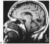
1. Hoyt W, Luis O: The primate chiasm: details of visual fiber organization studied by silver impregnation techniques. Arch Ophthalmol 70:69, 1963

2. Tassinari G, Campara D, Balercia G, Chilosi M, Martignoni G: Magno- and parvocellular pathways are segregated in the human optic tract. Neuroreport 5:1425, 1994

3. Savino P, Paris M, Schatz N, Orr L, Corbett J: Optic tract syndrome. A review of 21 patients. Arch Ophthalmol 96:656, 1978

4. Newman S, Miller N: Optic tract syndrome. Neuro-ophthalmologic considerations. Arch Ophthalmol 101:1241, 1983

5. Frisèn L: The neurology of visual acuity. Brain 13:639, 1980

6. Paul T, Hoyt W: Funduscopic appearance of papilledema with optic tract atrophy. Arch Ophthalmol 94:467, 1976

7. Czarnecki J, Weingeist T, Burton J, Thompson H: “Twin peaks” papilledema: The appearance of papilledema with optic tract atrophy. Can J Ophthalmol 11:279, 1976

8. Bell R, Thompson H: Relative afferent pupillary defect in optic tract hemianopias. Am J Ophthlamol 85:538, 1978

9. O'Connor P, Mein C, Hughes J, Dorwart R, Shacklett D: The Marcus Gunn pupil in incomplete optic tract hemianopias. J Clin Neuroophthalmol 2:227, 1982

10. Klein L, Fermaglich J, Kataah J, Luessenhop A: Cavernous hemangioma of optic chiasm, optic nerves and right optic tract: Case report and review of the literature. Virchows Arch A Pathol Pathol Anat 383:225, 1979

11. Rosenblatt M, Behrens M, Zweufach P, et al: Magnetic resonance imaging of optic tract involvement in multiple sclerosis. Am J Ophthalmol 104:74, 1987

12. Youl B, Plant G, Stevens J, et al: Three cases of craniopharyngioma showing optic tract hypersignal on MRI. Neurology 40:1416, 1990

13. Tachibana O, Yamaguchi N, Yamashima T, Yamashita J: Radiation necrosis of the optic chiasm, optic tract, hypothalamus, and upper pons after radiotherapy for pituitary adenoma, detected by gadolinium-enhanced, T1-weighted magnetic resonance imaging: case report. Neurosurgery 27:640, 1990

14. Beck R, Schatz N, Savino P: Involvement of the optic chiasm, optic tract, and geniculo-calcarine visual system in multiple sclerosis. Bull Soc Belge Ophtalmol 208:159, 1983

15. Plant G, Kermode A, Turano G, et al: Symptomatic retrochiasmal lesions in multiple sclerosis. Clinical features, visual 
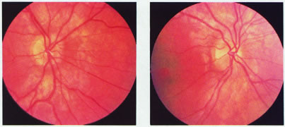
evoked potentials, and magnetic resonance imaging. Neurology 42:68, 1992

16. McLaurin E, Harrington D: Intracranial sarcoidosis with optic tract and temporal lobe involvement. Am J Ophthalmol 86:656, 1978

17. Zentner J, Grodd W, Hassler W: Cavernous angioma of the optic tract. J Neurol 236:117, 1989

18. Kupersmith M, Vargas M, Hoyt W, Berenstein A: Optic tract atrophy with cerebral arteriovenous malformations: direct and transsynaptic degeneration. Neurology 44:80, 1994

19. Anderson D, Trobe J, Hood T, Gebarski S: Optic tract injury after anterior temporal lobectomy. Ophthalmology 96:1065, 1989

20. Manesis E, Petrou C, Brouzas D, Hadziyannis S: Optic tract neuropathy complicating low-dose interferon treatment. J Hepatol 21:474, 1994

21. Margo C, Hamed L, McCarty J: Congenital optic tract syndrome. Arch Ophthalmol 109:1120, 1991

22. Bender M, Bodis-Wollner I: Visual dysfunctions in optic tract lesions. Ann Neurol 3:187, 1978

23. Sherman S, Koch C: The control of retinogeniculate transmission in the mammalian lateral geniculate nucleus. Exp Brain Res 63:1, 1986

24. Sillito A, Murphy P: The modulation of the retinal relay to the cortex in the dorsal lateral geniculate nucleus. Eye 2(Suppl):S221, 1988

25. Harting J, Huerta M, Hashikawa T, van Lieshout D: Projection of the mammalian superior colliculus upon the dorsal lateral geniculate nucleus: organization of the tectogeniculate pathways in nineteen species. J Comp Neurol 304:275, 1991

26. Frisèn L, Holmegaard L, Rosenkrantz M: Sectoral optic atrophy and homonymous horizontal sectoranopia: a lateral choroidal artery syndrome? J Neurol Neurosurg Psychiatry 41:374 , 1978

27. Frisèn L: Quadruple sectoranopia and sectorial optic atrophy. A syndrome of the distal anterior choroidal artery. J Neurol Neurosurg Psychiatry 42:590, 1979

28. Helgason C, Caplan L, Goodwin J, Hedges T: Anterior choroidal artery-territory infarction. Report of cases and review. Arch Neurol 43:681, 1986

29. Donahue S, Kardon R, Thompson H. Hourglass-shaped visual fields as a sign of bilateral lateral geniculate myelinolysis. Am J Ophthalmol 119:378, 1995

30. Barton J: Bilateral sectoranopia from osmotic demyelination. Neurology 57:2318, 2001

31. Greenfield D, Siatkowski R, Schatz N, Glaser J: Bilateral geniculitis associated with severe diarrhea. Am J Ophthalmol 122:280, 1996

32. Goldman J, Horoupian D: Demyelination of the lateral geniculate nucleus in central pontine myelinolysis. Ann Neurol 9:185, 1981

33. Jacobson D: The localizing value of a quadrantanopia. Arch Neurol 54:401, 1997

34. Carter J, O'Connor P, Shacklett D, Rosenberg M: Lesions of the optic radiations mimicking lateral geniculate nucleus visual field defects. J Neurol Neurosurg Psychiatry 48:982, 1985

35. Smith C, Richardson W: The course and distribution of the arteries supplying the visual (striate) cortex. Am J Ophthalmol 61:1391, 1966

36. Horton J, Hoyt W: The representation of the visual field in human striate cortex: a revision of the classis Holmes map. Arch Ophthalmol 109:816, 1991

37. Inouye T: Die Sehstorungen bei Schussverletzungen der kortikalen Sesphare. Leipzig: Engelmann, 1909

38. Holmes G, Lister W: Disturbances of vision from cerebral lesions with special reference to the cortical representation of the macula. Brain 39:34, 1916

39. McFadzean R, Brosnahan D, Hadley D, Mutlukan E: Representation of the visual field in the occipital striate cortex. Br J Ophthalmol 78:185, 1994

40. Trauzettel-Klosinski S, Brendler K: Eye movements in reading with hemianopic field defects: the significance of clinical parameters. Graefe's Arch Clin Exp Ophthalmol 236:91, 1998

41. Huber A: Homonymous hemianopia after occipital lobectomy. Am J Ophthalmol 54:623, 1962

42. Tootell R, Switkes E, Silverman M, Hamilton S: Functional anatomy of macaque striate cortex. II. Retinotopic organization. J Neurosci 8:1531, 1988

43. McAuley D, Russell R: Correlation of CAT scan and visual field defects in vascular lesions of the posterior visual pathways. J Neurol Neurosurg Psychiatry 42:298, 1979

44. Gray L, Galetta S, Siegal T, Schatz N: The central visual field in homonymous hemianopia. Evidence for unilateral foveal representation. Arch Neurol 54:312, 1997

45. Trauzettel-Klosinski S, Reinhard J: The vertical field border in hemianopia and its significance for fixation and reading. Invest Ophthalmol Vis Sci 39:2177, 1998

46. Reinhard J, Trauzettel-Klosinski S: Nasotemporal overlap of retinal ganglion cells in humans: a functional study. Invest Ophthalmol Vis Sci 44:1568, 2003

47. Heller-Bettinger I, Kepes J, Preskorn S, Wurster J: Bilateral altitudinal anopia caused by infarction of the calcarine cortex. Neurology 26:1176, 1976

48. Rush J: Nonbacterial thrombotic endocarditis and cortical blindness. Am J Ophthalmol 114:643, 1992

49. Horton JC, Hoyt WF: Quadrantic visual field defects. A hallmark of lesions in extrastriate (V2/V3) cortex. Brain 114:1703, 1991

50. Spalding J: Wounds of the visual pathway. Part II. The striate cortex. J Neurol Neurosurg Psychiatry 15:169, 1952

51. Benton S, Levy I, Swash M: Vision in the temporal crescent in occipital infarction. Brain 103:83, 1980

52. Chavis P, Al-Hazmi A, Clunie D, Hoyt W: Temporal crescent syndrome with magnetic resonance correlation. J Neuroophthalmol 17:151, 1997

53. Pessin M, Lathi E, Cohen M, et al: Clinical feature and mechanism of occipital lobe infarction. Ann Neurol 21:290, 1987

54. Hornstein S, Chamberlin W, Conomy J: Infarctions of the fusiform and calcarine regions: agitated delirium and hemianopia. Trans Am Neurol Assoc 92:85, 1967

55. Medina J, Chokroverty S, Rubino F: Syndrome of agitated delirium and visual impairment: a manifestation of medial temporo-occipital infarction. J Neurol Neurosurg Psychiatry 40:861, 1977

56. Vaphiades M, Celesia G, Brigell M: Positive spontaneous visual phenomena limited to the hemianopic field in lesions of central visual pathways. Neurology 47:408, 1996

57. Symonds C, McKenzie I: Bilateral loss of vision from cerebral infarction. Brain 80:415, 1957

58. Bougousslavsky J, van Melle G: Unilateral occipital infarction: evaluation of the risk of developing bilateral loss of vision. J Neurol Neurosurg Psychiatry 46:78, 1983

59. Halpern J, Sedler R: Traumatic bilateral homonymous hemianopic scotomas. Ann Ophthalmol 12:1022, 1980

60. Aldrich M, Alessi A, Beck R, Gilman S: Cortical blindness: etiology, diagnosis and prognosis. Ann Neurol 21:149, 1987

61. Chisholm I: Cortical blindness in cranial arteritis. Br J Ophthalmol 59:332, 1995

62. Naito H, Kurokawa K, Kanno T, Toya S, Osano M: Status epilepticus and cortical blindness due to subclavian steal syndrome in a girl with Blalock's operation. Surg Neurol 1:46, 1973

63. Carney A, Anderson E: Cortical blindness and tourniquet subclavian steal. JAMA 245:572, 1981

64. Wells T, Graham C, Moss M, Kearns G: Nifedipine poisoning in a child. Pediatrics 86:91, 1990

65. Morton C, Hickey-Dwyer M: Cortical blindness after nifedipine treatment. BMJ 305:693, 1992

66. Lawrence-Friedl D, Bauer K: Bilateral cortical blindness: an unusual presentation of bacterial endocarditis. Ann Emerg Med 21:1502, 1992

67. Morgan R, Nugent B, Harrison J, O'Connor P: Voluntary alteration of pattern visual evoked responses. Ophthalmology 92:1356, 1985

68. Spehlmann R, Gross R, Ho S, Leetsma J, Norcross K: Visual evoked potentials and postmortem findings in a case of cortical blindness. Ann Neurol 2:531, 1977

69. Celesia G, Archer C, Kuroiwa Y, Goldfader P: Visual function of the extra-geniculo-calcarine system in man: relationship to cortical blindness. Arch Neurol 37:704, 1980

70. Frank Y, Torres F: Visual evoked potentials in the evaluation of “cortical blindness” in children. Ann Neurol 6:126, 1979

71. Wong V: Cortical blindness in children. A study of etiology and prognosis. Pediatr Neurol 7:178, 1991

72. Gjerris F, Mellemgaard L: Transitory cortical blindness in head injury. Acta Neurol Scand 45:623, 1969

73. Griffith J, Dodge P: Transient blindness following head injury in children. N Engl J Med 278:648, 1968

74. Drubach D, Carmona S, Meyerrose G, Peralta L, Sostre S: Brain SPECT in a case of cortical blindness. Stroke 25:1061, 1994

75. Kooi K, Sharbrough F: Electrophyiological findings in cortical blindness. Electroencephalogr Clin Neurophysiol 20:260, 1966

76. Duchowny M, Weiss I, Majlessi H, Barnet A: Visual evoked responses in childhood cortical blindness after head trauma and meningitis. A longitudinal study of six cases. Neurology 24:933, 1974

77. Tepperberg J, Nussbaum D, Feldman F: Cortical blindness following meningitis due to Hemophilus influenzae type B. J Pediatr 91:434, 1977

78. Ramani V: Cortical blindness following ictal nystagmus. Arch Neurol 42:191, 1985

79. Skolnik S, Mizen T, Burde R: Transient post-ictal cortical blindness. J Clin Neuroophthalmol 7:151, 1987

80. Joseph J, Louis S: Transient ictal cortical blindness during middle age. A case report and review of the literature. J Neuroophthalmol 15:39, 1995

81. Miyata Y, Motomura S, Tsuji Y, Koga S: Hepatic encephalopathy and reversible cortical blindness. Am J Gastroenterol 83:780, 1988

82. Kupferschmidt H, Bont A, Schnorf H, et al: Transient cortical blindness and bioccipital brain lesions in two patients with acute intermittent porphyria. Ann Intern Med 123:598, 1995

83. Mukamel M, Weitz R, Nissenkorn I, Yassur I, Varsano I: Acute cortical blindness associated with hypoglycemia. J Pediatr 98:583, 1981

84. Marra T, Shah M, Mikus M: Transient cortical blindness due to hypertensive encephalopathy. Magnetic resonance imaging correlation. J Clin Neuroophthalmol 13:35, 1993

85. Tychsen L, Hoyt W: Hydrocephalus and transient cortical blindness. Am J Ophthalmol 98:819, 1984

86. Greenblatt S: Post-traumatic cerebral blindness: association with migraine and seizure diathesis. JAMA 225:1073, 1973

87. Patronas N, Argyropoulu M: Intravascular thrombosis as a possible cause of transient cortical brain lesions. CT and MRI. J Comput Assist Tomogr 16:849, 1992

88. Berman I, Mann M: Seizures and transient cortical blindness associated with cisplatinum (II) diamminedichloride (PPD) therapy in a thirty-year-old man. Cancer 45:764, 1980

89. Philip P, Carmichael J, Harris A: Convulsions and transient cortical blindness after cisplatin. BMJ 302:416, 1991

90. Rubin A: Transient cortical blindness and occipital seizures with cyclosporine toxicity. Transplantation 47:572, 1989

91. Byrd R, Rohrbaugh T, Raney R, Norris D: Transient cortical blindness secondary to vincristine therapy in childhood malignancies. Cancer 47:37, 1981

92. Shutter L, Green J, Newman N, Hooks M, Gordon R: Cortical blindness and white matter lesions in a patient receiving FK506 after liver transplantation. Neurology 43:2417, 1993

93. Studdard W, Davis D, Young S: Cortical blindness after cerebral angiography. Case report. J Neurosurg 54:240, 1981

94. Parry R, Rees J, Wilde P: Transient cortical blindness after coronary angiography. Br Heart J 70:563, 1993

95. Lantos G: Cortical blindness due to osmotic disruption of the blood–brain barrier by angiographic contrast material: CT and MRI studies. Neurology 39:567, 1989

96. Eldridge P, Punt J: Transient traumatic cortical blindness in children. Lancet 1:815, 1988

97. Liebowitz H, Hall P: Cortical blindness as a complication of eclampsia. Ann Emerg Med 13:365, 1984

98. Cunningham F, Fernandez C, Hernandez C: Blindness associated with preeclampsia and eclampsia. Am J Obstet Gynecol 172:1291, 1995

99. Gospe S: Transient cortical blindness in an infant exposed to methamphetamine. Ann Emerg Med 26:380, 1995

100. DeSousa A, Kleiman M, Mealey J: Quadriplegia and cortical blindness in Hemophilus influenzae meningitis. J Pediatr 93:253, 1978

101. Woodward G: Posttraumatic cortical blindness: are we missing the diagnosis in children? Ped Emerg Care 6:289, 1990

102. Barnet A, Manson J, Wilner E: Acute cerebral blindness in childhood. Neurology 20:1147, 1970

103. Moel D, Kwun Y: Cortical blindness as a complication of hemodialysis. J Pediatr 93:890, 1978

104. Hochstetler K, Beals R: Transient cortical blindness in a child. Ann Emerg Med 16:218, 1987

105. Rodriguez A, Lozano J, del Pozo D, Paez J: Post-traumatic transient cortical blindness. Int Ophthalmol 17:277, 1993

106. Carmola J, Harris B: Transient cortical blindness: still an overlooked syndrome? N Engl J Med 282:1325, 1970

107. Kaye E, Herskowitz J: Transient post-traumatic cortical blindness: brief versus prolonged syndromes in childhood. J Child Neurol 1:206, 1986

108. Makino A, Soga T, Obayashi M, et al: Cortical blindness causes by acute general cerebral swelling. Surg Neurol 29:393, 1989

109. Lambert S, Hoyt C, Jan J, Barkovitch J, Flodmark O: Visual recovery from hypoxic cortical blindness during childhood. Computed tomographic and magnetic resonance predictors. Arch Ophthalmol 105:1371, 1988

110. Goodlin R, Strieb E, Sun S, Cox T, Williams N: Cortical blindness as the initial symptom in severe preeclampsia. Am J Ophthalmol 147:841, 1983

111. Seaward G, England M, Nagar A, van Gelderen C: Transient post-partum amaurosis. A report of four cases. J Reprod Med 34:253, 1989

112. Duncan R, Hadley D, Bone I, et al: Blindness in eclampsia: CT and MR imaging. J Neurol Neurosurg Psychiatry 52:899, 1989

113. Lau S, Chan F, Yu Y, Woo E, Huang C: Cortical blindness in toxemia of pregnancy: findings on computed tomography. Br J Radiol 60:347, 1987

114. Herzog T, Angel O, Karram M, Evertson L: Use of magnetic resonance imaging in the diagnosis of cortical blindness in pregnancy. Obstet Gynecol 76:980, 1990

115. Beal M, Chapman P: Cortical blindness and homonymous hemianopia in the postpartum period. JAMA 244:2085, 1980

116. Stiller R, Leone-Tomaschoff S, Cuteri J, Beck L: Post-partum pulmonary embolus as an unusual cause of cortical blindness. Am J Obstet Gynecol 162:696, 1990

117. Branch D, Andres R, Digre K, Rote N, Scott J: The association of antiphospholipid antibodies with severe eclampsia. Obstet Gynecol 73:541, 1989

118. Carpenter E, Kava H, Plotkin D: The development of total blindness as a complication of pregnancy. Am J Obstet Gynecol 66:641, 1953

119. Beck R, Gamel J, Willcourt R, Berman G: Acute ischemic optic neuropathy in severe eclampsia. Am J Ophthalmol 90:342, 1980

120. Sandifer PH: Anosognosia and disorders of body scheme. Brain 69:122, 1946

121. McDaniel K, McDaniel L: Anton's syndrome in a patient with posttraumatic optic neuropathy and bifrontal contusions. Arch Neurol 48:101, 1991

122. Hess R, Pointer J: Spatial and temporal contrast sensitivity in hemianopia. Brain 112:871, 1989

123. Rizzo M, Robin D: Bilateral effects of unilateral occipital lobe lesions in humans. Brain 119:951, 1996

124. Barton JJ, Sharpe JA: Smooth pursuit and saccades to moving targets in blind hemifields. A comparison of medial occipital, lateral occipital and optic radiation lesions. Brain 120:681, 1997

125. Ball K, Owsley C, Sloane M, Roenker D, Bruni J: Visual attention problems as a predictor of vehicle crashes in older drivers. Invest Ophthalmol Vis Sci 34:3110, 1993

126. Rizzo M, Reinach S, McGehee D, Dawson J. Simulated car crashes and crash predictors in drivers with Alzheimer's disease. Arch Neurol 54:545, 1997

127. Zihl J, von Cramon D: Visual field recovery from scotoma in patients with post-geniculate damage. Brain 108:335, 1985

128. Parisi J, Bell R, Yassein H: Homonymous hemianopic field defects and driving in Canada. Can J Ophthalmol 26:252, 1991

129. Szlyk J, Brigell M, Seiple W: Effects of age and hemianopic visual field loss on driving. Optom Vision Sci 70:1031, 1993

130. Pommerenke K, Markowitsch J: Rehabilitation training of homonymous visual field defects in patients with postgeniculate damage of the visual system. Restor Neurol Neurosci 1:47, 1989

131. Kerkoff G, Munssinger U, Meier E: Neurovisual rehabilitation in cerebral blindness. Arch Neurol 51:474, 1994

132. Kasten E, Sabel B: Visual field enlargement after computer training in brain-damaged patients with homonymous deficits: an open pilot trial. Restor Neurol Neurosci 8:113, 1995

133. Kasten E, Wüst S, Behrens-Baumann W, Sabel B: Computer-based training for the treatment of partial blindness. Nature Med 4:1083, 1998

134. Schlageter K, Gray B, Hall K, Shaw R, Sammet R: Incidence and treatment of visual dysfunction in traumatic brain injury. Brain Injury 7:439, 1993

135. Behrmann M, Watt S, Black S, Barton J: Impaired visual search in patients with unilateral neglect: an oculographic analysis. Neuropsychologia 35:1445, 1997

136. Barton J, Behrmann M, Black S: Ocular search during line bisection. The effects of hemineglect and hemianopia. Brain 121:1117, 1998

137. Kerkhoff G, Münssinger U, Eberle-Strauss G, et al: Rehabilitation of hemianopic alexia in patients with post-geniculate visual field disorders. Neuropsychol Rehab 2:21, 1992

138. Rossi P, Kheyfets S, Reding M: Fresnel prisms improve visual perception in stroke patients with homonymous hemianopia or unilateral visual neglect. Neurology 40:1597, 1990

139. Barbur J, Ruddock K, Waterfield V: Human visual responses in the absence of the geniculo-calcarine projection. Brain 103:905, 1980

140. Barbur J, Watson J, Frackowiak R, Zeki S: Conscious visual perception without V1. Brain 116:1293, 1993

141. Blythe I, Kennard C, Ruddock K: Residual vision in patients with retrogeniculate lesions of the visual pathways. Brain 110:887, 1987

142. Sanders MD, Warrington E, Marshall J, Weiskrantz L: Blindsight: vision in a field defect. Lancet April 20:707, 1974

143. Stoerig P, Cowey A: Blindsight in man and monkey. Brain 120:535, 1997

144. Weiskrantz L: Residual vision in a scotoma: a follow-up study of “form” discrimination. Brain 110:77, 1987

145. Marcel AJ: Blindsight and shape perception: deficit of visual consciousness or of visual function? Brain 121:1565, 1998

146. Sahraie A, Weiskrantz L, Barbur J, et al: Pattern of neuronal activity associated with conscious and unconscious processing of visual signals. Proc Natl Acad Sci USA 94:9406, 1997

147. Weiskrantz L, Barbur JL, Sahraie A: Parameters affecting conscious versus unconscious visual discrimination with damage to the visual cortex (V1). Proc Natl Acad Sci USA 92:6122, 1995

148. Pöppel E, Held R, Frost D: Residual visual function after brain wounds involving the central visual pathways in man. Nature 243:295, 1973

149. Perenin M-T, Jeannerod M: Residual vision in cortically blind hemifields. Neurosychologia 13:1, 1975

150. Weiskrantz L, Warrington E, Sanders M, Marshall J: Visual capacity in the hemianopic field following a restricted occipital ablation. Brain 97:709, 1974

151. Blythe IM, Bromley JM, Kennard C, Ruddock KH: Visual discrimination of target displacement remains after damage to the striate cortex in humans. Nature 320:619, 1986

152. Barton J, Sharpe J: Smooth pursuit and saccades to moving targets in blind hemifields. A comparison of medial occipital, lateral occipital, and optic radiation lesions. Brain 120:681, 1997

153. Meienberg O, Zangemeister WH, Rosenberg M, et al: Saccadic eye movement stategies in patients with homonymous hemianopia. Ann Neurol 9:537, 1981

154. Corbetta M, Marzi C, Tassinari G, Aglioti S: Effectiveness of different task paradigms in revealing blindsight. Brain 113:603, 1990

155. Perenin M-T, Ruel J, Hécaen H: Residual visual capacities in a case of cortical blindness. Cortex 6:605, 1980

156. Perenin M-T, Jeannerod M: Visual functions within the hemianopic field following early cerebral hemidecortication in man-I. Spatial localization. Neuropsychologia 16:1, 1978

157. Bridgeman B, Staggs D: Plasticity in human blindsight. Vision Res 22:1199, 1982

158. Ptito A, Lepore F, Ptiito M, Lassonde M: Target detection and movement discrimination in the blind field of hemispherectomized patients. Brain 114:497, 1991

159. Riddoch G: Dissociation of visual perception due to occipital injuries with especial reference to appreciation of movement. Brain 40:15, 1917

160. Morland AB, Jones SR, Finlay AL, et al: Visual perception of motion, luminance and color in a human hemianope. Brain 122:1183, 1999

161. Perenin M-T: Discrimination of motion direction in perimetrically blind fields. NeuroReport 2:397, 1991

162. Heide W, Koenig E, Dichgans J: Optokinetic nystagmus, self-motion sensation and their aftereffects in patients with occipit-parietal lesions. Clin Vision Sci 5:145, 1990

163. Barton JJS, Sharpe JA: Motion direction discrimination in blind hemifields. Ann Neurol 41:255, 1997

164. Azzopardi P, Cowey A: Motion discrimination in cortically blind patients. Brain 124:30, 2001

165. ter Braak J, Schenk V, van Vliet A: Visual reactions in a case of long-lasting cortical blindness. J Neurol Neurosurg Psychiatry 34:140, 1971

166. Verhagen W, Huygen P, Mulleners W: Lack of optokinetic nystagmus and visual motion perception in acquired cortical blindness. Neuroophthalmology 17:211, 1997

167. Mestre D, Brouchon M, Ceccaldi M, Poncet M: Perception of optical flow in cortical blindness: a case report. Neuropyschologia 30:783, 1992

168. Perenin MT, Rossetti Y: Grasping without form discrimination in a hemianopic field. Neuroreport 7:793, 1996

169. Morland A, Ogilvie J, Ruddock K, Wright J: Orientation discrimination is impaired in the absence of the striate cortical contribution to human vision. Proc R Soc Lond B Biol Sci 263:633, 1996

170. Jackson SR: Pathological perceptual completion in hemianopia extends to the control of reach-to-grasp movements. Neuroreport 10:2461, 1999

171. Milner A, Goodale M: The Visual Brain in Action. Oxford: Oxford University Press, 1995

172. Stoerig P: Chromaticity and achromaticity. Evidence for a functional differentiation in visual field defects. Brain 110:869, 1987

173. Stoerig P, Cowey A: Increment-threshold spectral sensitivity in blindsight. Evidence for color opponency. Brain 114:1487, 1991

174. Guo K, Benson PJ, Blakemore C: Residual motion discrimination using color information without primary visual cortex. Neuroreport 9:2103, 1998

175. Morland AB, Ruddock KH: Retinotopic organisation of cortical mechanisms responsive to color: evidence from patient studies. Acta Psychol 97:7, 1997

176. Barbur JL, Harlow AJ, Weiskrantz L: Spatial and temporal response properties of residual vision in a case of hemianopia. Phil Trans R Soc Lond B Biol Sci 343:157, 1994

177. Hess RF, Pointer JS: Spatial and temporal contrast sensitivity in hemianopia. Brain 112:871, 1989

178. Weiskrantz L, Harlow A, Barbur JL: Factors affecting visual sensitivity in a hemianopic subject. Brain 114:2269, 1991

179. Weiskrantz L, Cowey A, Le Mare C: Learning from the pupil: a spatial visual channel in the absence of V1 in monkey and human. Brain 121:1065, 1998

180. Weiskrantz L, Cowey A, Barbur JL: Differential pupillary constriction and awareness in the absence of striate cortex. Brain 122:1533, 1999

181. Barbur JL, Weiskrantz L, Harlow JA: The unseen color aftereffect of an unseen stimulus: insight from blindsight into mechanisms of color afterimages. Proc Natl Acad Sci USA 96:11637, 1999

182. Morris JS, Ohman A, Dolan RJ: A subcortical pathway to the right amygdala mediating “unseen” fear. Proc Natl Acad Sci USA 96:1680, 1999

183. de Gelder B, Vroomen J, Pourtois G, Weiskrantz L: Non-conscious recognition of affect in the absence of striate cortex. Neuroreport 10:3759, 1999

184. Morris JS, DeGelder B, Weiskrantz L, Dolan RJ: Differential extrageniculostriate and amygdala responses to presentation of emotional faces in a cortically blind field. Brain 124:1241, 2001

185. Hamm AO, Weike AI, Schupp HT, et al: Affective blindsight: intact fear conditioning to a visual cue in a cortically blind patient. Brain 126:267, 2003

186. Marzi C, Tassinari G, Agliotti S, Lutzemberger L: Spatial summation across the vertical meridian in hemianopics: a test of blindsight. Neuropsychologia 24:749, 1986

187. Intriligator JM, Xie R, Barton JJ: Blindsight modulation of motion perception. J Cogn Neurosci 14:1174, 2002

188. Ward R, Jackson SR: Visual attention in blindsight: sensitivity in the blind field increased by targets in the sighted field. Neuroreport 13:301, 2002

189. Rafal R, Smith J, Krantz J, Cohen A, Brennan C: Extrageniculate vision in hemianopic humans: saccade inhibition by signals in the blind field. Science 250:118, 1990

190. Walker R, Mannan S, Maurer D, Pambakian AL, Kennard C: The oculomotor distractor effect in normal and hemianopic vision. Proc R Soc Lond B Biol Sci 267:431, 2000

191. Danziger S, Fendrich R, Rafal RD: Inhibitory tagging of locations in the blind field of hemianopic patients. Conscious Cogn 6:291, 1997

192. Danckert J, Maruff P, Kinsella G, de Graaff S, Currie J: Investigating form and color perception in blindsight using an interference task. Neuroreport 9:2919, 1998

193. Braddick O, Atkinson J, Hood B, et al: Possible blindsight in infants lacking one cerebral hemisphere. Nature 360:461, 1992

194. Ptito A, Lassonde M, Lepore F, Ptito M: Visual discrimination in hemispherectomized patients. Neuropsychologia 25:869, 1987

195. Tomaiuolo F, Ptito M, Marzi CA, Paus T, Ptito A: Blindsight in hemispherectomized patients as revealed by spatial summation across the vertical meridian. Brain 120:795, 1997

196. Wessinger CM, Fendrich R, Gazzaniga MS, Ptito A, Villemure J-G: Extrageniculostriate vision in humans: investigations with hemispherectomy patients. Prog Brain Res 112:405, 1996

197. Wessinger C, Fendrich R, Ptito A, Villemure J, Gazzaniga M: Residual vision with awareness in the field contralateral to a partial or complete functional hemispherectomy. Neuropsychologia 34:1129, 1996

198. Stoerig P, Faubert J, Ptito M, Diaconu V, Ptito A: No blindsight following hemidecortication in human subjects? Neuroreport 7:1990, 1996

199. Faubert J, Diaconu V, Ptito M, Ptito A: Residual vision in the blind field of hemidecorticated humans predicted by a diffusion scatter model and selective spectral absorption of the human eye. Vision Res 39:149, 1999

200. King S, Azzopardi P, Cowey A, Oxbury J, Oxbury S: The role of light scatter in the residual visual sensitivity of patients with complete cerebral hemispherectomy. Visual Neurosci 13:1, 1996

201. Faubert J, Diaconu V: From visual consciousness to spectral absorption in the human retina. Progr Brain Res 134:399, 2001

202. Balliet R, Blood KM, Bach-y-Rita P: Visual field rehabilitation in the cortically blind? J Neurol Neurosurg Psychiatry 48:1113, 1985

203. Campion J, Latto R, Smith YM: Is blindsight an effect of scattered light, spared cortex, and near-threshold vision? Behav Brain Sci 6:423, 1983

204. Fendrich R, Wessinger CM, Gazzaniga MS: Residual vision in a scotoma: implications for blindsight. Science 258:1489, 1992

205. Kasten E, Wuest S, Sabel B: Residual vision in transition zones in patients with cerebral blindness. J Clin Exp Neuropsychol 20:581, 1998

206. Scharli H, Harman A, Hogben J: Blindsight in subjects with homonymous visual field defects. J Cogn Neurosci 11:52, 1999

207. Scharli H, Harman AM, Hogben JH: Residual vision in a subject with damaged visual cortex. J Cogn Neurosci 11:502, 1999

208. Meeres SL, Graves RE: Localization of unseen stimuli by humans with normal vision. Neuropsychologia 28:1231, 1990

209. Kolb FC, Braun J: Blindsight in normal observers. Nature 377:336, 1995

210. Azzopardi P, Cowey A: Is blindsight like normal, near-threshold vision? Proc Natl Acad Sci USA 94:14190, 1997

211. Stoerig P, Hébner M, Pöppel E: Signal detection analysis of residual vision in a field defect due to a post-geniculate lesion. Neuropsychologia 23:589, 1985

212. Azzopardi P, Cowey A: Blindsight and visual awareness. Conscious Cogn 7:292, 1998

213. Dodds C, Machado L, Rafal R, Ro T: A temporal/nasal asymmetry for blindsight in a localisation task: evidence for extrageniculate mediation. Neuroreport 13:655, 2002

214. Bittar RG, Ptito M, Faubert J, Dumoulin SO, Ptito A: Activation of the remaining hemisphere following stimulation of the blind hemifield in hemispherectomized subjects. Neuroimage 10:339, 1999

215. Stoerig P, Kleinschmidt A, Frahm J: No visual responses in denervated V1: high-resolution functional magnetic resonance imaging of a blindsight patient. Neuroreport 9:21, 1998

216. Cowey A, Stoerig P: The neurobiology of blindsight. Trends Neurosci 14:140, 1995

217. Rodman H, Gross C, Albright T: Afferent basis of visual response properties in area MT of the macaque. I. Effects of striate cortex removal. J Neurosci 9:2033, 1989

218. Girard P, Salin P, Bullier J: Response selectivity of neurons in area MT of the macaque monkey during reversible inactivation of area V1. J Neurophysiol 67:1437, 1992

219. Rosa MG, Tweedale R, Elston GN: Visual responses of neurons in the middle temporal area of new world monkeys after lesions of striate cortex. J Neurosci 20:5552, 2000

220. Girard P, Salin P, Bullier J: Visual activity in areas V3a and V3 during reversible inactivation of area V1 in the macaque monkey. J Neurophysiol 66:1493, 1991

221. Gross C: Contributions of striate cortex and the superior colliculus to visual functions in area MT, the superior temporal polysensory area and inferior temporal cortex. Neuropsychologia 29:497, 1991

222. Payne BR, Lomber SG, Macneil MA, Cornwell P: Evidence for greater sight in blindsight following damage of primary visual cortex early in life. Neuropsychologia 34:741, 1996

223. Sorenson KM, Rodman HR: A transient geniculo-extrastriate pathway in macaques? Implications for ‘blindsight.”rsquo; Neuroreport 10:3295, 1999

224. ffytche D, Guy C, Zeki S: The parallel visual motion inputs into areas V1 and V5 of human cerebral cortex. Brain 118:1375, 1995

225. Beckers G, Zeki S: The consequences of inactivating areas V1 and V5 in visual motion perception. Brain 118:49, 1995

226. Hotson J, Braun D, Herzberg W, Boman D: Transcranial magnetic stimulation of extrastriate cortex degrades human motion direction discrimination. Vision Res 34:2115, 1994

227. Holliday IE, Anderson SJ, Harding GF: Magnetoencephalographic evidence for non-geniculostriate visual input to human cortical area V5. Neuropsychologia 35:1139, 1997

228. ffytche DH, Guy CN, Zeki S: Motion specific responses from a blind hemifield. Brain 119:1971, 1996

229. Magnussen S, Mathiesen T: Detection of moving and stationary gratings in the absence of striate cortex. Neuropsychologia 27:725, 1989

230. Moore T, Rodman H, Repp A, Gross C, Mezrich R: Greater residual vision in monkeys after striate damage in infancy. J Neurophysiol 76:3928, 1996

231. Zihl J: “Blindsight”: improvement of visually guided eye movements by systematic practice in patients with cerebral blindness. Neuropsychologia 18:71, 1980

232. Zihl J, Werth R: Contributions to the study of “blindsight.” I. Can stray light account for saccadic localization in patients with postgeniculate visual field defects? Neuropsychologia 22:1, 1984

233. Zihl J, Werth R: Contributions to the study of “blindsight.” II. The role of specific practice for saccadic localization in patients with postgeniculate visual field defects. Neuropsychologia 22:13, 1984

234. Albert ML, Reches A, Silverberg R: Hemianopic color blindness. J Neurol Neurosurg Psychiatry 38:546, 1975

235. Paulson HL, Galetta SL, Grossman M, Alavi A: Hemiachromatopsia of unilateral occipitotemporal infarcts. Am J Ophthalmol 118:518, 1994

236. MacKay G, Dunlop JC: The cerebral lesions in a case of complete acquired color-blindness. Scott Med Surg J 5:503, 1899

237. Pallis CA: Impaired identification of faces and places with agnosia for colors. J Neurol Neurosurg Psychiatry 18:218, 1955

238. Meadows JC: Disturbed perception of colors associated with localized cerebral lesions. Brain 97:615, 1974

239. Rizzo M, Smith V, Pokorny J, Damasio A: Color perception profiles in central achromatopsia. Neurology 43:995, 1993

240. Rizzo M, Smith V, Pokorny J, Damasio AR: Color perception profiles in central achromatopsia. Neurology 43:995, 1993

241. Damasio A, Yamada T, Damasio H, Corbett J, McKee J: Central achromatopsia: Behavioral, anatomic and physiologic aspects. Neurology 30:1064, 1980

242. Critchley M: Acquired anomalies of color perception of central origin. Brain 88:711, 1965

243. Sacks O: The case of the color-blind painter. An Anthropologist on Mars. New York: Alfred A Knopf, 1995

244. Land E: Recent advances in retinex theory. Vision Res 26:7, 1986

245. Zeki SM: A century of cerebral achromatopsia. Brain 113:1721, 1990

246. Land E, Hubel D, Livingstone M, Perry S, Burns M: Color-generating interactions across the corpus callosum. Nature 303:616, 1983

247. Kennard C, Lawden M, Morland AB, Ruddock KH: Color identification and color constancy are impaired in a patient with incomplete achromatopsia associated with prestriate lesions. Proc R Soc Lond B 260:169, 1995

248. Clarke S, Walsh V, Schoppig A, Assal G, Cowey A: Color constancy impairments in patients with lesions of the prestriate cortex. Exp Brain Res 123:154, 1998

249. Hurlbert AC, Bramwell DI, Heywood C, Cowey A: Discrimination of cone contrast changes as evidence for color constancy in cerebral achromatopsia. Exp Brain Res 123:136, 1998

250. D'Zmura MD, Knoblauch K, Henaff M-A, Michel F: Dependence of color on context in a case of cortical color vision deficiency. Vision Research 38:3455, 1998

251. Heywood CA, Cowey A, Newcombe F: Chromatic discrimination in a cortically color blind observer. Eur J Neurosci 3:802, 1991

252. Heywood CA, Nicholas JJ, Cowey A: Behavioural and electrophysiological chromatic and achromatic contrast sensitivity in an achromatopsic patient. J Neurol Neurosurg Psychiatry 61:638, 1996

253. Adachi-Usami E, Tsukamoto M, Shimada Y: Color vision and color pattern evoked cortical potentials in a patient with acquired cerebral dyschromatopsia. Doc Ophthalmol 90:259, 1997

254. Pearlman AL, Birch J, Meadows JC: Cerebral color blindness: an acquired defect in hue discrimination. Ann Neurol 5:253, 1979

255. Heywood CA, Kentridge RW, Cowey A: Form and motion from color in cerebral achromatopsia. Exp Brain Res 123:145, 1998

256. Cole G, Heywood C, Kentridge R, Fairholm I, Cowey A: Attentional capture by color and motion in cerebral achromatopsia. Neuropsychologia 41:1837, 2003

257. Cavanagh P, Hénaff M-A, Michel F, et al: Complete sparing of high-contrast color input to motion perception in cortical color blindness. Nature Neurosci 1:242, 1998

258. Green GJ, Lessell S: Acquired cerebral dyschromatopsia. Arch Ophthalmol 95:121, 1977

259. Mendola J, Corkin S: Visual discrimination and attention after bilateral temporal-lobe lesions: a case study. Neuropsychologia 37:91, 1999

260. Ogden JA: Visual object agnosia, prosopagnosia, achromatopsia, loss of visual imagery, and autobiographic amnesia following recovery from cortical blindness: case M.H. Neuropsychologia 31:571, 1993

261. Verrey D: Hemiachromatopsie droite absolue. Arch Ophthalmol (Paris) 8:289, 1888

262. Victor J, Maiese K, Shapley R, Sitdis J, Gazzaniga M: Acquired central dyschromatopsia: analysis of a case with preservation of color discrimination. Clin Vision Sci 4:183, 1989

263. Damasio H, Frank R: Three-dimensional in vivo mapping of brain lesions in humans. Arch Neurol 49:137, 1992

264. Dean P: Visual cortex ablation and thresholds for successively presented stimuli in Rhesus monkeys. II. Hue. Exp Brain Res 35:69, 1979

265. Wild HM, Butler SR, Carden D, Kulikowski JJ: Primate cortical area V4 important for color constancy but not wavelength discrimination. Nature 313:133, 1985

266. Heywood CA, Cowey A: On the role of cortical area V4 in the discrimination of hue and pattern in macaque monkeys. J Neurosci 7:2601, 1987

267. Heywood CA, Gadotti A, Cowey A: Cortical area V4 and its role in the perception of color. J Neurosci 12:4056, 1992

268. Walsh V, Kulikowski JJ, Butler SR, Carden D: The effects of lesions of area V4 on the visual abilities of macaques: color categorization. Behav Brain Res 7:1, 1992

269. Schiller P: The effects of V4 and middle temporal (MT) lesionson visual performance in the rhesus monkey. Vis Neurosci 10:717, 1993

270. Cowey A, Heywood C, Irving-Bell L: The regional cortical basis of achromatopsia: a study on macaque monkeys and an achromatopsic patient. Eur J Neurosci 14:1555, 2001

271. Heywood C, Gaffan D, Cowey A: Cerebral achromatopsia in monkeys. Eur J Neurosci 7:1064, 1995

272. Hadjikhani N, Liu A, Dale A, Cavanagh P, Tootell R: Retinotopy and color selectivity in human cortical visual area V8. Nature Neurosci 1:235, 1998

273. Bartels A, Zeki S: The architecture of the color centre in the human visual brain: new results and a review. Eur J Neurosci 12:172, 2000

274. Gulyás B, Roland P: Cortical fields participating in form and color discrimination in the human brain. Neuroreport 2:585, 1991

275. Gulyás B, Heywood C, Popplewell D, Roland P, Cowey A: Visual form discrimination from color or motion cues: functional anatomy by positron emission tomography. Proc Natl Acad Sci USA 91:9965, 1994

276. Beauchamp M, Haxby J, Jennings J, DeYoe E: An fMRI version of the Farnsworth-Munsell 100-Hue test reveals multiple color-selective areas in human ventral occipitotemporal cortex. Cereb Cortex 9:257, 1999

277. Merigan W: Human V4? Curr Biol 3:226, 1993

278. Wandell B, Wade A: Functional imaging of the visual pathways. Neurol Clin 21:417, 2003

279. Orrell R, James-Galton M, Stevens J, Rossor M: Cerebral achromatopsia as a presentation of Trousseau's syndrome. Postgrad Med J 71:44, 1995

280. Aldrich M, Vanderzant C, Alessi A, Abou-Khalil B, Sackellares J: Ictal cortical blindness with permanent visual loss. Epilepsia 30:116, 1989

281. Freedman L, Costa L: Pure alexia and right hemiachromatopsia in posterior dementia. J Neurol Neurosurg Psychiatr 55:500, 1992

282. Lawden M, Cleland P: Achromatopsia in the aura of migraine. J Neurol Neurosurg Psychiatr 56:708, 1993

283. Fine R, Parker G: Disturbance of central vision after carbon monoxide poisoning. Aust NZ J Ophthalmol 24:137, 1996

284. Kölmel HW: Pure homonymous hemiachromatopsia. Findings with neuroophthalmologic examination and imaging procedures. Eur Arch Psychiatr Neurol Sci 237:237, 1988

285. Linksz A: An Essay on Color Vision and Clinical Color Vision Tests. New York: Grune and Stratton, 1964

286. Working Group 41 N-NCoV: Procedures for Testing Color Vision. Washington, DC: National Academy Press, 1981

287. Wyszecki G, Stiles W: Color Science: Concepts and Methods, Quantitative Data and Formulae. New York: Wiley, 1982

288. Hardy L, Rand G, Rittler M: AO-HRR pseudoisochromatic plates. Buffalo, NY: American Optical Co, 1957

289. Ichikawa K, Hukame H, Tanabe S: Detection of acquired color vision defects by standard pseudoisochromatic plates, part 2. Doc Ophthalmol Proc 46:133, 1987

290. Frisèn L, Kalm P: Sahlgren's saturation test for detecting and grading acquired dyschromatopsia. Am J Ophthalmol 92:252, 1981

291. Verriest G, Uvijls A, Aspinall P, et al: The lightness discrimination test. Bull Soc Belge Ophtalmol 183:162, 1979

292. Pinckers A, Verriest G: Results of shorthand lightness discrimination test. In: Verriest G (ed): Color Vision Deficiencies VII. Dordrecht, The Netherlands: Martinus Nijhoff/Dr. W Junk, 1987:163

293. Heywood CA, Wilson B, Cowey A: A case study of cortical color ‘blindness’ with relatively intact achromatic discrimination. J Neurol Neurosurg Psychiatr 50:22, 1987

294. Beauchamp M, Haxby J, Rosen A, DeYoe E: A functional MRI case study of acquired cerebral dyschromatopsia. Neuropsychologia 38:1170, 2000

295. Holmes G: Pure word blindness. Folia Psychiatr Neurol Neurochir Neerl 53:279, 1950

296. Geschwind N, Fusillo M: Color-naming defects in association with alexia. Arch Neurol 15:137, 1966

297. Oxbury JM, Oxbury SM, Humphrey NK: Varieties of color anomia. Brain 92:847, 1969

298. de Vreese LP: Two systems for color-naming defects: verbal disconnection versus color imagery disorder. Neuropsychologia 29:1, 1991

299. Kinsbourne M, Warrington EK: Observations on color agnosia. J Neurol Neurosurg Psychiatry 27:296, 1964

300. Luzzatti C, Davidoff J: Impaired retrieval of object-color knowledge with preserved color naming. Neuropsychologia 32:933, 1994

301. Miceli G, Fouch E, Capasso R, et al: The dissociation of color from form and function knowledge. Nat Neurosci 4:662, 2001

302. Farah M: Visual Agnosia: Disorders of Visual Recognition and What They Tell Us about Normal Vision. Cambridge, MA: MIT Press, 1990

303. Riddoch M, Humphreys G: Visual agnosia. Neurol Clin 21:501, 2003

304. Teuber HL: Alteration of perception and memory in man. In: Weiskrantz L (ed): Analysis of Behavioral Change. New York: Harper & Row, 1968

305. Lissauer H: Einfall von Seelenblindheit nebst einem Bintrag zur Theorie derselben. Arch Psychiatr Nervenkr 2:22, 1890

306. Tranel D: Assessment of higher-order visual function. Curr Opin Ophthalmol 5:29, 1994

307. Anderson S, Rizzo M: Recovery and rehabilitation of visual cortical dysfunction. Neurorehabilitation 5:129, 1995

308. Lezak M: Neuropsychological Assessment. New York: Oxford University Press, 1995

309. Spreen O, Strauss E: A Compendium of Neuropsychological Tests. New York: Oxford University Press, 1991

310. Benton A, Van Allen M: Visuoperceptual, visuospatial, and visuoconstructive disorders. In Heilman K, Valenstein E (eds): Clinical Neuropsychology. London: Oxford University Press, 1985:161

311. Benton A, Hamsher J, Varney N, et al: Contributions to Neuropsychological Assessment. New York: Oxford University Press, 1983

312. Goodglass H, Kaplan E: The Assessment of Aphasia and Related Disorders. Philadelphia: Lea & Febiger, 1983

313. Hooper H: The Hooper Visual Organization Test Manual. Los Angeles: Western Psychological Services, 1958

314. Mooney C, Ferguson G: A new closure test. Can J Psychol 5:129, 1951

315. Newcombe F: Selective deficits after focal cerebral injury. In Dimond S, Beanmont J (eds): Hemisphere Function in the Human Brain. New York: Halsted Press, 1974:311

316. Bodamer J: Prosopagnosie. Arch Psychiatr Nervenkr 179:6, 1947

317. Tejeria L, Harper RA, Artes PH, Dickinson CM: Face recognition in age related macular degeneration: perceived disability, measured disability, and performance with a bioptic device. Br J Ophthalmol 86:1019, 2002

318. Mendez M, Martin R, Smyth K, Whitehouse P: Disturbances of person identification in Alzheimer's disease. A retrospective study. J Nerv Ment Dis 180:94, 1992

319. Roudier M, Marcie P, Grancher A, et al: Discrimination of facial identity and of emotions in Alzheimer's disease. J Neurol Sci 154:151, 1998

320. Cronin-Golomb A, Cronin-Golomb M, Dunne TE, et al: Facial frequency manipulation normalizes face discrimination in AD. Neurology 54:2316, 2000

321. Janati A: Kluver-Bucy syndrome in Huntington's chorea. J Nerv Ment Dis 173:632, 1985

322. Dewick H, Hanley J, Davies A, Playfer J, Turnbull C: Perception and memory for faces in Parkinson's disease. Neuropsychologia 29:785, 1991

323. Cousins R, Hanley JR, Davies AD, Turnbull CJ, Playfer JR: Understanding memory for faces in Parkinson's disease: the role of configural processing. Neuropsychologia 38:837, 2000

324. Young A, Ellis H: Childhood prosopagnosia. Brain Cognit 9:16, 1989

325. Kracke I: Developmental prosopagnosia in Asperger syndrome: presentation and discussion of an individual case. Dev Med Child Neurol 36:873, 1994

326. Takahashi N, Kawamura M, Hirayama K, Shiota J, Isono O: Prosopagnosia: a clinical and anatomic study of four patients. Cortex 31:317, 1995

327. Tranel D, Damasio A. Knowledge without awareness: an autonomic index of facial recognition by prosopagnosics. Science 228:1453, 1985

328. Young A, Aggleton J, Hellawell D, et al: Face processing impairments after amygdalotomy. Brain 118:15, 1995

329. de Haan E, Campbell R: A fifteen year follow-up of a case of developmental prosopagnosia. Cortex 27:489, 1991

330. Campbell R, Heywood C, Cowey A, Regard M, Landis T: Sensitivity to eye gaze in prosopagnosic patients and monkeys with superior temporal sulcus ablation. Neuropsychologia 28:1123, 1990

331. Bruyer R, Laterre C, Seron X, et al: A case of prosopagnosia with some preserved covert remembrance of familiar faces. Brain Cognit 2:257, 1983

332. Sergent J, Villemure J-G: Prosopagnosia in a right hemispherectomized patient. Brain 112:975, 1989

333. Sergent J, Poncet M: From covert to overt recognition of faces in a prosopagnosic patient. Brain 113:989, 1990

334. Evans J, Heggs A, Antoun N, Hodges J: Progressive prosopagnosia associated with selective right temporal lobe atrophy. Brain 118:1, 1995

335. Tranel D, Damasio AR, Damasio H: Intact recognition of facial expression, gender, and age in patients with impaired recognition of face identity. Neurology 38:690, 1988

336. Perrett D, Hietanen J, Oram M, Benson P: Organization and functions of cells responsive to faces in the temporal cortex. Phil Trans R Soc Lond B 335:23, 1992

337. Gauthier I, Logothetis N: Is face recognition not so unique after all? Cogn Neuropsychol 17:125, 2000

338. Haxby JV, Gobbini MI, Furey ML, et al: Distributed and overlapping representations of faces and objects in ventral temporal cortex. Science 293:2425, 2001

339. Bruyer R: Covert facial recognition in prosopagnosia: a review. Brain Cogn 15:223, 1991

340. Young A: Covert recognition. In Farah M, Ratcliff G (eds): The Neuropsychology of High-Level Vision. Hillsdale, NJ: LEA, 1994

341. Bauer R: Autonomic recognition of names and faces in prosopagnosia: a neuropsychological application of the guilty knowledge test. Neuropsychologia 22:457, 1984

342. Bauer R, Verfaellie M: Electrodermal discrimination of familiar but not unfamiliar faces in prosopagnosia. Brain Cogn 8:240, 1988

343. Renault B, Signoret J-L, DeBruille B, Breton F, Bolgert F: Brain potentials reveal covert facial recognition in prosopagnosia. Neuropsychologia 27:905, 1989

344. McNeil J, Warrington E: Prosopagnosia: a re-classification. Q J Exp Psychol 43A:267, 1991

345. Sergent J, Signoret J-L: Implicit access to knowledge derived from unrecognized faces. Cereb Cortex 2:389, 1992

346. Barton JJ, Cherkasova M, O'Connor M: Covert recognition in acquired and developmental prosopagnosia. Neurology 57:1161, 2001

347. Schweinberger S, Klos T, Sommer W: Covert face recognition in prosopagnosia: a dissociable function? Cortex 31:517, 1995

348. Rizzo M, Hurtig R, Damasio A: The role of scanpaths in facial recognition and learning. Ann Neurol 22:41, 1987

349. de Haan E, Young A, Newcombe F: Faces interfere with name classification in a prosopagnosic patient. Cortex 23:309, 1987

350. Young A, Hellawell D, de Haan E: Cross-domain semantic priming in normal subjects and a prosopagnosic patient. Q J Exp Psychol 40A:561, 1988

351. Farah M, O'Reilly R, Vecera S: Dissociated overt and covert recognition as an emergent property of a lesioned neural network. Psychol Rev 100:571, 1993

352. Young A, Burton A: Simulating face recognition: implications for modelling cognition. Cogn Neuropsychol 16:1, 1999

353. O'Reilly R, Farah M: Simulation and explanation in neuropsychology and beyond. Cogn Neuropsychol 16:49, 1999

354. Barton JJ, Cherkasova M: Face imagery and its relation to perception and covert recognition in prosopagnosia. Neurology 61:220, 2003

355. Kanwisher N: Domain specificity in face perception. Nat Neurosci 3:759, 2000

356. Tarr MJ, Gauthier I: FFA: a flexible fusiform area for subordinate-level visual processing automatized by expertise. Nat Neurosci 3:764, 2000

357. Lhermitte F, Chain F, Escourolle R, Ducarne B, Pillon B: Étude anatomo-clinique d'un cas de prosopagnosie. Rev Neurol 126:329, 1972

358. Whiteley A, Warrington E: Prosopagnosia: a clinical, psychological, and anatomical study of three patients. J Neurol Neurosurg Psychiatry 40:395, 1977

359. Damasio A, Damasio H, van Hoessen G: Prosopagnosia: anatomic basis and behavioral mechanisms. Neurology 32:331, 1982

360. de Renzi E: Prosopagnosia in two patients with CT scan evidence of damage confined to the right hemisphere. Neuropsychologia 24:385, 1986

361. McNeil J, Warrington E: Prosopagnosia: a face-specific disorder. Q J Exp Psychol 46A:1, 1993

362. Henke K, Schweinberger S, Grigo A, Klos T, Sommer W: Specificity of face recognition: recognition of exemplars of non-face objects in prosopagnosia. Cortex 34:289, 1998

363. Farah M, Levinson K, Klein K: Face perception and within-category discrimination in prosopagnosia. Neuropsychologia 33:661, 1995

364. Gauthier I, Behrmann M, Tarr M: Can face recognition really be dissociated from object recognition? J Cogn Neurosci 11:349, 1999

365. Barton J, Cherkasova M, Press D, Intriligator J, O'Connor M: Perceptual function in prosopagnosia. J Neuroophthalmol 2004

366. Albert M, Butters N, Levin J: Temporal gradients in retrograde amnesia of patients with alcoholic Korsakoff's disease. Arch Neurol 36:211, 1979

367. Warrington E: Warrington Recognition Memory Test. Los Angeles: Western Psychological Services, 1984

368. Benton A, van Allen M: Prosopagnosia and facial discrimination. J Neurol Sci 15:167, 1972

369. Parry F, Young A, Saul J, Moss A: Dissociable face processing impairments after brain injury. J Clin Exp Neuropsychol 13:545, 1991

370. Bruce V, Young A: Understanding face recognition. Br J Psychol 77:305, 1986

371. Damasio A, Tranel D, Damasio H: Face agnosia and the neural substrates of memory. Ann Rev Neurosci 13:89, 1990

372. de Renzi E, Faglioni P, Grossi D, Nichelli P: Apperceptive and associative forms of prosopagnosia. Cortex 27:213, 1991

373. Levine D, Calvanio R: Prosopagnosia: a defect in visual configural processing. Brain Cogn 10:149, 1989

374. Rentschler I, Treutwein B, Landis T: Dissociation of local and global processing in visual agnosia. Vision Res 34:963, 1994

375. de Renzi E, Faglioni P, Spinnler H: The performance of patients with unilateral brain damage on face recognition tasks. Cortex 4:17, 1968

376. Carlesimo G, Caltagirone C: Components in the visual processing of known and unknown faces. J Clin Exp Neuropsychol 17:691, 1995

377. Barton JJ, Press DZ, Keenan JP, O'Connor M: Lesions of the fusiform face area impair perception of facial configuration in prosopagnosia. Neurology 58:71, 2002

378. Joubert S, Felician O, Barbeau E, et al: Impaired configurational processing in a case of progressive prosopagnosia associated with predominant right temporal lobe atrophy. Brain 126:2537, 2003

379. Leder H, Bruce V: Local and relational aspects of face distinctiveness. Q J Exp Psychol 51A:449, 1998

380. Barton J, Keenan J, Bass T: Discrimination of spatial relations and features in faces: effects of inversion and viewing duration. Br J Psychol 92(Pt 3):527-49, 2001

381. Ellis H, Young A, Critchley E: Loss of memory for people following temporal lobe damage. Brain 112(pt 6):1469, 1989

382. Hanley J, Young A, Pearson N: Defective recognition of familiar people. Cogn Neuropsychol 6:179, 1989

383. Gorno-Tempini ML, Price CJ: Identification of famous faces and buildings: a functional neuroimaging study of semantically unique items. Brain 124:2087, 2001

384. Leveroni C, Seidenberg M, Mayer A, et al: Neural systems underlying the recognition of familiar and newly learned faces. J Neurosci 20:878, 2000

385. Levine D, Warach J, Farah M: Two visual systems in mental imagery: dissociation of “what” and “where” in imagery disorders due to bilateral posterior cerebral lesions. Neurology 35:1010, 1985

386. Landis T, Cummings J, Christen L, Bogen J, Imhof H-G: Are unilateral right posterior lesions sufficient to cause prosopagnosia ? Clinical and radiological findings in six additional patients. Cortex 22:243, 1986

387. de Haan E, Young A, Newcombe F: Face recognition without awareness. Cogn Neuropsychol 4:385, 1987

388. Meadows J: The anatomical basis of prosopagnosia. J Neurol Neurosurg Psychiatr 37:489, 1974

389. McCarthy G, Puce A, Gore J, Allison T: Face-specific processing in the human fusiform gyrus. J Cogn Neurosci 9:605, 1997

390. Kanwisher N, McDermott J, Chun M: The fusiform face area: a module in human extrastriate cortex specialized for face perception. J Neurosci 17:4302, 1997

391. Haxby J, Hoffman E, Gobbini M: The distributed human neural system for face perception. Trends Cogn Sci 4:223, 2000

392. Michel F, Perenin M-T, Sieroff E: Prosopagnosie sans hémianopsie après lésion unilatérale occipito-temporale droite. Rev Neurol 142:545, 1986

393. Barton JJ, Zhao J, Keenan JP: Perception of global facial geometry in the inversion effect and prosopagnosia. Neuropsychologia 41:1703, 2003

394. Malone D, Morris H, Kay M, Levin H: Prosopagnosia: a double dissociation between the recognition of familiar and unfamiliar faces. J Neurol Neurosurg Psychiatr 45:820, 1982

395. Tyrell P, Warrington E, Frackowiak R, Rossor M: Progressive degeneration of the right temporal lobe studied with positron emission tomography. J Neurol Neurosurg Psychiatr 53:1048, 1990

396. Martins IP, Cunha e Sa M: Loss of topographic memory and prosopagnosia during migraine aura. Cephalalgia 19:841, 1999

397. McConachie H: Developmental prosopagnosia: a single case report. Cortex 12:76, 1976

398. Ariel R, Sadeh M: Congenital visual agnosia and prosopagnosia in a child: a case report. Cortex 32:221, 1996

399. Barton JJ, Cherkasova MV, Press DZ, Intriligator JM, O'Connor M: Developmental prosopagnosia: a study of three patients. Brain Cogn 51:12, 2003

400. Polster M, Rapcsak S: Representations in learning new faces: evidence from prosopagnosia. J Int Neuropsychol Soc 2:240, 1996

401. Young A, Newcombe F, de Haan E, Small M, Hay D: Face perception after brain injury. Brain 116:941, 1993

402. de Renzi E, Bonacini M, Faglioni P: Right posterior brain-damaged patients are poor at assessing the age of a face. Neuropsychologia 27:839, 1989

403. Young A, Flude B, Hay D, Ellis A: Impaired discrimination of familiar from unfamiliar faces. Cortex 29:65, 1993

404. Rapcsak S, Polster M, Comer J, Rubens A: False recognition and misidentification of faces following right hemisphere damage. Cortex 30:565, 1994

405. Rapcsak SZ, Nielsen L, Littrell LD, et al: Face memory impairments in patients with frontal lobe damage. Neurology 57:1168, 2001

406. Rapcsak S, Polster M, Glisky M, Comer J: False recognition of unfamiliar faces following right hemisphere damage: neuropsychological and anatomical observations. Cortex 32:593, 1996

407. Binder J, Mohr J: The topography of callosal reading pathways. A case control analysis. Brain 115:1807, 1992

408. Horikoshi T, Asari Y, Watanabe A, et al: Music alexia in a patient with mild pure alexia: disturbed visual perception of non-verbal meaningful figures. Cortex 33:187, 1997

409. Beversdorf D, Heilman K: Progressive ventral posterior cortical degeneration presenting as alexia for music and words. Neurology 50:657, 1998

410. Black S, Behrmann M: Localization in alexia. In Localization and Neuroimaging in Neuropsychology. New York: Academic Press, 1994

411. Bub D, Black S, Howell J: Word recognition and orthographic context effects in a letter-by-letter reader. Brain Lang 36:357, 1989

412. Coslett H, Saffran E, Greenbaum S, Schwartz H: Reading in pure alexia. Brain 116:21, 1993

413. Albert M, Yamadori A, Gardner H, Howes D: Comprehension in alexia. Brain 96:317, 1973

414. Coslett H, Saffran E: Evidence for preserved reading in pure alexia. Brain 112:327, 1989

415. Caplan L, Hedley-White T: Cuing and memory dysfunction in alexia without agraphia: a case report. Brain 97:251, 1974

416. Feinberg T, Dyckes-Berke D, Miner C, Roane D: Knowledge, implicit knowledge and metaknowledge in visual agnosia and pure alexia. Brain 118:789, 1995

417. Damasio A, Damasio H: The anatomic basis of pure alexia. Neurology 33:1573, 1983

418. Lepore F: Visual deficits in alexia without agraphia. Neuroophthalmology 19:1, 1998

419. de Renzi E, Zambolin A, Crisi G: The pattern of neuropsychological impairment associated with left posterior cerebral artery infarcts. Brain 110:1099, 1987

420. Greenblatt S: Alexia without agraphia or hemianopia. Brain 96:307, 1973

421. Vincent F, Sadowsky C, Saunders R, Reeves A: Alexia without agraphia, hemianopia, or color-naming defect: a disconnection syndrome. Neurology 27:689, 1977

422. Uitti R, Donat J, Romanchuk K: Pure alexia without hemianopia. Arch Neurol 41:1130, 1984

423. Ajax E: Dyslexia without agraphia. Arch Neurol 17:645, 1967

424. Bub D, Arguin M: Visual word activation in pure alexia. Brain Lang 49:77, 1995

425. Henderson V, Friedman R, Teng E, Weiner J: Left hemisphere pathways in reading: inferences from pure alexia without hemianopia. Neurology 35:962, 1985

426. Erdem S, Kansu T: Alexia without either agraphia or hemianopia in temporal lobe lesion due to herpes simplex encephalitis. J Neuroophthalmol 15:102, 1995

427. Jonsdóttir M, Magnússon T, Kjartansson O: Pure alexia and word-meaning deafness in a patient with multiple sclerosis. Arch Neurol 55:1473, 1998

428. Freedman L, Selchen D, Black S, Garnett E, Nahmias C: Posterior cortical dementia with alexia: neurobehavioural, MRI and PET findings. J Neurol Neurosurg Psychiatry 54:443, 1991

429. Dejerine J: Contributions a l'étude anatomopathologique et clinique des differentes varietes de cecite verbale. Memoires de la Societé Biologique 44:61, 1892

430. Lanzinger S, Weder B, Oettli R, Fretz C: Neuroimaging findings in a patient recovering from global alexia to spelling dyslexia. J Neuroimaging 9:48, 1999

431. Silver F, Chawluk J, Bosley T, et al: Resolving metabolic abnormalities in a case of pure alexia. Neurology 38:731, 1988

432. Stommel E, Friedman R, Reeves A: Alexia without agraphia associated with spleniogeniculate infarction. Neurology 41:587, 1991

433. Behrmann M, Nelson J, Sekuler A: Visual complexity in letter-by-letter reading: “pure” alexia is not pure. Neuropsychologia 36:1115, 1998

434. Warrington E, Shallice T: Word-form dyslexia. Brain 103:99, 1980

435. Shallice T, Warrington. E: The possible role of selective attention in acquired dyslexia. Neuropsychologia 15:31, 1977

436. Levine D, Calvanio R: A study of the visual defect in verbal alexia-simultanagnosia. Brain 101:65, 1978

437. Mendez M, Cherrier M: The evolution of alexia and simultanagnosia in posterior cortical atrophy. Neuropsychiatry, Neuropsychol, Behav Neurol 11:76, 1998

438. Vaina L, Grzywacz N, Kikinis R: Segregation of computations underlying perception of motion discontinuity and coherence. Neuroreport 5:2289, 1994

439. Chanoine V, Ferreira C, Demonet J, Nespoulos J, Poncet M: Optic aphasia with pure alexia: a mild form of visual associative agnosia? A case study. Cortex 34:437, 1998

440. Moscovitch M, Winocur G, Behrmann M: What is special about face recognition? Nineteen experiments on a person with visual object agnosia and dyslexia but normal face recognition. J Cogn Neurosci 9:555, 1997

441. Beversdorf D, Ratcliffe N, Rhodes C, Reeves A: Pure alexia: clinical-pathologic evidence for a lateralized visual language association cortex. Clin Neuropathol 16:328, 1997

442. Benito-León J, Sanchez-Suarez C, Diaz-Guzman J, Martinez-Salio A: Pure alexia could not be a disconnection syndrome. Neurology 49:305, 1997

443. Beeson P, Insalaco D: Acquired alexia: lessons from successful treatment. J Int Neuropsychol Soc 4:621, 1998

444. Maher L, Clayton M, Barrett A, Schober-Peterson D, Rothi L: Rehabilitation of a case of pure alexia: exploiting residual reading abilities. J Int Neuropsychol Soc 4:636, 1998

445. Conway T, Heilman P, Rothi L, et al: Treatment of a case of phonologic alexia with agraphia using the Auditory Discrimination in Depth (ADD) program. J Int Neuropsychol Soc 4:608, 1998

446. Nitzberg Lott S, Friedman R: Can treatment for pure alexia improve letter-by-letter reading speed without sacrificing accuracy? Brain Language 67:188, 1999

447. Geschwind N: Disconnexion syndromes in animals and man. Brain 88:17, 1965

448. Gazzaniga M, Freedman H: Observations on visual processes after posterior callosal section. Neurology 23:1126, 1973

449. Molko N, Cohen L, Mangin J, et al: Visualizing the neural bases of a disconnection syndrome with diffusion tensor imaging. J Cogn Neurosci 14:629, 2002

450. Castro-Caldas A, Salgado V: Right hemifield alexia without hemianopia. Arch Neurol 41:84, 1984

451. Binder J, Lazar R, Tatemichi T, et al: Left hemiparalexia. Neurology 42:562, 1992

452. Benson D: Alexia. In Bruyn G, Klawans H, Vinken P (eds): Handbook of Clinical Neurology. New York: Elsevier, 1985

453. Kawahata N, Nagata K: Alexia with agraphia due to the left posterior inferiortemporal lobe lesion. Neuropsychological analysis and its pathogenetic mechanisms. Brain Lang 33:296, 1988

454. Ardila A, Rosselli M, Arvizu L, Kuljis R: Alexia and agraphia in posterior cortical atrophy. Neuropsychiatry, Neuropsychol, Behav Neurol 10:52, 1997

455. Ferracci F, Conte F, Gentile M, et al: Marchiafava-Bignami disease. Computer tomographic scan, HMPAO-SPECT, and FLAIR MRI findings in a patient with subcortical aphasia, alexia, bilateral agraphia, and left-handed deficit of constructional ability. Arch Neurol 56:107, 1999

456. Benson D, Brown J, Tomlinson E: Varieties of alexia. Word and letter blindness. Neurology 21:951, 1971

457. Benson D: The third alexia. Arch Neurol 34:327, 1977

458. Kirkham T: The ocular symptomology of pituitary tumors. Proc R Soc Med 65:517, 1972

459. de Luca M, Spinelli D, Zoccolotti P: Eye movement patterns in reading as a function of visual field defects and contrast sensitivity loss. Cortex 32:491, 1996

460. Zihl J: Eye movement patterns in hemianopic dyslexia. Brain 118:891, 1995

461. Behrmann M, Moscovitch M, Black S, Mozer M: Perceptual and conceptual factors in neglect dyslexia: two contrasting case studies. Brain 113:1163, 1990

462. Patterson K, Wilson B: A rose is a nose: a deficit in initial letter identification. Cogn Neuropsychol 13:447, 1990

463. Husain M, Stein J: Rezsö Bálint and his most celebrated case. Arch Neurol 45:89, 1988

464. Holmes G: Disturbances of visual orientation. Br J Ophthalmol 2:449, 1918

465. Pierrot-Deseilligny C, Gray F, Brunet P: Infarcts of both inferior parietal lobules with impairment of visually guided eye movements, peripheral inattention and optic ataxia. Brain 109:81, 1986

466. Baylis G, Driver J, Baylis L, Rafal R: Reading of letters and words in a patient with Balint's syndrome. Neuropsychologia 32:1273, 1994

467. Friedman D, Jankovic J, McCrary J: Neuro-ophthalmic findings in progressive supranuclear palsy. J Clin Neuroophthalmol 12:104, 1992

468. Beauvois M, Dérouesné J: Phonologic alexia: three dissociations. J Neurol Neurosurg Psychiatry 42:1115, 1979

469. Funnell E. Phonologic processes in reading: new evidence from acquired dyslexia. Br J Psychol 74:159, 1983

470. Friedman R, Kohn S: Impaired activation of the phonologic lexicon: effects upon oral reading. Brain Lang 38:278, 1990

471. Friedman R: Two types of phonologic alexia. Cortex 31:397, 1995

472. Shallice T, Warrington E, McCarthy R: Reading without semantics. Q J Exp Psychol 35A:111, 1983

473. Patterson K, Morton J: From orthograph to phonology: an attempt at an old interpretation. In Patterson K, Marshall J, Coltheart M (eds): Surface Dyslexia. London: LEA, 1985:335

474. Cummings J, Houlihan J, Hill M: The pattern of reading deterioration in dementia of the Alzheimer type. Brain Lang 29:315, 1986

475. Friedman R, Ferguson S, Robinson S, Sunderland T: Dissociation of mechanisms of reading in Alzheimer's disease. Brain Lang 43:400, 1992

476. Coltheart M: Deep dyslexia, a review of the syndrome. In Coltheart M, Patterson K, Marshall J (eds): Deep Dyslexia. London: Routledge & Kegan Paul, 1980

477. Aguirre G, D'Esposito M: Topographical disorientation: a synthesis and taxonomy. Brain 122:1613, 1999

478. Takahashi N, Kawamura M: Pure topographical disorientation: the anatomical basis of landmark agnosia. Cortex 38:717, 2002

479. Pai M: Topographic disorientation: two cases. J Formos Med Assoc 96:660, 1997

480. McCarthy R, Evans J, Hodges J: Topographic amnesia: spatial memory disorder, perceptual dysfunction, or category specific semantic memory impairment? J Neurol Neurosurg Psychiatry 60:318, 1996

481. O'Craven KM, Kanwisher N: Mental imagery of faces and places activates corresponding stimulus-specific brain regions. J Cogn Neurosci 12:1013, 2000

482. de Renzi E, Faglioni P, Villa P: Topographical amnesia. J Neurol Neurosurg Psychiatry 40:498, 1977

483. Takahashi N, Kawamura M, Shiota J, Kasahata N, Hirayama K: Pure topographic disorientation due to right retrosplenial lesion. Neurology 49:464, 1997

484. Sato K, Sakajiri K, Komai K, Takamori M: A patient with amnesic syndrome with defective route finding due to left posterior cerebral artery territory infarction. No To Shinkei 50:69, 1998

485. Habib M, Sirigu A: Pure topographical disorientation: a definition and anatomical basis. Cortex 23:73, 1987

486. Bálint R. Seelenlahmung des ‘Schauens,’ optische Ataxie, räumliche Storung der Aufmerksamkeit. Monatschrift für Psychiatrie und Neurologie 25:51, 1909

487. Hécaen H, de Ajuriaguerra J: Balint's syndrome (psychic paralysis of visual fixation) and its minor forms. Brain 77:373, 1954

488. Coslett H, Saffran E: Simultanagnosia. To see but not two see. Brain 114:1523, 1991

489. Rizzo M, Robin DA: Simultanagnosia: a defect of sustained attention yields insights on visual information processing. Neurology 40:447, 1990

490. Wolpert T: Die Simultanagnosie. Zeitschrift für Gesamte Neruologie und Psychiatrie 93:397, 1924

491. Luria AR, Pravdina-Vinarskaya EN, Yarbus AL: Disturbances of ocular movement in a case of simultanagnosia. Brain 86:219, 1962

492. Holmes G: Disturbances of vision caused by cerebral lesions. Br J Ophthalmol 2:353, 1918

493. Perenin M, Vighetto A: Optic ataxia: a specific disruption in visuomotor mechanisms. I. Different aspects of the deficit in reaching for objects. Brain 111:643, 1988

494. Jakobson L, Archibald Y, Carey D, Goodale M: A kinematic analysis of reaching and grasping movements in a patient recovering from optic ataxia. Neuropsychologia 29:803, 1991

495. Rizzo M, Rotella D, Darling W: Troubled reaching after right occipito-temporal damage. Neuropsychologia 30:711, 1992

496. Cogan DG: Congenital ocular motor apraxia. Can J Ophthalmol 1:253, 1965

497. Holmes G: Spasm of fixation. Trans Ophthalmol Soc UK 50:253, 1930

498. Johnston JL, Sharpe JA, Morrow MJ: Spasm of fixation: a quantitative study. J Neurol Sci 107:166, 1992

499. Mackworth NH: The breakdown of vigilance during prolonged visual search. Q J Exp Psychol 1:6, 1948

500. Mackworth NH, Kaplan IT, Matlay W: Eye movements during vigilance. Percept Mot Skills 18:397, 1964

501. Broadbent DE: Perception and Communication. New York: Pergamon Press, 1958

502. Luria AR: Disorders of simultaneous perception in a case of bilateral occipito-parietal brain injury. Brain 82:437, 1959

503. Castaigne P, Rondot P, Dumas J, Tempier P: Ataxie optique localisee au cote gauche dans les deux hemichamps visuels homonymes gauches. Rev Neurol (Paris) 131:23, 1975

504. Hecaen H, de Ajuriaguerra J: Bálint's syndrome (psychic paralysis of visual fixation) and its minor forms. Brain 77:373, 1954

505. Battaglia Mayer A, Ferraina S, Marconi B, et al: Early motor influences on visuomotor transformations for reaching: a positive image of optic ataxia. Exp Brain Res 123:172, 1998

506. Boller F, Cole M, Kim Y, Mack J, Patawaran C: Optic ataxia: clinical-radiological correlations with the EMI scan. J Neurol Neurosurg Psychiatry 38:954, 1975

507. Nagaratnam N, Grice D, Kalouche H: Optic ataxia following unilateral stroke. J Neurol Sci 155:204, 1998

508. Andersen R, Brotchie P, Mazzoni P: Evidence for the lateral intraparietal area as the parietal eye field. Curr Biol 2:840, 1992

509. Holmes G, Horrax G: Disturbances of spatial orientation and visual attention, with loss of stereoscopic vision. Arch Neurol Psychiatry 1:385, 1919

510. Rizzo M: Bálint's syndrome and associated visuospatial disorders. In Kennard C (ed):Bailliere's International Practice and Research. Philadelphia: WB Saunders, 1993:415

511. Kertesz A: Visual agnosia: the dual deficit of perception and recognition. Cortex 15:403, 1979

512. Onofrj M, Fulgente T, Thomas A: Event related potentials recorded in Dorsal Simultanagnosia. Brain Res Cogn Brain Res 3:25, 1995

513. Jarry D, Rigolet M-H, Rivaud S, Bakshine S: Diagnostic électrophysiologique de deux syndromes psycho-visuels: syndrome de Balint et cécité corticale. J Fr Ophtalmol 22:876, 1999

514. Auerbach S, Alexander M: Pure agraphia and unilateral optic ataxia associated with a left superior parietal lobule lesion. J Neurol Neurosurg Psychiatry 44:430, 1981

515. Ando S, Moritake K: Pure optic ataxia associated with a right parieto-occipital tumour. J Neurol Neurosurg Psychiatry 53:805, 1990

516. Ogren MP, Mateer CA, Wyler AR: Alterations in visually related eye movements following left pulvinar damage in man. Neuropsychologia 22:187, 1984

517. Hijdra A, Meerwaldt J: Balint's syndrome in a man with borderzone infarcts caused by atrial fibrillation. Clin Neurol Neurosurg 86:51, 1984

518. Montero J, Pena J, Genis D, et al: Balint's syndrome: report of four cases with watershed parieto-occipital lesions from vertebrobasilar ischemia or systemic hypotension. Acta Neurol Belg 82:270, 1982

519. Graff-Radford N, Bolling J, Earnest Ft, et al: Simultanagnosia as the initial sign of degenerative dementia. Mayo Clin Proc 68:955, 1993

520. Hof P, Bouras C, Constandinidis J, et al: Selective disconnection of specific visual association pathways in cases of Alzheimer's disease presenting the Balint's syndrome. J Neuropathol Exp Neurol 49:168, 1990

521. Iizuka O, Soma Y, Otsuki M, et al: Posterior cortical atrophy with incomplete Balint's syndrome. No To Shinkei 49:841, 1997

522. Perez FM, Tunkel RS, Lachman EA, Nagler W: Balint's syndrome arising from bilateral posterior cortical atrophy or infarction: rehabilitation strategies and their limitation. Disabil Rehabil 18:300, 1996

523. Trobe J, Bauer R: Seeing but not recognizing. Surv Ophthalmol 30:328, 1986

524. Truffert A, Dumas J, Dandelot J: Dysconnexion interhemispherique, syndrome de Balint et troubles arthriques persistants: maladie de Marchiafava-Bignami avec hemorrhagie de la substance blanche. Rev Neurol (Paris) 152:174, 1996

525. Paytubi Gari C, Lopez-Balaguer J, Balmana J, Cadafalch J: Sindrome de Balint secundario a leucoencefalopatia multifocal progresiva en una paciente con sindrome de inmunodeficiencia adquirida. Med Clin (Barc) 111:357, 1998

526. Schnider A, Landis T, Regard M: Balint's syndrome in subacute HIV encephalitis. J Neurol Neurosurg Psychiatry 54:822, 1991

527. Shah P, Nafee A: Migraine aura masquerading as Balint's syndrome. J Neurol Neurosurg Psychiatry 67:554, 1999

528. Zihl J, von Cramon D, Mai N: Selective disturbance of movement vision after bilateral brain damage. Brain 106:313, 1983

529. Zihl J, von Cramon D, Mai N, Schmid C: Disturbance of movement vision after bilateral posterior brain damage. Further evidence and follow-up observations. Brain 114:2235, 1991

530. Hess R, Baker CJ, Zihl J: The “motion-blind” patient: low-level spatial and temporal filters. J Neurosci 9:1628, 1989

531. Baker CJ, Hess R, Zihl J: Residual motion perception in a “motion-blind” patient, assessed with limited-lifetime random dot stimuli. J Neurosci 11:454, 1991

532. Rizzo M, Nawrot M, Zihl J: Motion and shape perception in cerebral akinetopsia. Brain 118:1105, 1995

533. Marcar V, Zihl J, Cowey A: Comparing the visual deficits of a motion blind patient with the visual deficits of monkeys with area MT removed. Neuropsychologia 35:1459, 1997

534. Campbell R, Zihl J, Massaro D, Munhall K, Cohen M: Speechreading in the akinetopsic patient, L.M. Brain 120:1793, 1997

535. McLeod P, Heywood C, Driver J, Zihl J: Selective deficit of visual search in moving displays after extrastriate damage. Nature 339:466, 1989

536. Shipp S, de Jong B, Zihl J, Frackowiak R, Zeki S: The brain activity related to residual motion vision in a patient with bilateral lesions of V5. Brain 117:1023, 1994

537. Vaina L, Cowey A: Impairment of the perception of second order motion but not first order motion in a patient with unilateral focal brain damage. Proc R Soc Lond B 263:1225, 1996

538. Vaina L, Makris N, Kennedy D, Cowey A: The selective impairment of the perception of first-order motion by unilateral cortical brain damage. Visual Neurosci 15:333, 1998

539. Barton J, Sharpe J, Raymond J: Directional defects in pursuit and motion perception in humans with unilateral cerebral lesions. Brain 119:1535, 1996

540. Vaina L: Functional segregation of color and motion processing in the human visual cortex: clinical evidence. Cereb Cortex 5:555, 1994

541. Zeki S: Cerebral akinetopsia (visual motion blindness). A review. Brain 114:811, 1991

542. Clarke S, Miklossy J: Occipital cortex in man: organization of callosal connections, related myelo- and cytoarchitecture, and putative boundaries of functional visual areas. J Comp Neurol 298:188, 1990

543. Tootell R, Taylor J: Anatomical evidence for MT and additional cortical visual areas in humans. Cerebr Cortex 5:39, 1995

544. Watson J, Myers R, Frackowiak R, et al: Area V5 of the human brain: evidence from a combined study using positron emission tomography and magnetic resonance imaging. Cereb Cortex 3:79, 1993

545. Barton J, Simpson T, Kiriakopoulos E, et al: Functional magnetic resonance imaging of lateral occipitotemporal cortex during pursuit and motion perception. Ann Neurol 40:387, 1996

546. Plant G, Laxer K, Barbaro N, Schiffman J, Nakayama K: Impaired visual motion perception in the contralateral hemifield following unilateral posterior cerebral lesions in humans. Brain 116:1303, 1993

547. Greenlee M, Lang H, Mergner T, Seeger W: Visual short-term memory of stimulus velocity in patients with unilateral posterior brain damage. J Neurosci 15:2287, 1995

548. Barton J, Sharpe J, Raymond J: Retinotopic and directional defects in motion discrimination in humans with cerebral lesions. Ann Neurol 37:665, 1995

549. Vaina L: Selective impairment of visual motion interpretation following lesions of the right occipito-parietal area in humans. Biol Cybern 61:347, 1989

550. Regan D, Giaschi D, Sharpe J, Hong X: Visual processing of motion-defined form: selective failure in patients with parietotemporal lesions. J Neurosci 12:2198, 1992

551. Vaina L, Cowey A, Kennedy D: Perception of first- and second-order motion: separable neurological mechanisms? Hum Brain Mapp 7:67, 1999

552. Braun D, Petersen D, Schonle P, Fahle M: Deficits and recovery of first- and second-order motion perception in patients with unilateral cortical lesions. Eur J Neurosci 10:2117, 1998

553. Greenlee M, Smith A: Detection and discrimination of first- and second-order motion in patients with unilateral brain damage. J Neurosci 17:804, 1997

554. Smith A, Greenlee M, Singh K, Kraemer F, Hennig J: The processing of first- and second-order motion in human visual cortex assessed by functional magnetic resonance imaging. J Neurosci 18:3816, 1998

555. Batelli L, Cavanagh P, Intriligator J, et al: Unilateral right parietal brain damage leads to bilateral deficit for high-level motion. Neuron 32:985, 2001

556. Batelli L, Cavanagh P, Martini P, Barton J: Bilateral deficits of transient visual attention in right parietal patients. Brain 126:2164, 2003

557. Freitag P, Greenlee M, Lacina T, Scheffler K, Radü E: Effect of eye movements on the magnitude of functional magnetic resonance imaging responses in extrastriate cortex during visual motion perception. Exp Brain Res 119:409, 1998

558. Haarmeier T, Thier P, Repnow M, Petersen D: False perception of motion in a patient who cannot compensate for eye movements. Nature 389:849, 1997

559. Barton J, Sharpe J: Ocular tracking of step-ramp targets by patients with unilateral cerebral lesions. Brain 121:1165, 1998

560. Yamasaki D, Wurtz R: Recovery of function after lesions in the superior temporal sulcus in the monkey. J Neurophysiol 66:651, 1991

561. Rizzo M, Damasio H: Impairment of stereopsis with focal brain lesions. Ann Neurol 18:147, 1985

562. Patterson R, Fox R: The effect of testing method in stereoanomaly. Vision Res 24:403, 1984

563. Critchley M: Types of visual perseveration: ‘paliopsia’ and ‘illusory visual spread.’rsquo; Brain 74:267, 1951

564. Bender M: Polyopia and monocular diplopia of cerebral origin. Arch Neurol Psychiatry 54:323, 1945

565. Michel EM, Troost BT: Palinopsia: cerebral localization with computed tomography. Neurology 30:887, 1980

566. Meadows J: Observations on a case of monocular diplopia of cerebral origin. J Neurol Sci 18:249, 1973

567. Lopez JR, Adornato BT, Hoyt WF: ‘Entomopia’: a remarkable case of cerebral polyopia. Neurology 43:2145, 1993

568. Kinsbourne M, Warrington E: A study of visual perseveration. J Neurol Neurosurg Psychiatr 26:468, 1963

569. Bender MB, Feldman M, Sobin AJ: Palinopsia. Brain 91:321, 1968

570. Cummings JL, Syndulko K, Goldberg Z, Treiman DM: Palinopsia reconsidered. Neurology 32:444, 1982

571. Meadows JC, Munro SS: Palinopsia. J Neurol Neurosurg Psychiatry 40:5, 1977

572. Blythe IM, Bromley JM, Ruddock KH, Kennard C: A study of systematic visual perseveration involving central mechanisms. Brain 106:661, 1986

573. Swash M: Visual perseveration in temporal lobe epilepsy. J Neurol Neurosurg Psychiatr 42:569, 1979

574. Young WB, Heros DO, Ehrenberg BL, Hedges TR 3rd: Metamorphopsia and palinopsia. Association with periodic lateralized epileptiform discharges in a patient with malignant astrocytoma. Arch Neurol 46:820, 1989

575. Muller T, Buttner T, Kuhn W, Heinz A, Przuntek H: Palinopsia as sensory epileptic phenomenon. Acta Neurol Scand 91:433, 1995

576. Lefebre C, Kolmel HW: Palinopsia as an epileptic phenomenon. Eur Neurol 29:323, 1989

577. Joseph AB: Cotard's syndrome in a patient with coexistent Capgras' syndrome, syndrome of subjective doubles, and palinopsia. J Clin Psychiatry 47:605, 1986

578. Kawasaki A, Purvin V: Persistent palinopsia following ingestion of lysergic acid diethylamide (LSD). Arch Ophthalmol 114:47, 1996

579. McGuire P, Cope H, Fahy T: Diversity of psychopathology associated with use of 3,4-methylenedioxymethamphetamine (‘Ecstasy’). Br J Psychiatry 165:391, 1994

580. Purvin V: Visual disturbances secondary to clomiphene citrate. Arch Ophthalmol 113:482, 1995

581. Friedman D, Hu E, Sadun A: Neuro-ophthalmic complications of interleukin 2 therapy. Arch Ophthalmol 109:1679, 1991

582. Hughes MS, Lessell S: Trazodone-induced palinopsia. Arch Ophthalmol 108:399, 1990

583. Faber RA, Benzick JM: Nafazodone-induced palinopsia. J Clin Psychopharmacol 20:275, 2000

584. Ihde-Scholl T, Jefferson JW: Mitrazapine-associated palinopsia. J Clin Psychiatry 62:373, 2001

585. Terao T: Palinopsia and paroxetine withdrawal. J Clin Psychiatry 63:368, 2002

586. Johnson SF, Loge RV: Palinopsia due to nonketotic hyperglycemia. West J Med 148:331, 1988

587. Marneros A, Korner J: Chronic palinopsia in schizophrenia. Psychopathology 26:236, 1993

588. Gates TJ, Stagno SJ, Gulledge AD: Palinopsia posing as a psychotic depression. Br J Psychiatry 153:391, 1988

589. Arnold RW, Janis B, Wellman S, Crouch E, Rosen C: Palinopsia with bacterial brain abscess and Noonan syndrome. Alaska Med 41:3, 1999

590. Werring DJ, Marsden CD: Visual hallucinations and palinopsia due to an occipital lobe tuberculoma. J Neurol Neurosurg Psychiatry 66:684, 1999

591. Hayashi R, Shimizu S, Watanabe R, Katsumata Y, Mimura M: Palinopsia and perilesional hyperperfusion following subcortical hemorrhage. Acta Neurol Scand 105:228, 2002

592. Purvin V, Bonnin J, Goodman J: Palinopsia as a presenting manifestation of Creutzfeldt-Jakob disease. J Clin Neuroophthalmol 9:242, 1989

593. Cleland P, Saunders M, Rosser R: An unusual case of visual perseveration. J Neurol Neurosurg Psychiatry 44:262, 1981

594. Lazaro RP: Palinopsia: rare but ominous symptom of cerebral dysfunction. Neurosurgery 13:310, 1983

595. Jacome DE: Palinopsia and bitemporal visual extinction on fixation. Ann Ophthalmol 17:251, 1985

596. Pomeranz HD, Lessell S: Palinopsia and polyopia in the absence of drugs or cerebral disease. Neurology 54:855, 2000

597. ffytche DH, Howard RJ: The perceptual consequences of visual loss: ‘positive’ pathologies of vision. Brain 122:1247, 1999

598. Auzou P, Parain D, Ozsancak C, Weber J, Hannequin D: [EEG recordings during episodes of palinacousis and palinopsia]. Rev Neurol (Paris) 153:687, 1997

599. Silva JA, Tekell JL, Penny G, Bowden CL: Resolution of palinopsia with carbamazepine. J Clin Psychiatry 58:30, 1997

600. Jones MR, Waggoner R, Hoyt WF: Cerebral polyopia with extrastriate quadrantanopia: report of a case with magnetic resonance documentation of V2/V3 cortical infarction. J Neuroophthalmol 19:1, 1999

601. Gottlieb D: The unidirectionality of cerebral polyopia. J Clin Neuroophthalmol 12:257, 1992

602. Kölmel H: Colored patterns in hemianopic fields. Brain 107:155, 1984

603. Plant G: A centrally generated colored phosphene. Clin Vision Sci 1:161, 1986

604. Anderson S, Rizzo M: Hallucinations following occipital lobe damage: the pathological activation of visul representations. J Clin Exp Neurol 16:651, 1994

605. Manford M, Andermann F: Complex visual hallucinations. Clinical and neurobiological insights. Brain 121:1819, 1998

606. Weinberger L, Grant F: Visual hallucinations and their neuro-optical correlates. Arch Ophthalmol 23:166, 1940

607. Cogan D: Visual hallucinations as release phenomenon. Albrecht von Graefe's Arch klin exp Ophthal 188:139, 1973

608. Lance J: Simple formed hallucinations confined to the area of a specific visual field defect. Brain 99:719, 1976

609. Lepore F: Spontaneous visual phenomena with visual loss: 104 patients with lesions of retinal and neural afferent pathways. Neurology 40:444, 1990

610. Schultz G, Melzack R: The Charles Bonnet syndrome: ‘phantom visual images.’rsquo; Perception 20:809, 1991

611. Teunisse RJ, Cruysberg JR, Hoefnagels WH, Verbeek AL, Zitman FG: Visual hallucinations in psychologically normal people: Charles Bonnet's syndrome. Lancet 347:794, 1996

612. Ames D, Wirshing W, Szuba M: Organic mental disorders associated with bupropion in three patients. J Clin Psychiatry 53:53, 1992

613. Rivas D, Chancellor M, Hill K, Freedman M: Neurological manifestations of baclofen withdrawal. J Urol 150:1903, 1993

614. Lera G, Vaamonde J, Rodriguez M, Obeso J: Cabergoline in Parkinson's disease: long-term follow-up. Neurology 43:2587, 1993

615. Zoldan J, Friedberg G, Livneh M, Melamed E: Psychosis in advanced Parkinson's disease: treatment with ondansetron, a 5-HT3 receptor antagonist. Neurology 45:1305, 1995

616. Chen J, Brocavitch J, Lin A: Psychiatric disturbances associated with ganciclovir therapy. Ann Pharmacother 26:193, 1992

617. Gosh K, Sivakumaran M, Murphy P, Chapman C, Wood J: Visual hallucinations following treatment with vincristine. Clin Lab Hematol 16:355, 1994

618. Bourgeois JA, Thomas D, Johansen T, Walker DM: Visual hallucinations associated with fluoxetine and sertraline. J Clin Psychopharmacol 18:482, 1998

619. Platz W, Oberlaender F, Seidel M: The phenomenology of perceptual hallucinations in alcohol-induced delirium tremens. Psychopathology 28:247, 1995

620. Gaillard MC, Borruat FX: Persisting visual hallucinations and illusions in previously drug- addicted patients. Klin Monatsbl Augenheilkd 220:176, 2003

621. Oliveri M, Calvo G: Increased visual cortical excitability in ecstasy users: a transcranial magnetic stimulation study. J Neurol Neurosurg Psychiatry 74:1136, 2003

622. Lerner A, Koss E, Patterson M, et al: Concomitants of visual hallucinations in Alzheimer's disease. Neurology 44:523, 1994

623. Chapman FM, Dickinson J, McKeith I, Ballard C: Association among visual hallucinations, visual acuity, and specific eye pathologies in Alzheimer's disease: treatment implications. Am J Psychiatry 156:1983, 1999

624. Holroyd S, Shepherd ML, Downs JH, Downs JH 3rd: Occipital atrophy is associated with visual hallucinations in Alzheimer's disease. J Neuropsychiatry Clin Neurosci 12:25, 2000

625. Paulsen J, Salmon D, Thal L, et al: Incidence of and risk factors for hallucinations and delusions in patients with probable AD. Neurology 54:1965, 2000

626. Ballard C, McKeith I, Harrison R, et al: A detailed phenomenological comparison of complex visual hallucinations in dementia with Lewy bodies and Alzheimer's disease. Int Psychogeriatr 9:381, 1997

627. Harding AJ, Broe GA, Halliday GM: Visual hallucinations in Lewy body disease relate to Lewy bodies in the temporal lobe. Brain 125:391, 2002

628. Barnes J, David AS: Visual hallucinations in Parkinson's disease: a review and phenomenological survey. J Neurol Neurosurg Psychiatry 70:727, 2001

629. Sanchez-Ramos J, Ortoll R, Paulson G: Visual hallucinations associated with Parkinson disease. Arch Neurol 53:1265, 1996

630. Holroyd S, Currie I, Wooten G: Prospective study of hallucinations and delusions in Parkinson's disease. J Neurol Neurosurg Psychiatry 70:734, 2001

631. Klein C, Kompf D, Pulkowski U, Moser A, Vieregge P: A study of visual hallucinations in patients with Parkinson's disease. J Neurol 244:371, 1997

632. Arnulf I, Bonnet A-M, Damier P, et al: Hallucinations, REM sleep, and Parkinson's disease. Neurology 55:281, 2000

633. Nomura T, Inoue Y, Mitani H, et al: Visual hallucinations as REM sleep behavior disorders in patients with Parkinson's disease. Mov Disord 18:812, 2003

634. Barnes J, Boubert L, Harris J, Lee A, David AS: Reality monitoring and visual hallucinations in Parkinson's disease. Neuropsychologia 41:565, 2003

635. Lepore FE: Visual loss as a causative factor in visual hallucinations associated with Parkinson disease. Arch Neurol 54:799, 1997

636. Bullock R, Cameron A: Rivastigmine for the treatment of dementia and visual hallucinations associated with Parkinson's disease: a case series. Curr Med Res Opin 18:258, 2002

637. Menon GJ, Rahman I, Menon SJ, Dutton GN: Complex visual hallucinations in the visually impaired: the Charles Bonnet Syndrome. Surv Ophthalmol 48:58, 2003

638. Scott IU, Schein OD, Feuer WJ, Folstein MF: Visual hallucinations in patients with retinal disease. Am J Ophthalmol 131:590, 2001

639. Kölmel H: Complex visual hallucinations in the hemianopic field. J Neurol Neurosurg Psychiatry 48:29, 1985

640. Teunisse RJ, Cruysberg JR, Verbeek A, Zitman FG: The Charles Bonnet syndrome: a large prospective study in The Netherlands. A study of the prevalence of the Charles Bonnet syndrome and associated factors in 500 patients attending the University Department of Ophthalmology at Nijmegen [see comments]. Br J Psychiatry 166:254, 1995

641. Adair D, Keshaven M: The Charles Bonnet syndrome and grief reaction. Am J Psychiatry 145:895, 1988

642. Siatkowski RM, Zimmer B, Rosenberg PR: The Charles Bonnet syndrome. Visual perceptive dysfunction in sensory deprivation. J Clin Neuroophthalmol 10:215, 1990

643. Cohen SY, Bulik A, Tadayoni R, Quentel G: Visual hallucinations and Charles Bonnet syndrome after photodynamic therapy for age related macular degeneration. Br J Ophthalmol 87:977, 2003

644. Nesher G, Nesher R, Rozenman Y, Sonnenblick M: Visual hallucinations in giant cell arteritis: association with visual loss. J Rheumatol 28:2046, 2001

645. Cole MG: Charles Bonnet hallucinations: a case series. Can J Psychiatry 37:267, 1992

646. Heron W: The pathology of boredom. Sci Am 196:52, 1957

647. Schultz G, Melzack R: Visual hallucinations and mental state. A study of 14 Charles Bonnet syndrome hallucinators. J Nerv Ment Dis 181:639, 1993

648. Shedlack K, McDonald W, Laskowitz D, Krishnan K: Geniculocalcarine hyperintensities on brain magnetic resonance imaging associated with visual hallucinations in the elderly. Psychiatry Res 54:283, 1994

649. Schwartz TL, Vahgei L: Charles Bonnet syndrome in children. J Aapos 2:310, 1998

650. Mewasingh LD, Kornreich C, Christiaens F, Christophe C, Dan B: Pediatric phantom vision (Charles Bonnet) syndrome. Pediatr Neurol 26:143, 2002

651. Fernandes LH, Scassellati-Sforzolini B, Spaide RF: Estrogen and visual hallucinations in a patient with Charles Bonnet syndrome. Am J Ophthalmol 129:407, 2000

652. ffytche DH, Howard RJ, Brammer MJ, et al: The anatomy of conscious vision: an fMRI study of visual hallucinations. Nat Neurosci 1:738, 1998

653. Wunderlich G, Suchan B, Volkmann J, et al: Visual hallucinations in recovery from cortical blindness: imaging correlates. Arch Neurol 57:561, 2000

654. Adachi N, Watanabe T, Matsuda H, Onuma T: Hyperperfusion in the lateral temporal cortex, the striatum and the thalamus during complex visual hallucinations: single photon emission computed tomography findings in patients with Charles Bonnet syndrome. Psychiatry Clin Neurosci 54:157, 2000

655. Assadi M, Baseman S, Hyman D: Tc SPECT scan in a patient with occipital lobe infarction and complex visual hallucinations. J Neurosci Nurs 35:175, 2003

656. Batra A, Bartels M, Wormstall H: Therapeutic options in Charles Bonnet syndrome. Acta Psychiatr Scand 96:129, 1997

657. Bhatia MS, Khastgir U, Malik SC: Charles Bonnet syndrome. Br J Psychiatry 161:409, 1992

658. Hori H, Terao T, Shiraishi Y, Nakamura J: Treatment of Charles Bonnet syndrome with valproate. Int Clin Psychopharmacol 15:117, 2000

659. Paulig M, Mentrup H: Charles Bonnet's syndrome: complete remission of complex visual hallucinations treated by gabapentin. J Neurol Neurosurg Psychiatry 70:813, 2001

660. Maeda K, Shirayama Y, Nukina S, Yoshioka S, Kawahara R: Charles Bonnet syndrome with visual hallucinations of childhood experience: successful treatment of 1 patient with risperidone. J Clin Psychiatry 64:1131, 2003

661. Panayiotopoulos CP: Elementary visual hallucinations, blindness, and headache in idiopathic occipital epilepsy: differentiation from migraine. J Neurol Neurosurg Psychiatry 66:536, 1999

662. Bien C, Benninger F, Urbach H, et al: Localizing value of epileptic visual auras. Brain 123:244, 2000

663. Penfield W, Perot P: The brain's record of auditory and visual experience. Brain 86:595, 1963

664. Panayiotopoulos C: Elementary visual hallucinations in migraine and epilepsy. J Neurol Neurosurg Psychiatry 57:1371, 1994

665. Williamson P, Thadani V, Darcey T, et al: Occipital lobe epilepsy: clinical characteristics, seizure spread patterns, and results of surgery. Ann Neurol 31:3, 1992

666. Lance J, Smee R: Partial seizures with visual disturbance treated by radiotherapy o cavernous hemangioma. Ann Neurol 26:782, 1989

667. Sveinbjornsdottir S, Duncan J: Parietal and occipital lobe epilepsy: a review. Epilepsia 34:493, 1993

668. Rousseau M, Debrock D, Cabaret M, Steinling M: Visual hallucinations with written words in a case of left parietotemporal lesion. J Neurol Neurosurg Psychiatry 57:1268, 1994

669. Gastaut H: A new type of epilepsy: benign partial epilepsy of childhood with occipital spike-waves. Clin Electroencephalogr 13:13, 1982

670. Hupp S, Kline L, Corbett J: Visual disturbances of migraine. Surv Ophthalmol 33:221, 1989

671. Lance J, Anthony M: Some clinical aspects of migraine. A prospective survey of 500 patients. Arch Neurol 15:356, 1966

672. Plant G: The fortification spectra of migraine. BMJ 293:1613, 1986

673. Richards W: The fortification illusions of migraine. Sci Am 224:89, 1971

674. Grüsser O-J.: Migraine phosphenes and the retino-cortical magnification factor. Vision Res 35:1125, 1995

675. Walker M, Smith S, Sisodiya S, Shorvon S: Case of simple partial status epilepticus in occipital lobe epilepsy misdiagnosed as migraine: clinical, electrophysiological, and magnetic resonance imaging characteristics. Epilepsia 36:1233, 1995

676. Schulze-Bonhage A: [Differential diagnosis of visual aura in migraine and epilepsy]. Klin Monatsbl Augenheilkd 218:595, 2001

677. Sharma K, Wahi J, Phadke RV, Varma A, Jain VK: Migraine-like visual hallucinations in occipital lesions of cysticercosis. J Neuroophthalmol 22:82, 2002

678. Lhermitte J: Syndrome de la calotte du pedoncle cerebral: Les troubles psycho-sensoriels dans les lesions du mescephale. Rev Neurol (Paris) 38:1359, 1922

679. van Bogaert L: L'hallucinose pedonculaire. Rev Neurol (Paris) 43:608, 1927

680. Geller T, Bellur S: Peduncular hallucinosis: magnetic resonance imaging confirmation of mesencephalic infarction during life. Ann Neurol 21:602, 1987

681. Tsukamoto H, Matsushima T, Fujiwara S, Fukui M: Peduncular hallucinosis following microvascular decompression for trigeminal neuralgia. Surg Neurol 40:31, 1993

682. McKee A, Levine D, Kowall N, Richardson E: Peduncular hallucinosis associated with isolated infarction of the substantia nigra pars reticulata. Ann Neurol 27:500, 1990

683. de la Fuente Fernandez R, Lopez J, Rey del Corral P, de la Iglesia Martinez F: Peduncular hallucinosis and right hemi-parkinsonism caused by left mesencephalic infarction. J Neurol Neurosurg Psychiatry 57:870, 1994

684. Nadvi S, van Dellen J: Transient peduncular hallucinosis secondary to brain stem compression by a medulloblastoma. Surg Neurol 41:250, 1994

685. Noda S, Mizoguchi M, Yamamoto A: Thalamic experiential hallucinosis. J Neurol Neurosurg Psychiatry 56:1224, 1993

686. Rozanski J: Peduncular hallucinosis following vertebral angiography. Neurology 2:341, 1952

687. Feinberg W, Rapcsak S: ‘Peduncular hallucinosis’ following paramedian thalamic infarction. Neurology 39:1535, 1989

688. Cascino G, Adams R: Brainstem auditory hallucinosis. Neurology 36:1042, 1986

689. Serra Catafau J, Rubio F, Peres Serra J: Peduncular hallucinosis associated with posterio thalamic infarction. J Neurol 239:89, 1992

690. Caplan L: “Top of the basilar” syndromes. Neurology 30:72, 1980

691. Dunn D, Weisberg L, Nadell J: Peduncular hallucinations cause by brainstem compression. Neurology 33:1360, 1983

692. Fisher C: Visual hallucinations and racing thoughts on eye closure after minor surgery. Arch Neurol 48:1091, 1991

693. Fisher C: Visual hallucinations on eye closure associated with atropine toxicity: a neurologic analysis and comparison with other visual hallucinations. Can J Neurol Sci 19:18, 1991

694. Heinemann E, Tulving E, Nachmias J: The effects of oculomotor adjustments on apparent size. Am J Psychol 72:32, 1959

695. Alexander K: On the nature of accommodative micropsia. Am J Optom Physiol Opt 52:79, 1975

696. Hollins M: Does accommodative micropsia exist? Am J Psychol 89:443, 1976

697. Frisen L, Frisen M: Micropsia and visual acuity in macular edema. A study of the neuro- retinal basis of visual acuity. Albrecht Von Graefes Arch Klin Exp Ophthalmol 210:69, 1979

698. Sjostrand J, Anderson C: Micropsia and metamorphopsia in the re-attached macula following retinal detachment. Acta Ophthalmol (Copenh) 64:425, 1986

699. Thiébaut F, Matavulj N: Hémi-micropsie relative homonyme driote en quadrant inférieur. Rev OtoNeuroOphthalmol 21:245, 1949

700. Cohen L, Gray F, Meyrignac C, Dehaene S, Degos J-D: Selective deficit of visual size perception: two cases of hemi-micropsia. J Neurol Neurosurg Psychiatry 57:73, 1994

701. Ebata S, Ogawa M, Tanaka Y, Mitzuno Y, Yoshida M: Apparent reduction in the size of one side of the face associated with a small retrosplenial hemorrhage. J Neurol Neurosurg Psychiatry 54:68, 1991

702. Touge T, Takeuchi H, Yamada A, Miki H, Nishioka M: [A case of posterior cerebral artery territory infarction with micropsia as the chief complaint]. Rinsho Shinkeigaku 30:894, 1990

703. Abe K, Oda N, Araki R, Igata M: Macropsia, micropsia, and episodic illusions in Japanese adolescents. J Am Acad Child Adolesc Psychiatry 28:493, 1989

704. Wilson S: Dysmetropsia and its pathogenesis. Trans Ophthalmol Soc UK 36:412, 1916

705. Iruela L, Ibanez-Rojo V, Baca E: Zolpidem-induced macropsia in an anorexic woman. Lancet 342:443, 1993

706. Brégeat P, Klein M, Thiébaut F, et al: Hémi-macropsia homonyme droite et tumeur occipitale gauche. Rev OtoNeuroOphthalmol 21:245, 1949

707. Ardila A, Botero M, Gomez J: Palinopsia and visual allesthesia. Int J Neurosci 32:775, 1987

708. Enoch JM, Schwartz A, Chang D, Hirose H: Aniseikonia, metamorphopsia and perceived entoptic pattern: some effects of a macular epiretinal membrane, and the subsequent spontaneous separation of the membrane. Ophthalmic Physiol Opt 15:339, 1995

709. Amemiya T, Iida Y, Yoshida H: Subjective and objective ocular disturbances in reattached retina after surgery for retinal detachment, with special reference to visual acuity and metamorphopsia. Ophthalmologica 186:25, 1983

710. Ida Y, Kotorii T, Nakazawa Y: A case of epilepsy with ictal metamorphopsia. Folia Psychiatr Neurol Jpn 34:395, 1980

711. Nass R, Sinha S, Solomon G: Epileptic facial metamorphopsia. Brain Dev 7:50, 1985

712. Imai N, Nohira O, Miyata K, Okabe T, Hamaguchi K: [A case of metamorphopsia caused by a very localized spotty infarct]. Rinsho Shinkeigaku 35:302, 1995

713. Brau RH, Lameiro J, Llaguno AV, Rifkinson N: Metamorphopsia and permanent cortical blindness after a posterior fossa tumor. Neurosurgery 19:263, 1986

714. Krizek G: Metamorphopsias caused by pontine and peduncular lesions. Am J Psychiatry 142:999, 1985

715. Palca J: Insights from broken brains. Science 248:812, 1990

716. Weinberg J, Piasetsky E, Diller L, Gordon W: Treating perceptual organization deficits in non-neglecting RBD stroke patients. J Clin Neuropsychol 4:59, 1982

717. Carlsson G, Svardsudd K, Welin L: Long-term effects of head injuries sustained during life in three male populations. J Neurosurg 67:197, 1987

718. Feigenson J, McCarthy M, Greenberg S, et al: Factors influencing outcome and length of stay in a stroke rehabilitation unit. Stroke 8:657, 1977

719. Jongbloed L: Prediction of function after stroke: a critical review. Stroke 17:765, 1986

720. Kerkhoff G: Neurovisual rehabilitation: recent developments and future directions. J Neurol Neurosurg Psychiatry 68:691, 2000

Back to Top