Chapter 45
Infectious Retinitis
MOHAMED H. EL-BRADEY and WILLIAM R. FREEMAN
Main Menu   Table Of Contents

Search

ACUTE RETINAL NECROSIS SYNDROME
CYTOMEGALOVIRUS RETINITIS
HERPES SIMPLEX VIRUS RETINITIS
SUBACUTE SCLEROSING PANENCEPHALITIS
EPSTEIN-BARR VIRUS
RIFT VALLEY FEVER RETINITIS
OTHER FORMS OF RETINITIS IN IMMUNOSUPPRESSED PATIENTS
SURGICAL APPROACHES TO RETINITIS
REFERENCES

Retinitis refers to diseases that produce inflammation of the retina. This chapter deals primarily with infectious causes of retinitis and the differential diagnosis of these diseases. Each disease is described in detail including clinical presentations, pathophysiology, and diagnostic techniques. Therapeutic interventions including chemotherapy of each entity, as well as surgical interventions, are also discussed. Infectious causes of retinitis include diseases caused by viruses such as herpes simplex virus (HSV), varicella zoster virus (VZV), and cytomegalovirus (CMV). Less common vital agents that may cause disease of the posterior segment include subacute sclerosing panencephalitis (SSPE) and Rift Valley fever virus (RVFV). Bacterial and fungal infections of the retina (endogenous endophthalmitis) are discussed elsewhere in this text; however, they are briefly discussed here because they are causes of retinitis.
Back to Top
ACUTE RETINAL NECROSIS SYNDROME
The acute retinal necrosis (ARN) syndrome is classically described as a fulminant retinitis with moderate to severe uveitis that usually occurs in otherwise healthy patients. Mild forms of the disease1 and occurrence of ARN in immunocompromised hosts2,3 have been reported. Since the first description by Urayama and colleagues in 1971,4 ARN has been shown to be due to viral infection of the retina. Specific antiviral therapy may improve the visual outcome as compared with the outcome in older reports.

Since 1971, various terms have been used to describe ARN including Kirasawa's uveitis,4 necrotizing vaso-occlusive retinitis,5 peripheral retinal necrosis,6 and bilateral ARN.7 Behçet's disease, a noninfectious retinal vasculitis with systemic involvement of mucous membranes including recurrent oral and genital ulcers, has an extended course with numerous exacerbations and remissions and for these reasons is a distinct entity. ARN classically consists of uveitis and peripheral necrotizing retinitis often with secondary retinal vasculitis and optic nerve inflammation. In Behçet's disease, retinal vasculitis is the primary cause of retinal disease.

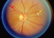
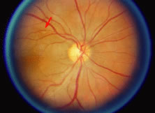
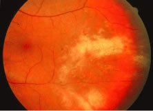
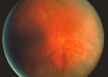
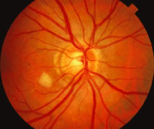
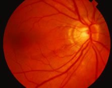
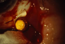
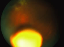
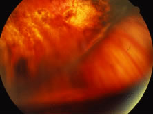
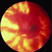
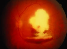
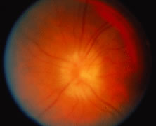
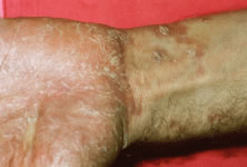
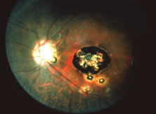
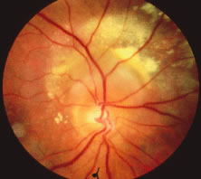
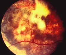
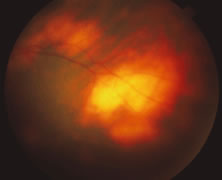
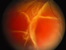
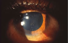
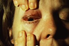
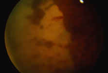
The ARN patient typically presents with progressive visual blurring in one or both eyes occurring over several weeks. These patients often are initially treated with corticosteroids, antitoxoplasmosis drugs, and other medications before arriving at the correct diagnosis. Examination reveals a prominent anterior uveitis that may be granulomatous or nongranulomatous (Fig. 1). Inflammatory signs may be prominent and cause severe pain (Fig. 2). The uveitis may be diffuse and so severe that it causes proptosis. These signs, and the diffuse vitreitis that makes the view of the retina difficult, may contribute to the high degree of delayed and/or misdiagnosis that occurs in the early stages of the disease. Significant vitreous cellular infiltration is seen in the presence of retinitis that is manifest by opacification of the retina, often most prominently in the periphery. Posterior pole involvement may include retinitis, as well as inflammation of the optic nerve head. Optic neuropathy might be the first sign of ARN with subsequent development of other retinal manifestations.8 Ultrasonography and computed tomography (CT) might be helpful in cases of ARN associated with optic nerve edema revealing enlargement of the optic nerve sheath.9 Even in ARN patients who are not immunocompromised and who have no clinical evidence of encephalitis, magnetic resonance imaging of selected cases has shown lesions of the lateral geniculate, optic tracts, and chiasma, which suggests that the virus spreads through the central nervous system (CNS) by axoplasmic transport from the retinal ganglion cells.10 A secondary retinal vasculitis is common, often accompanied by a mild number of retinal hemorrhages. Days to weeks after onset of the infection, the discrete peripheral lesions typically coalesce into a white or yellow ring of infected retina, and the associated vasculature is obliterated (Fig. 3). Necrotic retina desquamates into the vitreous resulting in vitreous sheets.3,6 Eventually, most untreated eyes can be expected to develop retinal detachment resulting from development of multiple full-thickness retinal breaks accompanied by traction or exudation.11 Giant retinal pigment epithelial tears have also been reported.12

Fig. 1. Granulomatous keratic precipitates seen in a patient with acute retinal necrosis syndrome.

Fig. 2. Severe inflammation in a patient with acute retinal necrosis causing chemosis of the conjunctiva and lid edema.

Fig. 3. Confluent peripheral retinitis seen in acute retinal necrosis. The perivascular lucent areas with some evidence of hemorrhage are typical of this disease.

In 1994, the American Uveitis Society published its criteria for the diagnosis of ARN. These criteria (classified into mandatory and supporting categories) were dependent on the clinical signs of the disease, and no laboratory or investigative results were included in these criteria.

The required clinical criteria include the following:

  1. One or more foci of retinal necrosis with discrete borders, located in the peripheral retina.
  2. Rapid progression of the disease in the absence of therapy.
  3. Circumferential spread of the disease.
  4. Evidence of occlusive vasculopathy and arteriolar involvement.
  5. A prominent inflammatory reaction in the vitreous and anterior chamber.

The supporting clinical criteria include the following:

  1. Optic neuropathy or atrophy.
  2. Scleritis.
  3. Pain.13

A human leukocyte antigen (HLA) association between ARN and HLA-DQw7, and HLA-Bw62 and DR4 has recently been demonstrated. An immunogenetic predisposition may explain why only some individuals who have been exposed to these ubiquitous herpesviruses develop the disease.14 In Japanese patients, the risk of ARN infection increases with HLA-Aw33, B44, and DRw6.15 No predilection for sex, race, or age has been identified. ARN has been reported in patients ranging from 9 to 89 years old.16 Before knowledge of the vital etiology, there was documentation of a variety of antecedent systemic vital or bacterial infections that were suspected to have a causal role.7,17,18 A history of prior exposure to herpesviruses, particularly VSV, may be elicited in some but not all cases. In some cases, unilateral ARN may be followed by involvement of the contralateral eye up to 10 years after the first episode. Recurrences in the initially involved eye may occur but they are rare.16,19–21

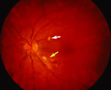
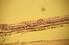
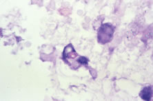
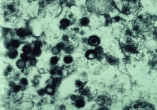
Both herpes simplex virus type 1 and herpes simplex virus type 2 (HSV-1, HSV-2) may cause ARN.20–22 In a single report, CMV particles were identified in and cultured from the retina of an enucleated eye of a nonimmunosuppressed patient suffering from bilateral ARN.23 VZV has been reported most frequently as the viral etiologic agent of ARN.2,3,24–27 We have demonstrated herpesvirus family viral particles in endoretinal biopsy specimens taken from patients in the active stage of the disease who showed an enormous viral load (Fig. 4). These studies, combined with the failure of many enucleated eyes with ARN to demonstrate evidence of viral particles, indicate that the virus is present only in the active stages of the disease and that a gliotic retina will not demonstrate the etiologic agent.28

Fig. 4. An electron micrograph showing abundant herpesvirus particles from a retinal biopsy taken at the active stage of acute retinal necrosis.

Some cases of ARN follow or precede zoster dermatitis,2,29,30 leading some to conclude that the retina is infected secondary to neural spread from sensory ganglia in the CNS. However, most patients with herpes zoster ophthalmicus do not develop ARN,3 suggesting that transmission of virus to the retina associated with trigeminal nerve reactivation is uncommon. The site of latency and mechanism of viral infection remain unknown.

The polymerase chain reaction (PCR) is a highly sensitive, specific, and rapid means of detecting small amounts of viral deoxyribonucleic acid (DNA) in intraocular fluid samples. Recently, PCR-based assays have been used as aids for the diagnosis of ARN and the determination of the specific virus causing the syndrome.31–33 Ganatra and associates conducted an interesting study about the viral causes of ARN using PCR-based assay of various viruses DNA in aqueous and vitreous. They found that VZV or HSV-1 cause ARN in patients older than 25 years, whereas HSV-2 causes ARN in patients younger than 25 years. In this study, VZV DNA was recovered in 15 out of 30 eyes. HSV-2 DNA was detected in six eyes, and HSV-1 was detected in seven eyes. CMV DNA was detected in only one eye, and no virus DNA was recovered in one eye in which the sample was taken after 6 weeks of acyclovir therapy. Also, no sample was positive for DNA from more than one virus.34

Light microscopic examination of these retinas reveals full-thickness necrosis and loss of the retinal architecture.25,30,35 Within the necrotic retina, macrophages, plasma cells, and other inflammatory cells are found in addition to cells containing eosinophilic inclusions. There is a sharp demarcation between affected and nonaffected retina, suggesting cell-to-cell transmission of the virus. Arteritis manifests as endothelial cell swelling with occlusion of the vessel lumen, infiltration of the subendothelium with plasma cells, and inflammatory cell thrombi. When healing occurs, a thin glial scar replaces the necrotic neural elements. Herpesvirus capsids may be observed in electron microscopic specimens of actively infected retinal tissue but are not seen once healing has occurred and glial scar tissue replaces the necrotic retina.28,30

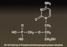
Given the evidence that most cases of ARN are due to VZV or HSV, acyclovir therapy is recommended.35–38 Acyclovir is taken up by infected cells and phosphorylated into a proactive form by virally encoded thymidine kinase. Subsequent phosphorylation by cell coded kinases results in formation of the active triphosphate form of acyclovir that inhibits viral replication by blocking DNA polymerase activity. Hung and coworkers39 demonstrated that oral administration of acyclovir (400 mg five times per day) to normal volunteers can produce mean plasma levels of 8.7 μm and mean aqueous levels of 3.3 μm.

Some strains of herpesviruses known to have caused ARN are inhibited only by acyclovir concentrations greater than the achievable aqueous concentrations after oral administration (400 mg five times per day) of acyclovir. Intravenous therapy with 1500 mg/m2/day in three divided doses achieves peak plasma levels of 30 to 40 μm without significant toxicity to the patient and is, therefore, the recommended regimen. Aqueous acyclovir levels of 8.7 to 11.2 μm are achievable using intravenous acyclovir (1500 mg/m2/day), and these levels are sufficient to treat most cases of ARN. Prodrugs of acyclovir are being developed that yield serum levels after oral administration that are as high as serum levels after intravenous infusions.

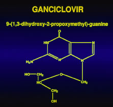
The duration of therapy is generally dependent on the response of the infection. Ten days is probably sufficient because progression of retinal lesions usually stops within several days of initiation of intravenous therapy.39 In cases that are deemed unresponsive to acyclovir, cultures of a retinal biopsy specimen may provide evidence of the presence of an acyclovir-resistant strain of VZV, HSV, or CMV, which in resistant to acyclovir but sensitive to ganciclovir.23,40

The use of steroid and/or anticoagulant therapy remains controversial. Optic nerve infiltration with inflammatory cells and occlusions of arteriolar vessels may result in optic nerve ischemia, which leads some to treat patients with oral prednisone at doses up to 120 mg daily.35,40 Some have suggested that sudden visual loss out of proportion to retinal findings is suggestive of optic nerve ischemia caused by mechanical compression and/or edema and have advocated optic nerve sheath decompression in selected cases.41 To avoid vaso-occlusion and because of a report of altered platelet aggregation in this disease,42 some investigators have advocated the use of aspirin (500 mg/day), heparin, sodium warfarin (Coumadin), or other antiplatelet agents as adjunct therapy to hopefully diminish the vascular complications of ARN.35 Finally, high-dose steroid administration, for example, methylprednisolone 250 mg intravenously every 6 hours, may diminish the generalized ocular inflammatory response resulting in less serum exudation and hence a decrease in the concentration of chemotactic agents in the vitreous that are responsible for causing migration of pigment epithelium cells. This would have the theoretic advantage of decreasing the chances for developing proliferative vitreoretinopathy. Controlled studies evaluating the efficacy of steroids in preventing proliferative vitreoretinopathy or comparing acyclovir versus acyclovir with antiplatelet therapy in preventing ARN-related optic neuropathy are lacking, and the evidence that corticosteroid use worsens the course of active herpetic corneal disease must be remembered.

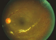
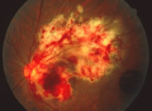
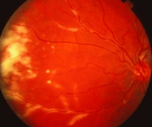
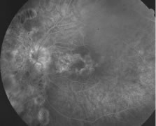
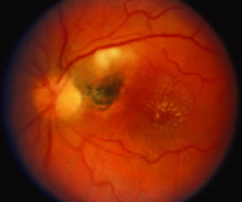
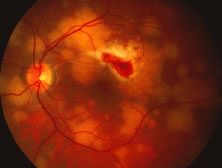
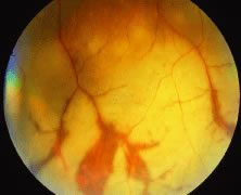
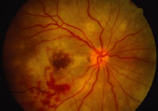
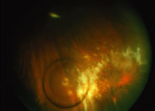
A variable number of eyes, up to 75%, develop retinal detachments (Fig. 5). Prophylactic laser for demarcating the areas of active retinitis from normal retina has been advocated to create chorioretinal adhesions that prevent retinal detachments around sites of retinal break formation (which usually occur at the zone between affected and healthy retina). Han and associates43 reported five cases treated with prophylactic laserpexy in addition to antiviral, steroid, and antiplatelet therapy. After 15 months of follow-up, no retinal detachments were noted. Sternberg and coworkers44 described a 75% decrease in the rate of retinal detachment using prophylactic photocoagulation. Some have advocated the creation of a “new ora serrata” by applying confluent rows of laser burns posterior to the areas of retinitis. However, if traction forces from vitreous organization, epiretinal membrane (ERM) formation, or proliferative vitreoretinopathy develop as they commonly do, the contractile forces will be able to overcome any increased chorioretinal adhesion created by the laserpexy. However, McDonald and associates45 reported failure of prophylactic peripheral laserpexy to prevent retinal detachment in ARN patients. In addition, many cases of ARN result in a severe vitritis, limiting the view for a planned laserpexy (Fig. 6). Therefore, vitrectomy with endolaser and concomitant encircling of the eye with a scleral buckle to reduce traction may be required in some patients.46,47 Decisions relating to the need for scleral buckling to support retinal breaks and the use of silicone oil or long-acting gases to repair retinal detachments should be made by an experienced vitreoretinal surgeon.40,48 Selection of cases to undergo operation should be made with consideration given to optic nerve function, visual potential, and medical control of retinitis.

Fig. 5. A patient with funnel-shaped retinal detachment and proliferative vitreoretinopathy complicating acute retinal necrosis syndrome.

Fig. 6. Failure of prophylactic laser treatment to stop the progression of a retinal detachment associated with retinal necrosis resulting from viral retinitis with progression of the detachment beyond the line of laser demarcation.

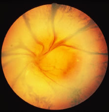
The ocular complications of ARN are myriad. During the active infection, uveitis is a prominent feature. Secondary glaucoma may develop because of inflammatory debris in the trabecular meshwork. Posterior synechiae may form with resulting pupillary block and occlusion of the angle by forward movement of the peripheral iris. Pupillary membranes and cataract development have been described. Optic nerve atrophy has been noted and may be due to loss of ganglion cell bodies in the retina or direct invasion of the nerve head by the virus with subsequent inflammation and swelling. Iris neovascularization and neovascular glaucoma may occur. Disc and retinal neovascularization with subsequent vitreous hemorrhage have also been noted.16,49 Finally, a blind painful eye may develop, necessitating retrobulbar alcohol injection or enucleation.

Recently, a mild variant of ARN was described in Japan that may represent a self-limited form of the disease.1 We recently reported a series of cases in which the retinitis was limited and in which the visual prognosis was significantly better than is commonly reported.50 These reports would seem to indicate that ARN manifested by limited peripheral involvement may portend a better prognosis, particularly when treated early with appropriate antiviral therapy.

Perhaps the most significant complication of a unilateral case of ARN is subsequent involvement of the fellow eye. Sternberg and associates16 reported the cases of six patients, four of whom went on to bilateral involvement anywhere from 3 weeks to 22 months following diagnosis of ARN in the first eye. Matsuo and associates1 described the onset of ARN in the fellow eye after a 3-year interval. In one case, a 10-year quiescent interval was reported between the onset of ARN in the first eye and involvement of the fellow eye. Culbertson and coworkers25 reported that 36% of their patients eventually had bilateral ARN syndrome. Interestingly, two independent laboratories studying the electrophysiologic aspects of ARN have found that the electroretinograms of unaffected eyes may be abnormal.51 The prognostic significance for eventual contralateral involvement has not been investigated. Prolonged oral acyclovir or valacyclovir therapy probably reduces the risk of fellow eye involvement.

In summary, ARN is primarily seen in otherwise healthy persons but has been reported in immunocompromised hosts as well; in HIV patients it is termed progressive outer retinal necrosis. Patients typically present with progressive visual blurring and may carry numerous prior misdiagnoses. The infection causes a severe retinitis, vitreitis, and uveitis that may result in numerous secondary ocular complications. Although the viral infection is treatable, preferably with intravenously administered acyclovir, eventual retinal detachment occurs in most cases. Hence, surgical interventions to preserve vision are usually warranted. Prophylactic laserpexy to wall off the area of infection and prevent total retinal detachment may be helpful if adequate visualization of the area to be treated is possible. However, for patients in whom adequate visualization of the retina is not possible or if retinal detachment is identified, pars plana vitrectomy with endolaser and possible scleral buckling should be considered. Most importantly, frequent dilated retinal examination of both eyes must be performed during the convalescent period and for several years thereafter.

Back to Top
CYTOMEGALOVIRUS RETINITIS
CMV is a ubiquitous, species-specific DNA-containing herpesvirus. Epidemiologic studies examining the temporal development of CMV antibody seropositivity in the general population reveal that approximately 1% of newborns contract CMV while in utero, and a larger group acquires the virus at birth through an infected birth canal or in early infancy. Thereafter a steady increase in the prevalence of antibody-positive individuals occurs from adolescence into adulthood. It is postulated that contact with CMV-contaminated urine, saliva, and semen is the major mode of transmission, because it has been demonstrated that crowding, sexual contact, and exposure of children to day care centers can result in conversion to antibody positivity.52 Eventually up to 90% of the general population develops antibody to CMV, suggesting widespread exposure to this agent.53

Despite a high prevalence of antibody positivity in the population, clinical ocular disease secondary to CMV infection in adults is rare or nonexistent in the absence of immunosuppression resulting from malignancies, autoimmune disease, acquired immunodeficiency syndrome (AIDS), or therapeutic intervention. Before 1970, only a small number of CMV retinitis cases had been described. In 1971, deVenecia54 elegantly documented the first histologically proved case of CMV retinitis. Since then, the increase in chemotherapeutic abilities and the emergence of AIDS as a leading cause of morbidity and mortality has caused the number of cases of CMV-related ocular disease to increase drastically.

Groups of patients at higher risk for contracting CMV-related disease include neonates born to CMV-infected mothers and immunosuppressed adults. In neonates, de novo infection with CMV during the last two trimesters leads to cytomegalic inclusion disease. Cytomegalic inclusion disease manifests as microcephaly, psychomotor retardation, hepatosplenomegaly, thrombocytopenia, and other systemic abnormalities in a small-for-gestational-age infant. In adults, the etiology and spectrum of CMV-related disease are less well defined. Secondary reactivation of primary latent infection has been documented and postulated to be a major source of CMV-related disease. The site of latency, however, has yet to be identified. De novo infection with a second strain of CMV has also been documented.55 Heterophil-negative mononucleosis manifest as lymphadenopathy, malaise, and atypical lymphocytosis is the most well-described CMV-related disease in otherwise healthy adolescents and is a relatively benign illness without retinal involvement. In contrast, in immunosuppressed persons and neonates, CMV-associated disease causes significant morbidity and mortality. For the ophthalmologist, CMV retinitis presents a formidable albeit treatable cause of blindness.

CMV retinitis was first described in 1959 by Foerster.56 Since then it has become obvious that a characteristic retinitis occurs in neonates with cytomegalic inclusion disease and in CMV-infected immunocompromised persons. In transplant patients, CMV retinitis may be the initial clinical manifestation of life-threatening viremia and indicates the need for immediate aggressive lifesaving intervention. For AIDS patients, CMV retinitis often is documented after other opportunistic infections, typically when the CD4 lymphocyte count falls below 100; however, CMV retinitis as the initial manifestation of AIDS has been well described.57 Early retinitis may present with small white lesions that may be confused with cotton-wool spots. However, unlike cotton-wool spots, which are seen in the nerve fiber layer, CMV retinitis is seen as granular whitening located deeper in the retina (Figs. 7, 8, and 9). These same areas of retinitis are often associated with intraretinal hemorrhage and vasculitis of nearby retinal vessels. Although the lesions of CMV retinitis are located in the posterior pole more often than in ARN, peripheral retinitis is not uncommon.58 The lesions can be multifocal as well, pointing out the need for thorough examination of the entire peripheral retina. The vitreous may be relatively quiet allowing excellent visualization of the underlying retina; however, significant vitreitis may occur. The pars plana is relatively spared,54,59 although it is unclear why this should be so, because the pars plana epithelium and the retina share a common neuroectodermal origin. On occasion, stellate keratic precipitates with cell and flare can be observed on the cornea and in the anterior chamber. In some cases a prominent retinal vasculitis may be seen posterior to the area of retinitis (Fig. 10).

Fig. 7. A patient with cotton-wool spot (white arrow) and early cytomegalovirus (CMV) retinitis (yellow arrow). Note the granular pattern of the CMV lesion.

Fig. 8. A patient with cytomegalovirus (CMV) lesion superotemporal to the optic nerve (red arrow) and cotton-wool lesion (yellow arrow) superonasal to the optic nerve. The CMV lesion is granular and deep.

Fig. 9. Same patient as in Figure 8 seen 1 month later shows resolution of cotton-wool patch and some enlargement of the area of infectious cytomegalovirus retinitis (red arrow).

Fig. 10. A patient with vasculitis secondary to cytomegalovirus (CMV) retinitis. The vasculitis is typically along the veins draining the lesion.

CMV retinitis occurs in 15% to 40% of AIDS patients58,60–62 and, in contrast to the noninfectious lesions of AIDS, demands aggressive treatment to prevent severe visual loss.60,63,64 Patients with active CMV disease may present with systemic symptoms of fever, arthralgia, leukopenia, pneumonitis, retinitis, or hepatitis. Blood cultures and urine specimens are usually positive for CMV. CMV infects the retina, as well as the CNS, the reticuloendothelial system, kidneys, adrenals, lungs, and gastrointestinal system.63,65 CMV retinitis is often the presenting sign of systemic CMV infection, and all patients should have a thorough systemic evaluation performed, including “buffy coat” culture of the blood and possible evaluation of bone marrow aspirates and cerebrospinal fluid. Thus, screening for CMV retinitis is very important in AIDS patients. Urine is culture positive for CMV in 50% of homosexual men and in most AIDS patients, and, thus, urine culture may not be of diagnostic value. Serology in AIDS patients is notoriously unreliable, and it is unusual to document rising CMV titers.60,63,65 CD4 cell count might be used as a practical test for screening because the risk of CMV retinitis increases at CD4 cell count below 50/mm3.66 Techniques for detection of CMV DNA based on PCR in ocular fluids have become more popular. One PCR-based study for the detection of CMV DNA in vitreous samples had an estimated sensitivity of 95% in detecting untreated CMV retinitis and a sensitivity of 48% in detecting CMV retinitis that had been treated with systemic ganciclovir or foscarnet or both.67 A variant of the Amsler grid has been used as a screen for central CMV retinitis within a 22-degree radius (3 disc diameters) of the fovea. This technique may detect up to two thirds of the cases of CMV.68 Recently, we reported the use of a new technique, entoptic perimetry, as a screening test in patients with CMV retinitis. This technique employs patient visualization of moving particles on a computer monitor. This test has a very high sensitivity and specificity (more than 90%) in detecting CMV retinitis within the central 30-degrees radius of fixation.69

The clinical presentation of CMV retinitis in AIDS is similar in many respects to CMV retinitis found in iatrogenically immunosuppressed patients and in infants with cytomegalic inclusion disease.57,70,71 Correlation of the clinical and typical pathologic findings at autopsy has been demonstrated.72 Specifically, it is known that CMV is a neurotropic virus with a tendency to infect neural tissues and the retina. Necrosis of the retina in AIDS-associated CMV retinitis is typical, with pathognomonic cytomegaly and only a few inflammatory cells in the lesions. Choroidal involvement is rare; however, retinal vascular endothelium may be infected. These lesions may also appear as noncontiguous patches rather than the contiguous, spreading lesion more commonly seen. Antigens to CMV have been found by immunofluorescence, immunoperoxidase staining, and DNA hybridization techniques (Fig. 11).65,73

Fig. 11. Histopathologic specimen showing immunohistochemical staining (red areas) in retinal cells infected with cytomegalovirus (CMV). This is in an area approximately 1 mm posterior to an area of confluent retinal destruction seen at the left of the figure. The small lesions probably correspond to the white dots that are seen just beyond the area of confluent CMV retinitis clinically. These areas of cytomegalic changes are associated with retinal edema and opacification. They are approximately 200 to 300 μm and would be visible as small white areas of opacification.

Progression of the retinitis generally occurs with a leading edge of active retinitis and a trailing region of thin gliotic retina. This pattern indicates cell to cell transmission of the virus. CMV is a slowly progressive, necrotizing retinitis that may affect the posterior pole, the peripheral retina, or both and may be unilateral or bilateral. Involved areas appear as white intraretinal lesions; areas of infiltrate; and often necrosis, along the vascular arcades in the posterior pole. In addition, prominent retinal hemorrhages are often seen within the necrotic area or along its leading edge (Figs. 12 and 13). Peripherally, CMV retinitis tends to have a less intense white appearance, with areas of granular, white retinitis that may or may not demonstrate associated retinal hemorrhage (Fig. 14). As the retinitis progresses, an area of atrophic, avascular retina may remain with underlying retinal pigment epithelial atrophy and/or hyperplasia (Fig. 15).57,58,60,62 Peripheral CMV retinitis may be the most common form of CMV retinitis seen. Patients may initially complain only of floaters with or without a visual field deficit.57 Wide-angle fundus photography and fluorescein angiography may be of benefit when the diagnosis is uncertain. These techniques may be used to document progression of retinitis, and fluorescein leakage in areas of retinitis may be helpful in confirming the diagnosis. Although peripheral CMV retinitis may not pose an immediate threat to vision, initiation of treatment should be seriously considered because several investigators have shown that untreated CMV retinitis is inexorably progressive and becomes bilateral in virtually all untreated patients.59,60,65,74–76 Occasionally, noncontiguous areas of new retinitis may be seen. As healing takes place, a thin glial scar replaces the necrotic retinal tissue. The transition from infected to noninfected retina is usually sharply demarcated and well seen during the active phase of the disease. When healing takes place, the transition zone is between a glial scar and normal retina and may be difficult to visualize. Clinically, the pigment epithelium tends to develop a characteristic spiculated granular appearance directly under the residual glial scar. The retina in the immediately adjacent uninvolved areas is thick in comparison to the glial scar, thus, differing light reflexes occur with indirect ophthalmoscopy. Using the pigment epithelium pigment changes and light reflexes, one can define the extent of prior retinal involvement. More importantly, defining the edge of prior involvement allows one to predict with great accuracy the sites of new involvement, allowing for more effective monitoring of therapeutic efforts to control the infection. Other manifestations of CMV retinitis include retinal edema, attenuated vessels, perivascular sheathing, and exudative retinal detachment.77 In addition, vitreitis and anterior uveitis are often seen,70 and optic atrophy may occur as a late manifestation secondary to widespread retinal destruction. CMV may be demonstrated in vitreous biopsy specimens in these patients.65 The yield may be higher in the presence of marked vitreitis because CMV is a cell-associated virus. Other causes of retinitis, including herpes simplex retinitis,78,79 toxoplasmosis,80 Candida, Behçet's disease, syphilis, ARN,7,28,81 and SSPE82 can usually be distinguished from CMV on clinical grounds. Cotton-wool spots are nonspecific and may be seen in diabetes mellitus, hypertension, severe anemia, systemic lupus erythematosus, dermatomyositis, and leukemia,83,84 as well as AIDS. It may be difficult to distinguish a small focus of nonhemorrhagic CMV retinitis from a cotton-wool spot corresponding to the area of peripapillary optic nerve fiber layer that was destroyed (Figs. 16 and 17). In eyes in which CMV infection initially involves the optic nerve head, the visual acuity may be normal early; however, the visual prognosis may be poor because these eyes often go on to loss of light perception (see Fig. 13). When retinitis is in close proximity to the optic nerve head or macula, a serous retinal detachment may be seen that will resolve with therapy.

Fig. 12. A patient with a relatively nonhemorrhagic variant of cytomegalovirus retinitis. Note at the center of the lesion (oldest area of infection) the retina is atrophic and pigmentary changes can be seen. Peripheral to it the retina is edematous, and this is the area of active infection.

Fig. 13. A patient with hemorrhagic variant of cytomegalovirus (CMV) retinitis that affected the posterior pole and optic nerve. Hemorrhages are most often intraretinal, and white areas correspond histologically to intracellular and extracellular edema and necrosis of the neurosensory retina. Eyes with extensive optic nerve involvement secondary to retinal involvement may still retain good central vision unless the papillomacular bundle is involved. When retinitis starts in the optic nerve head, the prognosis for vision is poor.

Fig. 14. Cytomegalovirus retinitis in the periphery may have minimal hemorrhagic components.

Fig. 15. Area of healed cytomegalovirus retinitis showing characteristic pigmentary changes. In the area of retinal necrosis, the retinal pigment epithelium (RPE) is also thinned and bare choroidal vessels are visible.

Fig. 16. A patient with cotton-wool spots near the optic nerve. The nerve fiber layer swelling obscures the details of retinal vessels.

Fig. 17. Same patient (see Fig. 16) 6 weeks later showing disappearance of the cotton-wool spot. A residual nerve fiber layer defect is seen as are some small areas of residual vascular changes and hemorrhage.

Histologic findings in CMV retinitis include the presence of typical cytomegalic cells with prominent intranuclear inclusions resulting in a pathognomonic “owl's eye” appearance.57 Closer examination of the owl's eye of infected cells reveals a dense central nuclear inclusion body with clearing of nucleoplasm immediately around the inclusion (Fig. 18). Inflammation, manifesting as mononuclear cell infiltration, is usually scant, and the choroid is spared. Endothelial cells of retinal vessels may be infected with CMV.54,59

Fig. 18. Typical cytomegalovirus (CMV) cells with prominent intranuclear inclusions. This retina was double immunostained with an anti-CMV antibody (blue) and an anti-endothelial cell antibody (red factor-VIII associated antigen). Thus, the figure illustrates that CMV can infect vascular endothelial cells. There is abundant retinal necrosis also present.

Left untreated, CMV retinitis may progress to loss of light perception. Reinfection of previously infected areas does not occur, apparently because the virus does not infect the tissue forming the glial scar. Areas of old retinitis remain quiet, with activity only at the junction of normal retina. Retinal photocoagulation to uninvolved retina immediately in front of the advancing edge to prevent the virus from proceeding is not efficacious.85 The mainstay of treatment for exogenously immunosuppressed persons remains amelioration of their immunosuppression in conjunction with the internist or multidisciplinary team. In most instances, reduction of the immunosuppression results in healing of the retinitis.

Immunoaugmentation, even in AIDS patients, may result in healing of CMV retinitis. Guyer and associates86 documented regression of active CMV after prolonged administration of azidothymidine in one patient. Because azidothymidine has no activity against CMV, this group postulated that azidothymidine induced reconstitution of the patient's immune system, resulting in healing of the retinitis. We have reported three similar cases87; most AIDS patients, however, develop CMV retinitis while being treated with azidothymidine.75 We previously reported that human immunodeficiency virus (HIV)-positive patients who receive highly active antiretroviral therapy (HAART) and who have CMV retinitis healed by anti-CMV therapy are likely to remain healed if the anti-CMV therapy is withdrawn. However, continuous monitoring of these patients with indirect ophthalmoscopy is mandatory because HAART failure may occur allowing CMV retinitis reactivation. If a patient with CMV retinitis undergoes immune reconstitution with HAART therapy, it typically takes 3 months of a sustained elevation of CD4 count above 100 to cause healing of the retinitis.88

Neonates with cytomegalic inclusion disease and immunosuppressed persons in whom treatment to ameliorate the immunosuppression is not possible require the use of antiviral agents. In 1980, Pollard and coworkers89 described the use of adenine arabinoside to treat seven transplant patients with progressive CMV retinopathy despite minimal immunosuppression. The results using this drug were suboptimal; significant gastrointestinal, hematologic, and neurologic side effects were observed.

Ganciclovir, also known as dihydroxy propoxymethyl guanine (DHPG) and BW759U (Cytovene), is a hydroxylated derivative of acyclovir; it was licensed for use in the United States in 1989 (Fig. 19). Treatment with ganciclovir is usually successful at controlling CMV infection but is difficult and requires a multidisciplinary team approach. Intravenous ganciclovir has a short half-life (3.6 hours) and must be given on a once- or twice-daily basis intravenously.90 Side effects are numerous; myelosuppression and life-threatening neutropenia are the most difficult to manage. In many patients, concurrent treatment with azidothymidine, which has been shown to prolong life in HIV-infected individuals, is often not possible.

Fig. 19. The antiviral drug ganciclovir is a hydroxylated derivative of acyclovir that has greatly enhanced activity against cytomegalovirus. Oral ganciclovir has poor bioavailability, but the introduction of a prodrug of ganciclovir, valganciclovir, has allowed the achievement of high blood levels of ganciclovir with oral therapy.

Photographs to document regression or progression of lesions are most helpful. Close collaboration with the internist or infectious disease specialist is important and should include monitoring of the peripheral blood throughout the course of the treatment. If the drug must be stopped secondary to granulocytopenia, relapse of the retinitis can be expected to occur within weeks.58 If induction and maintenance are initially successful, reinduction can be attempted for retinitis no longer controlled by daily therapy. However, resistance to ganciclovir has been documented, and this should be kept in mind during reinduction. Should resistance be suspected, alternative salvage therapies may be considered such as intravitreal therapy91 or alternative agents such as foscarnet.

Ganciclovir-based induction therapy begins with twice-daily dosing with 5 to 6 mg/kg per dose for a 2-week interval. Baseline wide-angle fundus photographs with overlapping fields should be obtained before induction for documentation of future progression or regression of the retinitis. After 2 weeks of induction, a repeat ophthalmoscopy should be performed that should show clear evidence of healing with decreasing opacity of the lesions along with less retinal edema. Thereafter, maintenance therapy with 5 mg/ kg/day prevents reactivation in most patients. However, reactivation rates of 27% while using 5 days per week maintenance therapy are common; in addition, the drug is virustatic58,75 rather than virucidal. Thus, 7 day per week therapy seems more rational and may be more efficacious for suppressing the retinitis. Clinical reevaluation of patients should take place every 2 weeks, to assess response to therapy. Early signs of regression of retinitis include nonadvancement of edges, decreased satellite lesions or confluence of retinitis to the initial satellite lesions, thinning, increased granularity and yellowing or graying of the white fluffy exudative areas, improved demarcation of the lesions, and decreased hemorrhage and vessel wall sclerosis. Late signs of healing include increased pigmentation and thinning of the atrophic retina, gliosis, and increased refractile deposits. Ghost vessels with sclerotic-appearing walls are seen late. Although oral and intravenous ganciclovir have been reported to have a comparable efficacy as a maintenance therapy for CMV retinitis, patients taking oral ganciclovir have the tendency to develop earlier reactivation. This could be attributed to the relatively poor and variable gastrointestinal absorption of the oral preparation.92–94 The main advantage of oral over intravenous ganciclovir is that an indwelling catheter is unnecessary.95 However, for oral ganciclovir to be effective the patient must take 12 capsules daily, which might be a problem for many patients. Thus, most clinicians reserve oral ganciclovir for maintenance therapy for patients with peripheral lesions or as a prophylaxis to protect nonocular tissue and the uninvolved eye in patients receiving local anti-CMV therapy. Recently, a prodrug of ganciclovir, valganciclovir, has become available. Clinical trials have shown blood levels of ganciclovir after oral administration of valganciclovir are comparable to that of intravenous ganciclovir. In our hands, valganciclovir given orally has replaced the use of intravenous ganciclovir unless a patient has a severe malabsorption syndrome.

Granulocyte-monocyte colony stimulating factor (GMCSF) is a cytokine that increases the peripheral blood granulocyte count. Recent trials have suggested that it may allow ganciclovir therapy even in neutropenic patients by increasing the absolute neutrophil count to safe levels. The drug is given subcutaneously and if these preliminary observations are confirmed, GMCSF will be an important addition to the anti-CMV armamentarium. rG-CSF (recombinant granulocyte colony-stimulating factor) has largely replaced rGM-CSF (recombinant granulocyte macrophage colony-stimulating factor) to improve the white blood count in patients who developed neutropenia resulting from the marrow toxicity of ganciclovir or its prodrug. rG-CSF is administered subcutaneously and is extremely helpful in treating neutropenia.

Because of the difficulties associated with systemic ganciclovir, there is much interest in its local injection into the vitreous cavity. In rabbits, intravitreal ganciclovir injections of up to 400 mg produced no ophthalmoscopic, histologic, or electroretinographic changes. Multiple intraocular injections of ganciclovir have been successful in controlling CMV retinitis in at least one patient who was treated for 3 months with a total of 28 intravitreal injections of 200 mg each.96 No evidence of retinal toxicity was noted, and the estimated half-life of the drug in the human vitreous was 13 hours. Several investigators have found intravitreal injection to be efficacious in controlling CMV retinitis.97–99 The usual dose is 200 mg in 0.1 ml intravitreally. Injections have been performed in the outpatient setting under local anesthesia, in the clinic and under sterile conditions in the operating room. Those injections performed in the clinic have used topical gentamicin drops the night before the injection. However, sterilization of the conjunctiva with povidone-iodine is preferable. The complications of intraocular injections are well known and must be considered carefully.100 Review of the literature shows that intravitreal injection of ganciclovir is controversial. Between 1 and 58 injections per patient have been administered. Of 463 intravitreal injections in 30 patients, two cases of endophthalmitis (Staphylococcus aureus and Staphylococcus epidermidis) and one retinal detachment have been described.98 Laboratory studies of multiple intraocular ganciclovir injections of 200 mg have suggested toxicity.

An intraocular sustained-release ganciclovir delivery implant that releases drug into the vitreous is commercially available. The rate of drug release is set at the drug diffusion port to approximately 1μg/hr. The implant is inserted through a sclerotomy at the pars plana and then secured with scleral sutures (Figs. 20 and 21). Studies have shown the median time to progression of CMV retinitis to be about three times longer than with intravenous ganciclovir, or approximately 7 to 8 months.101,102 Thus, replacement of the implant at 7 to 8 months intervals is necessary.103 Serious side effects of implant insertion or exchange are infrequent but include endophthalmitis, retinal detachment, vitreous hemorrhage, and cataract formation.104 Recent reports suggest that there is an increased risk of endophthalmitis in these patients and that such patients may have a poor prognosis for vision. The incidence of endophthalmitis may be as high as 1% to 2%.

Fig. 20. Ganciclovir implant before insertion through the sclerotomy site. Site preparation should include good hemostasis. Care must be taken to insert the implant into the vitreous and not into the subretinal space.

Fig. 21. Colored fundus photo showing the intravitreal ganciclovir implant after insertion. An area of active cytomegalovirus (CMV) retinitis is seen in the upper portion of the illustration.

FOSCARNET THERAPY FOR CYTOMEGALOVIRUS RETINITIS

Foscarnet (trisodium phosphonoformate hexahydrate) is a potent virustatic agent with in vitro antiviral activity against HIV and herpesviruses (Fig. 22). It inhibits ribonucleic acid (RNA) and DNA virus polymerases and has been found to be efficacious in CMV retinitis patients. Tremor, nausea, serum calcium and/or phosphorus changes, and anemia have been noted in association with foscarnet treatment. Foscarnet does not cause myelosuppression and is, therefore, compatible with concurrent use of azidothymidine.

Fig. 22. Structure of foscarnet, an antiviral drug with activity against cytomegalovirus, herpes simplex virus, varicella zoster virus, and human immunodeficiency virus.

Virtually all patients placed on foscarnet have shown improvement of CMV retinitis with partial or complete resolution of retinitis. Response to foscarnet has been described in patients who have retinitis unresponsive to ganciclovir.105,106 Doses found to be efficacious were a bolus of 20 mg/kg intravenously over 30 minutes followed by maintenance with constant infusion at a rate of 0.16 mg/ kg/minute. Infusion was decreased by 0.02 mg/kg/ minute for each 20-mM increase in serum creatinine and discontinued if the creatinine was more than 250 mM. Unfortunately, intravenous saline infusions are required to prevent nephrotoxicity and can cause edema and cardiac complications. The currently recommended dose is a 180 mg/kg/day 2-week induction course followed by maintenance therapy of 90 to 120 mg/kg/day. In 1992, the Study of the Ocular Complication of AIDS (SOCA) Research Group compared the efficacy and toxicity of ganciclovir and foscarnet. They found no apparent difference in the efficacy of the two drugs. However, a statistically significant difference was demonstrated between treatment groups with regard to patient survival. Patients received foscarnet had a median survival of approximately 12.6 months versus 8.5 months for those who received ganciclovir.107 The cause of this difference could be attributed to the antiretroviral replication effect of foscarnet (it has intrinsic antiretroviral activity) and could be because patients taking ganciclovir were less able to tolerate zidovudine.108 A subsequent large prospective study failed to show a difference in mortality between patients treated with ganciclovir versus foscarnet monotherapy.109 The reasons for this difference is not known but might be related to the improved compliance with ganciclovir because of the greater use of leucocyte growth factors. Currently, there is strong evidence that ganciclovir and foscarnet act synergistically against CMV. The Cytomegalovirus Retinitis Retreatment Trial (CRRT) found that the time to subsequent relapse in patients with active CMV retinitis was 4.3 months in patients treated with both ganciclovir and foscarnet, as compared with 2.0 months and 1.3 months, respectively, in patients treated with either ganciclovir or foscarnet alone. Moreover, side effects were similar in the monotherapy and combination therapy treatment groups.109

Cidofovir, (S)-1-[3-hydroxy-2-(phosphonylmethoxy)propyl] cytosine, formerly known as HPMPC (Fig. 23), was the first antiviral nucleotide analogue available for the treatment of CMV retinitis. The treatment of CMV retinitis with intravenous cidofovir was found to be effective in slowing the progression of peripheral CMV retinitis patients with previously untreated CMV retinitis and AIDS. Intravenous cidofovir also has been used for long-term suppression of CMV retinitis. Biweekly therapy (after induction) was reported to have a time to progression of CMV retinitis of 120 days.110 Unfortunately, cidofovir has been found to have limitations, including nephrotoxicity, as well as a very high incidence of iritis (up to 50%) and a risk of profound ocular hypotony with vision loss.111 Oral administration of probenecid before and after the intravenous infusion appears to help ameliorate the nephrotoxicity of the drug, but the ocular side effects of iritis and hypotony occur despite concomitant probenecid administration.

Fig. 23. Structure of Cidofovir. Cidofovir has an extremely long intracellular half-life and can be administered intravenously once every other week. Intravitreal injections of the drug can be given every 6 weeks. Toxicity with this compound includes iritis and hypotony. Hypotony may be more common after intravitreal administration.

It was also shown that injections of 20 μg of intravitreal cidofovir resulted in complete suppression of CMV replication with no advancement of retinitis borders when given every 6 weeks.112,113 However, the therapeutic benefit of intravitreal cidofovir is limited by the development of severe uveitis and hypotony that might occur with doses slightly above those needed to achieve viral suppression.112,114

RHEGMATOGENOUS RETINAL DETACHMENT IN TREATED CYTOMEGALOVIRUS RETINITIS

Others and we have reported a high incidence (up to 22%) of rhegmatogenous retinal detachment in association with ganciclovir treatment for CMV retinitis.115 The presence of chronic inflammation in these eyes may be important in the pathogenesis of proliferative vitreoretinopathy and retinal detachment (Figs. 24, 25, and 26). Inflammatory products and cells may contribute to contraction of the vitreous gel by stimulating migration of contractile cells or by metaplasia of macrophages into contractile elements that contribute to posterior vitreous detachment.105

Fig. 24. Rhegmatogenous retinal detachment associated with cytomegalovirus (CMV) retinitis. The retina is extremely thin and filled with atrophic breaks; these are best seen in the superior aspect of the illustration.

Fig. 25. Proliferative vitreoretinopathy (PVR) manifest by star fold is seen in a patient with rhegmatogenous retinal detachment associated with healed cytomegalovirus retinitis. PVR is very unusual in cytomegalovirus (CMV) retinitis, however, it may occur in associated with immune recovery uveitis.

Fig. 26. A case of rhegmatogenous retinal detachment associated with cytomegalovirus (CMV) treated successfully with peripheral retinal laser, vitrectomy, and buckling. Note that the silicone oil does not provide inferior tamponade. There is a shallow detachment between the posterior edge of the scleral buckle and the area of peripheral scatter laser that has prevented progressive retinal detachment.

Treatment of retinal detachment in these eyes must address the multiple retinal breaks, many of which may be difficult to visualize, as well as the propensity of these eyes to develop proliferative vitreoretinopathy. In addition, these patients suffer from serious concurrent systemic diseases. For these reasons, primary pars plana vitrectomy with internal retinal tamponade is appropriate. Silicone oil offers long-acting tamponade to the extensive areas containing retinal breaks, as well as to areas of potential future retinal breaks in zones of retinitis. This tamponade makes destructive and inflammatory use of widespread retinopexy unnecessary. Extensive scleral buckling may be avoided. In our experience with this technique, eyes with silicone oil completely filling the vitreous cavity have retained clear lenses from 1 to 7 months after surgery. These lenses may remain clear for a longer time than do other phakic eyes containing silicone oil because they have not suffered the trauma of multiple prior surgical procedures. In all cases in our series, retinas have remained attached after vitrectomy and repair with silicone oil. Treatment of the retinitis with foscarnet or ganciclovir appears not to be affected by the use of intravitreal silicone oil.

Although laser treatment of CMV retinitis has been unsuccessful in preventing the spread of CMV, laserpexy may be a useful adjunct to “wall off” necrotic areas of healed retinitis.85 The prophylactic laser should be considered in the fellow eye of patients with rhegmatogenous retinal detachment associated with healed peripheral CMV retinitis.115 Further work is necessary to evaluate the potential benefit of this technique.

IMMUNE RECOVERY UVEITIS

Others and we have described a new intraocular inflammatory syndrome in patients with AIDS and inactive CMV retinitis. The syndrome is associated with increased immunocompetence as a result of HAART including protease inhibitors.116,117 This inflammatory syndrome was initially termed immune recovery vitreitis. More recently it has become known as immune recovery uveitis (IRU), a term that may more accurately describes the range of inflammatory manifestations involved. The clinical picture of IRU is still evolving, and the long-term clinical course of the disease is unclear. We previously reported that IRU is a persistent condition and is characterized clinically by a decrease of vision, floaters, vitreitis, papillitis, and macular changes including macular edema and ERM (Fig. 27). However, Zegan and coworkers117 reported that IRV is a transient vitreitis with improvement in visual acuity within 6 weeks of initial diagnosis regardless of treatment.

Fig. 27. Midphase fluorescein angiogram showing typical cystoid macular edema in a patient with immune recovery uveitis and healed cytomegalovirus (CMV) retinitis.

We recently performed a longitudinal cohort study of 18 eyes in 13 patients having IRU associated with macular complications to evaluate the course and the treatment options for this syndrome. The first group included the mild cases of IRU whose visual acuity was better than 20/30. This group showed spontaneous improvement in their visual acuity and vitreitis at the end of their follow-up period. Five of six eyes had visual acuity of 20/20 at the end of the follow-up. The vitreitis also resolved in four of six eyes. The other two eyes had only mild grade 1 residual vitreitis. In contrast, CME showed spontaneous improvement in only three eyes (50%) with complete resolution of the edema in two of them. In the remaining three eyes, CME was persistent. ERM was not recorded as a major problem in this group. Only one eye developed mild ERM in the form of parafoveal surface changes that required no intervention. The second group in this study included patients with more severe inflammatory changes with visual acuity of 20/30 or worse (10 patients). This group was treated with a series of posterior subtenon injections of repository corticosteroids. The main advantage of periocular corticosteroids is the production of therapeutic local drug levels to avoid the potential problems of systemic corticosteroids in these immunosuppressed patients. In our study, repository corticosteroids appeared to improve vitreitis with decline in inflammatory cells in 60% of treated eyes. However, this treatment had a lesser effect on the visual acuity with improvement in only 40% of the treated eyes. In addition, macular edema was resistant to steroid injections. None of the eyes showed complete resolution of the edema and partial improvement was reported in only two eyes (20%)118 (Figs. 28 and 29).

Fig. 28. A. A late-phase fluorescein angiogram of a patient with immune recovery uveitis and cystoid macular edema (CME) before repository corticosteroids. B. Late-phase fluorescein angiogram of the same patient showing persistence of the CME 6 months after its onset and after four injections of repository corticosteroids. The ETDRS visual acuity declined from 20/40 to 20/60 despite this intensive treatment.

Fig. 29. A. A late-phase fluorescein angiogram of a patient with immune recovery uveitis and cystoid macular edema (CME) before repository corticosteroids. His best corrected ETDRS visual acuity was 20/25, and he had symptoms of vision decline and floaters. B. Late-phase fluorescein angiogram of the same patient 6 month later after four injections of repository corticosteroids. The CME improved but the visual acuity did not.

Henderson and Mitchell119 reported improvement in four patients with cystoid macular edema (CME) associated with IRU after repository corticosteroid injection. However, in Henderson's study, the CME was resistant to steroid injection, and improvement occurred after the addition of oral acetazolamide to the treatment. Oral acetazolamide may be of limited value in these patients because the drug can have long-term side effects and the CME associated with IRV has a chronic course. In a recent study by Nguyen and coworkers,120 four eyes were reported to have IRV associated with CME. The CME improved in two of them (50%). In the other two patients, the CME persisted despite aggressive therapy with topical, periocular, and systemic corticosteroids. The exact cause of resistance of CME associated with IRU to various modalities of treatment is unknown. Canzano and colleagues reported two cases of IRV associated with CME, which failed to respond to all modalities of treatment including topical nonsteroidal anti-inflammatory, topical prednisolone acetate drops, and posterior subtenon injection of triamcinolone, 40 mg/ml. He suggested that the reason for this persistence of CME was the vitreomacular traction syndrome, as proved by dynamic B scan ultrasonography. TPPV was performed in both eyes with subsequent improvement.121 Kuppermann and Holland122 suggested that continued, aggressive anti-CMV therapy for a prolonged time after initiation of potent antiretroviral therapy may reduce the rate or severity of IRU. They recommended further investigation to clarify this point. We found that TPPV is mainly indicated for peeling of dense ERM developed as a complication of IRU (group 3). Vitreitis improved in all eyes after surgery. Visual acuity also improved in three out of four eyes, which may be attributed to the successful peeling of the ERM and also to the removal of the vitreous inflammatory cells. However, TPPV did not improve the angiographic CME in this group of patients. Partial improvement in CME occurred only in one case. Therefore, if the cortical vitreous condensation and traction were the main pathogenic factors associated with CME, we would expect improvement in CME, which did not occur.118

In conclusion, mild cases of IRU have the tendency to improve in visual acuity and vitreous inflammation without treatment. Corticosteroid injection may be effective in reducing vitreitis but does not cause resolution of angiographic CME. TPPV may be useful for peeling of dense ERM that develops as a complication of IRV. CME associated with IRV appears to have a chronic course that is resistant to treatment.

Back to Top
HERPES SIMPLEX VIRUS RETINITIS
HSV may cause some cases of ARN; however, two distinct clinical syndromes may be associated with this virus: congenital retinitis and adult retinitis. The congenital/infantile form of HSV retinitis is seen during the neonatal period with or without associated systemic findings. Most of these retinal infections are due to HSV-2 acquired during passage through the birth canal. A smaller percentage is believed to occur from infection in utero, presumably transplacentally. Of the two genetically distinct varieties of HSV, HSV-2 is more commonly genitally associated; hence 80% of infantile HSV disease is secondary to HSV-2.123 In the past it was estimated that neonates have a minimum of 40% risk of acquiring HSV from a genitally infected mother.124 Today the risk is much lower because of sophisticated methods for detecting the shedding of HSV-2 virus. Of those neonates infected, it is apparent that various severities of herpetic disease are possible.125

A disseminated form of congenital HSV occurs with involvement of the brain and viscera, principally the liver, adrenal glands, kidneys, lungs, and gastrointestinal tract. The child typically presents with fever, poor feeding, hypoactivity, and respiratory difficulty (Fig. 30). This is the most severe form of congenital herpetic disease with the poorest prognosis. More localized forms of congenital HSV infection may include the CNS, eyes, skin, or oral cavity. Overall, 20% of neonates contracting herpetic infections have ocular involvement.125

Fig. 30. Herpes simplex retinitis in a newborn manifested by fulminant panretinal involvement of white areas of retinitis and variable hemorrhage. The child also suffered from herpetic encephalitis and skin disease.

The patterns of ocular involvement may be dependent on the time of exposure to the virus.126 First trimester infection may result in gross ocular congenital malformations. Second trimester infection can cause quiescent retinal and anterior segment scarring without gross malformations. Infection in the third trimester or during delivery is believed to result in active herpetic disease.

Herpetic retinitis in neonates has a characteristic appearance. Cogan and colleagues78 first described this in 1964. Generally, the lesions consist of large patches of yellow to white retinitis in an equatorial distribution, although more anterior or posterior lesions may be observed. Coalescence of the lesions may occur resulting in massive involvement of the retina (see Fig. 27). A marked vitreitis and retinal vasculitis may be seen. If a diagnosis of neonatal HSV retinitis is made, intravenous treatment with acyclovir is recommended.

Diagnoses to be considered when confronted with a severe retinitis in a neonate should include HSV, toxoplasmosis, rubella, CMV, and syphilis.126 Diagnoses of HSV retinitis is made by the appearance of a fulminant, active, usually diffuse retinitis and when other causes of chorioretinitis are ruled out. Often one can elicit a history of antecedent herpetic infection from the parents, or on gynecologic examination of the mother, active vesicular disease may be discovered. Brain biopsy in cases of concomitant encephalitis may be necessary for definitive diagnosis.

Adult HSV retinitis in immunocompetent persons is extremely rare and is usually due to HSV-1 infection, often with an accompanying temporal lobe encephalitis.79,127–129 Isolated HSV-1 retinitis may occur, but it is unclear whether this disease actually represents ARN.130 Although encephalitis resulting from HSV is the most common cause of sporadic fatal encephalitis,131,132 only a small percentage of patients progress to retinal involvement. Retinal involvement may occur by spread down the optic nerves from the temporal lobes or hematogenously. Retinal involvement includes fluffy white areas of retinitis with engorged retinal vasculature mimicking an impending vein occlusion. Flame-shaped, red intraretinal hemorrhages in association with the white lesions may be seen. Swelling of the optic nerve head may be seen either secondary to increased intracranial pressure from the encephalitis or as a result of direct nerve head invasion by the virus.127 The vitreous is inflamed. Neovascularization of the retina may also be seen.133 Retinal detachment may ensue, resulting either from massive exudation from the inflammation or from rhegmatogenous disease. After healing, the retina is reduced to a thin glial scar tissue with hyperpigmentation of the underlying pigment epithelium.

Histologically, full-thickness retinal necrosis occurs with variable pigment epithelium and choroidal involvement and eventual destruction of infected areas.79 Extensive hemorrhagic necrosis of the retina with preferential involvement of the inner layers was noted in one well-studied case with infiltration of polymorphonuclear leukocytes (PMNs), plasma cells, and lymphocytes. Less involved areas demonstrated sparing of the photoreceptors, perhaps suggesting that the inner retina was involved first because of viral spread from the optic nerves to the ganglion cell layer. Cowdry type A inclusion bodies were noted in some residual retinal cells. Local retinal vessels were necrotic with swollen endothelial cells obstructing their lumens. Because minimal vascular inflammation may be present, it has been suggested that direct vital invasion of the vascular endothelium occurs. The retinal pigment epithelium may also be found to demonstrate focal necrosis. Choroidal involvement of areas corresponding to infected retina has been documented with focal necrosis of the choriocapillaris, choroidal infiltration of plasma cells and PMNs, and the presence of intranuclear inclusions in formalin-fixed choroidal cells. Reports have shown similar findings in cases of culture and histologically proved HSV-1 encephalitis. Full-thickness invasion of the retina with preferential involvement of the ganglion cell layer was documented but choroidal involvement was not.127 Given the histologic findings, the term retinochoroiditis might be more precise, because the retina appears to be preferentially involved.129

Antiviral treatment in patients with HSV encephalitis and retinitis should include intravenous acyclovir 30 mg/kg/day in three divided doses for 14 days during which reexamination and reassessment of the activity of the disease should occur. Although vidarabine has been used in the past with some success, acyclovir is a more specific agent for HSV infection and is better tolerated when administered intravenously.59 Acyclovir-resistant HSV has been recovered in patients with AIDS who have received long-term suppressive therapy with the drug. Foscarnet has been used in patients with AIDS and acyclovir-resistant HSV infection and has been shown to be superior to vidarabine.134 The newly approved oral product of acyclovir, Valtrex, can also be used for herpetic retinitis in some cases.

Back to Top
SUBACUTE SCLEROSING PANENCEPHALITIS
Retinal and optic nerve disease are seen in 30% to 50% of SSPE-affected patients.135 SSPE is an uncommon encephalitis seen in school-aged children involving both gray and white matter. The viral etiology of SSPE was first suggested in 1933 and again in 1934 by Dawson on his discovery of intranuclear and intracytoplasmic inclusion bodies in brain tissue from children who eventually succumbed to this disease.136,137 Relentless progression to vegetative states or death occurs over months to years.138 The first stage is usually seen in a school-aged child, or rarely a young adult, who manifests changes in personality, adopts bizarre behavior, and develops declining cognitive function.139,140 Evolution to the second stage is characterized by the onset of involuntary movements of the extremities and generalized seizures. Characteristic electroencephalograms with rhythmic bursts of high-voltage spikes corresponding to the myoclonic movements and slow wave activity are seen during this stage. During the final stage, corticate rigidity and death occur.

The etiologic agent responsible for SSPE is the measles virus. From most but not all cases of SSPE, a history of a measles-like illness can be elicited. The presence of increased titers of antibody to the measles virus in patients suffering from SSPE has been demonstrated.140 Since then, numerous laboratory studies have confirmed that the encephalitis occurs as an unusual manifestation of measles virus infection.141–143 Ocular involvement may follow or precede development of systemic neurologic changes.144

The diagnosis of SSPE requires a high level of suspicion, and definitive diagnosis requires brain biopsy with pathologic demonstration of acidophilic, measles-like intracytoplasmic and intranuclear inclusion bodies. Early and late serologies may demonstrate increasing titers of antibody to the virus.140 Cerebrospinal fluid may also contain increased quantities of anti-measles antibody. CT scans will generally be nondiagnostic. Magnetic resonance imaging may be able to identify focal cerebral abnormalities early in the course of the disease.145 Papilledema from increased intracranial pressure, optic atrophy from chronic nerve swelling, and nystagmus and cortical blindness all result from the underlying panencephalitis. However, in some cases, retinal lesions are observed. Typically, ground-glass whitening of the retina in macular and paramacular areas is observed along with rare retinal hemorrhage and mottling of the underlying pigment epithelium. No association with the retinal vasculature has been noted. It is unknown if these lesions correspond to areas of direct invasion by the measles virus or represent a secondary immune reaction. Direct invasion of the retina by the SSPE virus is suggested by identification of inclusion bodies in the retinas of two SSPE patients82; there is minimal evidence of vitreitis or choroiditis.91 There is currently no effective antiviral therapy for SSPE.146

Back to Top
EPSTEIN-BARR VIRUS
Epstein-Barr virus (EBV) has been implicated in causing a rare form of multifocal choroiditis.147,148 Serologic evidence of high or elevated titers of EBV was reported in patients with idiopathic multifocal choroiditis, and a suggestion of response to acyclovir was made.147,148 EBV serologies are very difficult to interpret, however, and the virus has not been histopathologically proved to cause either retinal or choroidal infection.
Back to Top
RIFT VALLEY FEVER RETINITIS
Rift Valley fever is a vital disease transmitted by a mosquito vector and is endemic to the African continent.149 RVFV is an arbovirus capable of infecting higher mammals and humans and causing a potentially fatal febrile illness. Human epidemics of Rift Valley fever have occurred in South Africa, Egypt, and Mauritania. Following these epidemics, retinopathy was recognized as a complication of human Rift Valley fever.

In most cases, Rift Valley fever presents as an acute febrile illness with a benign outcome. More severe hemorrhagic and encephalitic forms exist, which can be fatal. Overall fatality rates for Rift Valley fever are as high as 10% to 20%.150 In a small percentage of cases, an exudative retinopathy occurs, which has a variable visual prognosis. It is not clear if the observed fundus lesions are due to direct invasion of the retina by RVFV or represent a secondary manifestation of the disease.

In 1980, Siam and coworkers151 described the ocular aspects of Rift Valley fever in a series of seven serologically diagnosed patients. Visual acuity at presentation was variable. The most common ocular manifestations were white macular, paramacular, and/or extramacular retinal lesions. Retinal thickening at the site of the lesions was noted, and the authors postulated that this represented either edema secondary to exudation or local swelling of axons. Unfortunately, histologic examination of the active lesions was not performed. These lesions were accompanied by occasional intraretinal hemorrhages, and nearby vessels were sheathed suggestive of vasculitis. Anterior uveitis and vitreitis were also described. Fluorescein angiography generally demonstrated blockage in the area of the lesions with extensive vascular leakage and/or occlusion (Fig. 31).

Fig. 31. Colored fundus photography and fluorescein angiography of the right eye of a male patient who presented with sudden drop of vision following high fever 3 days previously. In the ophthalmoscopic view of the right eye there is extensive retinal whitening in the macular area (necrotic retina) with macular edema. Angiography reveals early blocked fluorescence corresponding to retinal necrosis seen in the central fundus. In the late arteriovenous phase of the angiogram, there is complete occlusion of the some perifoveal capillary arterioles, which end abruptly. During the very late phase of the angiogram, the picture of blocked fluorescence by the retinal necrosis is still seen centrally, however, there is late staining of some macular capillary arterioles. Late dye leakage from these damaged perifoveal capillaries with marked perivascular staining of these capillaries is indicative of perivasculitis. This fundus picture of acute necrotizing retinitis with perivasculitis and multiple arteriolar occlusion gives the typical picture of Rift Valley Fever retinitis. (Courtesy of M Moussa, Tanta University, Egypt.)

Rapid diagnosis of Rift Valley fever is now possible using monoclonal antibodies and enzyme-linked immunosorbent assay (ELISA) techniques.152 Treatment, however, has proved more difficult. Antiviral therapy with ribavirin, a guanosine analog, may be effective, although no clinical trials have been conducted.153 Systemic management includes general supportive measures. Over a several-month period the lesions resorb, with occasional residual retinal scarring. Approximately one half of affected patients can be expected to have permanent reduction of visual acuity following resorption of their macular lesions.154 Further definition of this interesting viral retinitis requires postmortem studies involving microscopic analysis of the retinal lesions.

Back to Top
OTHER FORMS OF RETINITIS IN IMMUNOSUPPRESSED PATIENTS
HIV may infect retinal tissue. HIV capillary endothelial infection and associated immune complex deposition may play a role in the formation of cotton-wool spots.155 HIV does not appear to cause a progressive retinitis, and retinal cotton-wool spots do fade and become invisible over a period of 6 to 14 weeks.

There have been a small number of nonviral intraocular infections in AIDS70,156 patients reported, including Toxoplasma gondii,157,158Cryptococcus neoformans,156,159 and Histoplasma capsulatum.160 Most of these cases were not clinically apparent, except for ocular toxoplasmosis. Our group has seen fungal and bacterial ocular infections that resulted in severe visual loss in three AIDS patients161 (Fig. 32).

Fig. 32. A patient with preretinal abscess resulting from streptococcal septicemia.

Syphilis may be seen in immunosuppressed or healthy persons and is treatable with systemic penicillin. Optic nerve swelling, retinitis, subretinal opacities, retinal vasculitis, vitreous inflammation, and iridocyclitis may be present (Figs. 33 and 34). Isolated iridocyclitis may be an early precursor of ocular syphilis; thus, the diagnosis must be suspected in patients with iridocyclitis and AIDS. A typical presentation may occur in immunosuppressed patients.162 Treatment may be prolonged and recurrence is possible in AIDS patients.163

Fig. 33. A patient with luetic papillitis.

Fig. 34. Patient with immunocompromise resulting from acquired immunodeficiency syndrome (AIDS) showing the cutaneous manifestations of secondary syphilis.

Toxoplasmosis is the most common cause of retinitis in healthy patients in developed countries and is the second most common cause of infectious retinitis in AIDS but is relatively rare given the high frequency of CNS toxoplasmosis. In contrast to immunocompetent patients in whom lesions arise by reactivation of latent toxoplasmic cysts in the retina (Figs. 35 and 36), infections in AIDS patients are probably from newly acquired primary infection or dissemination from extraocular sites to the retina. This is supported by the lack of associated preexisting chorioretinal scars. Ten percent to 20% of patients with toxoplasmosis of the CNS have retinitis,164–166 but more than 50% of those with retinitis have encephalitis. Therefore, a contrast-enhanced CT scan of the head is indicated in patients diagnosed with toxoplasmic retinitis. The ocular findings of toxoplasmosis in immunosuppressed patients may include single, multifocal, or diffuse yellow-white intraretinal lesions in one or both eyes with minimal hemorrhage, vascular sheathing, and prominent vitreous and anterior chamber inflammatory reactions. The infection leads to full-thickness retinal necrosis. Occasionally toxoplasmic retinitis, if not accompanied by significant vitreitis, may be difficult to distinguish from peripheral, nonhemorrhagic CMV retinitis. Treatment of toxoplasmosis is effective with pyrimethamine, clindamycin, and other antibiotics. Recurrence is common, and chronic maintenance therapy is usually required.164

Fig. 35. Healed toxoplasmic chorioretinal lesions in a nonimmunosuppressed patient. Focal area of each lesion and the presence of a satellite lesion is obvious.

Fig. 36. A patient showing reactivation of toxoplasmosis at the edge of prior lesion. Note the presence of associated neuroretinitis.

Cryptococcal infection is a common occurrence in immunosuppressed patients, resulting in meningitis and secondary ocular involvement. Chorioretinitis, endophthalmitis, or both, resulting from direct intraocular invasion by the organism, have been described in healthy and immunosuppressed transplant patients.4,167 Visual loss resulting from cryptococcal infection has been demonstrated to be a result of invasion of the visual pathways, including the optic nerve, tract, and chiasm.168 We have seen a patient who progressed to loss of light perception bilaterally, with amaurotic pupils and optic atrophy; India ink stain and culture of the cerebrospinal fluid were positive for cryptococcal organisms and magnetic resonance imaging was consistent with neural invasion by Cryptococcus.161

Endogenous staphylococcal endophthalmitis is a rare cause of retinitis and may present as a focal retinitis. Endogenous bacterial retinitis should be considered in the differential diagnosis of opportunistic ocular infections.

Pneumocystis carinii pneumonia occurs in 80% of patients with AIDS and is the initial opportunistic infection in up to 60% of these patients.169 Although previously rare, extrapulmonary sites of P. carinii infection appear to be increasing because of prolonged survival of AIDS patients with subsequent development of multiple immunocompromising factors and/or leukopenia from toxicity of antibiotic and antineoplastic drugs or because of a change in the intrinsic virulence of these organisms. Pneumocystis choroiditis is seen almost exclusively in individuals given inhalational pentamidine as prophylaxis. That form of therapy is less frequently used because it did allow for dissemination of Pneumocystis; systemic disease was not covered by the antibiotic.

Patients with clinical and histopathologically confirmed P. carinii choroiditis have been reported. Clinically the patient may be asymptomatic. Ophthalmoscopically, the lesions appear as multiple subretinal, slightly elevated plaque-like, yellow-white lesions in the choroid (Fig. 37). The lesions may appear round or oval or geographic or multilobulated and are most commonly between ½ to 2 disc diameters in size located primarily in the posterior pole. Significantly, there is no evidence of intraocular inflammation clinically, and, therefore, P. carinii choroiditis may be easily distinguished from other processes that may be similar in appearance except for the presence of vitreous cells. Even if the lesions affect the macula, vision may be relatively well preserved. Fluorescein angiography also shows a lack of inflammation and early hypofluorescence and late staining of the lesions. There are as yet no serologic or other tests to detect disseminated P. carinii infection. The differential diagnosis of P. carinii choroiditis includes cryptococcal choroiditis, reticulum cell carcinoma,170,171 metastatic lesions, atypical mycobacterial infection, Dalen-Fuchs nodules of Vogt-Koyanagi-Harada syndrome, sympathetic ophthalmia, and sarcoid granulomata172 of sarcoidosis. Patients with P. carinii choroiditis have a pre-existing Pneumocystis pneumonia and are severely immunocompromised.

Fig. 37. A patient with acquired immunodeficiency syndrome (AIDS) developed Pneumocystis carinii choroidopathy seen as irregularly shaped orange lesions of the choroid deep to an area of cytomegalovirus (CMV) retinitis.

Histopathologically, the lesions appear eosinophilic and acellular in areas of necrotic choroid with vacuolated and frothy material. The yellow subretinal material may simulate retinitis; however, the disease is primarily choroidal. Electron microscopic examination allows definitive diagnosis; however, staining of mature cysts by light microscopy and Gomori's methenamine silver stain is possible.169,172 Ocular findings may be the first to suggest disseminated P. carinii infection. Presumptive diagnosis of Pneumocystis infection may allow more aggressive treatment and possibly longer survival. Treatment for P. carinii choroiditis includes trimethoprim-sulfamethoxazole and pentamidine parenterally or orally. Side effects of pentamidine are well described elsewhere.

To our knowledge, no cases of ocular coccidioidomycosis have been described in patients with AIDS. Ocular involvement in disseminated disease has been reported.173 Uveal tissue is the usual site of intraocular affection. Fungi may infect the iris and ciliary body leading to severe granulomatous iridocyclitis.174 The typical posterior segment involvement is in the form of multifocal choroiditis with numerous scattered, discrete, yellow-white lesions less than one disc diameter.175 The disease may be presented by large choroidal fundus lesion, which may also involve the optic nerve head176 (Fig. 38).

Fig. 38. Intraocular coccidioidomycosis with choroidal lesions and optic nerve head involvement.

Blastomycosis is a granulomatous disease caused by fungus named Blastomyces dermatitidis. Patients usually have cutaneous lesions in exposed area associated with lymphatic involvement Disseminated disease usually arises from respiratory involvement. Ocular findings can include severe anterior segment inflammation, single or multiple choroidal lesions, and perivascular infiltrates177 (Fig. 39). Systemic amphotericin B was reported to be an effective treatment modality leading to regression of the choroidal lesion with excellent visual outcome.178

Fig. 39. Color fundus photograph of a patient with blastomycosis showing multiple choroidal lesions.

Progressive outer retinal necrosis (PORN) syndrome is a devastating disease caused by VZV and is actually a form of ARN that occurs in AIDS patients. It is characterized by in the early stage by rapidly progressive, patchy choroidal and deep retinal lesions followed by diffuse retinal thickening in the late stage.179 It results in large zones of central and peripheral retinal necrosis. Retinal atrophy and breaks formation similar to that in CMV retinitis occurs, and retinal detachment is common. Most patients with this syndrome had low CD4 cell counts (below 50/mm3). PORN is distinguished from ARN by the absence of inflammation and early involvement of the posterior pole. It also differs from CMV retinitis because it is multifocal, has diffuse deep retinal opacification, and progresses more rapidly (Figs. 40, 41, and 42).

Fig. 40. Color fundus photograph of an acquired immunodeficiency syndrome (AIDS) patient with progressive outer retinal necrosis (PORN) syndrome showing deep retinal opacification and overlying vasculitis.

Fig. 41. Color fundus photograph of an acquired immunodeficiency syndrome (AIDS) patient with progressive outer retinal necrosis (PORN) syndrome involving the posterior pole.

Fig. 42. Color fundus photograph of zoster choroiditis.

Back to Top
SURGICAL APPROACHES TO RETINITIS

DIAGNOSTIC ASPIRATION

Vitreous aspiration in cases of bacterial retinitis with vitreitis may be performed immediately after this diagnosis is suspected. It should be emphasized that in chronic cases it is safer to enter the vitreous cavity and sample the gel with a vitrectomy instrument so that vitreous traction and retinal breaks and dialysis may be avoided. After thorough examination of the peripheral retina, a site for pars plana entry is selected; the superotemporal or inferotemporal quadrant is preferred because of the wider dimensions of the pars plana in these quadrants. The horizontal meridian should be avoided in all cases to avoid damage to the long posterior ciliary structures. Retrobulbar anesthesia with 2% lidocaine may be used, although subconjunctival anesthesia may be preferred when orbital infection is suspected. In these cases, 4% lidocaine may be applied topically before injection of 2% lidocaine with epinephrine. Epinephrine is used to limit the bleeding seen in inflamed eyes after subconjunctival injection. The presence of blood in the vitreous cavity of inflamed eyes is associated with ERM formation, retinal detachment, and proliferative vitreoretinopathy and must be avoided.180 In phakic eyes, entry is made 4 mm posterior to the limbus and in aphakic eyes, 3.5 mm posterior to the limbus. Because sterile calipers may be unavailable in a clinic situation, we use the outer hub of a plastic syringe of either the Luer-Lok or non-Luer-Lok type. In the United States, the hub of all plastic (semirigid) syringes has an outer diameter of 4 mm and can be used accurately as a caliper measuring 4 mm by pressing the syringe firmly on the conjunctiva so that a 4-mm ring imprint is left with the anterior border at the limbus and the posterior border at the entry site 4 mm posterior to the limbus.181

The eye is entered with a sterile 22-gauge needle that has been marked with a sterile clamp 1 cm from the tip. This mark should be observed when advancing the needle toward the center of the vitreous cavity. A common error here is directing the needle tip too far anteriorly. If the tip of the needle is directed toward the optic nerve and it is not advanced past the 1-cm mark, the tip of the needle will be safely in the center of the vitreous cavity. The needle should be visualized during passage with the indirect ophthalmoscope using a high-power [28 diopter (D)] lens so that a wide field-of-view is obtained. Aspiration through the 3-ml Luer-Lok syringe attached to the needle is best done by an assistant while the surgeon observes the tip of the needle in the center of the vitreous cavity. If no vitreous is obtained after an initial attempt at aspiration, the tip of the needle should be slowly moved to another area in the vitreous cavity.

Postoperative examination of the peripheral retina, preferably with scleral depression, should be performed as the media clears to observe for retinal breaks in relation to the vitreous aspiration site. Appropriate cryopexy and scleral buckling with or without pars plana vitrectomy must be performed as is appropriate if retinal breaks or dialysis are noted in the postoperative period.

A small portable vitrector with a 25-gauge pointed tip was recently made available. This allows aspiration for vitreous biopsy in the office or clinic using this small gauge cutter. It is possible that this may reduce the incidence of retinal breaks or tears that can be seen after aspiration through a 22-gauge needle.

Appropriate processing of vitreous obtained as part of a diagnostic procedure is mandatory. Plating of undiluted vitreous on appropriate culture media for aerobic bacteria (chocolate and blood agar, brain heart media), anaerobic bacteria (thioglycolate media and cooked meat broth), acid-fast bacilli, and fungus is imperative. Smears of the undiluted gel should be stained for these etiologic agents as well. In addition, histopathologic stains are useful in ruling out intraocular neoplasms, particularly intraocular lymphoma, and in confirming by the presence of large numbers of PMNs, which are diagnostic for acute endophthalmitis.

PARS PLANA VITRECTOMY

Pars plana vitrectomy is the diagnostic procedure of choice for intraocular inflammation when acute bacterial endophthalmitis is not considered likely. In these cases, a standard three-port system is used, and undiluted vitreous should be obtained for analysis. A 4-mm inferotemporal infusion cannula is sewn in place and is not turned on until it is visualized with the indirect ophthalmoscope. Sterile undiluted vitreous is obtained by aspirating through the vitrectomy probe into a sterile syringe. This technique is continued after the infusion cannula can be visualized until approximately 1 ml of undiluted vitreous is obtained or until the globe begins to collapse. At this point the infusion is turned on and the vitrectomy probe removed from the eye and the sclerotomies plugged. All undiluted vitreous in the probe and tubing is aspirated into the syringe, and then the tubing is reconnected to the vitrectomy machine's suction port. The undiluted vitreous may be processed as outlined earlier. The remaining vitreous may be removed using the vitrectomy machine if indicated clinically. In many eyes with chronic or subacute retinitis, a posterior vitreous detachment has occurred and the total volume of the gel may be easily removed. If a posterior vitreous detachment has not occurred, no attempt should be made to remove the cortical vitreous gel from the surface of the retina because it is often strongly adherent and retinal breaks may occur.115 Most modern vitrectomy machines use sterile, disposable tubing and cassettes so that vitreous washings obtained are sterile. These washings may be filtered and/or centrifuged for appropriate stains, cultures, and cytology.

TRANSSCLERAL BIOPSY

Choroidal biopsies may be indicated when infiltrative processes of the choroid are seen clinically. Fungal and bacterial disease may cause metastatic focal or diffuse infiltrative lesions in the choroid and produce a secondary retinitis.182 Tuberculous disease and neoplastic disorders including leukemia, lymphoma, and metastatic disease may also produce such lesions. All such diseases respond temporarily, if at all, to steroid therapy, and in many cases the diagnosis cannot be made by study of vitreous cells. Before undertaking this diagnostic procedure, prior arrangements for appropriate histologic examination of all materials obtained must be made because the amount of material that can be obtained is usually small. Tissue specimens should be placed on small slips of paper so that they can be found and sectioned appropriately by the laboratory. The agar-albumin sandwich technique is very useful when dealing with small pieces of tissue.183 Any cultures to be taken should be performed by direct plating of the tissue onto the appropriate media in the operating room as mentioned earlier.

Thorough preoperative examination of the posterior segment as previously outlined is necessary. The choroidal lesion to be biopsied must be well localized. Echography can be used to gain further information on the consistency of the lesion and the presence of subretinal or suprachoroidal fluid. It is best to select a site distant from the macular area; a nasal area of involvement is best.

In the operating room, the conjunctiva is removed from the limbus 360 degrees and all four rectus muscles are isolated on 2-0 silk sutures. Tenon's capsule is cleaned from the quadrant to be biopsied and the margins of the lesion are marked with a blunt diathermy or a marking scleral depressor and a marking pen. It is advantageous to choose a site where a serous or exudative retinal detachment is present overlying the lesion in question because this provides an added margin of safety. A half-thickness scleral dissection is performed 10 to 30 mm2 and fashioned into a “trap door.” A smaller area of underlying sclera is diathermized to prevent bleeding, and then an area 3 to 4 mm on each side to resected. Care is taken to remove choroid and not retina. The trap door is then closed with 5-0 polyester sutures, and the area of resection is examined with the indirect ophthalmoscope. Some sclera should be visible if a full-thickness choroidal biopsy was performed. Retinal incarceration in the biopsy site is a potential source of problems and may have to be addressed using advanced pars plana surgical techniques. Vitreous in the biopsy implies retinal incarceration or a retinal break that must be repaired using pars plana vitrectomy techniques. Using this technique, embolic bacterial endophthalmitis may be diagnosed in immunosuppressed patients, as well as intraocular neoplastic disorders including ocular reticulum cell sarcoma that may be missed despite prior vitreous biopsy attempts.171

Full-thickness eye-wall biopsy has been used to obtain sclera, choroid, and retina for examination. In our experience this is rarely indicated except in the case of ocular melanoma in which occasionally this technique can be both diagnostic and therapeutic.172 The procedure is similar to choroidal biopsy except for the use of preoperative retinopexy, usually using laser photocoagulation to ensure that retinal detachment will not occur from the margins of the resected retina. Some surgeons perform this procedure under hypotensive anesthesia.

ENDORETINAL BIOPSY

Endoretinal biopsy is an exciting new technique that has been reported to be of value in the diagnosis of vital retinitis. We first reported this technique in 198628 when we proved that the cause of two cases of ARN was a member of the herpesvirus family. One patient was healthy, and the second had a history of a systemic malignancy but was in remission. We pursued this technique in patients with AIDS suffering from CMV retinitis. After healing of retinitis with ganciclovir, a large number of these patients develop rhegmatogenous retinal detachment secondary to numerous breaks in areas of necrotic and healed retina. We showed in several eyes that persistent infection is present because viral particles were seen on electron microscopy, some of which appeared complete and presumably infectious. This technique documented that ganciclovir is only a virostatic drug and provided histopathologic evidence to explain the recurrences of retinitis once the drug is discontinued.

Currently we do not recommend endoretinal biopsy for all cases of viral retinitis. We treat such cases with the appropriate antiviral drug and wait for a response. This response may not indicate a specific vital etiology, however, because some antiviral drugs are broad spectrum. For example, acyclovir is active against both HSV and VZV, although higher doses are necessary (intravenous treatment) in treating the latter infection. Similarly, ganciclovir is active against both of these viruses and against human CMV as well. Both drugs demonstrate activity against EBV, although ganciclovir is more active. Foscarnet is active against HSV, VZV, CMV, hepatitis B, and HIV viruses. Problems arise because ganciclovir and foscarnet are much more toxic drugs than acyclovir and can only be administered intravenously. In certain cases, therefore, it is helpful to obtain an etiologic diagnosis. In addition, we have seen several cases of atypical retinitis in immunosuppressed patients. In these patients, apparently healed retinitis consistent with CMV was seen after treatment with antiretroviral drugs, which may stimulate the immune systems of AIDS patients. In these cases, the typical, slowly progressive course of necrotizing retinitis was not seen, and, thus, the diagnosis of CMV was doubted. As new antiviral and immunostimulating drugs become available, viral retinitis may not take on its classic clinical appearance and aggressive diagnostic techniques may become more important. At the present we obtain endoretinal biopsies at the time of pars plana vitrectomy to repair rhegmatogenous retinal detachments in unusual patients. During these procedures, undiluted vitreous specimens are taken for viral cultures and in some cases for in situ nucleic acid hybridization studies. The retinal biopsy (technique described in next section) is divided into three small pieces, and each is placed on a small wedge of sterile paper in the operating room using the operating microscope. Tissue is fixed in glutaraldehyde for electron microscopic studies, as well as in buffered formalin for light microscopic and some immunologic studies. The third piece of tissue may be frozen for further immunologic studies or cultured for virus.

RETINAL DETACHMENT REPAIR

The repair of retinal detachment in eyes with retinitis and intraocular inflammation should be approached more cautiously than in conventional settings. The causes of retinal detachment in inflamed eyes include rhegmatogenous retinal detachment, traction retinal detachment, and serous or exudative detachments. Treatment of the latter is medical, and there is no need for drainage because fluid will reaccumulate if the inciting inflammatory stimulus is not eliminated. In rhegmatogenous retinal detachment, standard scleral buckling may be performed if the media allows evaluation of the entire retina and identification of the causative retinal breaks. Patients with CMV retinitis characteristically have thin necrotic retinas that may contain multiple breaks not visible on clinical examination. If multiple retinal breaks or areas of retinal necrosis requiring treatment are seen preoperatively, pars plana vitrectomy, an encircling scleral buckle, and photocoagulation with endolaser through the indirect ophthalmoscope should be considered. Photocoagulation with endolaser causes less inflammation and scatter of retinal pigment epithelial cells and, thus, may be safer in these eyes. Commercially available lasers that operate through the indirect ophthalmoscope have become widely available. Alternatively, with a curved endolaser probe in phakic eyes, the surgeon can apply the peripheral retinal laser to the ora serrata using the indirect ophthalmoscope at the time of vitrectomy surgery. This is done with the endoprobe while the surgeon observes through the indirect ophthalmoscope and the assistant provides gentle scleral depression with a cotton-tip applicator. In all cases, the operator must wear laser goggles.

The repair of retinal detachment in eyes with viral retinitis is a complex subject and must be performed using a combination of pars plana vitrectomy, internal tamponade (usually with silicone oil or a long-acting gas such as perfluoropropane), and photocoagulation with endolaser combined with scleral buckling. The most common causes of rhegmatogenous retinal detachment in patients with vital retinitis are ARN and treated CMV retinitis.28,115 In the former, retinal detachment is part of the natural course of the disease; in the latter it appears to be associated with healing usually after treatment with appropriate antiviral therapy. In these eyes, proliferative vitreoretinopathy often is established or has the potential to occur secondary to multiple retinal breaks and necrosis combined with intraocular inflammation (Fig. 43). In CMV retinitis, retinal detachment may also complicate IRU. In this condition, proliferating membranes are formed leading to retinal detachment or retinal re-detachment of previously repaired eyes. Scleral buckling has been largely unsuccessful in these cases because of the numerous areas of retinal necrosis and break formation. Initially, retinal detachment in these eyes may appear exudative; however, this is probably due to the relatively young age of these patients and the relatively intact vitreous.115 Retinal breaks are often not apparent until the time of vitrectomy, and the configuration of the retinal detachments are atypical because of peripheral retinal scarring and adhesion to the pigment epithelium and choroid. Thus, in these eyes, rhegmatogenous retinal detachments may not extend to the ora serrata.

Fig. 43. Color fundus photograph of a patient with retinal detachment complicating cytomegalovirus (CMV) retinitis showing star fold formation.

We approach these eyes by pars plana vitrectomy and leave the lens intact whenever possible. After the vitreous gel is removed, all ERMs are segmented and traction is removed allowing the retina to become mobile. In some cases, the peripheral vitreous gel is adherent to the necrotic peripheral retina and cannot be removed without causing further retinal damage. The soft-tipped extrusion needle may be used cautiously to gently separate the vitreous from the retina. A posterior retinotomy is made, and if an endoretinal biopsy is to be performed, it is done at the location of the posterior retinotomy that will be used for internal drainage. It is important to perform the biopsy at the junction of healed and active retinitis to be able to determine the etiologic diagnosis. Vessels are cauterized only posterior to the biopsy site. We prefer to use the 20-gauge intraocular unimanual bipolar cautery that attaches to the standard operating room cautery unit. Low power must be used, and major vessels posterior to the biopsy site are cauterized. Using a motorized vertical cutting scissors, a rectangular strip of retina is excised that crosses the site of active retinitis or the leading border. The strip may be 2 mm wide and 3 to 5 mm long. It is chosen from an area of necrotic or gliotic nonfunctional retina so that no visual loss will occur. Removal of the tissue from the eye through the 20-gauge sclerotomy must not be done with a forceps because this will crush and mutilate the tissue. Instead, the tissue is guided toward the sclerotomy with a pick or pick forceps. The tissue is released, and the instrument is then removed from the eye while the infusion bottle is elevated to the high position. In this way, the tissue is hydraulically directed into the sclerotomy site and plugs it. The tissue is gently teased out of the sclerotomy with a 0.12-mm forceps and spread over the cornea removing all folds. The small piece of tissue can be floated onto an agar-coated paper strip and then fixed to the agar by a drop of albumin fixative or with a drop of warm solidifying agar. This preserves orientation and maximizes the number of histologic sections available for special studies.183

After processing, the eye is reentered and a pneumohydraulic exchange is made through the biopsy site attaching the retina and filling the eye with air using a constant-pressure sterile air delivery pump. Retinopexy is placed around all breaks in eyes to be treated with a long-acting gas. In all cases the peripheral retina is encircled with either a small buckle or a band to relieve vitreous base traction, which may become a problem later in these inflamed eyes. In eyes with widespread retinal necrosis, we prefer the use of silicone oil because it will permanently tamponade all retinal breaks including future sites of retinal necrosis and break formation (Fig. 44).

Fig. 44. A case of retinal detachment resulting from cytomegalovirus (CMV) retinitis successfully treated with TPPV and silicone oil tamponade. Note the presence of small cidofovir bubble injected intravitreally.

The role of prophylactic laser photocoagulation to prevent retinal detachment in patients with viral retinitis is currently under investigation. Certainly in cases of ARN it may be useful because a large number of these cases develop rhegmatogenous retinal detachment. In cases of bilateral healed CMV retinitis similar considerations apply. The difficulty in both diseases is that all areas of retinal involvement must be surrounded with three rows of argon laser treatment. It is often impossible to carry out treatment to the ora serrata, however, and fluid may, thus, leak anteriorly and cause retinal detachment despite treatment. The widespread availability of the indirect laser ophthalmoscopic delivery system should solve this problem. In addition, subretinal fluid may break through a wall of laser treatment if the mass of detached retina and subretinal fluid is relatively large. For this reason studies must be performed to determine the optimal type of laser photocoagulation.

Back to Top
REFERENCES

1. Matsuo T, Nakayama T, Koyama T et al: A proposed mild type of acute retinal necrosis syndrome. Am J Ophthalmol 105:579, 1988

2. Jabs DA, Schachat AP, Liss R et al: Presumed varicella zoster retinitis in immunocompromised patients. Retina 7:9, 1987

3. Chess J, Marcus DM: Zoster-related bilateral acute retinal necrosis syndrome as presenting sign in AIDS. Ann Ophthalmol 20:431, 1988

4. Urayama A, Yamada N, Sasak T: Unilateral acute uveitis with periarteritis and detachment. Jpn J Clin Ophthalmol 25:607, 1971

5. Willerson D, Aaberg TA, Reeser FH: Necrotizing vaso-occlusive retinitis. Am J Ophthalmol 84:209, 1977

6. Martenet A: “Necrose” retinienne peripherique et decollement retinier total d'origine vasculaire. 5th Congress of the Society of European Ophthalmology, Hambourg, 1976. Stuttgart, Enke, 1978, pp 180–182

7. Young NJA, Bird AC: Bilateral acute retinal necrosis. Br J Ophthalmol 62:581, 1978

8. Kang SW, Kim SK: Optic neuropathy and central retinal vascular obstruction as initial manifestations of acute retinal necrosis. Jpn J Ophthalmol 45:425, 2001

9. Sergott RC, Belmont JB, Savino PJ: Optic nerve involvement in the acute retinal necrosis. Arch Ophthalmol 103:1160, 1985

10. Farrell TA, Wolf MD, Folk JC: Trigeminal nerve involvement by herpes zoster leading to keratouveitis and contralateral acute retinal necrosis. Ophthalmology 97(Suppl):152,1990

11. Carney MD, Peyman GA, Goldberg MF et al: Acute retinal necrosis. Am J Ophthalmol 93:403, 1982

12. Fox JM, Blumenkranz M: Giant retinal pigment epithelial tears in acute retinal necrosis syndrome. Am J Ophthalmol 116:302, 1993

13. Holland GN: Standard diagnostic criteria for the acute retinal necrosis syndrome. Am J Ophthalmol 117:663, 1994

14. Holland GN, Cornell PJ, Park MS et al: An association between acute retinal necrosis syndrome and HLA-DQw7 and phenotype Bw62, DR4. Am J Ophthalmol 108:370, 1989

15. Ichikawa T, Sakai J, Usui M: HLA antigen in patients with Kirisawa's uveitis and herpetic keratitis. Atarashii Ganka 6:107, 1989

16. Steinberg P, Knox DL, Finkelstein D et al: Acute retinal necrosis syndrome. Retina 2:145, 1982

17. Saari KM, Boke W, Manthey KF: Bilateral acute retinal necrosis. Am J Ophthalmol 93:403, 1982

18. Sergott RC, Anand R, Belmont JB et al: Acute retinal necrosis neuropathy: Clinical profile and surgical therapy. Arch Ophthalmol 107:692, 1989

19. Matsuo T, Nakayama T, Koyama T et al: Mild type acute retinal necrosis syndrome involving both eyes at three-year interval. Jpn J Ophthalmol 31:455, 1987

20. Ludwig IH, Zegarra H, Zakov ZN: The acute retinal necrosis syndrome: Possible herpes simplex retinitis. Ophthalmology 91:1659, 1984

21. Matsuo T, Date S, Toshihiko TT et al: Immune complex containing herpesvirus antigen in a patient with acute retinal necrosis. Am J Ophthalmol 101:368, 1986

22. Margolis T, Irvine AR, Hoyt WF et al: Acute retinal necrosis syndrome presenting with papillitis and arcuate neuroretinitis. Ophthalmology 95:937, 1988

23. Rungger-Brandle E, Roux L, Leuenberger PM: Bilateral acute retinal necrosis (BARN): Identification of the presumed infectious agent. Ophthalmology 91:1648, 1984

24. Chambers RB, Derick RJ, Davidorf FH et al: Varicella zoster retinitis in human immunodeficiency virus infection. Arch Ophthalmol 107:960, 1989

25. Culbertson WW, Blumenkranz MS, Pepose JS et al: Varicella zoster virus is a cause of the acute retinal necrosis syndrome. Ophthalmology 93:559, 1986

26. Moriyama K, Asano Y, Fujimoto K et al: Successful combination therapy with acyclovir and vidarabine for disseminated varicella zoster virus with retinal involvement in a patient with B-cell lymphoma and adult T-cell leukemia. Am J Med 85:885, 1988

27. Soushi S, Ozawa H, Matsuhashi M et al: Demonstration of varicella-zoster virus antigens in the vitreous aspirates of patients with acute retinal necrosis syndrome. Ophthalmology 95:1394, 1988

28. Freeman WR, Thomas EL, Rao NA et al: Demonstration of herpes group virus in acute retinal necrosis syndrome. Am J Ophthalmol 102:701, 1986

29. Browning D J, Blumenkranz MS, Culbertson WW et al: Association of varicella zoster dermatitis with acute retinal necrosis syndrome. Ophthalmology 94:602, 1987

30. Culbertson WW, Blumenkranz MS, Haines H: The acute retinal necrosis syndrome. Part 2: Histopathology and etiology. Ophthalmology 89:1317, 1982

31. de Boer JH, Verhagen C, Bruinenberg M et al: Serologic and polymerase chain reaction analysis of intraocular fluids in the diagnosis of infectious uveitis. Am J Ophthalmol 121:650, 1996

32. Knox CM, Chandler D, Short GA et al: Polymerase chain reaction-based assays of vitreous samples for diagnosis of viral retinitis. Ophthalmology 105:37, 1998

33. Short GA, Margolis TP, Kuppermann BD et al: A polymerase chain reaction-based assay for the diagnosis of varicella zoster virus retinitis in patients with AIDS. Am J Ophthalmol 123:157, 1997

34. Gantara GB, Chandler D, Santos C et al: Viral causes of the acute retinal necrosis syndrome. Am J Ophthalmol 129:166, 2000

35. Blumenkranz MS, Culbertson WW, Clarkson JG et al: Treatment of the acute retinal necrosis syndrome with acyclovir. Ophthalmology 93:296, 1986

36. Drew WL, Buhles W, Erlich KS: Herpesvirus infections (cytomegalovirus, herpes simplex virus, varicella zoster virus): The use of ganciclovir (DHPG) and acyclovir. Infect Dis Clin North Am 2:495, 1988

37. Hirst LW, Beyer TL, Waters D et al: Successful management of acute retinal necrosis with intravenous acyclovir. Ann Ophthalmol 19:445, 1987

38. Immonen I, Laatikainen L, Linnanvuon K: Acute retinal necrosis syndrome treated with vitrectomy and intravenous acyclovir. Acta Ophthalmol Copenh 67:106, 1989

39. Hung SO, Patterson A, Rees PJ: Pharmacokinetics of oral acyclovir (Zovirax) in the eye. Br J Ophthalmol 68:192, 1984

40. Blumenkranz M, Clarkson J, Culbertson W et al: Visual results and complications after retinal reattachment in the acute retinal necrosis syndrome. Retina 9:170, 1989

41. Sergott RC, Belmont JB, Savino PJ et al: Optic nerve involvement in the acute retinal necrosis syndrome. Arch Ophthalmol 103:1160, 1985

42. Ando FA, Kato M, Goto S et al: Platelet function in bilateral acute retinal necrosis. Am J Ophthalmol 96:27, 1983

43. Han DP, Lewis H, Williams GA et al: Laser photocoagulation in the acute retinal necrosis syndrome. Arch Ophthalmol 105:1051, 1987

44. Sternberg P, Han DP, Yeo JH: Photocoagulation to prevent retinal detachment in acute retinal necrosis. Ophthalmology 95:1389, 1988

45. McDonald HR, Lewis H, Krieger AE et al: Surgical management of retinal detachment associated with the acute retinal necrosis syndrome. Ophthalmology 96(Suppl):111, 1989

46. Blumenkranz MS, Clarkson JG, Culbertson WW et al: Vitrectomy for retinal detachment associated with acute retinal necrosis. Am J Ophthalmol 106:426, 1988

47. Peyman GA, Goldberg MF, Uninsky E et al: Vitrectomy and intravitreal antiviral drug therapy in acute retinal necrosis syndrome. Arch Ophthalmol 102:1618, 1984

48. Clarkson JG, Blumenkranz MS, Culbertson WW et al: Retinal detachment following the acute retinal necrosis syndrome. Ophthalmology 91:1665, 1984

49. Han DP, Abrams GW, Williams GA: Regression of disc neovascularization by photocoagulation in the acute retinal necrosis syndrome. Retina 8:244, 1988

50. Crapotta JA, Freeman WR, Lowder CY et al: Prompt treatment of early acute retinal necrosis: A more favorable prognosis. Ophthalmology 97(Suppl):117, 1990

51. Lewis ML, Culbertson WW, Post JD et al: Herpes simplex virus type 1: A cause of the acute retinal necrosis syndrome. Ophthalmology 96:875, 1989

52. Boniuk I: The cytomegaloviruses and the eye: Rubella and other intraocular viral diseases in infancy. Int Ophthalmol Clin 12:169, 1972

53. Rowe WP, Hartley JW, Waterman S et al: Cytopathogenic agent resembling human salivary gland virus recovered from tissue cultures of human adenoids. Proc Soc Exp Biol Med 92:418, 1956

54. deVenecia G, Zu Rhein GM, Pratt MV et al: Cytomegalic inclusion retinitis in an adult: A clinical, histopathologic and ultrastructural study. Arch Ophthalmol 86:44, 1971

55. Chou S: Acquisition of donor strains of cytomegalovirus by renal-transplant recipients. N Engl J Med 314:1418, 1986

56. Foerster HW: Pathology of granulomatous uveitis. Surv Ophthalmol 4:283, 1959

57. Henderly DE, Freeman WR, Smith RE et al: Cytomegalovirus retinitis as the initial manifestation of the acquired immune deficiency syndrome. Am J Ophthalmol 103:316, 1987

58. Henderly DE, Freeman WR, Causey DM et al: Cytomegalovirus retinitis and response to therapy with ganciclovir. Ophthalmology 94:425, 1987

59. Egbert PR, Pollard RB, Gallagher JG et al: Cytomegalovirus retinitis in immunosuppressed hosts: II. Ocular manifestations. Ann Intern Med 93:664, 1980

60. Freeman WR, Lerner CW, Mines JA et al: A prospective study of the ophthalmologic findings in the acquired immune deficiency syndrome. Am J Ophthalmol 97:133, 1984

61. Holland GN, Pepose JS, Pettit TH et al: Acquired immune deficiency syndrome: Ocular manifestations. Ophthalmology 90:859, 1983

62. Palestine AG, Rodriguez MM, Macher AM et al: Ophthalmic involvement in acquired immune deficiency syndrome. Ophthalmology 91:1092, 1984

63. Collaborative DHPG Treatment Study Group: Treatment of serious cytomegalovirus infections with 9-(1,3-dihydroxy-2-propoxymethyl) guanine in patients with AIDS and other immunodeficiencies. N Engl J Med 314:801, 1986

64. Rosecan LR, Stahl-Bayliss CM, Kalman CM et al: Antiviral therapy for cytomegalovirus retinitis in AIDS with dihydroxy propoxymethyl guanine. Am J Ophthalmol 101:405, 1986

65. Friedman AH, Oreliana J, Freeman WR et al: Cytomegalovirus retinitis: A manifestation of the acquired immune deficiency syndrome (AIDS). Br J Ophthalmol 67:372, 1983

66. Kuppermann BD, Petty JG, Richman DD et al: Cross sectional prevalence of CMV retinitis in AIDS patients: correlation with CD4 counts. Invest Ophthalmol Vis Sci 33:750, 1992

67. McCann JD, Margolis TP, Wong MG et al: A-sensitive and specific polymerase chain reaction-based assay for the diagnosis of cytomegalovirus retinitis. Am J Ophthalmol 120:219, 1995

68. Teich SA, Saltzman BR: Evaluation of a new self-screening chart for cytomegalovirus retinitis in patients with AIDS. J Acquir Immune Def Syndr 13:336, 1996

69. Plummer DJ, Arevalo JF, Fram N et al: Effectiveness of entoptic perimetry for locating peripheral scotomas caused by cytomegalovirus retinitis. Arch Ophthalmol 114:828, 1996

70. Chumbley LC, Robertson DM, Smith TF et al: Adult cytomegalovirus inclusion retino-uveitis, Am J Ophthalmol 80:807, 1975

71. Cogan DG: Immunosuppression and eye disease. Am J Ophthalmol 83:777, 1977

72. De Venecia G, Zu Rhein GM, Pratt MV et al: Cytomegalic inclusion retinitis in an adult: A clinical, histopathologic, and ultrastructural study. Arch Ophthalmol 86:44, 1971

73. Kennedy PGE, Newsome DA, Hess J et al: Cytomegalovirus but not human T lymphotropic virus type III/lymphadenopathy associated virus detected by in situ hybridization in retinal lesions in patients with the acquired immune deficiency syndrome. Br Med J 293:162, 1986

74. Holland GN, Buhles WC, Mastre B et al: The UCLA CMV Retinopathy Study Group: A controlled retrospective study of ganciclovir treatment for cytomegalovirus retinopathy. Arch Ophthalmol 107:1759, 1989

75. Jabs DA, Enger C, Bartlett JG: Cytomegalovirus retinitis and acquired immunodeficiency syndrome. Arch Ophthalmol 107:75, 1989

76. Masur H, Lane HC, Palestine A et al: Effect of 9-(1,3-dihydroxy-2-propoxymethyl) guanine on serious cytomegalovirus disease in eight immunosuppressed homosexual men. Ann Intern Med 104:41, 1986

77. Broughton WL, Cupples HP, Parver LM: Bilateral retinal detachment following cytomegalovirus retinitis. Arch Ophthalmol 96:618, 1978

78. Cogan DG, Kuwabara T, Young GF et al: Herpes simplex retinopathy in an infant. Arch Ophthalmol 72:641, 1964

79. Minckler DS, McLean EB, Mei Shaw C et al: Herpes-virus hominis encephalitis and retinitis. Arch Ophthalmol 94:89, 1976

80. Friedmann CT, Knox DL: Variations in recurrent active toxoplasmic retinochoroiditis. Arch Ophthalmol 81:481, 1969

81. Price JW Jr, Schlaegel TJ Jr: Bilateral acute retinal necrosis. Am J Ophthalmol 89:419, 1980

82. Nelson D, Weiner A, Yanoff M et al: Retinal lesions in subacute sclerosing panencephalitis. Arch Ophthalmol 84:613, 1970

83. Aronson AJ, Ordonez NG, Diddle KR et al: Immune-complex deposition in the eye in systemic lupus erythematosus. Arch Intern Med 139:1312, 1982

84. Yanoff M, Fine BS: Ocular Pathology. 2nd ed. Philadelphia, Harper & Row, 1982, p 486

85. Stevens G, Palestine AG, Rodrigues MM et al: Failure of argon laser to halt cytomegalovirus retinitis. Retina 6:119, 1986

86. Guyer DR, Jabs DA, Brant AM et al: Regression of cytomegalovirus retinitis with zidovudine: A clinicopathologic correlation. Arch Ophthalmol 107:868, 1989

87. Fay MT, Freeman WR, Wiley CA et al: Atypical retinitis in patients with the acquired immunodeficiency syndrome. Am J Ophthalmol 105:483, 1988

88. Macdonald JC, Karavellas MP, Torriani FJ et al: Highly active antiretroviral therapy-related immune recovery in AIDS patients with cytomegalovirus retinitis. Ophthalmology 107:877, 2000

89. Pollard RB, Egbert PR, Gallagher JG et al: Cytomegalovirus retinitis in immunosuppressed hosts. I: Natural history and effects of treatment with adenine arabinoside. Ann Intern Med 93:655, 1980

90. Somadosi JP, Bevan R, Ling T et al: Clinical pharmacokinetics of ganciclovir in patients with normal and impaired renal function. Rev Infect Dis 10(Suppl 3):S507, 1988

91. Henry K, Cantrill H, Fletcher C et al: Use of ganciclovir (dihydroxy propoxymethyl guanine) for cytomegalovirus retinitis in a patient with AIDS. Am J Ophthalmol 103:17, 1987

92. Danner SA, Matheron S: Cytomegalovirus retinitis in AIDS patients: A comparative study of intravenous and oral ganciclovir maintenance therapy. AIDS 10(Suppl 4):7, 1996

93. Drew WL, Ives D, Lalezari JP et al: Oral ganciclovir as maintenance treatment for cytomegalovirus retinitis in patients with AIDS. N Engl J Med 333:615, 1995

94. Intravenous versus oral ganciclovir: European/Australian comparative study of efficacy and safety in the prevention of cytomegalovirus retinitis recurrence in patients with AIDS. The oral Ganciclovir European and Australian cooperative Study Group. AIDS 9:471, 1995

95. Prichard JG: Infections caused by central venous catheters in patients with acquired immunodeficiency syndrome. South Med J 81:1496, 1988

96. Henry K, Cantrill H, Fletcher C et al: Use of intravitreal ganciclovir (dihydroxy propoxymethyl guanine) for cytomegalovirus retinitis in a patient with AIDS. Am J Ophthalmol 103:17, 1987

97. Cantrill HL, Henry K, Melroe H et al: Treatment of cytomegalovirus retinitis with intravitreal ganciclovir. Ophthalmology 96:367, 1989

98. Heinemann MH: Long term intravitreal ganciclovir therapy for cytomegalovirus retinopathy. Arch Ophthalmol 107:1767, 1989

99. Ussery FM, Gibson SR, Conklin RH et al: Intravitreal ganciclovir in the treatment of AIDS-associated cytomegalovirus retinitis. Ophthalmology 95:640, 1988

100. Freeman WR: Intraocular antiviral therapy. Arch Ophthalmol 107:1737, 1989

101. Martin DF, Parks DJ, Mellow SD et al: Treatment of cytomegalovirus retinitis treated with sustained-release intravitreal ganciclovir. Ophthalmology 100:1023, 1993

102. Musch DC, Martin DF, Gordon JF et al: Treatment of cytomegalovirus retinitis with sustained release ganciclovir implant. N Engl J Med 337:83, 1997

103. Martin DF, Ferris FL, Parks DJ et al: Ganciclovir implant exchange: timing, surgical procedure, and complications. Arch Ophthalmol 115:1389, 1997

104. Engstrom RE Jr, Holland GN: Local therapy for cytomegalovirus retinopathy. Am J Ophthalmol 120:376, 1995

105. Hui YN, Sorgente N, Ryan SJ: Posterior vitreous separation and traction retinal detachment induced by intravitreal activated macrophages. Invest Ophthalmol Vis Sci 27(Suppl):304, 1986

106. Lehoang P, Girard B, Robinet P et al: Foscarnet in the treatment of cytomegalovirus retinitis in acquired immune deficiency syndrome. Ophthalmology 96:865, 1989

107. Studies of Ocular Complications of AIDS (SOCA) Research Group in collaboration with the AIDS Clinical Trial Group (ACTG): Mortality in patients with the acquired immunodeficiency syndrome treated with either foscarnet or ganciclovir for cytomegalovirus retinitis. N Engl J Med 326:213, 1992

108. Hochster H, Dieterich D, Bozzette S et al: Toxicity of combined ganciclovir and Zidovudine for cytomegalovirus disease associated with AIDS: An AIDS clinical trials group study, Ann Intern Med 113:111, 1990

109. Studies of Ocular Complications of AIDS (SOCA) Research Group in collaboration with the AIDS Clinical Trial Group (ACTG): Combination foscarnet and ganciclovir therapy vs monotherapy for the treatment of relapsed cytomegalovirus retinitis in patients with AIDS—The Cytomegalovirus Retinitis Retreatment Trial (CRRT). Arch Ophthalmol 114:23, 1996

110. Studies of Ocular Complications of AIDS Research Group in collaboration with the AIDS Clinical Trials Group: Parenteral cidofovir for cytomegalovirus retinitis in patients with AIDS: The HPMPC peripheral cytomegalovirus retinitis trial-a randomized, controlled trial. Ann Intern Med 126:264, 1997

111. Davis JL, Taskintuna I, Freeman WR et al: Iritis and hypotony after treatment with intravenous cidofovir for cytomegalovirus retinitis. Arch Ophthalmol 115:733, 1997

112. Banker AS, Arevalo JF, Munguia D et al: Intraocular pressure and aqueous humor dynamics in patients with AIDS treated with intravitreal cidofovir (HPMPC) for cytomegalovirus retinitis. Am J Ophthalmol 124:168, 1997

113. Kuppermann BD, Assil KK, Vuong C et al: Liposome-encapsulated (s)-1-(3-hydroxy-2-phosphonyl methoxypropyl) cytosine for long-acting therapy of viral retinitis. J Infect Dis 173:18, 1996

114. Chavez-de la Paz E, Arevalo JF, Kirsch LS et al: Anterior non granulomatous uveitis after intravitreal HPMPC (cidofovir) for the treatment of cytomegalovirus retinitis: Analysis and prevention. Ophthalmology 104:539, 1997

115. Freeman WR, Henderly DE, Wan WL et al: Prevalence, pathophysiology, and treatment of rhegmatogenous retinal detachments in treated cytomegalovirus retinitis. Am J Ophthalmol 103:527, 1987

116. Karavallas MP, Lowder CY, Macdonald JC et al: Immune recovery vitritis associated with inactive cytomegalovirus retinitis. Arch Ophthalmol 116:169, 1998

117. Zegans ME, Walton RC, Holland GN et al: Transient vitreous inflammatory reactions associated with combination antiretroviral therapy in patients with AIDS and cytomegalovirus retinitis. Am J Ophthalmol 125:292, 1998

118. El-Bradey MH, Cheng L, Song MK et al: Long term follow up of macular complication in eyes with immune recovery vitritis. American Academy of Ophthalmology, Dallas, TX, 2000, program 303

119. Henderson HWA, Mitchell SM: Treatment of immune recovery vitritis with local steroids. Br J Ophthalmol 83:540, 1999

120. Nguyen QD, Kempen JH, Bolton SG et al: Immune recovery uveitis in patients with AIDS and cytomegalovirus retinitis after highly active antiretroviral therapy. Am J Ophthalmol 129:634, 2000

121. Canzano JC, Reed JB, Morse LS: Vitreomacular traction syndrome following highly active antiretroviral therapy in AIDS patients with cytomegalovirus retinitis. Retina 18:443, 1998

122. Kuppermann BD, Holland GN: Immune recovery uveitis; editorial. Am J Ophthalmol 130:103, 2000

123. Yanoff M, Allman MI, Fine BS: Congenital herpes simplex virus, type 2, bilateral endophthalmitis. Trans Am Ophthalmol Soc 75:325, 1977

124. Nahmias AJ, Josey WE, Naib ZM et al: Perinatal risk associated with maternal genital herpes simplex virus infection. Am J Obstet Gynecol 110:825, 1971

125. Nahmias AJ, Hagler WS: Ocular manifestations of herpes simplex in the newborn (neonatal ocular herpes). Int Ophthalmol Clin 12:19i, 1972

126. Reynolds JD, Griebel M, Mallory S et al: Congenital herpes simplex retinitis. Am J Ophthalmol 102:33, 1986

127. Johnson BL, Wisotzkey HM: Neuroretinitis associated with herpes simplex encephalitis in an adult. Am J Ophthalmol 83:481, 1977

128. Bloom JN, Katz JI, Kaufman HE: Herpes simplex retinitis and encephalitis in an adult. Arch Ophthalmol 95:1798, 1977

129. Grutzmacher RD, Henderson D, McDonald PJ et al: Herpes simplex chorioretinitis in a healthy adult. Am J Ophthalmol 96:788, 1983

130. Partamian LG, Morse PH, Klein HZ: herpes simplex type 1 retinitis in an adult with herpes simplex zoster. Am J Ophthalmol 92:215, 1981

131. Notkins AL, Bankowski RA, Baron S et al: Workshop on the treatment and prevention of herpes simplex virus infections. J Infect Dis 127:117, 1973

132. Meyer HM, Johnson RY, Crawford IP et al: Central nervous system syndromes of “vital” etiology: A study of 713 cases. Am J Med 30:334, 1960

133. Savir H, Grosswasser Z, Mendelson L: Herpes virus hominis encephalomyelitis and retinal vasculitis in adults. Ann Ophthalmol 12:1369, 1980

134. Safrin S, Crumpacker CS, Chatis P et al: A controlled trial comparing foscarnet with vidarabine for acyclovir-resistant mucocutaneous herpes simplex in the acquired immune deficiency syndrome. N Engl J Med 325:551, 1991

135. Robb RM, Watters GW: Ophthalmic manifestations of subacute sclerosing panencephalitis. Arch Ophthalmol 83:426, 1970

136. Dawson JR Jr: Cellular inclusions in cerebral lesions of lethargic encephalitis. Am J Pathol 9:7, 1933

137. Dawson JR Jr: Cellular inclusions in cerebral lesions of epidemic encephalitis: Second report. Arch Neurol Psychiatry 31:685, 1934

138. Obenour LC: Subacute sclerosing panencephalitis. Int Ophthalmol Clin 12:215, 1972

139. Salib EA: Subacute sclerosing panencephalitis (SSPE) presenting at the age of 21 as a schizophrenic-like state with bizarre dysmorphophobic features. Br J Psychiatry 152:709, 1988

140. Connolly JH, Allen I, Hurwitz LJ et al: Measleslike antibody and antigen in subacute sclerosing panencephalitis. Lancet 1:542, 1967

141. Chen TT, Watanabe I, Zeman W et al: Subacute sclerosing panencephalitis: Propagation of measles virus from brain biopsy in tissue culture. Science 163:1 193, 1969

142. Katz M, Rorke LB, Masland WS et al: Transmission of an encephalitogenic agent from brains of patients with subacute sclerosing panencephalitis to ferrets: Preliminary report. N Engl J Med 279:793, 1968

143. Payne FE, Baublis JV, ltabshi HH: Isolation of measles virus in subacute sclerosing panencephalitis. N Engl J Med 281:585, 1969

144. Szreterowa M, Karkowska B: Vision disorders and fundus oculi changes as the first symptoms of subacute sclerosing panencephalitis in children. Klin Oczna 90:329, 1988

145. Geller TJ, Vern BA, Sarwar M: Focal MRI findings in early SSPE. Pediatr Neurol 3:310, 1987

146. Ogle JW, Toltzis P, Parker WD et al: Oral ribavirin therapy for subacute sclerosing panencephalitis. J Infect Dis 159:748, 1989

147. Tiedeman JS: Epstein-Barr antibodies in multifocal choroiditis and panuveitis. Am J Ophthalmol 103:659, 1987

148. Aaberg, TM, O'Brien WJ: Expanding ophthalmologic recognition of Epstein-Barr virus infections. Am J Ophthalmol 104:420, 1987

149. Diguotte JP, Peters CJ: General aspects of the 1987 Rift Valley fever epidemic in Mauritania. Res Virol 140:27, 1989

150. Walsh J: Rift Valley fever rears its head. Science 240:1397, 1988

151. Siam AL, Meegan JM, Gharbawi KF: Rift Valley fever ocular manifestations: Observations during the 1977 epidemic in Egypt. Br J Ophthalmol 64:366, 1980

152. Meegan J, LeGuenno B, Ksiazek T et al: Rapid diagnosis of Rift Valley fever: A comparison of methods for the direct detection of viral antigen in human sera. Res Virol 140:59, 1989

153. Huggins JW: Prospects for treatment of viral hemorrhagic fevers with ribavirin, a broad spectrum antiviral drug. Rev Infect Dis 2(Suppl 4):S750, 1989

154. Siam AL, Meegan JM: Ocular disease resulting from infection with Rift Valley fever virus. Trans R Soc Trop Med Hyg 74:539, 1980

155. Pomerantz RJ, Kuritzkes DR, Dela Monte SM et al: Infection of the retina by human immunodeficiency virus type 1. N Engl J Med 317:1643, 1987

156. Pepose JS, Holland GN, Nestor MS et al: Acquired immune deficiency syndrome: Pathogenic mechanisms of ocular disease. Ophthalmology 92:472, 1985

157. Parke DW II Font RL: Diffuse toxoplasmic retinochoroiditis in a patient with AIDS. Arch Ophthalmol 104:571, 1986

158. Weiss A, Margo CE, Ledford DK et al: Toxoplasmic retinochoroiditis as an initial manifestation of the acquired immune deficiency syndrome. Am J Ophthalmol 101:248, 1986

159. Newman NM, Mandel MR, Gullett J et al: Clinical and histologic findings in opportunistic ocular infections: Part of a new syndrome of acquired immunodeficiency. Arch Ophthalmol 101:396, 1983

160. Macher A, Rodrigues MM, Kaplan W et al: Disseminated bilateral chorioretinitis due to Histoplasma capsulatum in a patient with the acquired immunodeficiency syndrome. Ophthalmology 92:1159, 1985

161. Lipson BK, Freeman WR, Beniz JB et al: Optic neuropathy associated with cryptococcal arachnoiditis in AIDS patients. Am J Ophthalmol 107:523, 1989

162. Passo MS, Rosenbourn JT: Ocular syphilis in patients with human immunodeficiency virus infection. Am J Ophthalmol 106:1, 1988

163. Weinberg RS, Zaidman GW: Acquired syphilitic uveitis in homosexuals. In Saari KM (ed): Uveitis Update. Amsterdam, Elsevier Science, 1984, pp 269–273

164. Holland GN, Engstrom RE Jr, Glasgow BJ et al: Ocular toxoplasmosis in patients with the acquired immunodeficiency syndrome. Am J Ophthalmol 106:653, 1988

165. Luft BJ, Conley F, Remington JS et al: Outbreak of central nervous system toxoplasmosis in Western Europe and North America. Lancet 1:781, 1983

166. Snider WD, Simpson DM, Nielsen S et al: Neurological complications of acquired immunodeficiency syndrome: Analysis of 50 patients. Ann Neurol 14:403, 1983

167. O'Dowd GJ, Frable WJ: Cryptococcal endophthalmitis: Diagnostic vitreous aspiration cytology. Am J Clin Pathol 79:382, 1983

168. Kupfer C, McCrane E: A possible cause of decreased vision in cryptococcal meningitis. Invest Ophthalmol 13:801, 1974

169. Freeman WR, Gross JG, Labelle J et al: Pneumocystis carinii choroidopathy. Arch Ophthalmol 107:863, 1989

170. Gass JD, Sever RJ, Grizzard SW et al: Multifocal pigment epithelial detachments by reticulum cell sarcoma. Retina 4:135, 1984

171. Kirmani MH, Thomas EL, Rao NA et al: Intraocular reticulum cell sarcoma: Diagnosis by choroidal biopsy. Br J Ophthalmol 71:748, 1987

172. Rao NA, Zimmerman PL, Boyer D et al: A clinical, histopathologic, and electron microscopic study of Pneumocystis carinii choroiditis. Am J Ophthalmol 107:218, 1989

173. Blumenkranz MS, Stevenz DA: Endogenous coccidioidal endophthalmitis. Ophthalmology 87:974, 1980

174. Rodenbiker HT, Ganley JP: Ocular coccidioidomycosis. Surv Ophthalmol 24:263, 1980

175. Glasgow BJ, Brown HH, Foos RY: Miliary retinitis in coccidioidomycosis. Am J Ophthalmol 104:24, 1987

176. Zaka KA, Foos RY, Brown WJ: Intra-ocular coccidioidomycosis. Surv Ophthalmol 22:313, 1978

177. Bond WI, Sanders CV, Joffe L et al: Presumed blastomycosis endophthalmitis. Ann Ophthalmol 14:1183, 1982

178. Lewis H, Aaberg TM, Fary DRB et al: Latent disseminated blastomycosis with choroidal involvement. Arch Ophthalmol 106:527, 1988

179. Pavesio CE, Mitchell SM, Barton K et al: Progressive outer retinal necrosis (PORN) in AIDS patients: A different appearance of varicella-zoster retinitis. Eye 9:271, 1995

180. Steinberg RH: Research update: Report from a workshop on cell biology of retinal detachment. Exp Eye Res 43:695, 1986

181. Gross JG, Freeman WR, Goldbaum MH et al: Useful adjuncts for vitreoretinal surgery. Br J Ophthalmol 73:435, 1989

182. Greenwald MJ, Wohl LG, Sell CH: Metastatic bacterial endophthalmitis: A contemporary reappraisal. Surv Ophthalmol 31:81, 1986

183. Schneiderman TE, Faber DW, Gross JG et al: The agar-albumin sandwich technique for processing retinal biopsy specimens. Am J Ophthalmol 108:567, 1989

Back to Top