Chapter 57
Ocular Angiography in Uveitis
MAURICE F. RABB , WICO W. LAI and TOM JENNINGS
Main Menu   Table Of Contents

Search

INTERPRETATION OF HYPOFLUORESCENCE AND HYPERFLUORESCENCE
UVEITIS OF UNKNOWN ETIOLOGY
UVEITIS SECONDARY TO VIRUSES
UVEITIS SECONDARY TO BACTERIA
UVEITIS SECONDARY TO FUNGUS
UVEITIS SECONDARY TO PROTOZOA: TOXOPLASMOSIS
UVEITIS SECONDARY TO PARASITES
REFERENCES

Fluorescein angiography was initially developed to study retinal vascular flow patterns but now also serves as an important way to diagnose ocular fundus disease. Although often used today in ophthalmology, this technique is not particularly useful in defining the choroidal circulation.1 The rapid leakage of fluorescein from the choriocapillaris and the scattering of fluorescence by pigmented layers of the fundus have limited its ability to study the choroid in both the normal and pathologic states. Indocyanine green (ICG), a water-soluble tricarboncyanine dye, possesses unique optical and biophysical properties, including its near-infrared absorption peak and its high plasma protein-binding capacity. Coupled with the development of high-resolution imaging systems, ICG angiography has become a useful adjunct or alternative to fluorescein angiography in imaging the choroidal circulation.

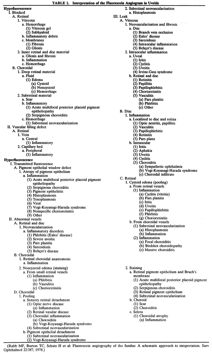
In this chapter, we describe the characteristic angiographic findings in many common uveitic syndromes using abnormal hypofluorescence or hyperfluorescence patterns with both fluorescein (Table 1)2,3 and ICG (Fig. 1).4 Many of these clinical entities and their treatments are described in more detail in other chapters in these volumes.


Fig. 1. Schematic interpretation of indocyanine angiogram. (Adapted from Freund KB, Yannuzzi LA, Schneider U et al: A schematic approach to clinical interpretation. In Yannuzzi LA, Flower RW, Slakter JS (eds): Indocyanine Angiography. St Louis: Mosby, 1997:129.)

Back to Top
INTERPRETATION OF HYPOFLUORESCENCE AND HYPERFLUORESCENCE

HYPOFLUORESCENCE

Hypofluorescence may be caused by blocked fluorescence, which is synonymous with transmission decrease, or by a vascular filling defect. Blocked fluorescein corresponds to the size, shape, and location of the blocking material seen ophthalmoscopically. Any hypofluorescence that does not correspond to the ophthalmoscopic picture must then be considered a vascular filling defect.

With ICG angiography, hypofluorescence needs to be analyzed by taking into consideration its evolution during the different phases of the angiogram. It is important to distinguish between hypofluorescence observed in both the early and late phases and when it is observed only in the later phases. The blockage depends on the nature of the blocking material and on the background choroidal fluorescence.

Blocked Fluorescence

Retinal. Any opacification in front of the retinal vessels that reduces media clarity causes blocked retinal vascular hypofluorescence on fluorescein angiogram (FA). Many inflammatory conditions produce hazy media, causing poor visualization of fundus detail. Fibrotic or gliotic membranes and inflammatory material on the retina or involving its inner layers obstruct the view of the retinal vasculature and cause blocked fluorescence.

CHOROIDAL.

Hypofluorescence can result from blocked choroidal vasculature due to fluid, exudate, hemorrhage, pigment, or other material accumulating in front of the choroidal vasculature deep to the retinal vasculature. Any opaque substance located beneath the retina but in front of the choroid blocks fluorescence of the choroidal vasculature; however, it does not block retinal vascular fluorescence. Fluorescence can be blocked by substances such as fluid in retinal pigment epithelial detachments and inflammatory material in acute multifocal posterior placoid pigment epitheliopathy (AMPPPE).

In general, the blockage to ICG is less than that to fluorescein. For example, although blood at any level can lead to blockage throughout the FA, blockage may occur only in the later phases in ICG angiography. However, inflammatory diseases that primarily affect the choroid, such as birdshot chorioretinopathy and multifocal choroiditis, may result in profound and more dramatic blockage than that observed with FA.

Vascular Filling Defect

RETINAL.

A vascular filling defect is the other cause of hypofluorescence in the retina if blocked fluorescence is not present. For a retinal vascular filling defect to be diagnosed, the location of the defect must be determined. It may arise from inflammatory obstruction of the artery, vein, or capillary or from a combination of these. Eales' disease, sarcoidosis, and other inflammatory diseases also cause peripheral capillary nonperfusion.

CHOROIDAL.

In ICG angiography, vascular filling defects have been subdivided into those that are physiologic, those resulting from a vascular occlusion, and those that are caused by tissue atrophy (see Fig. 1).4 Physiologic filling defects may be the result of choroidal watershed zones (early defects) and choroidal vascular silhouette against the background choroidal fluorescence (late defect). In contrast to FA, ICG studies are better at delineating choroidal vascular occlusion than are retinal vascular defects. Patients with AMPPPE may show persistent vascular filling defects after resolution of the condition. Choroidal vascular atrophy may develop in patients with a condition such as age-related macular degeneration, which results in vascular bed closure from scar formation.

HYPERFLUORESCENCE

Hyperfluorescence is any abnormally light area on the positive print of an angiogram. There are three types of hyperfluorescence in uveitis: transmitted fluorescence, abnormal vessels, and leakage.

Transmitted Fluorescence

Increased visibility of fluorescence from the choroidal vasculature caused by the absence of pigment in the pigment epithelium is called transmitted fluorescence (i.e., a pigment epithelial window defect) on FA. Various types of inflammation at the level of the pigment epithelium, choriocapillaris, or choroid produce pigment epithelial window defects such as AMPPPE, serpiginous choroiditis, and histoplasmosis. Each of these inflammatory conditions adversely affects the pigment epithelium, resulting in depigmentation and atrophy. Window defect is a very frequent finding in a fundus that has undergone choroidal inflammation.

In ICG angiography, transmitted fluorescence may present as one of two forms depending on the integrity of the choriocapillaris. When the choriocapillaris is intact, transmission hyperfluorescence of the larger choroidal vessels is noted in the early phases of the angiogram. In the late phases of the study, the fluorescence pattern appears normal. In conditions associated with atrophy of the choriocapillaris, hyperfluorescence of the larger choroidal vessels is noted in the early phase of the angiogram. However, this area may become relatively hypofluorescent in the later phases because of decreased or absent perfusion of the choriocapillaris.

Abnormal Vessels

RETINAL AND DISC.

There are many diseases that produce neovascularization. Retinal ischemia is theorized to be the primary pathogenic stimulus. Most of the disease processes resulting in neovascularization show some degree of ischemia by fluorescein angiography, with capillary nonperfusion an almost constant finding. The diseases that are known to result in neovascularization are phlebitis (Eales' disease), severe uveitis, pars planitis, sarcoidosis, and Behçet's disease.

CHOROIDAL.

Fluorescein angiography has proved to be of great value as an aid in the recognition and demonstration of normal and abnormal vascular patterns in the subretinal and choroidal tissues. The demonstration of subretinal neovascularization (SRNV) by fluorescein angiography (early lacyhyperfluorescence) represents one of the most significant advances in ophthalmic knowledge.

Although ICG angiography can identify abnormal vessels in the retina, it is more helpful in imaging changes in the choroidal circulation and is most useful in situations in which occult or poorly defined SRNV is present. Often the neovascular complex is obscured by blood or fluid and is poorly visualized on FA. ICG angiography has been shown to be efficacious in identifying occult SRNV and has increased the likelihood of identifying potentially treatable lesions in patients who are otherwise considered ineligible for laser treatment under conventional guidelines.5,6

Leakage

VITREOUS.

In uveitis, there are two major causes of vitreous leakage seen on fluorescein angiography: (1) neovascularization and fibrosis and (2) intraocular inflammation. Disc and retinal neovascularization may be associated with uveal inflammation. Inflammation of the retina or the disc, such as that seen in toxoplasmic chorioretinitis, is an example of intraocular inflammation leading to vitreous leakage.

DISC.

Inflammations of the optic nerve head, such as papillitis, vasculitis, and papillophlebitis, show a fuzzy hyperfluorescence of the disc in the later phases of the FA because of the staining of the edematous inflamed tissue.

RETINAL.

In the late stages of the normal angiogram, the retinal vessels contain a minimal concentration of fluorescein and the retina is normally dark. Retinal hyperfluorescence in the late stages indicates extravasation of fluorescein into the retinal extravascular space. The leakage occurs in a cystoid or noncystoid pattern and arises from leakage of retinal or choroidal vessels. Similar findings may be observed in ICG angiograms.

In the macula, cystoid retinal edema assumes a stellate or flower-petal appearance because of the obliquity of the outer plexiform layer. Outside the macula, there is a honeycomb appearance. Causes of cystoid edema rising from retinal vessels include inflammatory conditions, such as uveitis, chorioretinitis, and aphakia (Irvine-Gass syndrome). Cystoid retinal edema may be caused by leakage from choroidal vessels, such as with SRNV. Inflammation, such as toxoplasmic retinitis, is one of the most common causes of noncystoid retinal edema. On the FA, the lesion leaks fluorescein profusely into the surrounding retina and overlying vitreous, resulting in a fuzzy cotton-ball fluorescence. Marked staining of the retina (noncystoid edema) is then apparent.

When a large retinal vessel is inflamed, as in phlebitis or vasculitis, the endothelium of the involved large retinal vessel leaks fluorescein, resulting in hyperfluorescence. On the angiogram, this appears as a line of hyperfluorescence along the wall of the vessel. Retinal vessels traversing an area of chorioretinitis show reactive perivascular staining.

CHOROIDAL.

Choroidal inflammatory diseases, as in focal or massive choroiditis (Harada's syndrome), can change or damage the overlying pigment epithelium sufficiently to produce a sensory retinal detachment. In some cases, marked leakage occurs, which results in heavy pooling (defined as leakage of fluorescein dye) under the sensory retinal detachment.

Back to Top
UVEITIS OF UNKNOWN ETIOLOGY

VOGT-KOYANAGI-HARADA SYNDROME

Vogt-Koyanagi-Harada syndrome, also known as uveomeningoencephalitis syndrome, is a systemic disease often found in young, darkly pigmented whites, blacks, native Indians, Hispanics, and Asians. It presents as an anterior and posterior uveitis with disc hyperemia, disc edema, and exudative retinal detachment (Figs. 2A and B and 3A). Systemic signs and symptoms include headache, hearing loss, poliosis, vitiligo, nuchal rigidity, seizures, and even coma. SRNV and disciform scars are late complications. The differential diagnosis should include AMPPPE and sympathetic ophthalmia.

Fig. 2. Vogt-Koyanagi-Harada syndrome. (A) Fundus photograph of the right eye and (B) the left eye showing bullous retinal detachments. (C) Fluorescein angiogram showing multiple hyperfluorescent leaks at the level of the retinal pigment epithelium. (D) Multiple coalescing leaks and fluorescein staining of the subretinal fluid. Large areas of hyperfluorescence involving the entire right posterior pole due to pooling of dye beneath the subretinal space.

Fig. 3. Vogt-Koyanagi-Harada syndrome. A. Fundus photograph of the left eye showing multifocal serous retinal detachment. B. Late fluorescein angiogram showing pooling of dye in the areas of serous detachment. C. Indocyanine angiography showing areas of blocked fluorescence corresponding to serous detachment. More areas of hypofluorescence are noted than those observed on fluorescein angiogram.

There is a characteristic fluorescein angiographic appearance in the early phase showing multiple discrete hyperfluorescent dots at the retinal pigment epithelial level that enlarge over time (see Fig. 2C and D). In the late phase, if there is a serous detachment, the fluorescein pools beneath the subretinal space (see Fig. 3B). The edematous disc hyperfluoresces in the late phase. Generally, the retinal vessels are unaffected.7,8 Retinochoroidal anastomoses also have been documented by fluorescein angiography.9

ICG angiography shows multiple hypofluorescent spots in the posterior pole (see Fig. 3C). These spots may coalesce and obscure the filling of large choroidal vessels. When serous retinal detachment develops, the marked hyperfluorescence observed on FA is not as prominently noted in ICG angiography, presumably because of the highly protein-bound nature of ICG. In fact, diffuse late hypofluorescence may be observed in patients with serous retinal detachment.10 Ill-defined areas of hyperfluorescence corresponding to diffuse choroidal staining may be seen in some cases in the late phases of the angiogram. The optic disc may stain with ICG in the acute phase but usually is minimal compared to that observed on FA.

SYMPATHETIC OPHTHALMIA

Sympathetic ophthalmia is a rare, bilateral granulomatous, paninflammatory disease that can occur several weeks to years after an ocular injury. Sympathetic ophthalmia is hypothesized to be an autoimmune reaction against a normally sequestered antigen unmasked by an injury. Retinal findings in sympathetic ophthalmia are perivasculitis, papillitis, and yellowish subretinal nodules known as Dalen-Fuchs nodules. Dalen-Fuchs nodules are believed to be transformed retinal pigment epithelium cells or T lymphocytes or both. Areas of choroiditis or granuloma simulating AMPPPE may be seen. Pigmentary disturbance may be noted in the chronic phase of the disease (Fig. 4A and B). Choroidal neovascularization also may develop (see Fig. 4C).

Fig. 4. Sympathetic ophthalmia. A. Areas of pigmentation and small areas of focal atrophy. B. Fluorescein angiogram showing blocked fluorescein corresponding to the areas of pigmentary disturbance with areas of hyperfluorescence representing old areas of inflammation. C. Blocked fluorescence due to subretinal blood in the juxtafovea with associated choroidal neovascularization.

One of two possible fluorescein angiographic findings may be evident. The more common one is identical to Vogt-Koyanagi-Harada syndrome, with early multiple pinpoint retinal pigment epithelial leaks that enlarge with time.11 The second, less common, manifestation is early choroidal hypofluorescence of the Dalen-Fuchs nodules followed by hyperfluorescence in the later phase.12 When exudative retinal detachment occurs, there are confluent areas of subretinal fluorescein pooling.

Two patterns have been identified on ICG angiography.13 The first pattern, observed in a patient with chronic disease, showed hypofluorescent spots in both the intermediate and late phases of the angiogram. The second pattern, reported in a patient with recent onset of the disease, showed hypofluorescent spots in the intermediate phase but faded in the late phase of the angiogram.

POSTERIOR SCLERITIS

Posterior scleritis is a generally unilateral condition that usually is seen in patients 40 to 60 years old. Patients present with pain, decreased vision, and decreased visual fields. It generally is associated with anterior scleritis. Posterior scleritis may cause exudative retinal detachment resembling Harada's disease (Fig. 5A), cystoid macular edema, retinal pigment epithelial detachments, choroidal folds, and subretinal masses. Thickening of the choroid and posterior sclera can be seen with ultrasound, magnetic resonance imaging, and computed tomography.

Fig. 5. Posterior scleritis. A. Fundus photograph with multifocal exudative retinal detachment involving the macula. B. Multiple pinpoint retinal pigment epithelial leaks with late staining of the subretinal fluid can be seen.

Fluorescein angiography can show several manifestations. Frequently, the angiographic picture of posterior scleritis is identical to that of Vogt-Koyanagi-Harada syndrome, except that posterior scleritis usually is unilateral. Multiple early pinpoint retinal pigment epithelial leaks with late staining of the subretinal fluid can be seen (see Fig. 5B).14 Another pattern is early multifocal choriocapillaris filling defects followed by overlying retinal pigment epithelial staining, late staining of the subretinal fluid, and disc staining.15 Cystoid macular edema16 and multiple discrete retinal pigment epithelial detachments can be seen.

ICG angiography shows diffuse zonal hyperfluorescence in the intermediate and late phases. Pinpoint areas of hyperfluorescence also may be seen in the areas of zonal hyperfluorescence in the later phases of the angiogram. Delayed choroidal perfusion and hypofluorescent spots present up to the intermediate phase and faded in the late phase of the angiogram may be visualized. These spots are noted to be smaller and more irregular in size than those observed in Vogt-Koyanagi-Harada syndrome.17

RETICULUM CELL SARCOMA

Reticulum cell sarcoma, a non-Hodgkin's lym-phoma, is a systemic disease that can affect many organs, including the eyes and central nervous system. Seventy-five percent of the patients with ocular and central nervous system involvement have no other manifestation. Patients with ocular reticulum cell sarcoma tend to be older and have painless loss of vision. Findings include vitreous cells that are not responsive to corticosteroids, multiple subretinal pigment epithelial detachments with yellow infiltrates, exudative retinal detachment, and retinal vascular sheathing. On fluorescein angiography, patients with reticulum cell sarcoma with multiple retinal pigment epithelial detachments have an irregular, incomplete staining of these detachments.18

BEHÇET'S DISEASE

Behçet's disease is a systemic occlusive vasculitis that presents predominately in young middle Eastern and Japanese men. The classic features include acute hypopyon, iritis, aphthous stomatitis, and genital ulceration. Skin lesions and strokes also occur frequently. There often is an acute recurrent bilateral panuveitis. Ocular findings include retinal vasculitis (Fig. 6A) with an occlusive arteritis, vitritis, macular edema, ischemic retinitis, ischemic optic neuropathy, peripheral neovascularization, and occasionally SRNV.

Fig. 6. Behçet's disease. A. Fundus photograph showing vasculitis of the superotemporal vascular arcade. Intraretinal hemorrhages also are seen. B. Fluorescein staining of the vessel wall and adjacent areas of blocked fluorescence corresponding to the intraretinal hemorrhage.

On fluorescein angiography (see Fig. 6B), during the active phases of the disease, capillary dropout and dilated retinal capillaries are seen. Dilated retinal capillaries (particularly peripapillary capillaries) leak dye and cause retinal and disc staining.19 Cystoid macular edema,19 SRNV, and disciform scars often are seen.20 Leakage of peripheral capillaries can be seen in patients with normal-appearing fundi.21

ICG angiography shows hyperfluorescent spots from the early to late phases and hypofluorescent plaques, both of which are not evident on FA. Staining of choroidal vessel walls and leakage of ICG from the choroidal vessels also have been described.22–24

SARCOIDOSIS

Sarcoidosis is a systemic, idiopathic, noncaseating, granulomatous disease that affects various organs, including the eye, brain, lung, and skin. Ocular findings may include conjunctival nodules; anterior iridocyclitis; vitreous cells; retinal, optic nerve, and choroidal granulomas (Fig. 7A); retinal vasculitis (venules are preferentially affected); cystoid macular edema; retinal vessel occlusion; and disc and retinal neovascularization.

Fig. 7. Sarcoidosis. A. Choroidal granuloma. B. Fluorescein angiogram shows staining of the choroidal granuloma.

The FA reflects the various clinical entities. Retinal venular walls stain, particularly where there are perivenular exudates.25–27 More extensive venous involvement can produce a picture of dilated veins and perivenous leakage.27 Peripheral neovascularization occurs near areas of retinal capillary nonperfusion.25,28 Optic disc granulomas and optic disc neovascularization both leak extensively (Fig. 8A); sarcoid retinal lesions also stain (see Fig. 7B).27 In disc edema, the disc is hyperfluorescent and leaks fluorescein (see Fig. 8B).25

Fig. 8. Sarcoidosis. A. Fluorescein angiography shows two separate areas of neovascularization of the peripheral retina. B. Extreme leakage of dye from the right disc with cystoid macular edema of the right macular region in a patient with chronic sarcoidosis.

Four main patterns can be identified with ICG angiography. The first and most common pattern is hypofluorescent dark spots in the early and intermediate phases of the angiogram. These spots either become isofluorescent or remain hypofluorescent in the late phases. The second pattern is focal hyperfluorescent spots seen in the intermediate and late phases. The third pattern is fuzzy choroidal vessels due to perivascular choroidal leakage in the intermediate phase. Finally, the fourth pattern is characterized by diffuse zonal hyperfluorescence representing choroidal staining in the late phase of the angiogram. The latter two patterns resolved after systemic corticosteroid treatment.29

INTERMEDIATE UVEITIS

Intermediate uveitis is also known as pars planitis, peripheral uveitis, and chronic cyclitis. Intermediate uveitis is an inflammatory disease that affects young adults, causing symptoms of photophobia, floaters, and blurry vision. Clinically, mild anterior chamber inflammation, vitreous cells, vitreous snowballs, inflammatory membranes on the pars plana, phlebitis, cystoid macular edema, and, rarely, choroidal and retinal neovascularization are seen.

On fluorescein angiography, there is venular wall staining (Fig. 9),30 hyperfluorescence, and leakage of the peripheral inflammatory membranes.31 Cystoid macular edema often is evident.30 Optic disc, peripheral retinal, and subretinal32–34 neovascularization are rare.

Fig. 9. Intermediate uveitis. A. Red-free photography of the peripheral retina shows sheathing of the retinal venules. B. Fluorescein angiography shows staining of the vessel walls with leakage from the peripheral venules.

BIRDSHOT RETINOCHOROIDOPATHY

Birdshot retinochoroidopathy (also known as vitiliginous chorioretinitis) presents bilaterally, generally in middle-aged women, causing floaters and decreased vision, night blindness, and color blindness. Clinically, there are patches of postequatorial choroidal and retinal pigment epithelial depigmentation (Fig. 10A), vitreous cells, macular and disc edema, and venous sheathing. SRNV frequently is a late sequela. Often central vision may be preserved in at least one eye.

Fig. 10. Birdshot retinochoroidopathy. A. Multiple cream-color deep choroidal lesions. B. Fluorescein angiogram shows leakage of the optic disc and the retinal vasculature. C. Indocyanine green angiography showing areas of hypofluorescence that exceed those observed on fluorescein angiography.

On fluorescein angiography, retinal vessel staining, disc leakage, and cystoid macular edema are found (see Fig. 10B). There often is generalized hypofluorescence of the retinal vessels and increased circulation time.35,36 Surprisingly, the patches of depigmentation may appear normal on angiography, although there can be mild late hyperfluorescence.35,36 Posterior pole choroidal hyperfluorescent lesions that correspond to the areas of depigmentation and SRNV also can be seen.37

On ICG angiography (see Fig. 10C), early and late hypofluorescent patches, exceeding the clinically detectable lesions, with a choroidal vasotropic distribution and relative sparing of the peripapillary area and the central macula, are noted. These findings differentiate this condition from AMPPPE, multifocal choroiditis, and other granulomatous conditions such as sarcoidosis and sympathetic ophthalmia. Rarely, hyperfluorescent spots are noted in the late phases of the angiogram, which correspond ophthalmoscopically to retinal inflammation or obstructive changes.38

MULTIFOCAL CHOROIDITIS

Multifocal choroiditis may mimic the typical clinical findings of presumed ocular histoplasmosis syndrome (discussed later) and has the additional finding of anterior chamber and vitreous cells. Multiple yellow or gray acute choroidal lesions measuring 50 to 350 μm, periphlebitis, and occasionally retinal neovascularization can be seen. Marked pigmentary disturbances may be seen in the chronic phase (Fig. 11A).

Fig. 11. Multifocal choroiditis. A. Fundus photograph showing pigmentary disturbances. B. Multiple areas of hypofluorescence and hyperfluorescence representing chorioretinal scars with associated atrophic areas. C. Indocyanine green angiogram shows multiple areas of hypofluorescence around the disc, the macula, and the midperipheral fundus. Some of these areas are not visible clinically or on fluorescein angiogram.

On fluorescein angiography (see Fig. 11B), the punched-out lesions show the typical window defects. Acute lesions block early choroidal fluorescence and stain late. Cystoid macular edema and prolonged arteriovenous circulation times may be seen.39 Progressive subretinal fibrosis is a reported sequela that presents as multiple stellar zones of subretinal fibrosis. This fibrosis can be surrounded by multiple atrophic punched-out lesions (Fig. 12).40

Fig. 12. Multifocal choroiditis with subretinal fibrosis in a 26-year-old woman. A and B. Color photographs show hypopigmented lesions representing subretinal fibrosis involving both macular lesions. Multiple punched-out lesions surround the bands of fibrosis. C. Staining of the large stellate fibrous lesion can be seen in the left macula. There are multiple punched-out lesions above and below the macular zone. Leakage from the optic disc and its vessels can also be seen.

ICG angiography shows large hypofluorescent spots in the posterior pole measuring 200 to 500 μm, which did not usually correspond to clinically or fluorescein angiographically detectable lesions (see Fig. 11C). Smaller hypofluorescent spots, less than 50 μm, also may be seen in the posterior pole. Both large and small lesions are best seen in the later phases of the angiogram. Confluent hypofluorescent areas may be seen around the optic nerve in patients reporting an enlarged blind spot on visual field testing.41

ACUTE MULTIFOCAL POSTERIOR PLACOID PIGMENT EPITHELIOPATHY

Acute multifocal posterior placoid pigment epitheliopathy is a bilateral systemic disease that generally affects young, healthy adults, often after a flulike viral illness. It presents as a rapid, painless loss of vision, which usually is bilateral. The characteristic findings of the disease are deep, multiple, discrete, flat, creamy-white subretinal lesions, usually centered in the posterior pole. Associated vitreous cells, serous retinal detachments, disc edema, and vasculitis have been reported. Shortly after the onset of symptoms, the acute yellow placoid lesions resolve, showing irregular deposition of pigmentation without corresponding atrophy of the choroid. Associated but infrequent systemic signs include hematuria, hearing loss, erythema nodosum, thyroiditis, and cerebral vasculitis. In general, vision returns to normal even when significant retinal pigment epithelial mottling is present. SRNV is a rare complication. AMPPPE rarely recurs; however, there are some unilateral cases in which the second eye may become affected months later. When unilateral cases occur, it is necessary to differentiate acute multifocal posterior placoid pigment epitheliopathy from serpiginous choroiditis.

Angiographically, the creamy lesions initially block the choroidal background fluorescence. Later there is staining that is widely and evenly distributed (Fig. 13). The cause of the initial blocking is controversial. Some believe that this disorder is a systemic vasculitis with nonperfusion of the choriocapillaris. In support of this view, choroidal vessels occasionally can be seen through the hypofluorescent lesions.42,43 However, others believe that opaque retinal pigment epithelial cells block the choroidal fluorescence. The lesions do not correspond to the anatomy of the choriocapillaris and do not stain from the periphery as would be expected from the normal perfused neighboring choriocapillaris.44 Some cases of AMPPPE have been associated with serous detachment, showing late subretinal pooling of fluorescence. Old lesions show both hyperfluorescence and hypofluorescence depending on the retinal pigment epithelial integrity. Visual acuity usually returns to normal, even with clinically significant retinal pigment epithelial alteration.

Fig. 13. Acute multifocal posterior placoid pigment epitheliopathy. A. Red-free photograph of multifocal lesions at the level of the retinal pigment epithelium during the acute stage. B. Fluorescein angiography shows the multiple areas of hypofluorescence early in the arteriolar-venous phase. C. In the late venous phase, the hypofluorescent lesions have become hyperfluorescent.

ICG angiography shows well-demarcated areas of hypofluorescence of the acute and subacute lesions, especially in the late phases of the study. Similar findings are seen in healed AMPPPE lesions, but they tend to be smaller in size than in the acute lesions. The cause of the hypofluorescence seen with ICG angiography also is debatable. RPE edema, partial choroidal vascular occlusion, or complete choroidal vascular occlusion has been proposed, but none have been proved as the etiologic mechanism for the blockage.45

SERPIGINOUS CHOROIDOPATHY

Serpiginous choroidopathy, also known as serpiginous choroiditis, geographic, or helicoid choroidopathy, is an idiopathic, chronic, recurring peripapillary inflammation of the choroid and retinal pigment epithelium that spreads centrifugally. Frequently, only the macular region is affected, resulting in visual loss (Figs. 14 and 15). Although serpiginous choroidopathy can affect young persons, most patients are middle-aged or older. Active pigment epithelial lesions have a creamy geographic appearance and are at the level of the retinal pigment epithelium and choriocapillaris. Old lesions frequently show various amounts of retinal pigment epithelium/choriocapillaris loss with subretinal scarring and pigmentation surrounded by islands of normal choroid (Fig. 16A). There can be retinal vasculitis, iridocyclitis, disc swelling, periphlebitis, and, occasionally, vitreous cells. Disc neovascularization also has been seen. The disease usually begins unilaterally, although years later recurring episodes are common in one or both eyes. Asymmetric involvement also is very common.

Fig. 14. Macular serpiginous choroiditis. A. Red-free photograph of the left posterior pole shows the presence of a geographic lesion with diffuse borders at the level of the retinal pigment epithelium. B. Fluorescein angiogram of the left macular region shows a large area of choroidal hypofluorescence involving the macula. Other smaller hypofluorescent lesions surround the large lesion. Note pinpoint areas of hyperfluorescence representing drusen. C. The late venous phase shows diffuse staining of the left macular lesion.

Fig. 15. Subretinal neovascularization in serpiginous choroiditis. A. The arteriolar phase of the angiogram of the right eye shows a geographic center of choroidal hypofluorescence in the macula. There are additional areas of hypofluorescence superior to the right macular region. B. The venous phase of the angiogram shows a subretinal neovascular membrane just inferior and temporal to the macular zone (arrow). The hypofluorescence has decreased markedly in size compared with (A). The marked hypofluorescence masked the subretinal neovascular membrane initially.

Fig. 16. Serpiginous choroiditis. A. Old lesions show various amounts of retinal pigment epithelium/choriocapillaris loss with subretinal scarring and pigmentation. B. Fluorescein angiogram shows peripapillary hyperfluorescence corresponding to classic choroidal neovascularization.

Fluorescein angiography of the acute lesions shows early hypofluorescence due to blockage of the underlying choroidal fluorescence with eventual hyperfluorescent staining of the lesions' margins (see Fig. 14B and C). Later, there is patchy hyperfluorescence of the lesion due to leakage from the lesions' margins and from islands of normal choroid and choriocapillaris within the lesion. Sometimes the lesion becomes indistinguishable from the surrounding background fluorescence of the normal retina.46–48 The old lesions are hypofluorescent and are bordered by hyperfluorescence from the adjacent normal choriocapillaris. Hyperfluorescence of the old lesions occurs because of scar tissue that stains in the late phase of fluorescein angiography.

SRNV is a well-known complication of serpiginous choroidopathy. Occasionally, ophthalmoscopic signs such as subretinal blood and exudate can be seen. However, SRNV often appears like recurrent serpiginous lesions and can be diagnosed in patients only by fluorescein angiography, with persistent late enlarging hyperfluorescence of the lesions (see Fig. 15).49 The differential diagnosis includes AMPPPE, age-related macular degeneration, presumed ocular histoplasmosis, and choroidal ischemia.

In the acute stage, ICG angiography shows diffuse, homogeneous, marked, and persistent hypofluorescence during all phases of the angiogram in areas of active inflammation. Active choroidal involvement beyond the boundaries observed by ophthalmoscopy as well as those delineated by FA is noted. In the subacute phase of the disease, heterogeneous hypofluorescence with clear visualization of medium and large choroidal vessels is seen. In the healed stages, delayed or absent choroidal filling in the early transit phase with better visualization of deeper choroidal vessels because of loss of overlying RPE and inner choroid may be shown. If SRNV develops, hyperfluorescence of the lesion is noted.50,51

MULTIPLE EVANESCENT WHITE-DOT SYNDROME

Multiple evanescent white-dot syndrome causes acute unilateral or, rarely, bilateral vision loss in young, healthy adults, often after a viral illness. Clinically, small white dots are seen deep in the retina or at the level of the retina pigment epithelium. In addition, fine white or orange granularity seen in the macula is pathognomonic for the syndrome. Vitreous cells, blurred disc margins, and venous sheathing also can be seen. Vision improves to normal within several months and the lesions fade. Enlargement of the blind spot is a frequent sign during the active phase of the disease.

On fluorescein angiography, several findings are noted: early hypofluorescence and late staining of the dots and leakage of dye from the disc and retinal capillaries (Fig. 17). Retinal pigment epithelial window defects and late staining also may be seen. In one report, the lesions had a wreathlike configuration.52 As the lesions resolve, the angiographic changes are less pronounced and often return to normal.53,54

Fig. 17. Multiple evanescent white-dot syndrome. A. Fluorescein angiography in the early venous phase shows a few punctate hyperfluorescent spots (retinal pigment epithelium window defects) around the disc. B. Late venous phase shows staining of more lesions and the optic disc.

On ICG angiography, multiple small, deep hypofluorescent spots are seen in the early phase of the angiogram. These spots persist into the later phases of study and are more numerous and widely distributed throughout the fundus when compared with either funduscopic or fluorescein angiographic examination results. They are randomly scattered and, like birdshot chorioretinopathy, may have a vasotropic distribution in the peripheral retina. Areas of confluence hypofluorescence, like that observed in multifocal choroiditis, also may be seen.55

ACUTE RETINAL PIGMENT EPITHELIITIS

Acute retinal pigment epitheliitis is a relatively benign disorder that occurs in young, healthy adults often after a viral illness. Clinically, two to four gray or black deep retinal parafoveal lesions are seen, often surrounded by a yellow halo. The lesions resolve, although faint retinal pigment epithelial changes can be seen. Visual loss may be minimal or moderate, with recovery frequently in 6 to 12 weeks.

During the acute stage, the FA either can be normal or the lesions can hypofluoresce with lacy hyperfluorescence of the halo. There is no change in the size or outline of the lesion.56,57 Sometimes only hyperfluorescence of the halo is seen. Old le-sions may show diffuse, delicate hyperfluorescence.58

ACUTE MACULAR NEURORETINOPATHY

Acute macular neuroretinopathy is a bilateral disease that generally affects young, healthy adults, usually after a viral illness, and causes acute-onset decreased central or paracentral vision. Clinically, a petalloid, pinkish to grayish outer retinal macular lesion, which resolves over several weeks to months, is seen. The vision generally improves to baseline over a similar interval, although paracentral scotomas can persist.

On fluorescein angiography, there can be three presentations: a normal angiogram,59 nonleaking dilated perifoveal capillaries,59 and early hyperfluorescence followed by late staining of the macular lesions.60–62

Back to Top
UVEITIS SECONDARY TO VIRUSES

ACUTE RETINAL NECROSIS

Acute retinal necrosis is an often bilateral peripheral retinitis that generally occurs in young, healthy adults. Herpes simplex, cytomegalovirus retinitis, and herpes zoster virus have been implicated as the etiologic agents in various patients. Acute retinal necrosis has a similar clinical appearance to proven cytomegalovirus retinitis. It presents as peripheral white necrotic retinal lesions, often with arteriolar occlusions and sheathing (Fig. 18A). Disc edema and pallor can be seen. Retinal detachments are frequent sequelae of the infection.

Fig. 18. Acute retinal necrosis. A. Necrotizing retinitis with vitritis, papillitis, intraretinal hemorrhage, and retinal vasculitis. B. Fluorescein angiogram shows areas of hypofluorescence corresponding to choroidal hypoperfusion and intraretinal hemorrhage.

Fluorescein angiography (see Fig. 18B) shows multiple retinal vascular occlusions, particularly arteriolar. In the late phases, the retinal vessels stain, often proximal to the occlusion. Nonoccluded vessels also may show staining.63,64 Late staining of the disc can be seen during the acute phase.64 The differential diagnosis includes herpes zoster retinitis, toxoplasmosis, and Behçet's disease.

CYTOMEGALOVIRUS RETINITIS

Cytomegalovirus retinitis often is found in immunosuppressed patients and in patients with acquired immunodeficiency syndrome. Clinically, it presents a sharply demarcated necrotizing hemorrhagic retinitis with a vasculitis that often is occlusive. The necrotizing retinitis usually is located in the midperiphery and clinically manifests as whitish opacifications in the deep retina and in the retinal pigment epithelium. Retinal hemorrhages are seen in the necrotic area and surrounding the leading edge (Fig. 19A). Retinal vascular sheathing, usually periphlebitis, may be bilateral or unilateral (Fig. 20).65 Retinal detachments (both exudative and rhegmatogenous) and vitreous cells can occur.

Fig. 19. Cytomegalovirus retinitis. A. Fundus photograph of the left posterior pole shows necrotizing retinal changes with vascular sheathing and hemorrhages. B. Angiography shows large areas of hypofluorescence with a central region of multiple focal areas of staining within the lesion. Sheathed vessels stain but do not leak. C. Old cytomegalovirus retinitis. In an area of chronic retinitis, retinal pigment epithelium mottling is seen because of the necrosis.

Fig. 20. Frosted branch angiitis. A. Marked sheathing of retinal vessels with area of active retinitis and intraretinal hemorrhages is seen. B. Leakage of dye from the retinal venules representing retinal periphlebitis. Areas of hypofluorescence also are noted temporally corresponding to the retinitis.

On fluorescein angiography, findings that parallel the clinical appearance can be seen. Fusiform aneurysmal dilations of arteries, arteriole venules, and capillaries as well as capillary microaneurysms can be seen.66 The area of retinitis and necrosis blocks the choroidal fluorescence (see Fig. 19B); however, in the late venous phase, hyperfluorescence of the center lesion can be seen. Branch retinal artery occlusions may be seen.67 In addition, late dye leakage from arteries67 and optic disc occurs.

ACQUIRED IMMUNODEFICIENCY SYNDROME

Acquired immunodeficiency syndrome is caused by the human immunodeficiency virus. In addition to cytomegalovirus retinitis, the ocular complications of acquired immunodeficiency syndrome include Kaposi's sarcoma of the lids or conjunctive, Roth's spots, retinal hemorrhage, microaneurysms, telangiectasia, disc edema, cotton-wool spots, and cranial nerve palsies. On fluorescein angiography, microaneurysms, telangiectases, and small areas of capillary nonperfusion corresponding to the cotton-wool spots have been seen.68

HERPES ZOSTER

Herpes zoster can cause an iridocyclitis with sector iris atrophy and retinal arteritis, retinal necrosis, and central retinal vein occlusion.69 Sector iris atrophy can be seen on iris fluorescein angiography.70 With arteritis, arteriolar narrowing and slow dye transit with fluorescein leakage are evident.

SUBACUTE SCLEROSING PANENCEPHALITIS

Subacute sclerosing panencephalitis is a progressive neurologic disorder that occurs in children and young adults. Measles virus is believed to be the infectious agent. Personality and behavioral changes occur and are followed by neurologic symptoms of seizures, dementia, coma, and death. Half the patients have visual involvement, which often antedates the neurologic symptoms. Clinically, a focal posterior pole retinitis and flat focal white retinal lesions are seen (Fig. 21).

Fig. 21. Subacute sclerosing panencephalitis. A. Focal area of retinitis noted in the posterior pole.B. Fluorescein angiogram shows minimal blockage inferior to the macula corresponding to the areaof the retinitis. (Park DW, Boldt HC et al: Subacute sclerosing panencephalitis manifesting as viral retinitis: clinical and histopathologic findings. Am J Ophthalmol 123:533, 1997.)

With fluorescein angiography during the acute stage, the lesions mask the choroidal fluorescence. There is late venous leakage, vein staining, and arteriole and venule occlusions.71

Back to Top
UVEITIS SECONDARY TO BACTERIA

SYPHILIS

Luetic chorioretinitis can present as a myriad of clinical findings, such as a salt-and-pepper fundus, cotton-wool spots, exudative retinal detachment, periarteritis, periphlebitis, disc edema, neovascularization, occlusive arteritis, multiple flat gray chorioretinal lesions, and macular edema. Ocular syphilis may mimic retinitis pigmentosa or acute retinal necrosis. Disciform scars are sequelae of macular choroidal inflammation.

With cotton-wool spots and macular edema, capillary nonperfusion and diffuse leakage are seen, respectively, on fluorescein angiography. Patients with syphilis who have a retinitis pigmentosa-like fundus frequently show attenuated arterioles, delayed venular filling with late leakage of the peripapillary vessels, and mottled choroidal fluorescence.72 An edematous disc leaks dye.73 A fluorescein appearance similar to that of branch ret-inal vein obstruction can be seen.74 In rare cases, however, the FA in a patient with periarteritiscan be normal.75 Choroiditis can be seen as choroidal hyperfluorescence that resolves with treat-ment.72

SRNV presents with a typical appearance.76 Several cases of unilateral chorioretinitis presented as severe visual loss as the first manifestation of early secondary syphilis. Fluorescein angiography in these cases showed diffuse leakage under the neurosensory retina in the posterior pole associated with multifocal areas of staining along the retinal vessels (Fig. 22).77

Fig. 22. Syphilitic choroiditis. A. Fluorescein angiography of the left posterior pole shows peripapillary irregular mottling of the pigment epithelium with numerous areas of retinal pigment epithelial staining. B. Four months later, there is an increased subretinal hyperfluorescence temporal to the disc. The adjacent area of hypofluorescence represents subretinal blood.

A case of bilateral syphilitic posterior placoid chorioretinitis mimicking AMPPPE has been reported. ICG angiography showed hypofluorescent lesions during both early and late phases of the study, suggesting inflammation-associated perfusion abnormalities of the choriocapillaris.78

TUBERCULOSIS

Tuberculosis has numerous ocular presentations, including anterior uveitis, scleritis, sclerokeratitis, phlyctenulosis, interstitial keratitis, choroidal granulomas (often with serous elevations; Fig. 23A),retinal exudate, macular star formation, retinal vas-culitis, vitreous cells, SRNV, and optic nerve granulomas. Eales' disease, a bilateral peripheral vasculitis with peripheral neovascularization and vitreous hemorrhage, frequently occurs in young men who have positive purified protein derivative test results (Fig. 24).

Fig. 23. Tuberculoma. A. Subretinal tuberculoma. B. Multiple loculated areas of hyperfluorescence due to pooling of dye beneath the sensory retina.

Fig. 24. Eales' disease (retinal vasculitis). A. Angiography of the left disc shows staining of the retinal vessels, particularly the venules in the late venous stage. B. In the far periphery, there is staining of the peripheral vessels (particularly venules) just adjacent to an area of nonperfused retina. The hypofluorescent areas represent intraretinal hemorrhages.

On fluorescein angiography, a choroidal tuberculoma hyperfluoresces in the late phase (see Fig. 23B). There may be leakage and pooling of dye if there is a serous retinal detachment. As the tuberculoma responds to therapy, the hyperfluorescence decreases and pockets of hypofluorescence can be seen.79 Retinal arteriovenous shunts with dilated capillaries also can be seen in the granuloma.80 Peri-phlebitis causes capillary dropout and venous staining. Neovascularization of the peripheral retina is not uncommon.

Four angiographic signs have been described using ICG angiography: (1) irregularly distributed, hypofluorescent spots in the early and intermediate phases of angiography that either became isofluorescent (type 1 hypofluorescence) or remained hypofluorescent (type 2 hypofluorescence) in the late phase; (2) numerous, punctate hyperfluorescent spots; (3) fuzzy choroidal vessels in the intermediate phase due to leakage, leading in the late phase to (4) diffuse choroidal hyperfluorescence. Type 1 hypofluorescent lesions, fuzzy choroidal vessels, and diffuse choroidal hyperfluorescence tended to regress after the initiation of antituberculous and corticosteroid treatment. Focal hyperfluorescence tended to be associated with longstanding disease.81

Back to Top
UVEITIS SECONDARY TO FUNGUS

PRESUMED OCULAR HISTOPLASMOSIS SYNDROME

Presumed ocular histoplasmosis syndrome consists of the following triad: peripapillary atrophy, “punched-out” chorioretinal lesions, and disciform macular scarring in young and middle-aged adults (Fig. 25A). If punched-out lesions (atrophic spots) occur in the macula, the probability of SRNV occurring within 3 years increases from 3% to 25%. A systemic histoplasmosis infection during childhood is believed to cause the syndrome.

Fig. 25. A Fundus photograph showing multiple punched-out chorioretinal lesions and disciform scar in the macula. B. Arteriovenous phase shows areas of hypofluorescence representing atrophic areas. C. Late venous phase photograph shows staining of the atrophic spots and leakage in the macula corresponding to the choroidal neovascularization.

Fluorescein angiography is an important diagnostic test because SRNV is a major cause of vision loss (see Fig. 25B and C).82 The SRNV can be either peripapillary or macular. The choroidal neovascular membranes appear as a lacy network surrounded by a hypofluorescent ring in the early phase of angiography. A hallmark is the enlarging subretinalhyperfluorescence seen in the late venous phases. Punched-out lesions show early hypofluorescence followed by late scleral staining from the adjacent choriocapillaris,83 which can be differentiated from the progressively enlarging hyperfluorescence of SRNV.

With ICG angiography, focal hyperfluorescent spots, which are more numerous than those seen on FA, are noted in the posterior pole during the intermediate or late phase of the study. These lesions are not detectable in most cases on funduscopic examination. Smaller hypofluorescent spots also are seen in some patients with this condition. They are similar to those observed in multifocal choroiditis. However, the larger hypofluorescent spots seen in multifocal choroiditis are not found in ocular histoplasmosis. This latter finding in ICG angiography allows the two conditions to be distinguished. If SRNV develops, localized areas of hyperfluorescence with late staining are seen. This observation helps to distinguish neovascularization from localized inflammation, with the latter showing persistent late hypofluorescence.84

CANDIDA ENDOPHTHALMITIS

Candida endophthalmitis generally occurs in immunosuppressed patients and intravenous drug abusers. It also can occur in patients receiving hyperalimentation therapy and in patients after abdominal surgery. Candida endophthalmitis presents as white retinal abscesses with and without hemorrhage that extend into the vitreous and form vitreous snowballs (Fig. 26A). Roth's spots also can be seen.

Fig. 26. Candida endophthalmitis. A. Red-free photograph shows multiple retinal lesions. B. There are multiple areas of hyperfluorescence due to intraretinal and subretinal lesions. Some peripheral atrophic lesions represent age-related macular degeneration. Opacification of the vitreous inferior to the right macular region can be seen.

On fluorescein angiography, there are multiple early hypofluorescent deep retinal lesions that are not seen clinically. The white retinal lesions stain progressively in a centripetal fashion in the later phases (see Fig. 26B).85

Back to Top
UVEITIS SECONDARY TO PROTOZOA: TOXOPLASMOSIS
Toxoplasmosis is an intracellular parasite with a propensity to infect the inner retinal layers and cause an acute retinochoroiditis. Many cases are the sequelae of an acute intrauterine infection. The acute lesions appear white with prominent vitreous exudate. Often these acute lesions (satellite lesions) are adjacent to old chorioretinal toxoplasmosis scars. Toxoplasmosis also can present as an elevated optic disc swelling, papillitis, and old healed macular lesions (known as macular colobomas), which are sometimes seen with congenital toxoplasmosis (Fig. 27A). Segmental periarteritis, neovascularization of the disc and retina, SRNV, and branch retinal artery occlusion also can be seen (Fig. 28A). A variant form of toxoplasmosis shows multiple gray-white deep retinal or retinal pigment epithelial lesions with minimal overlying vitreous inflammation.

Fig. 27. Congenital toxoplasmosis. A. Recurrent toxoplasmosis retinochoroiditis adjacent to an old scar with surrounding serous retinal detachment. B. Late venous phase angiogram showing discrete pooling of dye in the subretinal space due to an active toxoplasmosis lesion in the superotemporal macula. (Courtesy of Helmut Buettner, MD.)

Fig. 28. Branch retinal arterial occlusion in acute toxoplasmosis. A. Red-free photograph of a superior temporal branch retinal arteriolar occlusion caused by acute Toxoplasma retinitis. B. Angiography shows evidence of a branch arteriolar obstruction with late staining surrounding the area of retinitis (arrow).

On fluorescein angiography in congenital toxoplasmosis, the acute macular lesions hypofluoresce early and hyperfluoresce late. Often there is extensive, complete loss of the retinal pigment epithelium and choriocapillaris, resulting in a hypofluorescent region with possible hyperfluorescence of scar tissue.

On fluorescein angiography, the acute lesions hypofluoresce in the early phase and hyperfluoresce in the late phase (see Fig. 27B).86,87 Old toxoplasmosis scars show hypofluorescence due to loss of the choriocapillaris.88 Various entities can be seen on fluorescein angiography, including branch retinal arteriole occlusion (see Fig. 28B),87,89,90 retinochoroidal anastomosis,91–93 retinal neovascularization,94 and SRNV.95 If papillitis occurs, there are dilated disc capillaries and late disc leakage.96 Segmental periarteritis seen clinically does not usually produce intravascular leakage, vessel staining, or alteration in flow.96 Cystoid macular edema is a rare finding.97 In the atypical deep retinal presentation of toxoplasmosis, there are minimal areas of hyperfluorescence that do not correspond to the clinical, deep retinal lesions.98

On ICG angiography, the main focus of retinochoroiditis is hypofluorescent in all phases of the angiogram, but late-phase (35- to 45-minute) hyperfluorescence also has been observed. The most striking feature, however, is multiple hypofluorescent satellite dark dots. In many cases, the hypofluorescent areas are not seen on FA and clinical examination, suggesting that toxoplasmic retinochoroiditis is a more widespread process than is clinically suspected because it extends beyond the visible lesions. After antitoxoplasma therapy, satellite dark dots disappeared from most of the cases reported. Further, the hypofluorescence under the main lesion was markedly reduced or disappeared in some cases.99–101 A case of toxoplasmic frosted branch angiitis was reported in both eyes of a 10-year-old boy. ICG angiography showed late-phase hyperfluorescence along the frosted vessels and in the disc. Delayed filling in the choriocapillaris also was identified in this child.102

Back to Top
UVEITIS SECONDARY TO PARASITES

CYSTICERCOSIS

Cysticercus is the larval form of Taenia solium, a tapeworm. Eggs are transformed 24 to 72 hours after ingestion into embryos that penetrate the intestinal wall and are distributed throughout the body via the circulatory system. The embryos then metamorphose into the cysticercus larval forms. The cysticercus can be found in the lids, conjunctive, anterior chamber, all layers and potential spaces of the retina and choroid, and the vitreous. The parasite often presents with amoeboid movements in a grayish white, semitransparent, well-defined cyst. A milky-white paracentral or central spot corresponding to the scolex often can be seen. If the cyst is subretinal, there may be hemorrhage, exudate, vasculitis, and venous tortuosity in the overlying retina. Early in the angiogram of subretinal lesions, the border of the cyst is not well-defined, but in the late phases, the cyst is outlined with fluorescence.103

DIFFUSE UNILATERAL SUBACUTE NEURORETINITIS

Diffuse unilateral subacute neuroretinitis is an ocular disease that occurs in otherwise healthy patients and is believed to be caused by chronic subretinal migration of one of at least two species of nematodes. Generally, visual acuity is poor (less than 20/200 [6/120]), and findings include pseudoretinitis pigmentosa, afferent papillary defects, anterior chamber and vitreous cells, disc edema, focal shifting, and recurrent deep retinal or subretinal yellow-white lesions. The lesions often are juxtamacular, and they resolve after several days. Occasionally, a subretinal worm (500 μm long and 25 μm wide) can be seen (Fig. 29A). There also may be diffuse retinal pigment epithelial atrophy with some hyperplasia. In the inactive stage, optic atrophy, retinal arterial narrowing and sheathing, and, rarely, SRNV can be seen.

Fig. 29. Diffuse unilateral subacute neuroretinitis. A. A coiled nematode (arrow) is seen in this red-free photograph just inferior to the right macula. Note the attenuation of the retinal arterioles. There are numerous subretinal grayish-white lesions involving the inferior portion of the right fundus. Five circular gray lesions are artifacts (arrowhead). B. Fluorescein angiography of the right posterior pole shows a hypofluorescent nematode (arrow) with multiple areas of hyperfluorescence due to diffuse retinal pigment epithelial changes. The nematode has now moved closer to the macula.

Many FAs obtained during the acute stage may show only perivenous leakage or may be normal. If optic disc edema is present, disc capillary leakage can be seen. The active deep retinal or subretinal lesions hypofluoresce early and stain late (see Fig. 29B). Mild but easily detectable retinal pigment epithelial can be seen angiographically. In the late stages of the disease, hyperfluorescence indicative of retinal pigment epithelial abnormalities occurs in the periphery and the peripapillary region. Generally, fine mottled hyperfluorescence is evident in the macular region.104 Cystoid macular edema occurs infrequently. Occasionally, the worm is seen as a hypofluorescent lesion on the FA (see Fig. 29B).

Back to Top
REFERENCES

1. Archer D, Krill AF, Newell FW: Fluorescein studies of normal choroidal circulation. Am J Ophthalmol 69:543, 1970

2. Rabb MF, Burton TC, Schatz H et al: Fluorescein angiography of the fundus: A schematic approach to interpretation. Surv Ophthalmol 22:387, 1978

3. Schatz H, Burton TC, Yannuzzi LA et al: Interpretation of Fundus Fluorescein Angiography. St Louis: CV Mosby, 1978

4. Freund KB, Yannuzzi LA, Schneider U et al: A schematic approach to clinical interpretation. In Yannuzzi LA, Flower RW, Slakter JS (eds): Indocyanine Angiography. St Louis: Mosby, 1997:129

5. Guyer DR, Yannuzzi LA, Slakter JS et al: Digital indocyanine green videoangiography of occult choroidal neovascularization. Ophthalmology 101:1727, 1994

6. Yaannuzzi LA, Hope-Ross M, Slakter JS et al: Analysis of vascularized pigment epithelial detachments using indocyanine green videoangiography. Retina 14:99, 1994

7. Shimizu K: Harada's, Behçet's, Vogt-Koyanagi syndromes: Are they clinical entities? Trans Am Acad Ophthalmol Otolaryngol 77:281, 1973

8. Saari M: Vogt-Koyanagi-Harada syndrome. Ophthalmologica 169:326, 1974

9. Ober RR, Smith RE, Ryan SJ: Subretinal neovascularization in the Vogt-Koyanagi-Harada syndrome. Int Ophthalmol 6:225, 1983

10. Freund KB, Yannuzzi LA: Vogt-Koyanagi-Harada syndrome. In Yannuzzi LA, Flower RW, Slakter JS (eds): Indocyanine Angiography. St Louis: Mosby, 1997:261

11. Dreyer WB, Zegarra H, Zakov ZN et al: Sympathetic ophthalmia. Am J Ophthalmol 92:816, 1981

12. Sharp DC, Bell RA, Patterson E et al: Sympathetic ophthalmia: Histologic and fluorescein angiographic correlation. Arch Ophthalmol 102:232, 1984

13. Bernasconi O, Auer C, Zografos L et al: Indocyanine green angiographic findings in sympathetic ophthalmia. Graefes Arch Clin Exp Ophthalmol 236:635, 1998

14. Benson WE, Shields JA, Tasman W et al: Posterior scleritis. Arch Ophthalmol 97:1482, 1979

15. Margargal LE, Donoso LA, Goldberg RE et al: Ocular manifestations of relapsing polychondritis. Retina 1:96, 1981

16. Cleary PE, Watson PG, McGill JI et al: Visual loss due to posterior segment disease in scleritis. Trans Ophthalmol Soc UK 95:297, 1975

17. Auer C, Herbort CP: Indocyanine green angiographic features in posterior scleritis. Am J Ophthalmol 126:471, 1998

18. Gass JDM, Sever RJ, Grizzard WS et al: Multifocal pigment epithelial detachments by reticulum cell sarcoma. Retina 4:135, 1984

19. Shimuzu K: Fluorescein fundus angiography. Mod Probl Ophthalmol 10:224, 1972

20. Michelson JB, Michelson PE, Chisari FV: Subretinal neovascular membrane and disciform scar in Behçet's disease. Am J Ophthalmol 90:182, 1980

21. Atmaca LS: Fundus changes associated with Behçet's disease. Graefes Arch Clin Exp Ophthalmol 227:340, 1989

22. Matsu T, Sato Y, Shiraga F et al: Choroidal abnormalities in Behçet's disease observed by simultaneous indocyanine green and fluorescein angiography with scanning laser ophthalmoscopy. Ophthalmology 106:295, 1999

23. Klaeger A, Tran VT, Hiroz C et al: Indocyanine green angiography in Behçet's uveitis. Retina 20:309, 2000

24. Atmaca LS, Batioglu F: Indocyanine green videoangiography and color Doppler imaging in Behçet's disease. Acta Ophthalmol Scand 77:444, 1999

25. Algvere P: Fluorescein studies of retinal vasculitis in sarcoidosis. Acta Ophthalmol Copenh 48:1129, 1970

26. Chumbley L, Kearns TP: Retinopathy of sarcoidosis. Am J Ophthalmol 73:123, 1972

27. Sanders MD, Shilling JS: Retinal, choroidal, and optic disc involvement in sarcoidosis. Trans Ophthalmol Soc UK 96:140, 1976

28. Asdourain GK, Goldberg MF, Busse BJ: Peripheral retinal neovascularization in sarcoidosis. Arch Ophthalmol 93:787, 1975

29. Wolfensberger TJ, Herbort CP: Indocyanine green angiographic features in ocular sarcoidosis. Ophthalmology 106:285, 1999

30. Pruett RC, Brockhurst RJ, Letts NF: Fluorescein angiography of peripheral uveitis. Am J Ophthalmol 77:448, 1974

31. Zenker HJ: Fluorescein angiography in inflammation of the peripheral fundus. III. Involvement of the pars plana corporis ciliaris. Ophthalmologica 190:205, 1985

32. Felder KS, Brockhurst RJ: Neovascular fundus abnormalities in peripheral uveitis. Arch Ophthalmol 100:750, 1982

33. Shorb SR, Irvine AR, Kimura SJ et al: Optic disk neovascularization associated with chronic uveitis. Am J Ophthalmol 82:175, 1976

34. Arkfeld DF, Brockhurst RJ: Peripapillary subretinal neovascularization in peripheral uveitis. Retina 5:157, 1985

35. Gass JDM: Vitiliginous chorioretinitis. Arch Ophthalmol 99:1778, 1981

36. Fuerst DJ, Tessler HT, Fishman GA et al: Birdshot retinochoroidopathy. Arch Ophthalmol 102:214, 1984

37. Brucker AJ, Deglin EA, Bene C et al: Subretinal choroidal neovascularization in birdshot retinochoroidopathy. Am J Ophthalmol 99:40, 1985

38. Chang B, Lumbroso L, Rabb MF et al: Birdshot chorioretinopathy. In Yannuzzi LA, Flower RW, Slakter JS (eds): Indocyanine Angiography. St Louis: Mosby, 1997:231

39. Dreyer RF, Gass JDM: Multifocal choroiditis and panuveitis. Arch Ophthalmol 102:1776, 1984

40. Cantrill HL, Folk JC: Multifocal choroiditis associated with progressive subretinal fibrosis. Am J Ophthalmol 101:170, 1986

41. Slakter JS, Giovannini A: Multifocal choroiditis and the presumed ocular histoplasmosis syndrome. In Yannuzzi LA, Flower RW, Slakter JS (eds): Indocyanine Angiography. St Louis: Mosby, 1997:271

42. Deutman AF, Lion F: Choriocapillaris nonperfusion in acute multifocal placoid pigment epitheliopathy. Am J Ophthalmol 84:652, 1977

43. Hedges TR, Sinclair S, Gragoudas ES: Evidence for vasculitis in acute posterior multifocal placoid pigment epitheliopathy. Ann Ophthalmol 10:539, 1978

44. Gass JDM: Stereo Atlas of Macular Disease. St Louis: CV Mosby, 1987:508

45. Schatz H, Park D, McDonald HR et al: Acute multifocal posterior placoid pigment epitheliopathy. In Yannuzzi LA, Flower RW, Slakter JS (eds): Indocyanine Angiography. St Louis: Mosby, 1997:239

46. Chisholm H, Gass JDM, Hutton WL: The late stage of serpiginous (geographic) choroiditis. Am J Ophthalmol 82:343, 1976

47. Schatz H, Maumenee AE, Patz A: Geographic helicoid peripapillary choroidopathy. Trans Am Acad Ophthalmol Otolaryngol 78:747, 1974

48. Hamilton AM, Bird AC: Geographical choroidopathy. Br J Ophthalmol 58:784, 1974

49. Laatikainen L, Erkkila H: Subretinal and disc neovascularization in serpiginous choroiditis. Br J Ophthalmol 66:326, 1982

50. Giovannini A, Ripa E, Scassellati-Sforzolini B et al: Indocyanine green angiographic findings in serpiginous choroidopathy. Eur J Ophthalmol 9:299, 1996

51. Mones JM, Slakter JS: Serpiginous choroidopathy. In Yannuzzi LA, Flower RW, Slakter JS (eds): Indocyanine Angiography. St Louis: Mosby, 1997:247

52. Mamalis N, Daily MJ: Multiple evanescent white dot syndrome: A report of eight cases. Ophthalmology 94:1209, 1987

53. Jampol LM, Sieving PA, Pugh D et al: Multiple evanescent white dot syndrome. Arch Ophthalmol 102:671, 1984

54. Aaberg TM, Campo RV, Joffe L: Recurrences and bilaterality in the multiple evanescent white dot syndrome. Am J Ophthalmol 100:29, 1985

55. Ie D, Glaser BM, Murphy RP et al: Indocyanine green angiography in multifocal evanescent white dot syndrome. Am J Ophthalmol 117:7, 1994

56. Krill AE, Deutman AF: Acute retinal pigment epitheliitis. Am J Ophthalmol 74:193, 1972

57. Chittum M, Kalina RE: Acute retinal pigment epitheliitis. Ophthalmology 94:1114, 1987

58. Deutman AF: Acute retinal pigment epitheliitis. Am J Ophthalmol 78:571, 1974

59. Bos PJM, Deutman AF: Acute macular neuroretinopathy. Am J Ophthalmol 80:573, 1975

60. Rush JA: Acute macular neuroretinopathy. Am J Ophthalmol 83:490, 1977

61. Priluck IA, Buettner H, Robertson DM: Acute macular neuroretinopathy. Am J Ophthalmol 86:775, 1978

62. Sieving PA, Fishman GA, Salzano T et al: Acute macular neuroretinopathy: Early receptor potential changes suggest photoreceptor pathology. Br J Ophthalmol 68:229, 1984

63. Price FW, Schlaegel TF: Bilateral acute retinal necrosis. Am J Ophthalmol 89:419, 1980

64. Hayreh SS: So-called acute retinal necrosis syndrome: An acute ocular panvasculitis syndrome. Dev Ophthalmol 10:40, 1985

65. Rabb MF, Jampol LM, Fish RH et al: Retinal periphlebitis in patients with acquired immunodeficiency syndrome with cytomegalovirus retinitis mimics acute frosted retinalperiphlebitis. Arch Ophthalmol 110:1257, 1992

66. Augsburger JJ, Henry RY: Retinal aneurysms in adult cytomegalovirus retinitis. Am J Ophthalmol 86:794, 1978

67. Digre KB, Blodi CF, Bale JF: Cytomegalovirus infection in a healthy adult associated with recurrent branch retinal artery occlusion. Retina 7:230, 1987

68. Newsome DA, Green WR, Miller ED et al: Microvascular aspects of acquired immune deficiency syndrome retinopathy. Am J Ophthalmol 98:590, 1984

69. Brown RM, Mendis U: Retinal arteritis complicating herpes zoster ophthalmicus. Br J Ophthalmol 57:344, 1973

70. Marsh RJ, Easty DL, Jones BR: Iritis and iris atrophy in herpes zoster ophthalmicus. Am J Ophthalmol 78:255, 1974

71. Kovacs B, Vastag O: Fluoroangiographic picture of the acute stage of the retinal lesion in subacute sclerosing panencephalitis. Ophthalmologica 177:264, 1978

72. Morgan CM, Webb RM, O'Connor GR: Atypical syphilitic chorioretinitis and vasculitis. Retina 4:225, 1984

73. Tait IA: Uveitis due to secondary syphilis. British Journal of Venereal Diseases 59:397, 1983

74. Folk JC, Weingeist TA, Corbett JJ et al: Syphilitic neuro-retinitis. Am J Ophthalmol 95:480, 1983

75. Crouch ER, Goldberg MF: Retinal periarteritis secondary to syphilis. Arch Ophthalmol 93:384, 1975

76. Saari M: Disciform detachment of the macula. Acta Ophthalmol Copenh 56:510, 1978

77. de Sousze EC, Jalkh AC, Trempe CL et al: Unusual central chorioretinitis as the first manifestation of early secondary syphilis. Am J Ophthalmol 105:271, 1988

78. Bellmann C, Holz FG, Breitbart A et al: Bilateral acute syphilitic posterior placoid chorioretinopathy: Angiographic and autofluorescence characteristics. Ophthalmologe 96:522, 1999

79. Cangemi FE, Friedman AH, Josephberg R: Tuberculoma of the choroid. Ophthalmology 87:252, 1980

80. Goldberg MF: Presumed tuberculous maculopathy. Retina 2:47, 1982

81. Wolfensberger TJ, Piguet B, Herbort CP: Indocyanine green angiographic features in tuberculous chorioretinitis. Am J Ophthalmol 127:350, 1999

82. Macular Photocoagulation Study: Argon laser photocoagulation for ocular histoplasmosis. Arch Ophthalmol 101:1347, 1983

83. Gass JDM: Stereo Atlas of Macular Disease. St Louis: CV Mosby, 1987:118

84. Slakter JS, Giovannini A: Multifocal choroiditis and the presumed ocular histoplasmosis syndrome. In Yannuzzi LA, Flower RW, Slakter JS (eds): Indocyanine Angiography. St Louis: Mosby, 1997:271

85. Robertson DM, Riley FC, Hermana PE: Endogenous Candida oculomycosis. Arch Ophthalmol 91:33, 1974

86. Schwartz PL: Segmental retinal periarteritis as a complication of toxoplasmosis. Ann Ophthalmol 9:157, 1977

87. Willerson D, Aaberg TM, Reeser F et al: Unusual ocular presentation of acute toxoplasmosis. Br J Ophthalmol 61:693, 1977

88. Gilbert H: Unusual presentation of acute ocular toxoplasmosis. Graefes Arch Clin Exp Ophthalmol 215:53, 1980

89. Braunstein RA, Gass JDM: Branch artery obstruction caused by acute toxoplasmosis. Arch Ophthalmol 98:512, 1980

90. Luxenburg MN: Branch retinal artery occlusion associated with recurrent toxoplasmic retinochoroiditis. Arch Ophthalmol 105:130, 1987

91. Saari M, Miettinen R, Nieminen H et al: Retinochoroidal vascular anastomosis in toxoplasmic chorioretinitis. Acta Ophthalmol Copenh 53:44, 1975

92. Owens PL, Goldberg MF, Busse BJ: Prospective observation of vascular anastomosis between the retina and choroid in recurrent toxoplasmosis. Am J Ophthalmol 88:402, 1979

93. Kennedy JE, Wise GN: Retinochoroidal vascular anastomosis in uveitis. Am J Ophthalmol 71:1221, 1971

94. Gaynon MW, Boldrey EE, Strahlman ER et al: Retinal neovascularization and ocular toxoplasmosis. Am J Ophthalmol 98:585, 1984

95. Fine SL, Owens SL, Haller JA et al: Choroidal neovascularization as a late complication of ocular toxoplasmosis. Am J Ophthalmol 91:318, 1981

96. Folk JC, Lobes LA: Presumed toxoplasmic papillitis. Ophthalmology 91:64, 1984

97. Schlaegel TF, Weber JC: The macula in toxoplasmosis. Arch Ophthalmol 102:697, 1984

98. Doft BH, Gass JDM: Punctuate outer retinal toxoplasmosis. Arch Ophthalmol 103:1332, 1985

99. Auer C, Bernasconi O, Herbort CP: Toxoplasmic retinochoroiditis: New insights provided by indocyanine green angiography. Am J Ophthalmol 123:131, 1997

100. Guex-Crosier Y, Auer C, Bernasconi O et al: Toxoplasmic retinochoroiditis: Resolution without treatment of the perilesional satellite dark dots seen by indocyanine green angiography. Graefes Arch Clin Exp Ophthalmol 236:476, 1998

101. Auer C, Bernasconi O, Herbort CP: Indocyanine green angiography features in toxoplasmic retinochoroiditis. Retina 19:22, 1999

102. Tanikawa H, Akimoto S, Honda M et al: Indocyanine green video angiographic findings of frosted branch angitis in a child. Nippon Ganka Gakkai Zasshi 102:399, 1998

103. Barsante CF: Cysticercus sub-retinalis. In Shimizu K (ed): Fluorescein Angiography. Tokyo: Igaku Shoin, 1974:193

104. Gass JDM, Gildberg WR, Guerry RK et al: Diffuse unilateral subacute neuroretinitis. Ophthalmology 85:521, 1978

Back to Top