Chapter 20
Functional Anatomy of the Retina
PAUL WITKOVSKY
Main Menu   Table Of Contents

Search

GENERAL ORGANIZATION OF THE RETINA
SYNAPSES IN THE RETINA
INFORMATION PROCESSING IN THE OUTER RETINA
NEURONS OF THE INNER RETINA: AMACRINE, GANGLION, AND INTERPLEXIFORM CELLS
INTERPLEXIFORM NEURONS OF THE RETINA
SIGNAL PROCESSING IN THE INNER RETINA
FUNCTIONAL CLASSES OF RETINAL GANGLION CELLS
RETINAL CIRCUITS UNDERLYING COLOR VISION
DIRECTIONALLY SELECTIVE GANGLION CELLS
PHARMACOLOGY AND ELECTROPHYSIOLOGY OF GANGLION CELLS
RETINAL NEUROMODULATORY SYSTEMS
ELECTRICAL SYNAPSES (GAP JUNCTIONS) IN THE RETINA AND THEIR MODULATION BY DOPAMINE
RETINAL GLIAL CELLS
OVERVIEW
ACKNOWLEDGMENTS
REFERENCES

The retina has been the focus of intensive investigation since the earliest days of neuroanatomical research. The great Spanish neuroanatomist Santiago Ramon y Cajal set forth reasons for the attractiveness of the retina as an experimental tissue in the introduction to his classic study1: the basic flow of information from photoreceptors toward ganglion cells was understood; the retinal neurons were arrayed in well-defined cellular layers, their contacts with other neurons were separated into clear zones (the inner and outer plexiform layers); and the compact nature of a cell's dendritic and axonal arbors facilitated study of its nervous connections. Cajal considered the retina to be a true nervous center, but one whose thinness and transparency made it ideal for histologic analysis.

Researchers working on the retina at the beginning of the 21st century find little to reject in Cajal's framework, but there is much to add. Electron microscopy provides a description of the organization of synapses. Neurochemistry and neurophysiology underlie our understanding of chemical and electrical synaptic transmission and the excitable properties of retinal membranes that contribute to their light-evoked responses. From molecular and cellular biology come tools to understand the many families of proteins that participate in retinal signaling, growth, normal and pathologic function, and cell death. One of the most important insights to arise from the studies of recent years is that the nervous system, including the retina, is highly plastic: that is, in addition to the “hard-wiring” of the system (i.e., the fixed pattern of connections between nerve cells and their defined neurotransmitters), there exists in parallel a system of neuromodulators that modifies the state of the circuits. Neuromodulation2 depends on a host of neuroactive substances, including peptides, amines, and metabolites, and it is linked through membrane-delimited or second-messenger pathways to a large variety of intracellular proteins (e.g., kinases and phosphatases), which exert a subtle but powerful control over membrane protein function, including neurotransmitter receptors and voltage-gated channels. In many cases the neuroactive substances reach their targets by diffusion from a more or less distant source,3 without the requirement for a morphologically defined synaptic contact.

A description of retinal circuitry is thus a much more complicated task today than in Cajal's era. He and Polyak4 asked the first question: what are the shape and retinal location of a cell's dendritic and axonal arborizations? We now can add: what are the patterns of electrical and chemical synapses the neuron makes and receives? What neurotransmitters does the neuron utilize? What receptors for neuroactive substances does the neuron possess? Into what functional retinal circuits is the neuron integrated? Are there plastic aspects of the neuron's organization and behavior? We have fairly complete answers for the first three questions, partial answers for questions four and five, and a growing but still sparse database for the last question. This essay surveys some of basic information about synapses and receptors, retinal cells, and circuits and then describes some newer findings in greater detail. The emphasis throughout is on the mammalian retina.

Back to Top
GENERAL ORGANIZATION OF THE RETINA
The retina contains five basic neuronal classes (photoreceptors, horizontal, bipolar, amacrine, and ganglion cells) and two types of glial cell (radial Mullerfiber and astrocytes). Their nuclei are arranged in regular layers (Fig. 1, left side): photoreceptors in the outer nuclear layer, horizontal cells in the distal portion of the inner nuclear layer (INL), amacrine cells at the proximal face of the INL where it abuts the inner plexiform layer (IPL), and bipolar cells distributed throughout the INL. Ganglion cells form a layer at the inner surface of the retina, separated from the vitreous body by a layer of optic nerve fibers and by the end feet of the Muller glial cells, whose nuclei are found in the INL. The astrocytes reside in the layer of optic nerve fibers and around retinal blood capillaries. There are some exceptions to the nuclear layering pattern. Amacrine perikarya occasionally are found within the IPL (interstitial amacrines) or in the ganglion cell layer (displaced amacrines) and the rarely encountered displaced ganglion cells have cell bodies at the INL/IPL border (i.e., mixed in among the amacrines). Figure 1, right side, depicts the shape of representative retinal neurons and illustrates that retinal neurons may have a primarily vertical orientation (photoreceptors and bipolar cells) or be oriented horizontally (horizontal and amacrine cells).

Fig. 1. General organization of the mammalian retina. The left side schematic is a vertical section through the retina showing the arrangement of the nuclei of retinal cells into discrete layers. The right side illustrates some representative shapes of each class of retinal neuron and the principal glial cell, the Muller cell, taken from Cajal's1 drawings of mammalian retinas.

SUBCATEGORIES OF THE BASIC RETINAL CELL TYPES

Each of the five basic classes of retinal neuron is further subdivisible into many subtypes, and the criteria for distinguishing subtypes are multiple. The most venerable is the shape of a cell's dendritic and axonal arbors (e.g., midget, diffuse, monostratified, multistratified), as epitomized by the studies of Cajal1 and Polyak.4 Further distinctions depend on the particular level or levels of the IPL in which the processes of the cell extend, as described in greater detail in subsequent sections. Recent studies provide new bases for subdividing retinal neurons, according to the neurotransmitters they utilize, the neurotransmitter receptors found in their processes, and the particular proteins identified in their cytoplasm. Examples in the latter category include calcium-binding proteins and enzymes such as protein kinase C.

In addition to these morphologic and neurochemical characteristics, one may refer to functional properties of the different cell groups. The older literature provides a description of their light-evoked responses as detected by extracellular or intracellular recording electrodes. For example, neurons in the outer retina, the photoreceptors and bipolar and horizontal cells, do not fire action potentials, instead responding to light with relatively slow changes of membrane potential. In contrast, most amacrine cells and all ganglion cells produce all-or-nothing action potentials. Other properties of light-evoked responses are described in more detail below. In more recent investigations, electrophysiologic measures are united with injection of a dye that reveals the cell's geometry. The most important of these are the fluorescent dye Lucifer Yellow and the opaque dye Neurobiotin. With the new power-ful electrophysiologic techniques of patch clamprecording, one can study the properties of ligand-gated and voltage-gated channels in retinal membranes. These membrane receptors can be examined in a living retinal slice, in a retinal cell isolated enzymatically and maintained in primary culture, or by pulling a small patch of membrane free from the cell with a suitable electrode.

RETINAL MOSAIC

As a general principle of retinal organization, each subtype of retinal neuron is distributed more or less evenly throughout the retina, giving rise to a network of like cells in the horizontal plane. The networks of different cell types overlap, creating the retinal mosaic. The density (number of cells/mm2) of neurons in one particular network need not be constant; in fact, it often reflects the organization of the photoreceptor layer. In the primate retina, for example, cones achieve their highest density in the center of the fovea,5 whereas for rods the highest density is found at about 20 degrees parafoveally in nasal and temporal retinas.6 Other mammalian retinas may lack foveas but often have some sort of central specialization (cat, area centralis; rabbit, visual streak) in which cones are found at a relatively high density. Thus, a bipolar cell that connects only to cones will be found at its highest density in central retina, whereas bipolar cells con-necting to rods are most concentrated in moreperipheral retinal regions.

A functional correlate of the central to peripheral axis is an increase in the size of neurons of the same type at increasingly more peripheral locations, measured both as an increase in the perikaryal diameter and an expansion of the dendritic and axonal arbors. This has been shown clearly for many specific types of retinal neuron, including horizontal cells and rod bipolar cells of the human retina,7 midget ganglion cells of primate retina,8 a variety of ganglion cells in the macaque retina,9 and for starburst amacrine cells of the rabbit retina10 (Fig. 2). Two functional consequences related to an increase in cell size are an increase in the dimensions of the neuron's receptive field center11 (described more fully below) and a fall in the resolving power (i.e., the acuity of the retina and visual pathway) as increasingly more peripheral portions of the retina are tested.12

Fig. 2. Camera lucida drawings of four starburst amacrine cells from the rabbit retina, located at increasing distance from the center of the eye. The basic shape of the cell remains the same, but the size increases. Marker bar is 100 μm. The numbers indicate the relative position of the drawn cell in the series. (Tauchi M, Masland RH: The shape and arrangement of the cholinergic neurons in the rabbit retina. Proc R Soc Lond B223:101, 1984, with permission of the authors)

Back to Top
SYNAPSES IN THE RETINA

ELECTRICAL SYNAPSES

Synapses in the nervous system are divisible into two basic functional classes: electrical and chemical. At electrical synapses a cytoplasmic bridge is formed by specialized proteins called connexins.13 Six connexins form a hexagonal tube that projects out of the plane of the plasma membrane of one participating cell; this unit is called a connexon. A similar connexon emerges from the membrane of the other participating cell to complete the junction (Fig. 3). The older name for such a synapse was the gap junction,14 so called because the two plasma membranes closely approached each other, but did not fuse as they do in a tight junction. We now understand that the “gap” provides the space for the connexons to project out of the plane of the membrane.

Fig. 3. Schematic of gap junctional channels in a biologic membrane. The membrane is schematized as a lipid bilayer, with circles indicating the polar head groups and squiggly lines the fatty acid tails of the lipid. The connexons forming the gap junction have been split down the center to reveal the aqueous pore. (Adapted from Kandel, Schwartz: Principles of Neural Science, 2d ed. Elsevier, 1985)

Electrical synapses can be large or small. For example, neighboring photoreceptors are joined by electrical synapses often less than 0.1 μm in diameter,15 whereas horizontal cells typically have patches of connexons greater than 0.5 μm16 (Fig. 4). Whatever the area of the electrical synapse, however, the connexons are present at a similar density of about 5,000/μm2. The central pore of the connexon is relatively large, permitting passage of all ions, whether positively or negatively charged, and also small metabolites, up to a molecular weight of about 1.5 kD.17 In many, perhaps most, cases, electrical current can flow through the electrical synapse in either direction (i.e., the synapse is unpolarized), but exceptions are known. The molecular biology of connexins shows that they form several families,18 distinguished by their molecular weight (in kilodaltons) and amino acid sequence. A recent study of mouse retina19 detected mRNAs for the following connexins: 26, 31, 32, 36, 37, 40, 43, 45, and 50, although a smaller number was identified by antibodies directed against connexin proteins, indicating that only a fraction of the genes for connexins is expressed.

Fig. 4. Gap junctions in retinal photoreceptors and horizontal cells. Part 1. Gap junctions viewed by transmission electron microscopy. Top. A gap junction (arrows) between a rod base (ROD) and a basal process emitted by a neighboring rod (left) in the Xenopus retina (× 180,000). Bottom. An extensive gap junction (arrows) between two horizontal cell axons in a Xenopus retina (× 216,000; data of P. Witkovsky and C.C. Powell). Part 2. Gap junctions viewed by freeze fracture. A. Gap junction (large arrows) on the protoplasmic face (PF) of the photoreceptor base in the Xenopus retina. At right the receptor cytoplasm is seen in cross fracture; synaptic vesi-cles (SV) are indicated by small arrows (× 109,500). B. Gap junction on the photoreceptor membrane in the Xenopus retina. The fracture plane passed through the junction, revealing both the protoplasmic face (PF), bearing particles, and the external face (EF), bearing pits associated with the gap junction (× 127,750). C. Gap junction on a horizontal cell in the Xenopus retina. The external membrane face contains a plaque of E-face pits (× 27,750; data of A. Nagy and P. Witkovsky).

Many connexins have been cloned and expressed in systems such as oocytes or cell lines, permitting a detailed study of their properties. When two cells with connexons in their membrane are brought together, gap junctions form spontaneously within a few minutes when the two connexons are compatible. This is invariably the case for identical (homologous) connexons, but is often also the case for different (heterologous) connexons.20 Unlike what was originally supposed, electrical synapses are far from being unchangeable structures with fixed properties. They may show voltage and pH dependence, can be sensitive to [Ca]21, and can be modified by dopamine and nitric oxide. Electrophysiologic investigations combined with dye injection reveal that many types of retinal neurons are electrically coupled through gap junctions. The coupling may be very great, as is found in the horizontal cell layer,22 or among AII amacrine cells23 (see below for a further description of cell types), or weak, as has been shown for cone photoreceptors,24 or alpha-ganglion cells of the cat retina.25 Recent findings in the retina indicate that cell-to-cell coupling may change according to its adaptational state, a topic to which we will return later.

CHEMICAL SYNAPSES

The chemical synapse is polarized: the presynaptic cell contains the transmitter, typically packaged in vesicles, whereas the subsynaptic membrane of the postsynaptic neuron contains receptors for the transmitter. The specialized pre- and postsynaptic membranes form a complex consisting of the specialized membranes themselves, together with the subjacent cytoplasm, which also contains a host of proteins unique to the synapse. On the presynaptic side, many proteins are involved in delivering vesicles to the active zone, docking them and priming them for exocytosis.26 The last-named process involves a calcium sensor protein, possibly synaptotagmin.27 On the postsynaptic side the transmitter receptor proteins are concentrated at a high density (up to several thousand per μm2) by anchoring proteins.28 Recent evidence indicates that the composition of receptors in the postsynaptic membrane can be regulated according to synaptic requirements, with particular proteins inserted into, or removed from, the synaptic zone, on a time scale of a few minutes.29 Such micromanagement of synaptic architecture permits rapid regulation of synaptic strength and organization.

In almost all cases, transmitter release is a calcium-dependent process.30 Voltage-gated calcium channels are found at high concentration just adjacent to the sites of vesicle docking. When these channels open, a very local region of presynaptic cytoplasm experiences a large increase in [Ca] amounting to 10 to 100 μM, which is a three tofour decade increase over the basal concentration.31 In the adjacent portion of the presynaptic terminal, however, the calcium concentration does not rise nearly so high because of extensive calcium buffering. The cytoplasm contains calcium-binding proteins, calcium pumps, and exchangers that rapidly remove calcium, and calcium may be sequestered in stores created from endoplasmic reticulum and/or by mitochondria.32 All of the above features are found in retinal synapses, clearly indicating that the retina shares the general functional properties of synaptic transmission known from studies of other regions of the central nervous system.

PHOTORECEPTOR SYNAPTIC COMPLEX

Each mammalian rod ends in a small terminal swelling, about 3 μm in diameter.1,33 The surface of the terminal facing the outer plexiform layer is indented (the invagination) to permit the entry of postsynaptic processes, which in the case of the rod are two horizontal and two bipolar cell dendrites.33 Almost all rods have only a single synaptic ribbon having a curved shape (Figs. 5 and 6) and positioned over a curved portion of the rod's terminal membrane, the arciform density, which overlies the active zone (Fig. 7). The synaptic ribbon is a flattened protein structure that tethers 700 to 800 synaptic vesicles by short protein strands. From the ribbon the vesicles are moved toward the active zone, where they undergo exocytosis, releasing their content of synaptic transmitter into the synaptic cleft. Cones have 20 to 40 synaptic ribbons;34 each is associated with an active zone and an invagination into which penetrate two horizontal cell and one bipolar cell dendrite that form a triad. The number of ribbons is smaller in the relatively slender foveal cones and increases in the larger cones located in more peripheral regions of the retina.

Fig. 5. Schematic of a rod photoreceptor terminal in the mammalian retina. The left drawing is perpendicular to the ribbon, the right drawing parallel to the ribbon. Presynaptically, a long active zone docks about 130 vesicles (yellow) and the extensive ribbon tethers about 770 vesicles. Postsynaptically, four processes (two horizontal cell and two bipolar cell [b]) occupy the invagination; the horizontal cell receptors (white speckling) arch parallel to the active zone and thus always lie near (16 nm) the docking sites; the bipolar cell receptors (white speckling distributed over entire surface) lie far (130 to 640 nm) from the docking sites. The mouth of the invagination is exaggerated because parts of the rod were cut away for clarity. (Drawing and description from Rao-Mirotznik R, Harkins A, Buchsbaum G, Sterling P: Mammalian rod terminal: architecture of a binary synapse. Neuron 14:561, 1995, with permission of the authors and Cell Press)

Fig. 6. A and B. Ribbon synapses in rod photoreceptor bases of the Xenopus retina. Note the fusion of a synaptic vesicle with the rod membrane adjacent to the synaptic ribbon (arrows). (A, × 72,000; B, × 120,000; courtesy of P. Witkovsky and C.C. Powell)

Fig. 7. A. A freeze-fracture view of the photoreceptor synaptic ridge characterized by a densely packed array of P-face particles (ridge). Adjacent to the ridge are three vesicle fusion sites (arrows). (× 100,000) B. A horizontal cell dendrite (h) showing a dense array of P-face particles (arrow), which may represent glutamate receptors (× 115,000). (Data of A.R. Nagy and P. Witkovsky)

The mechanism of transmitter release is thought to be essentially identical in rods and cones, and it is known that both types of photoreceptor utilize glutamate as the transmitter. This has been confirmed by immunocytochemistry,35 by fluorescence36 using enzyme systems coupled to glutamate dehydrogenase, and by electrophysiology in which cells or membrane patches bearing glutamate-sensitive channels are brought close to the terminals of isolated photoreceptors.37 To these methods can be added data from isolated bipolar and horizontal cells, maintained in short-term culture. Such cells invariably are very sensitive to glutamate,38 consistent with molecular and immunocytochemical studies showing that the dendrites of horizontal and bipolar cells contain a variety of ionotropic and metabotropic glutamate receptors.39–42

The release of glutamate by rods and cones is a calcium-dependent process. In the dark, when photoreceptors are relatively depolarized, the calcium channels have a higher probability of being in the open state43 and permit a greater influx of calcium44 (Fig. 8), and it is under these conditions that glutamate is released at a maximum rate45 (Fig. 9). Light causes a hyperpolarization of the photoreceptor and a closing of calcium channels, the degree of reduction being a function of the light intensity. Reduced calcium entry causes a fall in the rate of glutamate release, as illustrated in Figure 9. The calcium channels mediating the release process in photorecep-tors (and in bipolar cells) are of a subtype calleddihydropyridine-sensitive,43 referring to the family of pharmacologic agents that modify their activity, or high voltage-activated, meaning they open at relatively depolarized potentials. These calcium channels possess two crucial properties that make them appropriate for photoreceptor transmission: they are sensitive to voltage changes corresponding to the range of voltages over which photoreceptors operate, and they do not desensitize (i.e., the relation between voltage and calcium flux remainsapproximately constant for whatever period the voltage is maintained). The property of nondesensitization is important because photoreceptors remain steadily hyperpolarized when exposed to continuous light, which is a meaningful sensory signal for the retina. The calcium channels in cones have been visualized with a specific antibody and are concentrated in the synaptic region.46 Calcium pumps, on the other hand, are located in the terminal away from the synapse, and their role is to clear the cytoplasm of excess calcium. Calcium pumps act in conjunction with calcium-sequestering sites such as mitochondria and endoplasmic reticulum stores and calcium binding proteins (e.g., calmodulin) to regulate the calcium concentration of the teminal.

Fig. 8. Spatiotemporal dynamics of calcium changes in a rod photoreceptor. Sequential images of [Ca2+ ]i changes were recorded from a Fura 2-loaded rod. Between (a) and (b) the rod was superfused with high KCl. The images in (b) and (c) were captured 3 and 21 seconds after KCl application, respectively. The image in (d) was captured 15 seconds after KCl was replaced by LiCl. The image in (e) was captured 7 seconds after the return to control saline. These images show that KCl-evoked increases in [Ca2+ ]i occurred most rapidly in the synaptic terminal region and then in the basal region of the inner segment. In LiCl, the inner segment and synaptic terminal returned to baseline while [Ca2+ ]i in the outer segment rose, most notably at the tip. The pseudocolor scale representing the 256 gray levels of the 340/380 ratios is shown on the bottom; red indicates the largest changes. Scale bar, 10 μm. (Adapted from Krizaj, Copenhagen: Neuron 21:249, 1998, with permission of the authors and Cell Press)

Fig. 9. Dependence of glutamate release on light and dark adaptation. (a) Xenopus eyecups were treated to remove neural layers, leaving a layer of intact photoreceptors. These “reduced” retinas were maintained for 5 hours in white light, then dark-adapted for 5 hours. Dark and light periods are indicated by the horizontal bar. Each data point represents the glutamate content of 10-minute samples of superfusate (n = 12, mean ± SE). Data are normalized to the average of the light samples. Glutamate release increased markedly within the first 30 minutes of dark adaptation and slightly within the subsequent 4.5 hours. (b) Reduced retinas were dark-adapted overnight, stimulated by white light for 2 hours, then dark-adapted for 1 hour (n = 7). Data are normalized to the average of the first four dark samples. White light reduced release by about 50%. When the light was extinguished, the release rate increased about twofold. (Schmitz Y, Witkovsky P: Glutamate release by the intact light-responsive photoreceptor layer of the Xenopus retina. J Neurosci Meth 68:55, 1996, with permission of the authors and Elsevier Science B.V.)

POSTSYNAPTIC RECEPTORS ON HORIZONTAL AND BIPOLAR CELLS

A combination of molecular biologic and electrophysiologic research has shown that glutamate receptors are of two broad categories, ionotropic (iGluR) and metabotropic (mGluR).47 For iGluRs, the binding of glutamate directly opens the ion channel, which is an integral part of the ligand-gated receptor. The iGluRs are further divided into AMPA, kainate, and N-methyl-D-aspartate types, the names referring to the artificial agonists that most effectively activate them. Each kind of ionotropic receptor is made up of a family of subunits, with a single gene coding for each subunit protein. Subunits for all three major classes of iGluR have been identified in mammalian retinas. The number of subunits making up an iGluR is still not fully agreed on but is almost certainly either four or five.48

For an mGluR, the binding of glutamate does not act directly on an ion channel. Instead, the mGluR activates one of a large family of G-proteins, which in turn are coupled to a variety of intracellular biochemical pathways. The mGluRs differ from iGluRs in being composed of a single protein characterized by having seven transmembrane loops. Despite a common general structure, however, there is a large variety of metabotropic receptors;47 for glutamate alone eight groups are recognized,49 each of which has subtypes.

The physiologic properties of cloned iGluRs indicate that native channels probably are made up of a mixture of different subunits. We lack information about the particular composition of wild-type iGluRs, but immunocytochemical studies using antibodies directed against specific subunits have shown that multiple glutamate receptor subunits are found in the same retinal neuronal class (e.g., horizontal cells) or a subtype of ganglion cell. The situation for the rod bipolar cell is quite different in that it utilizes mGluR6. This protein is unique to the retina and is found in the dendritic tips of bipolar cells contacting rods.50–52 It has been reported that invaginating dendrites of bipolar cells in primate cones also express mGluR6.53

The glutamate receptors are clustered in the postsynaptic membrane at various distances from the active zone where glutamate is released54 (Fig. 10). Clustering involves association of the iGluR or mGluR with so-called scaffolding or anchoring proteins. Each category of glutamate receptor hasits specific scaffolding protein, as do GABA andglycine receptors. In one well-studied example, iGluR2, the scaffolding protein binds to the intracellularly located C-terminal of the GluR but in addition is linked to specific proteins, such as kinases, within the neuronal cytoplasm.55 This arrangement evidently is an adaptation to couple the activity of the GluR, whether ionic or metabotropic, to intracellular biochemical machinery.

Fig. 10. The synaptic complex of cone pedicles. A. Schematic drawing of the cone pedicle with the dendrites of horizontal (red), ON cone bipolar (green), and OFF cone bipolar (blue) cells. The desmosome-like junctions are indicated by the black double lines. B. Drawing of the cone pedicle and the precisely laminated expression of glutamate receptors and GABA receptors. (Haverkamp S, Grunert U, Wässle H: The cone pedicle, a complex synapse in the retina. Neuron 27:85, 2000, with permission of the authors and Cell Press)

At the photoreceptor terminal, the iGluRs of the more deeply invaginating horizontal cell terminals are positioned closer to the sites of glutamate release than are the mGluRs of the invaginating bipolar terminals.54 A small fraction of glutamate released is bound by receptors, but most diffuses away or is recaptured by glutamate transporters on the photoreceptors and glial cells.56 Thus, the farther the receptor from the site of release, the lower the concentration of glutamate it will experience. As a compensatory mechanism, the position of GluRs in postsynaptic terminals is correlated with their sensitivity. For example, AMPA receptors of horizontal cells, which are relatively insensitive to glutamate, are closer to the glutamate release sites than are the highly sensitive mGluR6 receptors of rod bipolar cells.

To summarize the description of a photoreceptor synapse: rods and cones both release glutamate. Glutamate release is higher in darkness than in light. Glutamate interacts with several different receptors, both iGluRs and mGluRs, located on the terminals of bipolar and horizontal cells, giving rise to light-induced voltage changes in these neurons. To understand the significance of photoreceptor synaptic transfer for information processing in the retina, we next review the varieties of horizontal and bipolar cells, and then the retinal circuits through which their signals flow.

VARIETIES OF BIPOLAR AND HORIZONTAL CELLS IN MAMMALIAN RETINAS

A typical mammalian retina has three or four categories of bipolar cells, one associated exclusively with rods and the other two or three only with cones.7 The rod bipolar cell has a dendritic arbor that is relatively extensive, contacting 20 to 30 rods at its most central location, which in the human retina is about 1 mm from the fovea, and approximately twice that number that in peripheral retina.7 The axon terminal of the rod bipolar cell ends deep in the inner plexiform layer; the functional consequences of its position and its connections to other retinal neurons are considered below in relation to the organization of the inner retina.

The bipolar cells associated with cones are of three sorts. Midget bipolar cells57 (found only in primate retinas) have a very restricted dendritic arbor that usually is completely confined to a single cone. In the peripheral retina, however, midget bipolars contacting more than one cone have been reported. Midget bipolar cells connect either to long-wavelength (L, or red-sensitive) or to middle-wavelength (M, or green-sensitive) cones, but not to the short-wavelength (S, or blue-sensitive) cones, which have their own bipolar cell type. The midget bipolar cells, moreover, have two types of endings with cones, either invaginating to form a member of the triad, or ending as a flat (basal) junction on the surface of the cone.57 Thus, each L and M cone innervates two midget bipolars, one making invaginating, the other flat contacts (Fig. 11).

Fig. 11. The midget pathways of the primate retina. The drawing in black is taken from Polyak S: The Retina. Chicago, University of Chicago Press, 1941. The cells drawn in red and green are midget bipolar cells and midget ganglion cells connecting to L and M cones, respectively. The cone synaptic bases are indicated at top. Each cone type connects to two midget bipolar cells, one of which makes invaginating contacts and ends in the proximal portion of the inner plexiform layer; the other makes basal contacts with cones and ends in the distal portion of the inner plexiform layer. In functional terms, these are, respectively, ON- and OFF-center bipolar cells, which contact ON- and OFF-center midget ganglion cells. (Drawing courtesy of Dr. Helga Kolb)

The dendrites of the bipolar cell contacting S cones58 are rather long and sparse compared with those of a midget bipolar cell, corresponding to the fact that blue cones are a minority (7%) of the cone population and so are more widely separated on the retinal surface than are L or M cones. Nevertheless, S-cone bipolars typically contact only a single S cone, or at most two S cones. The density of the S-cone bipolars is highest in central retina,58 although presumably these cells are not found in the very center of the fovea because blue cones are reported to be absent there. The dendrites of the S-cone bipolar are invaginating and its axons all arborize in the proximal portion of the IPL (Fig. 12).

Fig. 12. The blue cone pathway of the primate retina. Blue cone contacting bipolar cells (BCB) make invaginating contacts with blue cones (BC) in the outer retina. The blue cone bipolar axon terminal provides input to a blue/yellow color coded ganglion cell (B/Y GC) in the inner plexiform layer. The yellow surround is provided by diffuse cone bipolars (FB), which contact both L and M cones at basal junctions. Other cell types: C, cone; R, rod; DFIG and H2, horizontal cells, MB; midget bipolar; RB, rod bipolar; A, amacrine cell; MG, midget ganglion cell; DG, diffuse ganglion cell. (Drawing courtesy of Dr. Helga Kolb, based on Dowling JE, Boycott BB: Organization of the primate retina: Electron microscopy. Proc R Soc Lond Biol 166:80, 1966)

A class of diffuse bipolars is found that contacts all cone types indiscriminately.7 Several subtypes of diffuse bipolar are reported, according to whether they make synapses with cones using basal or invaginating contacts and also depending on the size of the dendritic arbor. In the human retina, small diffuse bipolars contact about 5 cones, whereas larger ones are associated with 10 to 15 cones. The axons of diffuse bipolar cells end in both distal and proximal portions of the IPL (see Fig. 11) and are associated with specific subsets of ganglion cell.59

Horizontal cells are of two types in many mammalian retinas: axonless and axon-bearing.60 Some rodent retinas, such as mouse and rat, appear to have only a single type of horizontal cell, the axon-bearing type.61 The dendrites of whatever type of horizontal cell contact only cones, whereas the axon terminals normally contact rods. The number of cones contacted by the dendrites of a horizontal cell varies according to the subtype of horizontal cell and its retinal position. In more central retina, the horizontal cell contacts fewer than 10 cones, but this increases to 15 to 20 cones in peripheral retina. With regard to the axon terminal, in some species (e.g., the cat62) the terminal arborization is enormous, contacting hundreds or thousands of rods. In the human retina, however, the situation is slightly different. On anatomical grounds, some workers distinguish three horizontal cell types, I, II, and III,7 and all three types possess axons. In the human retina, the HI and HIII horizontal cells obey the general rules that dendrites contact cones and axon terminals synapse with rods, but the axon terminal of the HII cell contacts only cones.

The axon that joins the two parts of the horizontal cell is so fine that it is thought not to permit meaningful electrical communication between the cell body and the axon terminal. Nevertheless, electrophysiologic recordings from mammalian horizontal cells show that signals from both rods and cones are expressed in the cell body.63 It appears that the rod signal reaches the horizontal cell through electrical synapses that couple rods to cones.64

Back to Top
INFORMATION PROCESSING IN THE OUTER RETINA
A first principle of information transfer relates to rods and cones. Rods operate in dim light, cones in bright light, and the requirements for vision are different in these two situations. Rod vision emphasizes sensitivity, whereas for cone vision, acuity and contrast are crucial. In the outer retina of mammals, bipolar cells are driven either by rods or by cones, but in the inner retina ganglion cells participate in both rod and cone circuits. Thus, a functional reorganization of retinal circuits driving ganglion cells occurs when the ambient light levels change substantially, a change referred to as adaptation, either dark-adaptation or light-adaptation, depending on the direction of the change.

The fact that cone bipolar cells have a relatively small dendritic extent compared with that of horizontal cells underlies the basic functional circuit of the outer retina under bright light conditions when cones govern vision, which is to create a sensory code for contrast. The organization of this circuit has been probed in two ways. Historically, it was first studied in lower vertebrate retinas by manipulating the light stimulus. Small spots of light centered over the cell elicit hyperpolarizations in so-called OFF-center bipolars of lower vertebrate retinas.65 Later studies in mammalian retinas with correlative anatomy showed that OFF-center bipolars in some cases contacted photoreceptors through basal junctions.66 A similar centered small spot evokes a depolarization in ON-center bipolars,67 and these are the neurons with dendrites that invaginate the cone terminals. Light that falls outside the central zone elicits a response of opposite polarity;65 the descriptive term for this arrangement is a concentric center-surround receptive field. The center and surround regions are mutually antagonistic (i.e., illuminating the surround diminishes the response evoked by light striking the center, and vice versa). Recently it was shown that some primate retinal bipolar cells have a center-surround organization.68

An alternative but complementary way of studying center-surround organization is through pharmacology. The bipolar cell responds to glutamatereleased by a small group of photoreceptors, this signal constituting the central mechanism of the center-surround receptive field. OFF-bipolars respond to light with a hyperpolarization, reflecting the fact that in darkness glutamate opens iGluRs, causing depolarization. Light, by hyperpolarizing photoreceptors, reduces their glutamate release, causing closure of some of the iGluRs in the OFF-bipolar cell. ON-bipolar cells are depolarized by light because activation of their mGluRs by glutamate is coupled through a biochemical cascade to closure of cation channels.69 Thus light, by reducing glutamate release, results in opening of the channels and an inflow of positive current.

The horizontal cell utilizes the inhibitory transmitter gamma-aminobutyric acid (GABA).70 Horizontal cells not only have more extensive dendritic arbors than do bipolar cells, but also are coupled to neighboring horizontal cells through electrical synapses.22 As a result they act as a functional syncytium that sums the average light signal over a large area. This summed response makes up the surround signal of the receptive field and is the neurophysiologic equivalent measure of the background light (e.g., a blue sky) against which the object of interest (e.g., a bird) is perceived. The inhibitory horizontal cell signal is conveyed to the bipolar cell, which calculates the difference between the local signal received from the few photoreceptors with which it is in direct contact and the background signal emanating from horizontal cells. In electrophysiologic terms, glutamate works on nonspecific cation channels, for which the equilibrium potential is near zero mV. GABA works on chloride channels whose equilibrium potential varies but is in the range -30 to -60 mV. When both glutamate- and GABA-operated channels are open at the same time, their respective signals oppose each other, leading to partial cancellation. The output of the bipolar cell, which reflects the difference between glutamate and GABA signals, is a neural equivalent measure of the stimulus contrast.

The neural pathway through which the GABA-mediated surround signal operates is not fully characterized. It might be a feedback signal to cones,71 opposing their light-evoked hyperpolarization. Alternatively, it could operate as a feed-forward signal to bipolar cell dendrites. The identification of GABA receptors on bipolar dendrites54 (see Fig. 10) indicates that the feed-forward pathway is operative, but the feedback pathway has neither been proven nor excluded in mammalian retinas.

The two bipolar cell classes ON and OFF represent pathways for signaling to the inner retina that the local light signal is either brighter or dimmer than the background. That these two pathways are separate and independent was shown in monkeys by a behavioral study.72 The animal was trained to distinguish a target that was either brighter or dimmer than adjacent targets and to indicate its choice by an eye movement. Monkeys performed this task with high fidelity. However, after a drug called alpha-phosphonobutyric acid73 was injected into the eye, the animal lost the ability to detect the brighter target but retained its ability to signal the dimmer target. Alpha-phosphonobutyric acid is a metabotropic glutamate analogue that blocks synaptic transfer from photoreceptors to ON bipolar cells but does not prevent photoreceptor to OFF bipolar cell signal transfer.

Back to Top
NEURONS OF THE INNER RETINA: AMACRINE, GANGLION, AND INTERPLEXIFORM CELLS
The neurons of the inner retina, amacrine and ganglion cells, make no direct contact with photoreceptors, depending instead on the axons of bipolar cells to relay the results of information processing in the outer plexiform layer. It was noticed by Kolb57,74 that the two types of midget bipolars, those with invaginating and those making flat contacts with cones, also had axons of different vertical extent. The invaginating bipolars had longer axons that terminated in the proximal layer of the IPL, close to the layer of ganglion cell bodies. The flat midget bipolar cells had shorter axons that terminated in the distal portion of the IPL. Further work showed that this distinction was fundamental, extending not only to bipolars (including both midget and all other types) but also to the dendritic arbors of amacrine and ganglion cells.75 Correlative functional studies established that approximately the outer third of the IPL corresponds to the OFF layer, whereas the inner two thirds represents the ON portion of the IPL (Fig. 13). The IPL is relatively thick, approximately 40 μm in its vertical extent, and so provides the space for a vertical segregation of neuronal arborizations, which are the axon terminals of bipolar cells and the arborizations of amacrine and ganglion cell processes. A scheme commonly encountered in the literature divides the IPL into five sublaminas, of which one and two are in the distal or OFF potion and three through five constitute the more proximal ON portion of the IPL. More recent work, building on the Kolb/Famiglietti scheme, suggests that within the larger ON and OFF subcategories are still finer horizontal sublaminas. For example, bipolar cells exist in about 10 subtypes,76 morphologically distinguished according to the substratum of the IPL in which the axonal arbor of the cell terminates. The amacrine and ganglion cells receiving input from bipolar axon terminals shw a similar precision in the vertical level(s) of the IPL in which their processes extend laterally.

Fig. 13. ON and OFF layering of the inner plexiform layer in all retinas. The axons of flat midget bipolar cells (FMB) terminate in the distal portion of the inner plexiform layer, which is the OFF zone, whereas those of invaginating midget bipolar cells (IMB) terminate in the proximal portion of the inner plexiform layer, the ON zone. Rod bipolar cell axons (RB) and those of invaginating diffuse bipolars end in the ON zone; those of flat diffuse bipolars (FDB) end in the OFF zone. Other cell labels as in Figure 12. (Drawing courtesy of Dr. Helga Kolb)

DIVERSITY OF AMACRINE CELLS

Using anatomical criteria alone, it is apparent that a typical mammalian retina has about 20 subtypes each of amacrine77 and ganglion cells. The question remained, however, whether all the subclasses of amacrine and ganglion cell had been identified. New quantitative approaches to this question have been taken by Masland and coworkers.78 Using a combination of methods, including differential interference contrast microscopy78 and oxidation of fluorescent dyes to yield insoluble products,79 these investigators set out to identify every type of amacrine cell in mouse and rabbit retinas. The results indicate that the mouse retina has 22 types of amacrine cell, the rabbit retina 28 types. The AII amacrines, which form part of the pathway for the processing of rod information, make up about 13% total amacrines, but no other amacrine cell type constitutes more than 5% of the total. An important result of these studies is to reveal previously unsuspected amacrine cell types. These unknown neurons had escaped detection because they did not react to the various immunochemical markers applied or were not singled out for electrophysiologic experiments, followed by cell marking. Another recently discovered feature of amacrine cells is that some of them have long, smooth axon-like processes that may extend up to a few millimeters from the cell.80 All of the axon-like processes are confined to the retina, unlike the axons of ganglion cells, which exit through the optic nerve. This new feature in fact contradicts the term “amacrine,” which means “without axon;” it remains for future studies to delineate the function of these axon-like processes.

The cellular profiles of amacrine cells provide baseline data for comparison with a recently performed, comprehensive immunocytochemical analysis of the mouse retina,81 using antibodies against calcium-binding proteins, neurotransmitters, and neurotransmitter receptors. Most antibodies react with multiple cell types and each cell type possesses multiple receptors, so it may not be possible to find antibodies unique to each cell class. Nevertheless, a substantial morphologic and histochemical “biography” of many retinal neurons is being created. Much emphasis currently is being placed on the mouse retina, because mice can be manipulated genetically with relative ease and the gestation period is brief.

Amacrine cells, like the other types of retinal neuron already discussed, can be subdivided on anatomical, electrophysiologic, and neurochemical grounds, and the interested reader may consult the following sources for more details.77,82,83 One special feature of amacrine cells is that almost all of the peptides identified in the retina are found in them. Wässle and Boycott82 point out that unlike cortical centers, which receive a diverse chemical input from subcortical structures, the retina is almost devoid of external innervation. They make the interesting speculation that the amacrines represent the internal retinal development of a multifaceted neuromodulatory system that is integrated into the adaptational responses the retina undergoes as the ambient light level changes. Collectively, amacrines have a much greater repertoire of light-evoked responses77,84 by comparison with the inhibitory interneuron of the outer retina, the horizontal cell.65 Horizontals have relatively slow response kinetics and so are unsuited for retinal circuits concerned with the neural coding of motion and direction of movement; this is accomplished by amacrine cells (see below).

Back to Top
INTERPLEXIFORM NEURONS OF THE RETINA
An interplexiform cell shares many characteristics of amacrine cells. As first categorized by Gallego,85 its cell body is located at the INL/IPL border and it sends processes into the IPL that make and receive synapses as amacrine cells do. Its distinguishing feature is an ascending process that reaches the outer plexiform layer (OPL), where it may arborize more or less profusely or remain as an unbranched process. In the mammalian retina, dopaminergic86,87 (Fig. 14) and GABAergic interplexiform neurons88 have been identified. In the Cebus monkey retina, the dopaminergic interplexiform cell synapses on horizontal and bipolar cells.86 In other retinas, the interplexiform processes end in close proximity to photoreceptors, horizontals, and bipolars without making a morphologically defined synapse.89 Whether interplexiform neurons should be considered a distinct class of retinal cell or a subtype of amacrine cell is hampered by our ignorance of the general organizing principles for amacrine cell function.

Fig. 14. Dopaminergic neurons of the rat retina revealed by an antibody against tyrosine hydroxylase. Two immunoreactive dopaminergic neuronal perikarya are seen at the border of the inner nuclear layer and the inner plexiform layer. Their dendrites extend primarily as a horizontal sheet in the distalmost portion of the inner plexiform layer, but occasional processes (arrows) pass through the inner nuclear layer to end in the vicinity of the OPL. (Unpublished data of R. Gabriel and P. Witkovsky; × 600)

Back to Top
SIGNAL PROCESSING IN THE INNER RETINA
Bipolar cell terminals, like those of photoreceptors, possess synaptic ribbons. Unlike the photoreceptor, however, there is no prominent invagination of the bipolar cell terminal. Instead, the postsynaptic processes, which are amacrine and ganglion cell dendrites,90 are aligned in shallow indentations of the bipolar cell surface. Most often two processes are postsynaptic to each bipolar cell ribbon, and typically they are either two amacrine cell processes or one each amacrine cell and ganglion cell dendrite. In central retina of primates, the midget system of invaginating and flat bipolars connect, respectively, to two types of midget ganglion cell. Serial reconstruction shows that each midget bipolar directs all of its ribbon synapses (55 to 80 in number) to the dendrites of a single ganglion cell, but in addition makes synaptic contact with an unknown number of amacrine cells.91 A study of a presumed rod bipolar cell showed that it made 23 ribbon synapses and received 32 amacrine synapses.92

These investigations point out an important feature of the bipolar axon terminal, which is that it receives a very large synaptic input from amacrine cells. Often the bipolar/amacrine synapses are arranged in a reciprocal manner: the same amacrine cell receiving input at a ribbon synapse makes a synaptic contact back onto the bipolar cell (Fig. 15). Amacrine cell synapses have all the features of typical chemical synapses found elsewhere in the brain, consisting of an accumulation of vesicles adjacent to a region of specialized membrane, and so are called conventional.90 Almost all amacrines contain either the inhibitory neurotransmitters GABA or glycine93,94, as shown in Figure 16 and as indicated in the schematic of Fig. 15, right side, and may contain one or more peptide neurotransmitters as well. Other amacrine neurons utilize acetylcholine, dopamine, or serotonin as neurotransmitters and may colocalize GABA.82

Fig. 15. Synaptic organization of the inner plexiform layer. The left side is an electron micrograph of a bipolar ribbon synapse in the inner plexiform layer of the carp retina. An amacrine process (A1) receiving input from the bipolar cell (B) makes a reciprocal synapse back onto the bipolar cell terminal. A second amacrine process (A2) synapses onto an unidentified process (× 25,000; data of P. Witkovsky and J. Dowling). The right side is a schematic of the synaptic arrangement at left with the addition of the different neurotransmitter receptor types participating in information exchange. (Courtesy of Dr. Helga Kolb)

Fig. 16. Goldfish retina. Light microscope autoradiographs of light-dark differences in the uptake of 3H-γ-aminobutyric acid (GABA). Top. GABA uptake by H1 horizontal cells denoted by downward-pointing arrows just below the outer nuclear layer. Uptake is induced by flickering green light. The axon terminals of H1 horizontal cells (H1AT) also label heavily. Other horizontal cells (H2/3) are unlabeled. bAT, bipolar axon terminals; INL, inner nuclear layer; IPL, inner plexiform layer; a, “off” portion; b, “on” portion; RH, rod horizontal cell; RL, receptor layer. Bottom. GABA uptake in darkness. No uptake is seen by horizontal cell bodies or axons. The Ab-type pyriform amacrine cells (Ab) do label under these conditions, however. HCAT, horizontal cell axon terminal; M, Müller fiber. Marker bars are 10 μm. (Marc RE, Stell WK, Bok D, Lam DM-K: GABA-ergic pathways in the goldfish retina. J Comp Neurol 182:221, 1978)

Bipolar cell axon terminals are very sensitive to GABA.95 In functional terms, a bipolar cell provides excitatory input to an amacrine cell/ganglion cell pair and then, after a short delay, receives a GABAergic input from the amacrine cell. The speed of the inhibitory effect reflects the physiology of the amacrine cell. As shown in the pioneering study of Werblin and Dowling,65 the amacrine cell, many subtypes of which are spiking neurons, begins to respond to bipolar input long before the relatively slow, nonspiking, bipolar cell response reaches its peak. The fast response properties of spiking amacrines result in a rapid feedback of the inhibitory GABA signal to the bipolar terminal, causing an attenuation of bipolar cell input to ganglion cells. Direct inhibition of ganglion cells by amacrines also contributes to the effect.96 The result is that the postsynaptic potential in a ganglion cell has an early peak initiated by bipolar cell input, but that falls back toward baseline, partly as a result of GABA-mediated inhibition.

Not all amacrine cells, however, are spiking neurons; some respond to light with slow, graded potentials that resemble those of bipolar cells.83 Moreover, the degree of amacrine cell inhibition of bipolar output also is variable, making it difficult to arrive at global generalizations about amacrine cell function in relation to information processing by the retina. The current approach is to establish the particular characteristics of each retinal microcircuit.

Back to Top
FUNCTIONAL CLASSES OF RETINAL GANGLION CELLS
Ganglion cells are the final output neurons of the retina, conveying to the brain the results of visual information processing by the retina. Their signals reach not only the primary visual cortex,97 via relays in the dorsal lateral geniculate nucleus and the superior colliculus of the midbrain, but also initiate reflex activity, such as the accommodation reflex and pupillary contraction, and a daily resetting of the circadian clock via a retinohypothalamic pathway to the suprachiasmatic nucleus.98 The fact that ganglion cells relay their signals over long distances explains why they have axons and fire self-propagating action potentials.

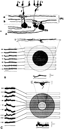
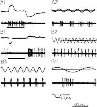
The existence of a large variety of ganglion cell forms has been known for a century, but the corresponding functional categories have emerged more recently. Hartline's pioneering work99 established the ON, OFF, and ON-OFF discharge patterns exhibited by single ganglion cell axons. Kuffler100 showed that many ganglion cells of the cat retina had the same mutually antagonistic, center-surround receptive fields that were described above for bipolar cells. He found two varieties of receptive field in about equal numbers: ON-center and OFF-center. Thus, in relation to the prior studies of Hartline, Kuffler showed that an individual ganglion cell can fire action potentials during the light and/or after the stimulus light is extinguished. The deciding factors are the distribution of the light over the retinal surface and the relative intensities of central versus peripheral illumination. Subsequent work integrated Kuffler's findings into the division of the IPL into ON and OFF zones.101 As illustrated in Figure 17, the dendrites of ON-center ganglion cells arborize in the proximal IPL, whereas those of OFF-center ganglion cells are found in the distal IPL. Naka102 showed, in a lower vertebrate retina, that current passed into ON bipolar cells drove ON ganglion cells; current injected into OFF bipolars drove OFF ganglion cells (Fig. 18). Collectively these findings indicate that the basic center-surround organization of a ganglion cell is established in the outer retina and is relayed to ganglion cells by the ON-center and OFF-center bipolars.

Fig. 17. A. Organization of cone bipolar cells and ganglion cells in the inner plexiform layer (IPL) of the cat retina. Flat cone bipolar cells (f) have axon terminals that end in sublamina a and contact the dendrites of Ga-type ganglion cells. Invaginating cone bipolar cells (i) have axon terminals that ramify at a more proximal level of the IPL in sublamina b, where they contact Gb-type ganglion cells. Ga cells are off-center and Gb cells are on-center ganglion cells. c, cones. B. Receptive field properties and morphology of an intracellularly stained off-center ganglion cell. Note the dendritic arborization in sublamina a. At left are shown responses to slits of light positioned at different levels of the receptive field. They show that a hyperpolarization (inhibitory postsynaptic potential) is elicited when the slit is positioned between 210 μm and -70 μm (shaded circle). The dotted circle gives the dendritic field of the cell as 490 μm. The response to a diffuse stimulus is shown at bottom. (Stimulus width, 50 μm; duration, 542 msec; wavelength, 441 nm; intensity, 2.7 log quanta μm-2 sec-1). c, cone. C. Concentrically organized on-center ganglion cell of the rat retina. The dendritic arbor of this cell is confined to sublamina b of the IPL. The membrane depolarization (excitatory postsynaptic potential) is elicited by light stimuli falling between 0 and 210 μm (open circle). The dotted circle indicates the subtense of the cell's dendritic arbor. The shaded circle indicates the spatial extent of the inhibitory surround. (Stimulus width, 100 μm; duration, 560 msec; wavelength, 647 nm; intensity, 4.6 log quanta μm-2 sec-1) (Nelson R, Famiglietti EV Jr, Kolb H: Intracellular staining reveals different levels of stratification for on- and off-center ganglion cells in cat retina. J Neurophysiol 41:472, 1978)

Fig. 18. Catfish retina. Activation of ganglion cells through light or an injection of extrinsic current into single bipolar cells. A1. On-center bipolar cell response (upper trace) and on-center ganglion cell response (lower trace) to a light stimulus. A2. Sinusoidal current passed through the bipolar cell membrane (upper trace) evokes ganglion cell spikes (lower trace) during the depolarizing phase. B1 through B4. Comparable sets of records for off-center bipolar cell and ganglion cell responses. (Naka K-I: Functional organization of catfish retina. J Neurophysiol 40:26, 1977)

A different sort of functional categorization began to emerge in 1966 with a study by Enroth-Cugell and Robson.103 They found that cat retinal ganglion cells could be classified into two groups, which they called X and Y. X cells responded in proportion to the total amount of light falling in the receptive field center, irrespective of its spatial distribution. Y cells, on the other hand, were exquisitely sensitive to the local light pattern. Other properties are associated with these two categories: X cells have a sustained discharge (i.e., spike discharges continue as long as the light is present), whereas the light-evoked response of Y cells is more transient. Stone and Hoffman104 added a third category, W cells, which have a slow conduction velocity, a sluggish discharge, and receptive fields that often lack a center-surround organization. Boycott and Wässle105 performed correlative morphologic studies on cat retinal ganglion cells, their work revealing three sizes of ganglion cell dendritic arbors: α, β, and γ, which they suggested correspond, respectively, to Y, X, and W ganglion cells.

The description of ganglion cell behavior would be incomplete without mentioning that many ganglion cells have complex trigger features,106 responding preferentially to stimulus color or to moving stimuli. The next two sections provide a brief overview of the organization of retinal circuits involved in color vision and in directionally selective ganglion cells.

Back to Top
RETINAL CIRCUITS UNDERLYING COLOR VISION

PHOTORECEPTOR CLASSES IN THE PRIMATE RETINA

In the primate retina, color vision is of primary importance to the animal's behavior. The trichromatic nature of primate color vision is based, in the first instance, on the three classes of cone photoreceptors described above, each containing a different visual pigment.107 Photocurrent recordings108 and microspectrophotometric studies109 revealed their absorbance maxima to lay near 445, 535, and 575 nm. The S cones constitute only about 7% of the total, with the remaining 93% divided approximately equally between M and L cones.110 S cones are excluded from the center of the fovea but appear on the foveal slopes. There is apparently no strict geometric mosaic in the arrangement of cones,111 but specificity is conferred in the patterns of connectivity between cones and particular subtypes of horizontal and bipolar cells, as described below. Marc and Sperling112,113 took advantage of the different spectral peaks of cone pigments to uncover the spatial distribution of the cone classes in the primate retina, using a histochemical method that stains the inner segment of photoreceptors. Figure 19 illustrates a baboon retina in flat mount that was exposed to a bright blue light while incubated with the nitroblue-tetrazolium stain. The blue-sensitive cone receptors are revealed. Suitable adjustment of the wavelength band of the stimulating light permitted identification of M- and L-cone distribution.

Fig. 19. Flat mount of baboon retina after stimulation with blue light of 5.1 × 10⁴ photons sec-1 μm-2 and incubation with nitroblue tetrazolium. Dark-staining cones are the blue receptors. (Marc RE, Sperling H: Chromatic organization of primate cones. Science 196:454, 1977)

CONE CONTACTS OF HORIZONTAL CELLS IN THE PRIMATE RETINA

In some lower vertebrates with highly developed color vision, horizontal cells are of two functional kinds. In one, light of any wavelength hyperpolarizes the cell (luminosity type); in the other, some wavelengths hyperpolarize and others depolarize the horizontal cell (chromaticity type).114 The potential relevance of chromaticity cells for color vision was tantalizing, and given that color vision is highly pronounced in primates, it was expected that chromaticity horizontal cells would be found. This turned out not to be the case, however. All the horizontal cells tested in primates are only hyperpolarized by lights of any spectral composition. Dacey and coworkers115 found evidence for only two functional classes: the HI and HIII anatomical types were grouped by them into one functional group characterized by input from L and M cones but very sparse input from S cones. The other type, corresponding to the HII anatomical category, receives a strong input from S cones but also processes inputs from L and M cones.

COLOR CODING BY GANGLION CELLS

Midget ganglion cells and ganglion cells receiving input from bipolars contacting blue cones often are color-coded.116 That is, cone specificity is superimposed on the center-surround organization, giving rise to a few color types of center-surround receptive fields. These are red-center, green surround, or the reverse, and blue-center, yellow surround, with the yellow reflecting summation of signals originating in L and M cones. Each of the red-green center-surround receptive fields is encountered in two forms, ON-center and OFF-center, and with either red or green input to the center.117 Thus, we end up with four subtypes: L+ /M-, L-/M+ , M+ /L-, and M-/L+ , with the first letter indicating the cone class providing input to the center of the field, the second to the surround, and the + /- sign indicating ON and OFF activity, respectively.

In their spatial summation properties, these color-coded midget ganglion cells resemble the X cells of the cat retina. In the primate, however, these color-coded cells are called P cells, reflecting the finding that they project to the parvocellular layers of the dorsal lateral geniculate nucleus.97 Parenthetically, the non-color-coded, transient ganglion cells of the primate are like the Y ganglion cells of the cat and are called M cells in primate because they project to the magnocellular layers of the dorsal lateral geniculate nucleus.

The blue-center/yellow surround type of color-coded ganglion cell differs somewhat from the red/green subtype in that blue input is always ON. The blue/yellow receptive field apparently is organized through bipolar cells.118 The S-cone-contacting bipolar cell forms the center of the receptive field, whereas diffuse bipolars summing L- and M-cone signals provide the input to the receptive field surround. The circuit underlying red/green opponent fields is unknown at present.

Back to Top
DIRECTIONALLY SELECTIVE GANGLION CELLS
One of the most intriguing properties of ganglion cells is their ability to respond to moving targets only in one direction (the preferred direction) while remaining silent when the target is moved in the opposite (null) direction.119 The circuitry underlying this behavior has been elucidated with pharmacologic and electrophysiologic tools and shown to involve interactions between glutamatergic bipolar cells and three amacrines, one cholinergic and the other two GABAergic.

Cholinergic amacrines have been well characterized.120 They comprise two groups of so-called star-burst amacrines: the cell bodies of the orthotopic variety lie at the border of the INL/IPL and all of their dendrites arborize in a thin stratum corresponding to sublamina 2 of the IPL. Their mirror image partners, called displaced, have cell bodies in the ganglion cell layer and processes in sublamina 4 of the IPL. In other words, one group is in the OFF and the other in the ON sublamina of the IPL. The precise subtype of GABAergic amacrine participating in this circuit has not yet been identified, but the functional roles of GABA are being worked out.121 One GABAergic neuron inhibits the cholinergic amacrine, whereas the other provides a direct inhibitory input to the directionally selective ganglion cell.

The essence of the circuit is that in the excitatory direction, a combination of glutamatergic and cholinergic signals arrives at the ganglion cell in advance of a direct GABA-mediated inhibition, thus providing excitation. The GABA input to the cholinergic cell appears to be important in shaping the excitatory input so as to process information emanating from slow- or fast-moving stimuli. But in the null direction, the GABA signal reaches the directionally selective ganglion cell ahead of the excitatory input and so cancels it out.

Back to Top
PHARMACOLOGY AND ELECTROPHYSIOLOGY OF GANGLION CELLS
The basic response of a ganglion cell to light is a change in membrane potential, either depolarizing or hyperpolarizing, called an excitatory postsynaptic potential in the former case and an inhibitory postsynaptic potential in the latter. Excitatory postsynaptic potentials evoke superimposed action potentials, whereas inhibitory postsynaptic potentials inhibit spike firing. The kinetics of the postsynaptic potentials plays a large role in determining the pattern of spike firing, which may be relatively sustained or relatively transient.65,100,103 Broadly speaking, three factors interact in this process: the intrinsic voltage-gated channels in the ganglion cell membrane, the pattern of excitatory (glutamatergic) and inhibitory (GABAergic and/or glycinergic) inputs the ganglion cell receives, and the kinds of ligand-gated channels the ganglion cell possesses, which dictates how it responds to glutamate, GABA, and glycine. Much of the relevant work on this topic has been carried out on amphibian retinas, but there is enough information about mammalian ganglion cells to suggest that generalizations are valid. The most important intrinsic voltage-gated channels in this context are the potassium (K) channels122,123 because they determine the rate at which the membrane repolarizes after spike initiation and so they place limits on the spike firing frequency. In relation to synaptic inputs, anatomical and electrophysiologic studies indicate that P-type ganglion cells have a relatively high proportion of synaptic inputs from bipolar cells compared with direct inputs from amacrine cells, whereas for M-type ganglion cells the relative inputs are reversed.124 A recent physiologic study125 suggests that ON bipolar cells themselves are separable into relatively transient and relatively sustained subtypes, which will of course affect the time course of glutamate release by their terminals.

With regard to the postsynaptic glutamate receptors, many ganglion cells have a mixture of AMPA and NMDA channels.126 The former tend to desensitize rapidly, and their dominance will favor a transient spike firing pattern. NMDA channels have several special properties. They do not open at relatively hyperpolarized potentials because a Mg ionblocks the channel. Thus, the initial depolarization elicited by glutamate depends on current flow through AMPA channels. When the NMDA channels open, after an initial AMPA receptor-mediated depolarization, their kinetics are much slower than those of AMPA channels, leading to more sustained spike firing. NMDA channels also permit Ca influx, which in turn activates a number of intracellular biochemical cascades affecting the behavior of the NMDA channels themselves, as well as GABA-activated channels. The inhibitory inputs to the ganglion cell, which are mediated primarily by GABA and glycine, also tend to reduce the depolarization induced by glutamate, as already mentioned.

THE ROD PATHWAY

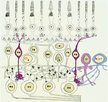
Rod signals are conveyed to the dendrites of rod bipolar cells7 and the axon terminals of horizontal cells.60 Additionally, the rod signal can reach cones through rod-cone gap junctions.127 The rod bipolar cell axon terminal does not contact ganglion cells directly; instead, it signals to two types of ama-crine cell, termed AII128 and A17129 in the namingscheme of Kolb (Fig. 20). The A17 amacrine is awide-field amacrine, with a dendritic arbor thatstretches 1 mm across the retina, in this way contact-ing more than 1,000 rod bipolar cells. Its output isdirected back onto the rod bipolar terminals. Thefunctional significance of this is not yet worked out in detail, but the A17 amacrine cell is one of the many GABAergic amacrines of the retina, and so its effect on the rod bipolar terminal is likely to be inhibitory. Because that inhibition is delayed, it will tend to shape the rod bipolar cell response, possibly making it more transient and/or smaller.

Fig. 20. The rod pathway of the mammalian retina. Rod bipolar cells (RB) contact multiple rod photoreceptors (R) in the outer retina. Their axon terminals end in the ON zone of the inner plexiform layer in close proximity to diffuse ganglion cells (DG) but without making a direct contact. Instead, the rod bipolar cells contact two types of amacrine cell, the AII and the A17. (Drawing courtesy of Dr. Helga Kolb)

The AII amacrine has an arbor that is restricted in its lateral extent but is bistratified, with terminals in both distal and proximal IPL.130 The proximal terminals receive the rod bipolar input, whereas the distal terminals are output sites to ganglion cells.110 The AII amacrine cell is glycinergic, and so its effect on ganglion cells also is expected to be inhibitory. In addition, the AII amacrine contacts ON bipolar cells through gap junctions. Because the light-evoked response of the AII amacrine is depolarizing, the current flowing through its gap junctions is probably excitatory to ON bipolar cells and thus to ON-center ganglion cells. In sum, therefore, the rod-to-rod bipolar-to-AII amacrine-to-ganglion cell path is excitatory for the ON system and inhibitory for the OFF system. The AII amacrine also receives input from the dopaminergic neurons,131,132 whose neuromodulatory actions are described below.

Back to Top
RETINAL NEUROMODULATORY SYSTEMS
A neuromodulator, as defined by Kaczmarek and Levitan,133 is a neuroactive substance that works through the intracellular biochemistry of the neuron to effect a change in neuronal function. The most commonly encountered biochemical pathway for neuromodulation is through a G protein that is functionally linked to a metabotropic receptor. Activation of a metabotropic receptor by its agonist results in activation of the G protein, but from this point the pathways are extremely diverse and may involve either up- or downregulation of other proteins. The biochemical pathway may involve a diffusible second messenger (e.g., cAMP, cGMP, or Ca) or it may be membrane delimited. In the latter case the G protein itself alters the function of a membrane-bound protein (e.g., a K channel).

Metabotropic receptors in the retina are extraordinarily diverse. The main excitatory transmitter, glutamate, has eight families of mGluRs,49,134 and the main inhibitory transmitter, GABA, has one, the GABA-B receptor. The muscarinic receptor of acetylcholine is metabotropic, and of the five classes of serotonin receptors, all but one are metabotropic. Dopamine, discussed in more detail below, works through two main classes of metabotropic receptor, D1 and D2. As far as we know, all the peptides in the retina work through metabotropic receptors and they constitute a very large population, numbering more than 30 different peptides, each probably working through multiple receptors. Common metabolites such as ATP and adenosine also activate membrane receptors, of which most are metabotropic.

When considered from the standpoint of the recipient neuron, it can be taken for granted that every retinal neuron has multiple metabotropic receptors, in addition to the ionotropic receptors it possesses, from which it follows that each neuron undergoes continuous fine-tuning, with each of its voltage-sensitive, ligand-gated, and gap junctional channels subject to modulation. The presynaptic process of neurotransmitter release also is sculpted by neuromodulatory influences.135

DOPAMINE: A MODEL NEUROMODULATOR

Dopaminergic retinal neurons are found throughout the vertebrate series.136 Invariably they are a class of amacrine and/or interplexiform cells, with large cells bodies that are regularly but sparsely distributed throughout the retina, at a density of 10 to 100 cells/mm2. The dendritic arbors of the dopaminergic cell are of two sorts. A network of fine dendrites expands in sublamina 1 of the IPL, where it forms rings that surround the AII amacrine cells. Stouter dendrites descend to more proximal layers of the IPL. In some retinas, ascending processes from the dopaminergic cell reach the outer retina (see Fig. 14).

There are two striking features of the dopaminergic cell system.136 First, most or all retinal neurons, from photoreceptors to ganglion cells, have D1 and/or D2 receptors, but in many cases dopamine reaches these receptors by diffusion rather than through a morphologically defined synapse.137–139 This is an example of volume transmission,3 which was formerly thought to apply principally to blood-borne hormones but now is known to occur throughout the nervous system. In the case of dopamine, the diffusion distances may be tens of microns.138,139 The second organizational principle of the dopaminergic cells is that synthesis and release are closely coupled and highly regulated.140 That is, when more dopamine is required, more is synthesized. This regulation occurs partly by phosphorylation.141 The rate-limiting enzyme for dopaminesynthesis is tyrosine hydroxylase, which has phosphorylation sites at serines 19, 31, and 40, near the N terminal of the molecule. Phosphorylation is under neural control and so is modified according to the state of activity of the neuron.142 Recently it was shown143 that the dopaminergic neuron fires nerve spikes continuously when freed from its synaptic inputs, and increased firing is associated with increased dopamine release. A neurotransmitter such as GABA, which suppresses spike firing, reduces dopamine release.

Another method of control is through upregulation of tyrosine hydroxylase synthesis. Prolonged (more than 1 hour) exposure to light results in activation of the gene responsible for tyrosine hydroxylase production.144 The exact pathway by which this activation occurs is still unknown, but clearly it depends on a linkage between neurochemical events at the cell surface with activation of a path to the cell's nucleus, resulting in the activation of gene expression.

The even distribution of dopaminergic nerve cells and processes throughout the retina means that their activation initiates a global change in retinal function, affecting all parts of the retina more or less equally. Among its many neuromodulatory activities, dopamine increases current flow through AMPA receptors at cone synapses,145 and it modifies the activities of calcium channels in ganglion cells.146 Dopamine also is involved in governing melatonin production147 and thus indirectly in controlling disk shedding by rods.148 Dopamine has even been implicated in eye growth in relation to myopia,149 although the pathway by which this regulation might occur is still quite unclear.

Back to Top
ELECTRICAL SYNAPSES (GAP JUNCTIONS) IN THE RETINA AND THEIR MODULATION BY DOPAMINE
Many classes of retinal neurons are joined by gap junctions.150 They occur between neurons of the same class, such as horizontal cells (Fig. 21), but also occur between neurons of different subtypes, such as between rods and cones, or between AII amacrine cells and cone ON bipolars. The same cell may be coupled to more than one cell type. For example, AII amacrines are coupled to each other as well as to certain bipolar cells.151 Although many different molecular categories of connexins in the nervous system have been identified, including in the retina, we lack information about the molecular identity of connexins in native gap junctions of retinal neurons and glial cells. Nevertheless, there is a substantial body of information concerning the modifiability of gap junctions. In the retina, the best-understood example is the modulation by dopamine of horizontal cell electrical synapses. The details were first worked out in fish152 and turtle retinas,153 but it has been shown subsequently that mammalian gap junctions are under similar control.23 Horizontal cells possess dopamine receptors of the D1 subtype. When activated by dopamine, D1 receptors upregulate adenylate cyclase, increasing the synthesis of cAMP, which in turn permits the activation of protein kinase A.154 It is presumed that protein kinase A phosphorylates some connexin, but that has not been conclusively shown. Nevertheless, the net effect is to reduce gap junctional conductance. Another pathway for modulation of gap junctional conductance is through nitric oxide,155 which activates soluble guanylate cyclase, leading to an increase in [cGMP] and a reduction in gap junctional conductance. Although both pathways lead to reduced current flow through the electrical synapse, they work through different mechanisms and presumably are turned on by different neuroactive substances.

Fig. 21. Neurobiotin coupling among horizontal cells of the rabbit retina. A. Tracer-coupled A-type horizontal cells in the dark-adapted rabbit retina after injection of Neurobiotin into a single cell. The micrograph shows only a portion of the network, which comprised more than 1,000 cells extending 2,350 μm across the axis parallel to the visual streak. B. Tracer-coupled B-type horizontal cells in the dark-adapted rabbit retina. This network included 227 cells extending 400 μm along the axis parallel to the visual streak. The asterisk indicates the point of injection. Scale bars are 50 μm. (Xin D, Bloomfield SA: Dark- and light-induced changes in coupling between horizontal cells in the rabbit retina. J Comp Neurol 383:512, 1998, with permission of the authors and Wiley-Liss)

Rod-cone gap junctions also are modulated by dopamine,156 but in this case it is through the intermediation of a D2 receptor. Activation of this receptor leads to increased rod-cone coupling, with the result that cone signals can flow through rods to modify the light-evoked responses of postsynaptic neurons. The mechanism of action of the D2 receptor has not been proven, but it is plausible tosuppose that it downregulates adenylate cyclase,leading to a reduction of cAMP (i.e., the same mechanism as D1-induced uncoupling, but working in the opposite direction). Yet another agent that induces uncoupling in horizontal cells is retinoic acid,157 a metabolite of the visual cycle that is produced within the pigment epithelium. Light increases the concentration of retinoic acid as a consequence of visual pigment bleaching and the conversion of retinaldehyde to retinoic acid.

Because dopamine is upregulated by bright light, whereas rod-mediated vision is decreased by bright light, one might expect an action of dopamine on coupling of neurons in the rod pathway (e.g., the AII amacrine cell). The experimental finding is that dopamine decreases AII coupling,23 but the relation between light and dopamine requires further work. Similarly, for horizontal cells, weak lights actually increase coupling, whereas strong lights uncouple horizontal cells,22 and it is only this latter action that is mimicked by dopamine. The pharmacology of dopamine's action on AII amacrine cells is the same as for horizontal cells, indicating a common underlying mechanism involving D1 receptors. Interestingly, dopamine does not seem to affect the coupling of the heterologous junctions between AII amacrines and cone bipolar cells.151 It has been suggested that these heterologous junctions permit passage of the neurotransmitter glycine from the AII amacrine cell, which synthesizes it, to the cone bipolar cell, which accumulates it, for an as yet undetermined function.

These several studies show that gap junctions in the retina are diverse and are under control by multiple agents, resulting in plasticity of coupling among multiple classes of retinal neurons. Future work will uncover the significance of a variable degree of coupling for information processing by the retina.

Back to Top
RETINAL GLIAL CELLS
The principal glial cell of the retina is the Muller cell. It extends vertically across the retina from the vitreal surface to about the level of photoreceptor nuclei (see Fig. 1). There the terminals of adjacent Muller cells abut, giving the impression, in a light microscopic view, that a membrane (outer limiting membrane, Fig. 1, left side) is formed. This apparent structure, however, is not a true membrane.

Muller cells have many functional roles, but two in particular have been well documented. The first of these is as a pathway for the regulation of extracellular potassium158 (Fig. 22). When neurons are depolarized they release K+ into the extracellular space. Neurons have their own Na/K exchangers to restore ion balances but are aided in this effort by Muller cells, which are highly permeable to K+ , particularly in the inner portion of the cell. The result is a circuit whereby K+ from the extracellular space enters the Muller cell and is moved into the vitreous body.

Fig. 22. Frog retina. A simplified diagram illustrating the postulated role of the Muller fiber in the generation of the ERG b wave. Light evokes an increase in the extracellular potassium concentration in both plexiform layers. Current flows into the Muller fiber at these regions and exits mainly at the Muller fiber vitreal end foot. The sink/source profile of the Muller fiber current plays a major role in the generation of the b wave. (Newman EA: Current-source density analysis of the b-wave of frog retina. J Neurophysiol 43:1355, 1980)

A second role for the Muller cell is in the removal of glutamate after its release at photoreceptor and bipolar cell synapses.159 The Muller glial cell has a glutamate transporter that is coupled to the sodium gradient. Once inside the Muller cell, glutamate is converted to glutamine by the glial cell-specific enzyme, glutamine synthetase.160 Parenthetically, this reaction removes one molecule of ammonia, a toxic substance. Glutamine is released and then enters neurons via another transporter, making it available anew as a substrate for glutamate synthesis.

Muller cells are far from indifferent to the various neurochemicals present in the extracellular space. On the contrary, it has been shown that they respond to glutamate161 and growth factors.162 The significance of this sensitivity to neuroactive substances is unknown, although one may speculate that it keeps the Muller cell informed about local neuronal activity and perhaps primes it for participation in K+ and glutamate clearance. Astroglia are present in the optic nerve.163 One of the unusual features of astroglia is that they exhibit calcium waves, which can be stimulated by mechanical activity. Activation of such waves has been shown to inhibit the activity of nearby retinal ganglion cells,164 an action postulated to depend on stimulation of inhibitory interneurons by glutamate released from glia. Like the Muller cells, astroglia are coupled by gap junctions to each other and also to Muller cells. The heterologous coupling, however, appears to work only in the direction of the Muller cell to astroglia. In any event, these two glial cell classes communicate and thus can interact in regulating neuronal function.

Back to Top
OVERVIEW
This essay has attempted to provide a review of retinal neurons, their connections, and their functional properties in relation to information processing by the retina. To return to Cajal's essential perception: the retina is a piece of the central nervous system organized into microcircuits, which he deciphered based solely on anatomical criteria. In fact, given the absence of functional data, Cajal had great difficulties with horizontal and amacrine cells, whose symmetric processes did not provide a clear indication as to the direction of signal propagation. From a modern perspective we can conclude that the microcircuits of the retina reduce the visual information to a series of abstractions termed contrast, color, and movement. The response kinetics of different retinal neurons revealed by electrophysiologic tests provides an understanding of which microcircuit a particular retinal cell fits into.

It is also important to realize that the retina is a tonic machine. Both rod and cone photoreceptors release their transmitter glutamate at a high rate in darkness, thereby activating retinal circuits, leading to tonic spike firing in darkness by many ganglion cells. The overall rate of retinal activity is subject to circadian control through a circadian pacemaker possibly located in the photoreceptor cells165 and is further modified by a changing ambient light level reflecting the passage from day to night. We can now appreciate that many neuroactive substances in the retina are influenced by these tonic changes, including melatonin and dopamine,166 to name only the best studied of these. Many activities of the retina appear to be rhythmic on an 24-hour schedule, including disk shedding by rods and the sensitivity of the photoreceptors. The circuits that respond to the more rapid light changes that underlie pattern vision, color perception, and motion detection function against a background of the slower changes that set the tone of the system through neuromodulatory pathways that influence the state of the hard-wired circuits. Understanding the molecular biology and function of the neuromodulatory pathways is the major challenge for future years. For example, it will begin to tell us how the many intrinsic peptides of the retina influence its behavior, a subject about which we understand almost nothing at present. Molecular biology studies also will continue to indicate the basis of many inherited and acquired diseases, such as retinitis pigmentosa, perhaps leading to new diagnostic and therapeutic tests. This newly acquired information, however, could only have been interpreted on the basis of the thorough understanding of retinal neurons, their circuits, and the biochemical and ionic bases of their light-evoked responses. That collected knowledge is an intellectual framework slowly and carefully erected from more than a century of anatomical, electrophysiologic, and pharmacologic studis.

Back to Top
ACKNOWLEDGMENTS
This study was supported by NIH grant EY 03570 and by the Helen Hoffritz Foundation, New York. I thank Drs. S. Bloomfield, S. Haverkamp, D. Krizaj, R. Masland, P. Sterling, and their publishers for permitting the reproduction of figures from their published work. Dr. Helga Kolb is owed special thanks for allowing me to use five unpublished figures.
Back to Top
REFERENCES

1. Ramón y Cajal S: The Structure of the Retina. Springfield, IL, Charles C. Thomas, 1972

2. Levitan IB: Modulation of ion channels by phosphorylation. How the brain works. Advances in Second Messenger and Phosphoprotein Research 33:3, 1999

3. Fuxe K, Agnati LF, eds: Volume Transmission in the Brain. Advances in Neuroscience, vol 1. New York, Raven Press, 1991

4. Polyak S: The Retina. Chicago, University of Chicago Press, 1941

5. Curcio CA, Sloan KR, Packer O et al: Distribution of cones in human and monkey retina: individual variability and radial asymmetry. Science 236:579, 1987

6. Adams CK, Perez JM, Hawthorne MN: Rod and cone densities in the rhesus monkey. Invest Ophthalmol Vis Sci 13:885, 1974

7. Kolb H, Linberg KA, Fisher SK: Neurons of the human retina: a golgi study. J Comp Neurol 318:147, 1992

8. Dacey DM: The mosaic of midget ganglion cells in the human retina. J Neurosci 13:5334, 1993

9. Perry VH, Oehler R, Cowey A: Retinal ganglion cells that project to the dorsal lateral geniculate nucleus in the macaque monkey. Neuroscience 12:1101, 1984

10. Tauchi M, Masland RH: The shape and arrangement of the cholinergic neurons in the rabbit retina. Proc R Soc Lond B223:101, 1984

11. Crook JM, Lange-Malecki B, Lee BB, Valberg A: Visual resolution of macaque retinal ganglion cells. J Physiol Lond 396:205, 1988

12. Pirenne MH: Visual acuity. In: Davson H, ed: The Eye, vol. 2, The Visual Process, pp 175–195. New York, Academic Press, 1962

13. Goodenough DA: Bulk isolation of mouse hepatocyte gap junctions. J Cell Biol 61:557, 1974

14. Revel JP, Karnovsky MJ: Hexagonal array of subunits in intercellular junctions of the mouse heart and liver. J Cell Biol 33:C7, 1967

15. Witkovsky P, Shakib M, Ripps H: Interreceptoral junctions in the teleost retina. Invest Ophthalmol Vis Sci 13:996, 1974

16. Witkovsky P, Owen WG, Woodwoth M: Gap junctions among the perikarya, dendrites, and axon terminals of the luminosity-type horizontal cell of the turtle retina. J Comp Neurol 216:359, 1984

17. Bruzzone R, Ressot C: Connexins, gap junctions and cell-cell signalling in the nervous system. Eur J Neurosci 9:1, 1997

18. Dermietzel R, Kremer M, Paputsoglu G et al: Molecular and functional diversity of neural connexins in the retina. J Neurosci 20:8331, 2000

19. Guldenagel M, Sohl G, Plum A et al: Expression patterns of connexin genes in mouse retina. J Comp Neurol 425:193, 2000

20. Verselis VK, Ginter CS, Bargiello TA: Opposite voltage gating polarities of two closely related connexins. Nature 368, 1994

21. Bennett MVL: Gap junctions as electrical synapses. J Neurocytol 26:349, 1997

22. Xin D, Bloomfield SA: Dark- and light-induced changes in coupling between horizontal cells in the rabbit retina.J Comp Neurol 383:512, 1998

23. Hampson ECGM, Vaney DI, Weiler R: Dopaminergic modulation of gap junction permeability between amacrine cells in mammalian retina. J Neurosci 12:4911, 1992

24. Baylor DA, Fuortes MGF: Electrical responses of single cones in the retina of the turtle. J Physiol 207:77, 1970

25. Mastronarde DN: Correlated firing of retinal ganglion cells. Trends Neurosci 12:75, 1989

26. Zucker RS: Exocytosis: a molecular and physiological perspective. Neuron 17:1049, 1996

27. Augustine GJ, Betz H, Bommert K et al: Molecular pathways for presynaptic calcium signaling. In: Stjarne L, Greengard P, Grillner S et al: Molecular and Cellular Mechanisms of Neurotransmitter Release. New York, Raven Press, 1994

28. Ziff EB: Enlightening the postsynaptic density. Neuron 19:1163, 1997

29. O'Brien RJ, Kamboj S, Ehlers MD et al: Activity-dependent modulation of synaptic AMPA receptor accumulation. Neuron 21:1067, 1998

30. Neher E: Vesicle pools and Ca2+ microdomains: new tools for understanding their roles in neurotransmitter release.Neuron 20:389, 1998

31. Llinas R, Sugimori M, Silver RB: The concept of calcium concentration microdomains in synaptic transmission. Neuropharmacology 34:1443, 1995

32. Golovina VA, Blaustein MP: Spatially and functionally distinct Ca2+ stores in sarcoplasmic and endoplasmic reticulum. Science 275:1643, 1998

33. Rao-Mirotznik R, Harkins A, Buchsbaum G, Sterling P: Mammalian rod terminal: architecture of a binary synapse. Neuron 14:561, 1995

34. Chun MH, Grunert U, Martin PR, Wässle H: The synaptic complex of cones in the fovea and in the periphery of the macaque monkey retina. Vision Res 36:3383, 1996

35. Marc RE, Liu W-LS, Kallionatis M et al: Patterns of glutamate immunoreactivity in the goldfish retina. J Neurosci 10:4006–4034, 1990

36. Ayoub GS, Copenhagen DR: Application of a fluorometric method to measure glutamate release from single retinal photoreceptors. J Neurosci Meth 37:7, 1991

37. Copenhagen DR, Jahr CE: Release of endogenous excitatory amino acids from turtle photoreceptors. Nature 342:536, 1989

38. Lasater EM, Dowling JE: Carp horizontal cells in culture respond selectively to L-glutamate and its agonists. Proc Natl Acad Sci USA 79:936, 1982

39. Vardi N, Morigiwa K, Wang T-L et al: Neurochemistryof the mammalian cone/synaptic complex. Vision Res 38:1359, 1998

40. Morigiwa K, Vardi N: Differential expression of ionotropic glutamate receptor subunits in the outer retina. J Comp Neurol 405:173, 1999

41. Vardi N, Morigiwa K: ON cone bipolar cells in rat express the metabotropic receptor mGluR6. Vis Neurosci 14:789, 1997

42. Brandstätter JH, Koulen P, Wässle H: Diversity of glutamate receptors in the glutamate retina. Vision Res 38:1385, 1997

43. Schmitz Y, Witkovsky P: Dependence of photoreceptor glutamate release on a dihydropyridine-sensitive calcium channel. Neuroscience 48:1209, 1997

44. Schmitz Y, Witkovsky P: Glutamate release by the intact light-responsive potoreceptor layer of the Xenopus retina. J Neurosci Meth 68:55, 1996

45. Wilkinson MF, Barnes S: The dihydropyridine-sensitive calcium channel subtype in cone photoreceptors. J Gen Physiol 107:621, 1996

46. Taylor WR, Morgans C: Localization and properties of voltage-gated calcium channels in cone photoreceptors of Tupaia belangeri. Vis Neurosci 15:541, 1998

47. Hollman M, Heinemann S: Cloned glutamate receptors. Ann Rev Neurosci 17:31, 1994

48. Rosenmund C, Stern-Bach Y, Stevens CF: The tetrameric structure of a glutamate receptor channel. Science 280:1596, 1998

49. Pin J-P, Duvoisin R: The metabotropic glutamate receptors: structure and functions. Neuropharmacology 34:1, 1995

50. Qin P, Pourcho RG: Distribution of AMPA-selective glutamate receptor subunits in cat retina. Brain Res 710:303, 1996

51. Nomura A, Shigeomoto R, Nakamura Y et al: Developmentally regulated postsynaptic localiztion of a metabotropic glutamate receptor in rat rod bipolar cells. Cell 77:361, 1994

52. Vardi N, Duvoisin R, Wu G, Sterling P: Localization of mGluR6 to dendrites of ON bipolar cells in primate retina. J Comp Neurol 423:402, 2000

53. Haverkamp S, Grunert U, Wässle H: The cone pedicle, a complex synapse in the retina. Neuron 27:85, 2000

54. Haverkamp S, Wässle H: Immunocytochemical analysis of the mouse retina. J Comp Neurol 424:1, 2000

55. Braithwaite SP, Meyer G, Henley JM: Interactions between AMPA receptors and intracellular proteins. Neuropharmacology 39:919, 2000

56. Rauen T, Rothstein JD, Wässle H: Differential expression of three glutamate transporter subtypes in the rat retina. Cell Tiss Res 286:325, 1996

57. Kolb H, Boycott BB, Dowling JE: A second type of midget bipolar cell in the primate retina. Philos Trans R Soc Lond Biol 255, 1969

58. Kouyama N, Marshak DW: Bipolar cells specific for blue cones in the macaque retina. J Neurosci 12:1233, 1992

59. Jacoby RA, Wiecmann AF, Amara SG et al: Diffuse bipolar cells provide input to OFF parasol ganglion cells in the macaque retina. J Comp Neurol 416:6, 2000

60. Peichl L, Sandmann D, Boycott BB: Comparative anatomy and function of mammalian horizontal cells. In Chalpua & Finlay, eds: Development and Organization of the Retina, pp 147–172. New York, Plenum Press, 1998

61. Peichl L, Gonzales-Soriano J: Morphological types of horizontal cell in rodent retinae: A comparison of rat, mouse, gerbil and guinea pig. Vis Neurosci 11:501, 1994

62. Boycott BB, Peichl L, Wassle H: Morphological types of horizontal cell in the retina of the domestic cat. Proc Roy Soc B 203:229, 1978

63. Steinberg RH: Rod and cone contributions to S-potentials from the cat retina. Vision Res 9:1319, 1969

64. Raviola E, Gilula NB: Gap junction between photoreceptor cells in the vertebrate retina. Proc Natl Acad Sci USA 70:1677, 1973

65. Werblin FS, Dowling JE: Organization of the retina of the mudpuppy Necturus maculosus: II. Intracellular recording. J Neurophysiol 32:339, 1969

66. Nelson R, Kolb H: Synaptic patterns and response properties of bipolar and ganglion cells in the cat retina. Vision Res. 23: 1183–1195, 1983

67. Dacheux RF, Raviola E: The rod pathway in the rabbit retina: a depolarizing bipolar and amacrine cell. J Neurosci 6:331, 1986

68. Dacey D, Packer OS, Diller L et al: Center surround receptive field structure of cone bipolar cells in primate retina. Vision Res 40:1801, 2000

69. Nawy S, Jahr CE: Suppression by glutamate of cGMP-activated conductance in retina bipolar cells. Nature 346:269, 1991

70. Marc RE, Stell WK, Bok D, Lam DM-K: GABAergic pathways in the goldfish retina. J Comp Neurol 182:221, 1978

71. Baylor DA, Fuortes MGF, O'Bryan PM: Receptive fields of cones in the retina of the turtle. J Physiol Lond 214:265, 1971

72. Schiller PH, Sandell JH, Maunsell JHR: Functions of the ON and OFF channels of the visual system. Nture 322:824, 1986

73. Slaughter MM, Miller RF: 2-Amino-4-phosphonobutyric acid: A new pharmacological tool for retina research. Science 211:182, 1981

74. Kolb H: Organization of the outer plexiform layer of the primate retina: Electron microscopy of Golgi-impregnated cells. Philos Trans R Soc 258:261, 1970

75. Famiglietti EV Jr, Kolb H: Structural basis for on- andoff-center responses in retinal ganglion cells. Science 194:193, 1976

76. Euler T, Wässle H: Immunocytochemical identification of cone bipolar cells in the rat retina. J Comp Neurol 361:461, 1995

77. Kolb H: Amacrine cells of the mammalian retina: neurocircuitry and functional roles. Eye 11:904, 1997

78. Jeon C-J, Strettoi E, Masland RH: The major cell populations of the mouse retina. J Neurosci 18:8936, 1998

79. MacNeil MA, Heussy JK, Dacheux RF et al: The shapes and numbers of amacrine cells: matching of photofilled with Golgi-stained cells in the rabbit retina and comparison with other mammalian species. J Comp Neurol 413:305, 1999

80. Dacey DM: Axon-bearing amacrine cells of the macaque monkey retina. J Comp Neurol 284:275, 1989

81. Haverkamp S, Wässle H: Immunocytochemical analysis of the mouse retina. J Comp Neurol 424:1, 2000

82. Wässle H, Boycott BB: Functional architecture of the mammalian retina. Physiol Rev 71:447, 1991

83. Ehinger B: Neurotransmitter systems in the retina. Retina 2:305, 1982

84. Ammermuller J, Kolb H: The organization of the turtle inner retina. I. ON- and OFF-center pathways. J Comp Neurol 358:1, 1995

85. Gallego A: Horizontal and amacrine cells in the mammal's retina. Vision Res (Suppl) 3:33, 1971

86. Dowling JE, Ehinger B: Synaptic organization of the amine-containing interplexiform cells of the goldfish and Cebus monkey retina. Science 188:270, 1975

87. Versaux-Botter C, Ngyuen-Legros J, Vigny A, Raoux N: Morphology, density and distribution of tyrosinehydroxylase-like immunoreacive cells in the retina of mice. Brain Res 301:192, 1984

88. Nakamura Y, McGuire BA, Sterling P: Interplexiform cell in cat retina: Identification by uptake of γ-[3H]aminobutyric acid and serial reconstruction. Proc Natl Acad Sci USA 77:658, 1980

89. Kolb H, Cuenca N, Wang HH, DeKorver L: The synaptic organization of the dopaminergic amacrine cell in the cat retina. J Neurocytol 19:343, 1990

90. Dowling JE, Boycott BB: Organization of the primate retina: Electron microscopy. Proc R Soc Lond Biol 166:80, 1966

91. Kolb H, DeKorver L: Midget ganglion cells of the parafovea of the human retina: a study by electron microscopy and serial section reconstructions. J Comp Neurol 303:617, 1991

92. Allen RA: The retinal bipolar cells and their synapses in the inner plexiform layer. In Straatsma BR, Hall MO, Allen RA, Crescitelli F, eds: The Retina: Morphology, Functional and Clinical Characteristics, vol. 3, pp 101–143. Los Angeles, University of California Press, 1969

93. Yazulla S: GABAergic mechanisms in the retina. Prog Retina Res 5:1, 1986

94. Poucho R, Goebel DJ: A combined Golgi and autoradiographic study of (3H)-glycine-accumulating amacrine cells in the cat retina. J Comp Neurol 233:473, 1985

95. Kaneko A, Tachibana M: GABA mediates the negative feedback from amacrine to bipolar cells. Neurosci Res (Suppl) 6:S239, 1987

96. Tauck DL, Frosch MP, Lipton SA: Characterization of GABA- and glycine-induced currents of solitary rodent retinal ganglion cells in culture. Neuroscience 27:193, 1988

97. Merigan WH, Maunsell JHR: How parallel are the primate visual pathways? Annu Rev Neurosci 16:369, 1993

98. Moore RY, Speh JC, Card JP: The retino-hypothalamic tract originates from a distinct subset of retinal ganglion cells. J Comp Neurol 352:351, 1995

99. Hartline HK: The response of single optic nerve fibers of the vertebrate eye to illumination of the retina. Am J Physiol 121:400, 1938

100. Kuffler SW: Discharge patterns and functional organization of mammalian retina. J Neurophysiol 16:37, 1953

101. Nelson R, Famiglietti EV Jr, Kolb H: Intracellular staining reveals different levels of stratification for on- and off-center ganglion cells in cat retina. J Neurophysiol 41:472, 1978

102. Naka K: Functional organization of catfish retina. J Neurophysiol 40:26, 1977

103. Enroth-Cugell C, Robson JG: The contrast sensitivity of retinal ganglion cells of the cat. J Physiol 187:517, 1966

104. Stone J, Hoffman K-P: Very slow conduction ganglion cells in the cat's retina: A major new functional type? Brain Res 43:610, 1972

105. Boycott BB, Wässle H: The morphological types of ganglion cells of the domestic cat's retina. J Physiol Lond 240:397, 1974

106. Levick WR: Receptive fields of retinal ganglion cells. In Fuortes MGF, ed: Handbook of Sensory Physiology, vol Vll/2, pp 531–566. Berlin, Springer-Verlag, 1972

107. Nathans J, Thomas D, Hogness DS: Molecular genetics of human color vision: the genes encoding blue, green and red pigments. Science 232:193, 1986

108. Baylor DA, Nunn BJ, Schnapf JL: Spectral sensitivity of cones of the monkey Macaca fascicularis. J Physiol Lond 390:145, 1987

109. Bowmaker JK, Dartnall HJA, Mollon JD: Microscopectrophotometric demonstration of four classes of photoreceptors in an old world primate, Macaca fascicularis. J Physiol Lond 298:131, 1980

110. Cicerone CM, Nerger JL: The relative numbers of long-wavelength-sensitive to middle-wavelength-sensitive cones in the human fovea centralis. Vision Res 29:115, 1989

111. Packer OS, Williams DR, Bensinger DG: Photopigment transmittance imaging of the primate photoreceptor mosaic. J Neurosci 16:2251, 1996

112. Marc RE, Sperling H: Chromatic organization of primate cones. Science 196:454, 1977

113. Marc RE: Chromatic pattern of cone photoreceptors. Am J Optom Physiol Opt 54:212. 1977

114. Svaetichin G, MacNichol EF: Retinal mechanisms for chromatic and achromaic vision. Ann NY Acad Sci 74:385, 1958

115. Dacey DM, Lee BB, Stafford DK et al: Horizontal cells of the primate retina: cone specificity without spectral opponency. Science 271:656, 1996

116. Gouras P: Identification of cone mechanisms in monkey ganglion cells. J Physiol Lond 199:533, 1968

117. Gouras P. Color vision. Prog Retina Res 3:227, 1984

118. Dacey DM, Lee BB: The blue-ON opponent pathway in primate retina originates from a distinct bistratified ganglion cell type. Nature 367:731, 1994

119. Wyatt HJ, Daw NW: Directionally sensitive ganglion cells in the rabbit: Specificity for stimulus direction, size and speed. J Neurophysiol 28:613, 1975

120. Famiglietti EV: “Starburst” amacrine cells and cholinergic neurons: mirror-symmetric ON and OFF amacrine cells of rabbit retina. Brain Res 261:138, 1983

121. Grzywacz NM, Amthor FR, Merwine DK: Necessity of acetylcholine for retinal directionally selective responses to drifting gratings in rabbit. J Physiol Lond 512:575, 1998

122. Eliasof S, Barnes S, Werblin F: The interaction of ionic currents mediating single spike activity in retinal amacrine cells of the tiger salamander. J Neurosci 7:3512, 1987

123. Lukasiewicz P, Werblin F: A slowly inactivating potassium current truncates spike activity in ganglion cells of the tiger salamander retina. J Neurosci 8:4470, 1988

124. Jacoby R, Stafford D, Kouyama N, Marshak D: Synaptic inputs to ON parasol ganglion cells in the primate retina. J Neurosci 16:8041, 1996

125. Awatramani GB, Slaughter MM: Origin of transient and sustained responses in ganglion cells of the retina. J Neurosci 20:7087, 2000

126. Mittman S, Taylor WR, Copenhagen DR: Concomitant activation of two types of glutamate receptor mediates excitation of salamander retinal ganglion cells. J Physiol 428:275, 1990

127. Schneeweis DM, Schnapf JL: Photovoltage of rods and cones in the macaque retina. Science 268:1053, 1995

128. Kolb H, Famiglietti EV: Rod and cone pathways in the inner plexiform layer of ct retina. Science 186:47, 1974

129. Nelson R, Kolb H: A17: a broad-field amacrine cell of the rod system in the retina of the cat. J Neurophysiol 54:592, 1989

130. Kolb H, Nelson R: Off-alpha and off-beta ganglion cells in the cat retina. II. Neural circuitry as revealed by electron microscopy of HRP stains. J Comp Neurol 329:85, 1993

131. Famiglietti EV, Kolb H: A bistratified amacrine cell and synaptic circuitry in the inner plexiform layer of the retina. Brain Res 84:293, 1975

132. Voigt T, Wässle H: Dopaminergic innervation of AII amacrine cells in mammalian retina. J Neurosci 7:4115, 1987

133. Kaczmarek LK, Levitan I: Neuromodulation. The Biochemical Control of Neuronal Excitability. New York, Oxford University Press, 1987

134. Thoreson WB, Witkovsky P: Glutamate receptors andcircuits in the vertebrate retina. Prog Retina Eye Res 18:765, 1999

135. Koulen P, Kuhn R, Wassle H, Brandstatter JH: Modulation of the intracellular calcium concentration in photoreceptor terminals by a presynaptic metabotropic glutamate receptor. Proc Natl Acad Sci USA 96:9909, 1999

136. Witkovsky P, Schutte M: The organization of doaminergic neurons in vertebrate retinas. Vis Neurosci 7:113, 1991

137. Witkovsky P, Dearry A: Functional roles of dopamine in the vertebrate retina. Prog Retina Res 11:247, 1991

138. Witkovsky P, Nicholson C, Rice ME et al: Extracellular dopamine concentration in the retina of the clawed frog, Xenopus laevis. Proc Natl Acad Sci USA 90:5667, 1993

139. Bjelke B, Goldstein M, Tinner B et al: Dopaminergic transmission in the rat retina: evidence for volume transmission. J Chem Neuroanat 12:37, 1996

140. Iuvone PM: Regulation of retinal dopamine biosynthesis and tyrosine hydroxylase activity by light. Fed Proc 43:2709, 1984

141. Haycock JW, Haycock DA: Tyrosine hydroxylase in rat dopaminergic nerve terminals: multiple phosphorylation in vivo and in synaptosomes. J Biol Chem 266:5650, 1991

142. Witkovsky P, Gabriel R, Haycock JW, Meller E: Ifluence of light and neural circuitry on tyrosine hydroxylase phosphorylation in the rat retina. J Chem Neuroanat 19:105, 2000

143. Gustincich S, Feigenspan A, Wu DK et al: Control of dopamine release in the retina: a transgenic approach to neural networks. Neuron 18:723, 1997

144. Iuvone PM, Galli CL, Garrison-Gund CK, Neff NH: Light stimulates tyrosine hydroxylase activity and dopamine synthesis in retinal amacrine neurons. Science 202:901, 1978

145. Knapp AG, Dowling JE: Dopamine enhances excitatory amino acid-gated conductances in cultured retinal horizontal cells. Nature 325:437, 1987

146. Liu Y, Lasater EM: Calcium currents in turtle retinal ganglion cells II. Dopamine modulation via a cyclic AMP-dependent mechanism. J Neurophysiol 71:743, 1994

147. Cahill GM, Besharse JC: Resetting the circadian clock in cultured Xenopus eyecups: regulation of retinal melatonin rhythms by light and D2 dopamine receptors. J Neurosci 11:2959, 1991

148. Besharse JC, Dunis DA. Methoxyindoles and photoreceptor metabolism: activation of rod shedding. Science 219:1341, 1983

149. Stone RA, Lin T, Laties AM, Iuvone PM: Retinal dopamine and form-deprivation myopia. Proc Natl Acad Sci USA 86:704, 1989

150. Vaney DI: Patterns of neuronal coupling in the retina. Prog Retina Eye Res 13:301, 1994

151. Mills SL, Massey SC: Differential properties of two gap junctional pathways made by AII amacrine cells. Nature 377:734, 1995

152. Lasater EM, Dowling JE: Dopamine decreases conductance of the electrical junctions between cultured horizontal cells. Proc Natl Acad Sci USA 82:3025, 1985

153. Piccolino M, Neyton J, Gerschenfeld HM: Decrease of gap junction permeability induced by dopamine and cyclic adenosine 3':5' monophosphate in horizontal cells of turtle retina. J Neurosci 4:2477, 1984

154. deVries SH, Schwartz EA: Modulation of an electrical synapse between solitary pairs of catfish horizontal cells by dopamine and second messengers. J Physiol 414:351, 1989

155. Lu C, McMahon DG: Moulation of retinal gap junction channel gating by nitric oxide. J Physiol Lond 499:689, 1997

156. Krizaj D, Gabriel R, Owen WG, Witkovsky P: Dopamine D2 receptor-mediated modulation of rod-cone coupling in the Xenopus retina. J Comp Neurol 398:529, 1998

157. Weiler R, He S, Vaney DI: Retinoic acid modulates gap junctional permeability between horizontal cellsof the mammalian retina. Eur J Neurosci 11:3346, 1999

158. Newman EA: Inward-rectifying potassium channels in retinal glial (Muller) cells. J Neurosci 13:3333, 1993

159. Barbour B, Brew H, Attwell D: Electrogenic uptake of glutamate and asparte into glial cells isolated from the salamander (Ambystoma) retina. J Physiol Lond 436:169, 1991

160. Riepe RE, Norenberg MD: Muller cell localization of glutamine synthetase in rat retina. Nature 268:654, 1977

161. Puro DG, Uyan JP, Sucher NJ: Activation of NMDA receptor-channels in human retinal Muller glial cells inhib-its inward-rectifying potassium currents. Vis Neurosci 13:319, 1996

162. Puro DG: Growth factors and Muller cells. Prog Retina Eye Res 15:89, 1995

163. Newman EA: High potassium conductance in astrocyteendfeet. Science 233:453, 1986

164. Newman EA, Zahs KR: Modulation of neuronal activity by glial cells in the retina. J Neurosci 18:4022, 1998

165. Tosini G, Menaker M: Circadian rhythms in cultured mammalian retina. Science 272:419, 1996

166. Cahill GM, Grace MS, Besharse JC: Rhythmic regulation of retinal melatonin: metabolic pathways, neurochemical mechanism, and the ocular circadian clock. Cell Mol Neurobiol 11:529, 1991

Back to Top