Chapter 39
Lymphocytic, Plasmacytic, Histiocytic, and Hematopoietic Tumors of the Orbit
HINDOLA KONRAD, BRIAN J. CLARK and GEOFFREY E. ROSE
Main Menu   Table Of Contents

Search

LYMPHOMA
ORBITAL LYMPHOID TUMORS
LEUKEMIA
HISTIOCYTIC DISORDERS
REFERENCES

LYMPHOMA
Lymphomas are solid malignant neoplasms that originate from leukocytes, mostly lymphocytes, dendritic histiocytes, and plasma cells. Lymphomas occur most commonly in lymph nodes and primary lymphoid tissue but also in extranodal sites, including the orbit, skin, oropharynx, gastrointestinal tract, and bone marrow. They comprise approximately 3% of all neoplasms. Most systemic lymphomas are of the nonHodgkin's B-cell variety, although 25% are Hodgkin's lymphomas. Most orbital lymphomas are low-grade, nonHodgkin's B-cell lymphomas, and Hodgkin's disease in the orbit is extremely rare. There are no lymphocytes or lymph nodes present in the deep orbit normally, although there are lymphocytes native to the substantia propria of the conjunctiva and the acini of the lacrimal gland. A study by Gausas and colleagues1 has shown convincing histochemical evidence on light microscopy of lymphatic vessels in the lacrimal gland and in the dura mater of the optic nerve in humans, and a previous study by Sherman and coworkers1a identified orbital lymphatic vessels in rhesus monkeys by light and electron microscopic histochemistry. However, any lymphocytes found in the orbit are, by definition, abnormal, whether part of an inflammatory, benign, or malignant process. Graves' disease and idiopathic orbital inflammation, although composed of lymphocytes, are classified as separate inflammatory disease entities from lymphoid processes because of their clinical behavior, treatment responses, and histologic appearance. Idiopathic orbital inflammation, in particular, is a hypocellular lesion consisting of mature lymphocytes, plasma cells, and histiocytes with scattered follicles and a significant amount of stroma with fibrosis increasing with chronicity (Fig. 1). Before advanced fibrosis occurs, the disease typically responds rapidly to steroid treatment. Other lymphoid infiltrates in the orbit exist in a spectrum from benign reactive hyperplasia, atypical lymphoid hyperplasia to low-grade and high-grade malignant lymphoma. These lesions tend to be hypercellular, to contain lymphoid cells of varying degrees of differentiation, rarely to regress spontaneously, and to require at least local treatment with radiation. The spectrum of lymphoid disease, from reactive hyperplasia to malignant lymphoma, is associated with an increasing incidence of systemic lymphoma.

Fig. 1. Examples of idiopathic orbital inflammation (orbital pseudotumor). Biopsy specimen from a patient with Tolosa-Hunt syndrome with normal orbital tissue, minimal fibrosis, and a sparse, diffuse infiltrate of lymphoid cells (A) (hematoxylin and eosin, × 10); higher power showing mature plasma cells and small lymphocytes (B) (hematoxylin and eosin, × 60). Chronic dacryoadenitis under low power showing an atrophic and inflamed lobule of the lacrimal gland (C) (hematoxylin and eosin, × 2); a lobule with interstitial fibrosis around viable acinar structures with a diffuse lymphoid infiltrate and no lymphoepithelial lesions (D) (hematoxylin and eosin, × 10); and lymphoid infiltrate consisting of small to medium-sized lymphocytes and mature plasma cells (E) (hematoxylin and eosin, × 60).

IMMUNOPATHOLOGY OF LYMPHOMA

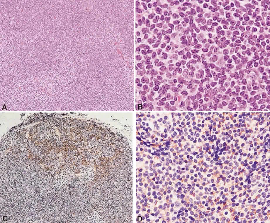
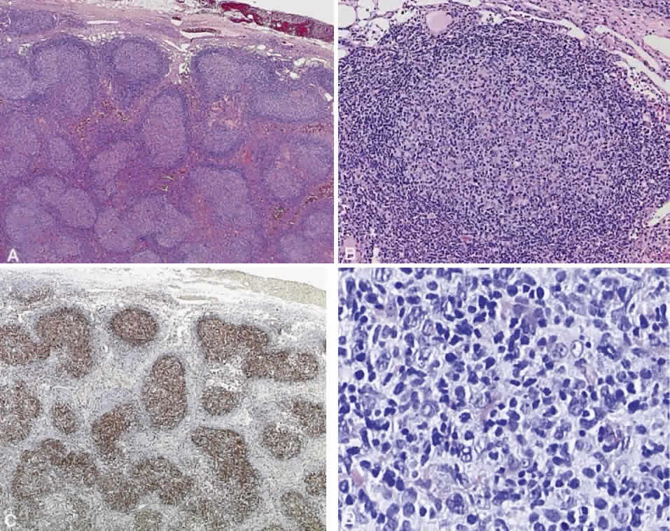
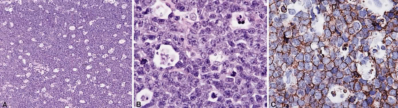
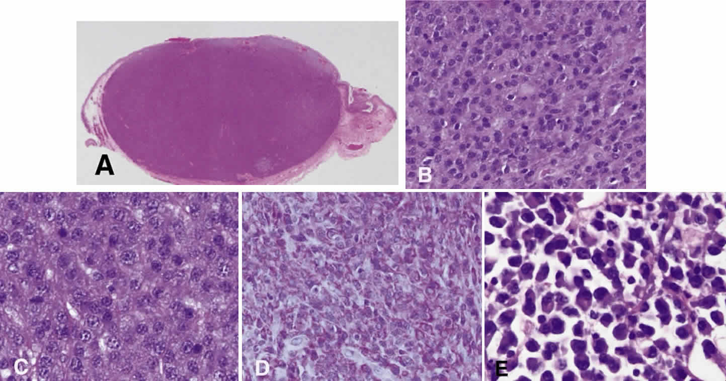
B cells arise from the bone marrow and produce immunoglobulin in a humoral response. T cells are thymus-derived and respond in a cell-mediated reaction via cells that are antigenically marked or produce lymphokines. Circulating blood and lymph nodes normally have a ratio of 70% T cells and 30% B cells, natural killer cells, or null cells. The normal lymph node is composed of follicles, each with germinal centers, mantle zones, and surrounding cortex (Fig. 2). The germinal center is composed of a framework of dendritic histiocytes, follicular dendritic cells, centroblasts, centrocytes, immunoblasts, small lymphocytes, and tingible body macrophages, whereas the surrounding cortex and paracortex consist mainly of B and T cells, respectively. Lymphomatous B cells were initially identified histologically using heterosera against immunoglobulin, which could then be stained with immunoperoxidase stain or rendered fluorescent with immunofluorescent markers.1b Plasma cells could be similarly identified by their cytoplasmic immunoglobulin. T cells were first identified cytologically by their rosette formation when combined with sheep erythrocytes.2 In addition, they were noted to uniquely contain cytoplasmic alphanaphthyl esterase, for which they could be stained histologically.3 Information regarding B-cell clonality could be obtained by staining for immunoglobulin, in particular, kappa and lambda light chains. In benign lesions, the kappa-lambda ratio of B-cell production is 2:1, whereas in malignant lesions, the ratio increases to 10:1.3 Benign and atypical reactive lesions have 50% or greater T cells, whereas malignant B-cell lesions usually consist of only 10% to 20% T cells.3 Malignant T-cell lymphomas, on the other hand, consist of 90% T cells.3 In reactive lesions, the T-cell helper-to-suppressor ratio is increased from the normal 2:1 to 5:1, whereas the normal ratio is maintained in malignant B-cell lymphoma.3 Thus, information about cell lines and immunoglobulin can be used to determine the predominant cell type, the cell type ratio, and the monoclonality or polyclonality of the B-cell population from the immunoglobulin expressed. Reactive processes have been thought to result from polyclonal or oligoclonal populations wherein every distinct clonal population expresses a distinct gene rearrangement, whereas a malignant lymphoma is thought to arise from a monoclonal clone population, which would express only a single gene rearrangement. Polymerase chain reaction has been used to specifically amplify clonal immunoglobulin gene rearrangements. In addition, cell surface markers of the various cell lines have been identified, beginning with the immunoglobulins and interleukins, and, later, cell surface antigen receptors, termed clusters of differentiation (CD), which are used to characterize the degree of differentiation of the cell type in question.4,5 The list of identified cell surface markers subsequently has expanded to over 100 and has aided in the characterization of Hodgkin's lymphoma and different subsets of nonHodgkin's B- and T-cell lymphomas6 (Table 1).

Fig. 2. The normal lymphoid follicle. A normal follicle is round or ovoid with a pale center containing tingible body macrophages. A. A well-defined zone of small dark lymphocytes surrounds the follicle separating it from the interfollicular zone (hematoxylin and eosin, × 10). B. A follicle center showing the follicle center cells with “cleaved” or “noncleaved” nuclei interspersed with tingible body macrophages (hematoxylin and eosin, × 20). C. CD20 demonstrates the follicle center cells, the mantle zone cells, and many of the interfollicular cells to be B cells (immunoperoxidase, × 10). D. CD21 staining illustrates the meshwork of dendritic follicular cells that provide a scaffold for the follicle center cells (immunoperoxidase, × 10). E. Follicle center cells express CD10 (immunoperoxidase, × 10). F. T cells are found in the interfollicular zone with few penetrating the follicle, as demonstrated by CD3 staining (immunoperoxidase, × 10). G. T cells express CD5 and also are predominantly found in the interfollicular zone, whereas some penetrate the follicle (immunoperoxidase, × 10). The oncoprotein bcl-2 is expressed normally by mantle zone and some interfollicular zone cells. H. Few cells of the follicle center express bcl-2, in contrast to follicle center cell lymphoma (immunoperoxidase, × 10).


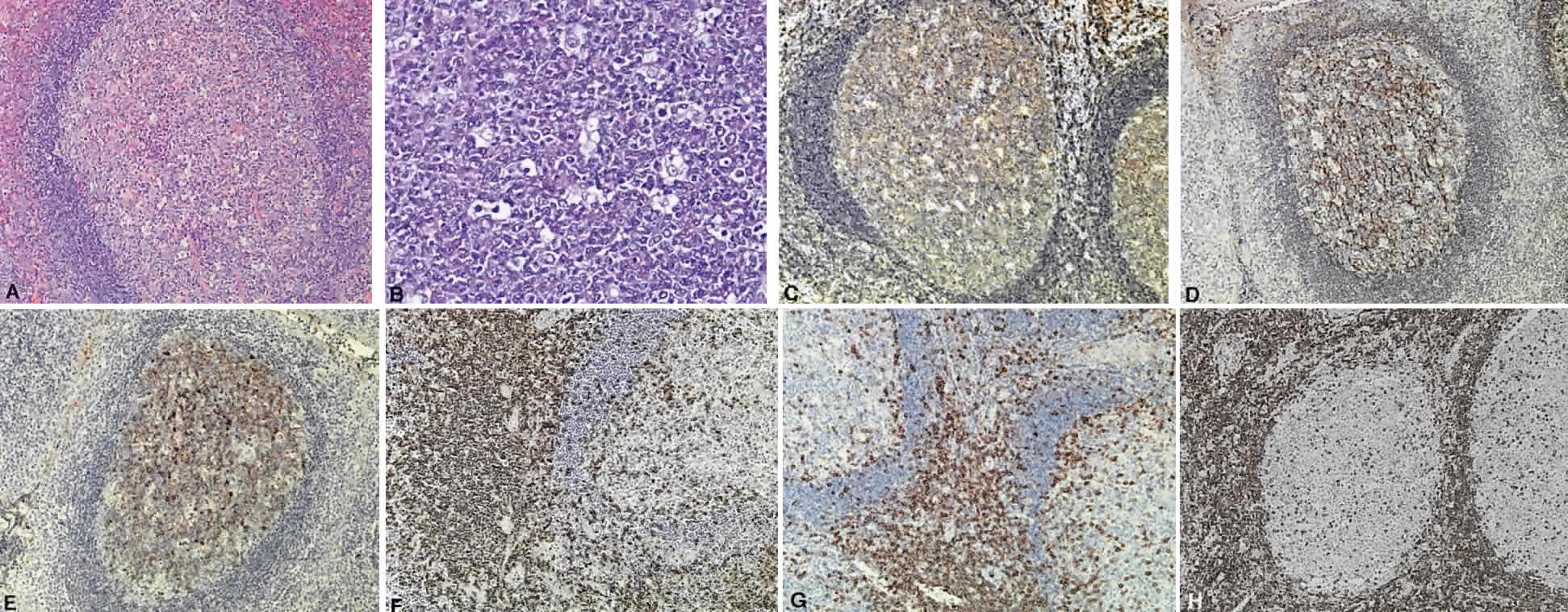
TABLE 1. List of Lymphoid Lesions and Their Characteristic Cell Markers


B-Cell Lymphomas 
Precursor B-cell neoplasmTdT+ CD19+ CD79a+ CD22+ CD20- /+ CD10+ /- HLA-DR+ SIg- cMu- /+ CD34+ /-
B-cell CLLBCAA+ SIgM+ CD5+ CD23+ CD43+ CD11c- /+ CD10-
LymphoplasmacytoidBCAA+ IgM+ CD22+ CD5- CD10- CD43+ /- CD25+ CD11c+
Mantle cell lymphomaBCAA+ SigM+ IgD+ CD5+ CD10- /+ CD23- CD43+ CD11c-
Follicle center lymphomaBCAA+ SIg+ CD10+ /- CD5- CD23- /+ CD43- CD11c-
Marginal zone lymphomaBCAA+ SIgM+ IgD- CIg+ CD5- CD10- CD23- CD43- /+ CD11c+ /-
Plasmacytoma/myelomaSig- CIg+ CD19- CD20- CD22- CD79a+ /- CD45+ /- HLA-DR-/+ CD38+ EMA-/+ CD43+ /- CD56+ /-
Large cell lymphomaBCAA+ SIg+ /- CIg-/+ CD45+ /- CD5-/+ CD10-/+
Burkitt's lymphomaBCAA+ SIgM+ CD22+ CD10+ CD5- CD38+ CD23-
T-Cell Lymphomas 
Precursor T-cell lymphomaCD7+ CD3+ TdT+ Ig- BCAA-
Peripheral T-cell CLLCD7+ CD2+ CD3+ CD5+
Mycosis fungoides/Sézary syndromeCD2+ CD3+ CD5+ CD7+ CD4+
Anaplastic large T-cellCD30+ CD45+ /- CD25+ /- EMA+ /- CD15-/+ CD3-/+ CD43-/+ CD45RO-/+ CD68-
Hodgkin's Disease 
Lymphocyte predominantBCAA+ CD45+ CDw75+ EMA-/+ CD15- CD30-/+ Ig-
Nodular sclerosisCD15+ /- CD30+ CD45-
Mixed cellularityCD15+ /- CD30+ CD45-
Lymphocyte depletionCD15+/- CD30+ CD45- BCAA- EMA-

CLL, chronic lymphocytic leukemia; LGL, lymphocytic granular leukemia; TdT, terminal deoxynucleotidyl transferase; IgH-R, immunoglobulin heavy chain rearrangement; IgL-R, immunoglobulin light chain rearrangement; TCR-R, T-cell receptor rearrangement; SIg, surface immunoglobulin; CIg, cytoplasmic immunoglobulin; EMA, epithelial membrane antigen; CD, cluster of differentiation; BCAA, CD19, CD20, CD79a (B-cell antigens); + , 90% incidence; + /—, over 50%; —/+ , less than 50%; —, 10% or less.
(Harris NL, Jaffe ES, Stein H et al: A revised European—American classification of lymphoid neoplasms: A proposal from the International Lymphoma Study Group. Blood 84: 1361, 1994)


GENETICS OF LYMPHOMA

Lymphomas are thought to occur as the result of an immunoregulatory defect, caused by somatic mutations resulting either in loss of a tumor suppressor gene or in the upregulation of a protooncogene or factor regulating mitosis and differentiation of naive lymphocytes to memory cells and plasma cells. As a result, patients who are immunosuppressed because of congenital immunodeficiencies, autoimmune diseases, cytotoxic agents after organ transplantation, or acquired immunodeficiency syndrome are thought to be at increased risk of developing nonHodgkin's lymphomas, and, in patients with acquired immunodeficiency syndrome in particular, lymphomas tend to be more aggressive with bone marrow, bowel, and central nervous system involvement and are associated with a poor prognosis despite treatment.7–12

Immunoglobulins have been studied at the molecular level in lymphoma. Monoclonal lymphoid lesions express gene rearrangements that can be detected with restriction endonuclease cleavage and Southern blot.13–25 The gene rearrangement appears as a unique band on the blot, known as a DNA fingerprint, and a polyclonal lesion, which expresses the immunoglobulins made by several different lymphocyte populations in undetectable amounts, then would demonstrate only the control band (Fig. 3). Many cytogenetic abnormalities that are characteristic of certain lymphoma subtypes have been isolated, mostly in the form of chromosomal translocations, some of which have been recognized to cause upregulation of protooncogenes, upregulation of transcription factors, and downregulation of factors involved in apoptosis. Chromosomal translocations are thought to occur so frequently in lymphoma because they take place during the normal process of B-cell differentiation. Examples of mutations and the affected gene include the c-myc oncogene (8q24) in Burkitt's Lymphoma, the bcl-1 gene (11q13) in mantle cell lymphoma, the bcl-2 (18q21) in follicular lymphoma, the bcl-2 t(14;18) gene in 70% to 90% of follicular lymphomas and 10% to 35% of nodal large cell lymphomas, the bcl-6 gene (3q27) in diffuse large cell lymphoma, and the PAX-5 gene t(9;14)(p13;q32) identified in 50% of lymphoplasmacytoid lymphomas25–37 (Table 2). The genes that are overexpressed or underexpressed, as a result of the mutations or translocations, have begun to be elucidated. The bcl-2 gene is a regulator of normal cell apoptosis.34 The t(14:18) translocation of bcl-2 is thought to allow unregulated cell longevity of the centrocytes in follicular lymphoma. The c-myc gene is known to code for a protooncogene, which, when upregulated, allows for unmitigated cell division.26,27 The PAX-5 gene belongs to a family of PAX genes, which have been identified as critical regulators of the cell cycle and differentiation.27,31,35 In particular, PAX-5 (BSAP) codes for the B-cell-specific transcription factor expressed in all stages of B-cell differentiation except in plasma cells.35 BSAP factor is suspected of enhancing B-cell lymphopoiesis, but the exact mechanism is not known. A mutation in bcl-1 allows for upregulation of the PRAD-1 gene, which codes for cyclin D1, a cell cycle regulator.26,27

Fig. 3. Diagram of DNA “fingerprint” from the Southern blot technique of DNA hybridization to distinguish monoclonal from polyclonal cell populations. The method is as follows: Double-stranded DNA is extracted from lymphocyte suspensions by cell lysis. Restriction endonucleases then are applied to the DNA to cleave it into several fragments. A. The fragments undergo electrophoresis on agarose gel slabs, which separates them based on their size and charge. The fragments then are denatured, which breaks them into single-stranded pieces of DNA. The fragments then are transferred to nitrocellulose filter. The fragments are incubated with 32p-labeled DNA probes, which are single-stranded nucleic acid fragments that hybridize specific base sequences from the original specimen. Probes then are used for regions known to encode for the kappa or gamma light chains, the immunoglobulin heavy chain region, and the T-cell receptor beta chain. Autoradiography is used to identify the radiolabeled material. This yields a blot that varies in intensity, depending on the amount of labeled material present in a given electrophoretic band. Each assessment is performed with a fibroblast control. Fibroblasts express the germline configuration of the light chain, heavy chain, and cell receptor DNA. B. Polyclonal populations express a variety of rearrangements but yield a germline band because it is the most frequent across the population, whereas a monclonal population yields a nongermline band, which represents all of the cells in the population.


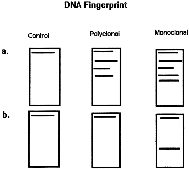
TABLE 2. Lymphoid Lesions and Their Characteristic Gene Rearrangements and Mutations


B-Cell Lymphomas 
Precursor B-cell/B-ALLIgH, IgL gene rearrangement
Peripheral B-cell B-CLLIgH, IgL gene rearrangement, trisomy 12
Lymphoplasmacytoid lymphoma/immunocytomaIgH, IgL gene rearrangement, 50% t(9;14) (p13;q32) rearrangement, PAX-5 on chr 9 to IgH on chr 14
Mantle cell lymphomat(11:14) involving IgH locus and bcl-1 on chr 11, overexpression of PRAD-1, codes for cyclinD1, p53 gene mutation (tumor suppressor gene)
Follicle center lymphoma grade I-III70–95% t(14;18) of bcl-2 gene, expresses antiapoptosis gene
Marginal zone lymphoma35% t(11;18) of bcl-2 gene, 60% trisomy 3, trisomy 18
Monocytoid and MALT-typep53 gene mutation
 plasmacytoma, multiple myelomaIgH and IgL genes rearrangement or deletion, t(9;14)(p13;q32), PAX-5 rearrangement
Diffuse large cell B-lymphoma45% bcl-6 (3q27) rearrangement
 30% bcl-2 rearrangement, 23% REL gene amplification (progression-associated marker), c-myc rearrangement
Burkitt's lymphomat(8;14) c-myc translocation from chr 8 to IgH region chr 14
 t(2;8) c-myc to IgL chr 2, t(8;22) c-myc to IgL chr 22
 25–40% African Burkitt's, some nonendemic EBV genome+
T-Cell Lymphomas 
Precursor T-cell/NK/T-ALLTCR gene rearrangement, IgH rearrangement
Peripheral T-cell lymphoma/T-CLLTCR gene rearrangement, trisomy 8q, 75% inv 14(q11;q32)
Mycosis fungoides/Sézary syndromeTCR gene rearrangement
Anaplastic large cell T-lymphoma50–60% TCR gene rearrangement, t(2;5) (p23;q35)
Hodgkin's Disease 
Lymphocyte predominantIgH or TCR gene rearrangement
Nodular sclerosisIgH or TCR gene rearrangement
Mixed cellularityIgH or TCR gene rearrangement
 60–70% EBV genome+
Lymphocyte depletionIgH or TR gene rearrangement

ALL, acute lymphocytic leukemia; CLL, chronic lymphocytic leukemia; IgH, immunoglobulin heavy chain; IgL, immunoglobulin light chain; TCR, T-cell receptor; EBV, Epstein-Barr virus; inv, inversion; MALT, mucosal-associated lymphoid tissue; chr, chromosome; NK, natural killer.
(Harris NL, Jaffe ES, Stein H et al: A revised European—Americcan classification of lymphoid neoplasms: A proposal from the International Lymphoma Study Group. Blood 84: 1361, 1994)


CLASSIFICATION OF LYMPHOMA

Numerous attempts have been made to classify lymphoid processes for clinical management and prediction of prognosis. Early classifications included only disease localized to the lymph node, which made classification of extranodal disease difficult and inaccurate. The Rappaport classification, first developed in 1956 and then modified in 1978, attempted to categorize lymphomas in two ways, first, using cytologic characteristics identified by conventional stains, and second, distinguishing between the follicular and diffuse growth pattern histologically38,39 (Table 3). The distinction of nodular, or follicular, and diffuse growth was considered useful because of the generally indolent nature of follicular growth, in which the tumor cell aggregates resemble germinal centers and disrupt the normal architecture of the node, compared with the appearance of diffuse growth, in which the lymph node is completely obliterated by a dense monotonous sheet of lymphocytes. In subsequent years, however, it was found that the descriptive growth pattern and cytogenetic characteristics of the Rappaport system did not predict prognosis reliably and were biologically inaccurate. The complexity of correlating degrees of differentiation, mitotic activity, and cytologic characteristics to prognosis have made lymphomas difficult to classify and have led to subsequent systems. The second system, proposed by Lukes-Collins in 1974, classifies lymphoma histologically according to its normal counterpart B-cell, T-cell, or null cell origin40,41 (see Table 3). Histologically, cells may appear small cleaved, large cleaved, small noncleaved, or large noncleaved, depending on the stage of B-cell arrest during normal transformation to immunoblast. Ninety percent of lymphomas are of B-cell origin, and the null cell also usually is of B-cell origin, although 10% may originate from T cells or histiocytes.42,43 Burkitt's lymphoma, the only lymphoma common in children, is a B-cell variant with a background of reactive histiocytes. As a result of histologic classification by Lukes-Collins, 76% of histiocytic lymphomas according to the Rappaport system were found to be not of histiocytic origin but of lymphocytic origin.40,44 The third system, the Working Formulation devised by the National Cancer Institute in 1982, attempted to predict prognosis by grouping lymphoma according to natural history, response to therapy, and overall survival.45 Three broad categories were established in terms of 5-year survival rates, the low-grade with a 50% to 70% survival rate, intermediate with 35% to 45%, and high grade with 23% to 32% (see Table 3). Orbital reactive hyperplasia, a relatively low-grade lesion, can be associated with systemic disease, whereas malignant or high-grade orbital lymphomas may be isolated findings. The Ann Arbor Staging Classification for Hodgkin's and non-Hodgkin's lymphomas was developed to stage disease based on systemic areas of involvement as a means of establishing a baseline for treating disease and following clinical progression46 (Table 4). Histologic classification, however, has been recognized as more useful than localization in the clinical management of nonHodgkin's lymphoma.47


TABLE 3. Rappaport, Lukes-Collins, and Working Formulation Classifications


5-y Survival Rate (%) Low GradeWorking FormulationRappaport ClassificationLukes-Collins Classification
50–70Small lymphocyticLymphocytic, well-differentiatedSmall lymphocyte and plasma-cytoid lymphocytic
 Follicular, predominantly small cleaved cellNodular, poorly differentiated lymphocyticFCC, small cleaved
 Follicular, mixed small and large cleavedNodular, mixed lymphocytic-histiocyticFCC, small and large cleaved
Intermediate Grade   
35–40Follicular, predominantly large cellNodular, histiocyticFCC, large cleaved and/or noncleaved
 Diffuse, small cleavedDiffuse, poorly differentiated lymphocyticFCC, small cleaved, diffuse
 Diffuse, mixed large and small cellDiffuse, mixed lymphocytic and histiocyticFCC, small cleaved, large cleaved, or large noncleaved
 Diffuse, large cellDiffuse histiocyticFCC, large cleaved or large noncleaved
High Grade   
23–32Large cell, immunoblasticDiffuse histiocyticImmunoblastic B- or T-cell type
 LymphoblasticLymphoblastic lymphomaConvoluted T-cell lymphoma
 Small noncleaved cellUndifferentiated Burkitt's and non-Burkitt'sFCC, small noncleaved

FCC, follicle center cell.


TABLE 4. Ann Arbor Staging of non-Hodgkin's Lymphoma


Clinical Staging (CS)
Stage I:Involvement of a single lymph node region (I) or of a single lymphatic organ or site (IE)
Stage II:Involvement of two or more lymph node regions on the same side of the diaphragm (II) or localized involvement of an extralymphatic origin or site of one or more lymph node regions on the same side of the diaphragm (IIE)
Stage III:Involvement of lymph node regions on both sides of the diaphragm (III), which may also be accompanied by localized involvement of thespleen (IIIS), extralymphatic site (IIIE), or both (IIISE)
Stage IV:Diffuse or disseminated involvement of one or more extralymphatic organs or tissue with or without associated lymph node enlargement
Pathologic Staging (PS)
Involvement found at primary laparotomy or by any further removal of tissue for histologic examination other than that taken for the original diagnosis. Include annotations for specific biopsy sites.
N+ or N-For other lymph node positive or negative by biopsy
H+ or H-For liver positive or negative by biopsy
S+ or S-For spleen positive or negative after splenectomy
L+ or L-For lung positive or negative by biopsy
M+ or M-For bone marrow positive or negative by biopsy or smear
P+ or P-For pleura or pleural fluid positive or negative by biopsy or by cytologic examination
O+ or O-For bone positive or negative by biopsy
D+ or D-For skin positive or negative by biopsy
(Carbone PP: Report of the committee on Hodgkin's disease staging classification. Cancer Res 31:1860, 1971)


While the Lukes-Collins and Working Formulation classifications were in wide use in the United States, the European literature made references to the Kiel and updated Kiel classifications, which led to disparities in classifying lymphoma. Another classification proposed by Jakobiec and coworkers was the most comprehensive classification available for orbital disease but failed to integrate systemic lymphoma, which is known to be associated in approximately half of cases.1 The most recent classification has made the system universal, comprehensive, and useful to interdisciplinary teams that characteristically manage patients with lymphoma. The International Lymphoma Study Group in 1994 developed the Revised European-American Lymphoma (REAL) classification (Table 5), which classifies lymphoid disease by the cell of origin into B-cell, T-cell, and natural killer cell lymphomas, leukemias, myeloma, and variants of Hodgkin's disease26 (Fig. 4). The identification of the putative benign progenitor cells has been inferred through the use of cell marker studies. The results of molecular genetic studies to identify immunoglobulin gene rearrangements and cytogenetic studies to detect chromosomal translocations in monoclonal proliferations have also been incorporated. A significant contribution of the REAL classification has been to incorporate primary extranodal lymphomas as recognizable and classifiable entities. As a result, new variants in this list include lymphoplasmacytic lymphoma, mantle cell lymphoma, marginal zone B-cell lymphoma, particularly mucosal-associated lymphoid tissue (MALT) lymphoma, subclasses of large cell lymphoma, and the natural killer cell lymphomas. Comparisons of the REAL classification to the Working Formulation and the Kiel classification are shown in Tables 6 and 7, respectively.48 The first series of 112 orbital lymphomas using the REAL classification reported the accuracy and utility of the system in classifying orbital lesions and predicting prognosis in combination with currently available immunophenotyping and immunocytogenetic studies.49 The REAL classification does not classify disease based on the degree of differentiation or clinical prognosis. However, a proposed prognostic scheme has been developed in accordance with the REAL classification28 (Table 8). In this text, tumor nomenclature adheres as strictly as possible to the REAL classification.


TABLE 5. Revised European—American Lymphoma (REAL) Classification

  B-Cell Neoplasms

  1. Precursor B-cell neoplasm: Precursor B-lymphoblastic leukemia/lymphoma
  2. Peripheral B-cell neoplasms:
    1. B-cell chronic lymphocytic leukemia/prolymphocytic leukemia/small lymphocytic lymphoma
    2. Lymphoplasmacytoid lymphoma/immunocytoma
    3. Mantle cell lymphoma
    4. Follicle center lymphoma, follicular
      Provisional cytologic grades: I (small cell), II (mixed small and large cell), III (large cell)
    5. Marginal zone B-cell lymphoma
      Extranodal (MALT-type + /— monocytoid B cells)
      Provisional subtype: diffuse, predominantly small cell type
    6. Provisional entity: Splenic marginal zone lymphoma (+ /— villous lymphocytes)
    7. Hairy cell leukemia
    8. Plasmacytoma/plasma cell myeloma
    9. Diffuse large B-cell lymphoma*
    10. Burkitt's lymphoma
    11. Provisional entity: high-grade B-cell lymphoma, Burkitt's-like


  T-Cell and Putative NK Cell Neoplasms
  1. Precursor T-cell neoplasm: Precursor T-cell lymphoblastic lymphoma/leukemia
  2. Peripheral T-cell and NK-cell neoplasms
    1. T-cell chronic lymphocytic leukemia/promyelocytic leukemia
    2. Large granular lymphocytic leukemia (LGL), T-cell and NK-cell types
    3. Mycosis fungoides/Sézary syndrome
    4. Peripheral T-cell lymphomas, unspecified.*
      Provisional cytologic catgories: Medium-sized cell, mixed medium and large cell, large cell, lymphoepithelioid cell
      Provisional subtype: Hepatosplenic γδ T-cell lymphoma
      Provisional subtype: Subcutaneous panniculitic T-cell lymphoma
    5. Angioimmunoblastic T-cell lymphoma (AILD)
    6. Angiocentric lymphoma
    7. Intestinal T-cell lymphoma (+ /— enteropathy associated)
    8. Adult T-cell lymphoma/leukemia (ATLL)
    9. Anaplastic large cell lymphoma (ALCL), CD30+ , T- and null-cell
    10. Provisional entity: Anaplastic large-cell lymphoma. Hodgkin's-like


  Hodgkin's Disease
  1. Lymphocyte predominance
  2. Nodular sclerosis
  3. Mixed cellularity
  4. Lymphocyte depletion
  5. Provisional entity: Lymphocyte-right classic


MALT, mucosal-associated lymphoid tissue; NK, natural killer.
*These categories are thought likely to include more than one disease entity.
(Harris NL, Jaffe ES, Stein H et al: A revised European—American classification of lymphoid neoplasms: A proposal from the International Lymphoma Study Group. Blood 84:1361, 1994)


Fig. 4. Diagram of a normal lymph node follicle indicating the germinal center, surrounding cortex, and marginal zone with the hypothetical cell origin of various lymphomas. (Ghia P, Nadler LM: Recent advances in lymphoma biology. Curr Opin Oncol 9:403, 1997)


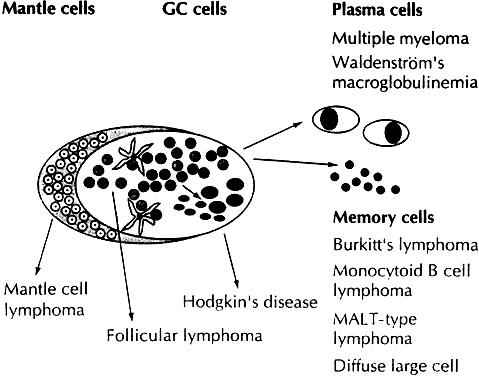
TABLE 6. Comparison of REAL Classification with Working Formulation


REAL ClassificationWorking Formulation
B-Cell
Low-Grade Lymphoma
CLLSmall lymphocytic consistent with CLL
Small lymphocytic/B-cell prolymphocytic leukemiaLymphoplasmacytoid/Immunocytoma
LymphoplasmacytoidSmall lymphocytic, plasmacytoid
Extranodal marginal zoneSmall lymphocytic
Nodal marginal zoneSmall lymphocytic
Splenic marginal zone/hairy cell leukemiaSmall lymphocytic
Plasmacytoma/myelomaPlasmacytoma
Follicle center, follicular, grade IFollicular, predominantly small cleaved
Follicle center, follicular, grade IIFollicular, mixed small and large cell
Intermediate-Grade Lymphoma 
Follicle center, follicular, grade IIIFollicular, predominantly large cell
Follicle center, diffuse, small cellDiffuse, small cleaved cell/diffuse, mixed small and large cell
Mantle cellDiffuse small cleaved cell
Diffuse large cellDiffuse, large cell
High-Grade Lymphoma
Diffuse large cellDiffuse large immunoblastic
Burkitt'sSmall noncleaved, Burkitt's
High-grade, Burkitt's-likeSmall noncleaved, non-Burkitt's
Precursor lymphoblasticLymphoblastic
T-Cell
Low-Grade Lymphoma
CL, prolymphocytic leukemiaSmall lymphocytic
Large granulocytic leukemia, T-Cell or NK cellSmall lymphocytic
Mycosis fungoides/Sézary syndromeMycosis fungoides
PeripheralDiffuse, small, cleaved
Diffuse, mixed small and large 
Diffuse, large cell 
Large cell, immunoblastic 
AngioimmunoblasticDiffuse mixed small and large, large cell immunoblastic
AngiocentricDiffuse mixed small and large, large cell immunoblastic
IntestinalLarge cell immunoblastic
Adult lymphoma/leukemiaDiffuse small cleaved, diffuse mixed small and large
Anaplastic large T- and null cellLarge cell immunoblastic

CLL, chronic lymphocytic leukemia; NK, natural killer; REAL, revised European—American Lymphoma classification.
(Harris NL, Jaffe ES, Stein H et al: revised European—American classification of lymphoid neoplasms: a proposal from the International Lymphoma Study Group. Blood 84:1361, 1994)


TABLE 7. Comparison of REAL Classification With Updated Kiel Classification


REAL ClassificationUpdated Kiel Classification
Precursor B-lymphoblastic leukemia/lymphomaB-lymphoblastic
B-cell chronic lymphocytic leukemia/promyelocytic leukemia/small lymphocytic lymphomaB-lymphocytic, CLL
 B-lymphocytic, prolymphocytic leukemia
 Lymphoplasmacytoid immunocytoma
Lymphoploasmacytoid lymphomaLymphoplasmacytic immunocytoma
Mantle cell lymphomaCentrocytic (mantle cell)
 Centroblastic, centrocytoid subtype
Follicle center lymphoma, follicularCentroblastic-centrocytic, follicular
 Grade I 
 Grade II 
 Grade IIICentroblastic, follicular
Follicle center lymphoma, diffuse, predominantly small cell (provisional)Centroblastic, follicular
 Centroblastic-centrocytic, diffuse
Extranodal marginal zone B-cell lymphoma (provisional)-
Nodal marginal zone B-cell lymphomaMonocytoid, including marginal zone
 Immunocytoma
Splenic marginal zone B-cell lymphoma (provisional)-
Hairy cell leukemiaHairy cell leukemia
Plasmacytoma/myelomaPlasmacytic
Diffuse large cellCentroblastic (monomorphic, polymorphic, and multilobated subtypes)
 B-immunoblastic
 B-large cell anaplastic
Primary mediastinal large B-cell lymphoma-
Burkitt's lymphomaBurkitt's lymphoma
High-grade B-cell lymphoma, Burkitt-like (Provisional)?
Precursor T-lymphoblastic lymphoma/leukemiaT-lymphoblastic
T-cell chronic lymphocytic leukemia/prolymphocytic leukemiaT-lymphocytic, CLL type
 T-lymphocytic, prolymphocytic leukemia
Large granular lymphocytic leukemia, 
 T-cell typeT-lymphocytic, CLL type
 NK-cell type-
Mycosis fungoides/Sézary syndromeSmall cell cerebriform (mycosis fungoides/Sézary syndrome)
Peripheral T-cell lymphomas, unspecifiedT-zone
 Lymphoepithelioid
 Including subtype: subcutaneous panniculytic T-cell lymphoma (provisional)Pleomorphic, small T-cell
 Pleomorphic, medium-sized and large T-cell
 T-immunoblastic
Hepatosplenic gd T-cell lymphoma (provisional)-
Angioimmunoblastic T-cell lymphoma with dysproteinemia)Angioimmunoblastic (angioimmuno-blastic lymphadenopathy
Angiocentric T-cell lymphoma-
Intestinal T-cell lymphoma-
Adult T-cell Lymphoma/LeukemiaPleomorphic small T-cell, HTLV1+
 Pleomorphic medium-sized and large T-cell, HTLV1+
Anaplastic large T- and null cellT-large cell anaplastic

AILD, ; CLL, chronic lymphocytic leukemia; HTLV1, human T-lymphotropic virus type 1; LgX, ; NK, natural killer; REAL, revised European—American Lymphoma classification.
(Chan JKC, Banks PM, Cleary ML et al: A proposal for classification of lymphoid neoplasms (by the international Lymphoma Study Group). Histopathology 25:517, 1994)


TABLE 8. Proposed Clinical Scheme for Malignancies of the Lymphoid System


B-Cell LineageT-Cell Lineage
I. Indolent lymphomas (low risk)I. Indolent lymphomas (low risk)
Chronic lymphocytic leukemia/small lymphocytic lymphomaLarge granular lymphocytic leukemia, T-cell and NK-cell
 Mycosis fungoides/Sézary syndrome
Lymphoplasmacytic lymphoma/immunocytoma/ Waldenstrom's macroglobulinemiaSmoldering and chronic adult T-cell leukemia/lymphoma, HTLV-1
Hairy cell leukemia 
Splenic marginal zone lymphoma 
Marginal zone B-cell lymphoma 
 Extranodal (MALT B-cell lymphoma) 
 Nodal (monocytoid) 
Follicle center lymphoma/follicular (small cell); grade I 
Follicle center lymphoma/follicular (mixed small and large cell); grade II 
II. Aggressive lymphomas (intermediate risk)II. Aggressive lymphomas (intermediate risk)
Prolymphocytic leukemiaProlymphocytic leukemia
Plasmacytoma/multiple myelomaPeripheral T-cell lymphoma, unspecified
Mantle cell lymphomaAngioimmunoblastic lymphoma
Follicle center lymphoma/follicular (large cell); grade IIIAngiocentric lymphoma
Diffuse large B-cell lymphoma (includes immunoblastic and diffuse large and centroblastic lymphoma)Intestinal T-cell lymphoma
Primary mediastinal (thymic) large B-cell lymphomaAnaplastic large cell lymphoma
High-grade B-cell lymphoma, Burkitt-like 
III. Very aggressive lymphomas (high risk)III. Very aggressive lymphomas (high risk)
Precursor B-lymphoblastic lymphoma/leukemiaPrecursor T-lymphoblastic
Burkitt's lymphoma/B-cell acute leukemiaAdult T-cell lymphoma/leukemia
Plasma cell leukemia 
IV. Hodkin's disease 

HTLV-1, human T-lymphotropic virus type 1; MALT, mucosal-associated lymphoid tissue. NK, natural killer.
(Harris NL, Jaffe ES, Stein H et al: A revised European—American classification of lymphoid neoplasms: A proposal from the International Lymphoma Study Group. Blood 84:1361, 1994)


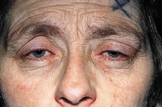
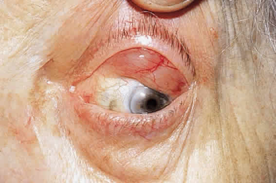
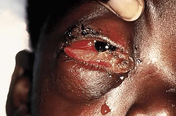
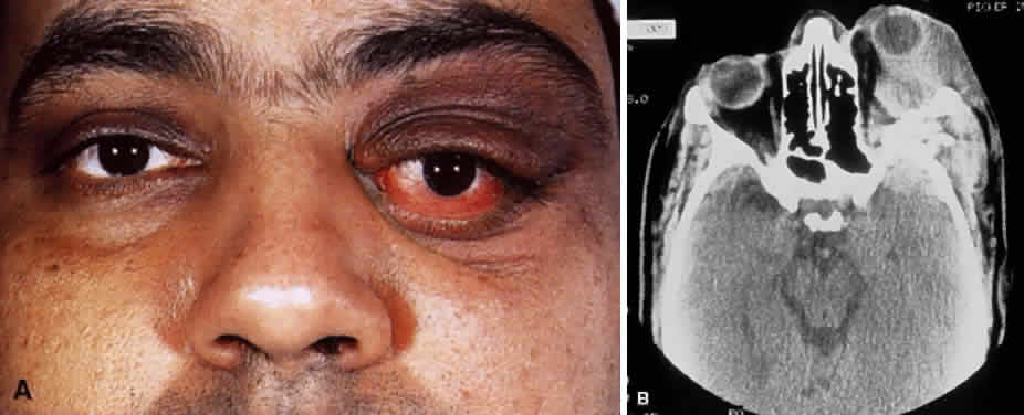
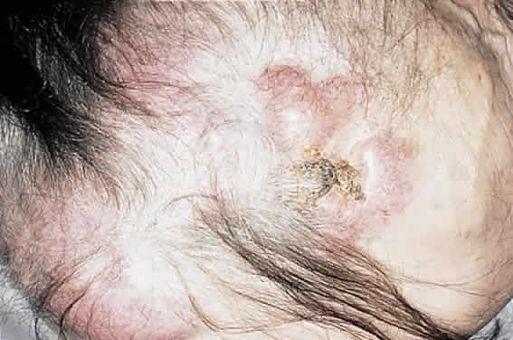
CLINICAL PRESENTATION OF SYSTEMIC NON-HODGKIN'S LYMPHOMA

Systemic non-Hodgkin's lymphomas usually present with painless lymph node enlargement in one or more nodes. Adults usually are in their fifth or sixth decade, and presentation in childhood is extremely rare. The fever, night sweats, and weight loss characteristic of Hodgkin's disease generally are absent. Splenomegaly may develop in 20% of patients. Patients usually have normal blood counts, although they may develop lymphocytosis, especially with well-differentiated lymphocytic lymphomas, or pancytopenia with anemia, hemorrhage, petechiae, ecchymosis, and infection—the pancytopenia occurring in one third of patients as a result of bone marrow involvement or chemotherapy and radiation. Patients with B-cell lymphomas tend to develop difficulty with bacterial infection, whereas those with T-cell lymphoma may develop difficulty with delayed-type hypersensitivity and viral infections. Leukemic conversion is rare in adults, although it occurs in 25% of children. In children, an acute leukemic phase may be the initial presentation. Furthermore, children are more likely to develop extranodal and aggressive disease, although they may respond well to therapy.50 In a series of 1269 patients, of whom only 3 (0.42%) presented initially with proptosis, one third of patients presented with extranodal disease and a few with bone marrow invasion.51 There was a 1.3% incidence rate of orbital disease secondary to systemic lymphoma.

Back to Top
ORBITAL LYMPHOID TUMORS
Although the orbit is an extranodal site where lymphocytes are not found under normal conditions, lymphoid tumors are the third most common cause of proptosis in the adult after inflammation, caused either by Graves' disease or orbital pseudotumor, and cavernous hemangioma.52 A series of 764 orbital tumors showed that 7.5% were lymphomas, and another series showed that 11% of 820 cases were classified as lymphoid disease.53,54

Of primary orbital lymphoid lesions, 50% have been shown to be reactive or atypical hyperplasia and 50%, malignant lymphoma.55 Recent evidence, however, has shown that there is a continuous spectrum of disease in terms of prognosis, with systemic disease occurring in 15% to 25% of reactive hyperplasias, 40% of atypical hyperplasias, 20% of well-differentiated lymphomas, and 60% of poorly differentiated lymphomas.1,55–60 In addition, more aggressive lesions may develop from a matrix of benign or reactive follicles, either simultaneously or as a recurrence. Bilateral lymphoma has a high incidence of systemic involvement, although systemic disease may be indolent and may not shorten life expectancy. Lymphoplasmacytoid lymphoma has been associated with Waldenström's macroglobulinemia, a paraproteinemia which produces IgM, a heavy protein resulting in a hyperviscosity syndrome with stasis retinopathy and ischemia.26 Mortality rates at 5 years are 6% for reactive hyperplasia, 19% for atypical hyperplasia, and 58% for malignant lymphoma.1

Benign reactive hyperplasia is histologically the most innocuous of the lymphoid lesions. It is hypercellular, with sheets of mature polyclonal lymphocytes, scattered plasma cells, and histiocytes in irregular follicles associated with hyperplastic capillaries lined by plump endothelial cells and no surrounding stroma.55 The germinal center consists of dendritic follicular cells, tingible body macrophages, and large lymphocytes with mitotic figures, and is surrounded by a mantle zone of small lymphocytes. Malignant orbital lymphoma, on the other hand, often shows little mitotic activity or endothelial proliferation. Cells tend to be monomorphous, immature, and small, medium, or large with a monoclonal population exhibiting either nodular or diffuse growth. Most are low grade. Atypical lymphoid hyperplasia shares features of both benign reactive hyperplasia and malignant lymphoma, in that it lacks mitotic activity and endothelial proliferation, but does exhibit a follicular polymorphous response.55 Essentially, lesions that appear reactive, but show atypical or aggressive histological features, are grouped as atypical hyperplasia.

Of malignant orbital lymphomas, 85% to 90% are diffuse, and only 10% to 15% follicular and 50% of the diffuse lesions are well differentiated.55 The follicular lesions tend to be located in orbital fat among the septa. Of the 30% of malignant lymphomas that develop systemic involvement, the extraorbital disease usually manifests within 2 years after diagnosis and, overall, approximately 50% of all orbital lymphoid lesions—reactive, benign, or malignant—are associated with subsequent systemic involvement over a 4-year follow-up.1,55

CLINICAL PRESENTATION OF ORBITAL LYMPHOMA

Most orbital lymphomas are non-Hodgkin's lymphomas and tend to present in the sixth and seventh decades. Presentation before the age of 20 is rare, and, in children, orbital leukemic infiltrates are much more common than lymphoma. Most subtypes of lymphoma have a small female-to-male preponderance of 1.5:1.

Lymphoid lesions usually are located superiorly and anteriorly in the orbit and present over several months with gradual painless proptosis of 5 mm or less, without conjunctival chemosis or injection (Fig. 5). There may be only mild dysmotility or displacement of the globe, since, with the exception of orbital fat, lymphoid lesions tend to mold around rather than infiltrate preexisting structures. Involvement of the extraocular muscles has been described and, when it occurs, most commonly involves the superior rectus-levator complex.61 Visual loss is uncommon, and bilateral blindness is rare.62 The lacrimal gland is involved in 30% of lesions, and this may be expected, since there are native lymphocyte populations in the lobules. Lymphoma involving the lacrimal gland feels rubbery to palpation and may have a nodular consistency, which likely results from lobular remnants of the gland. Lymphoid lesions localized to the epibulbar tissues, referred to as “salmon-patch” lesions because of their pink fleshy color, are freely mobile and may be isolated or extend posteriorly into the orbit (Fig. 6). Lymphadenopathy may be palpable and may indicate the presence of systemic disease. Of 117 patients reported by Knowles and Jakobiec, 64% of lesions were in the deep orbit, 28% subconjunctival, and 8% in the eyelid.60

Fig. 5. A 55-year-old patient with Sjögren's syndrome and bilateral orbital lymphoma and the associated anterior, superior soft tissue swelling, ptosis, orbital proptosis, and globe displacement.

Fig. 6. A 60-year-old patient with an epibulbar “salmon-patch” lesion, which can be the presenting sign of reactive hyperplasia or malignant lymphomas.

Computed tomography (CT) accurately demonstrates the molding of the mass to orbital structures, such as the globe and orbital bones, without bony erosion except in large cell lymphoma (Fig. 7). CT is used to localize the lesion, which tends to be unilateral and in both the intraconal and extraconal spaces. Lesions limited to the conjunctiva tend to be more benign with a better long-term prognosis, whereas those that extend into the orbit tend to be more malignant. Conjunctival lesions remain localized in 90% of cases, whereas orbital and lid lesions have a higher rate of systemic extranodal involvement.63 Lymphoid lesions of the lacrimal gland appear as a diffuse vertical expansion of the gland, which mold to both the globe and orbital bone without producing a bony fossa or erosion64 (Fig. 8). If the lesion extends beyond the orbital rim, the palpebral lobe of the gland is involved, and posterior or orbital lobe involvement appears as a straight line against orbital fat. Pleomorphic adenoma, on the other hand, appears as an oval, globular lesion with, in 80% of cases, adjacent bone changes caused by the firmer stroma of the tumor. Because epithelial tumors usually arise in the orbital lobe, extension beyond the orbital rim is not a feature.65 CT scan cannot distinguish between inflammatory and lymphoid lesions, because both lesions are homogeneous and enhance with intravenous contrast, and at biopsy, orbital lymphoid lesions are pink with a friable texture caused by the absence of stroma.66–68 The subtype and malignancy of the lesion can only be determined morphologically. The following subtypes of B-cell nonHodgkin's malignant lymphoma—extranodal B-cell marginal zone lymphoma, follicle center cell lymphoma, small lymphocytic lymphoma, lymphoplasmacytoid lymphoma, mantle cell lymphoma, large cell lymphoma, and Burkitt's lymphoma—are discussed in the approximate order of frequency with which they occur in the orbit.

Fig. 7. A CT scan, axial view, of the patient in Figure 6 showing molding of the lymphoma to adjacent extraocular muscle, globe, and medial orbital wall.

Fig. 8. A CT scan, axial view, of a B-cell lymphoma localized to the anterior orbit bilaterally, with left lacrimal gland involvement and a normal adjacent lateral wall.

EXTRANODAL B-CELL MARGINAL ZONE LYMPHOMA

Mucosa-associated lymphoid tissue normally is found in the conjunctiva and lacrimal gland, but not in the orbit, and conjunctival MALT secretes IgG, whereas the lacrimal gland produces IgA; the latter is bound to a secretory component made by lacrimal gland MALT and subsequently secreted into the tear film.69 Both conjunctival and lacrimal gland MALT contribute lysozyme to the tear film.69 MALT is the immune secretory system also found in the tracheobronchial tree, salivary gland, thyroid gland, gastrointestinal tract, and breast, and lymphoma of MALT has been incorporated for the first time by the REAL classification and termed extranodal marginal zone B-cell lymphoma, which occurs in the extranodal areas mentioned earlier as well as in the orbit.26,70 Extranodal B-cell marginal zone lymphoma in the stomach has been associated with Helicobacter pylori infection in as many as 90% of patients, and the lymphoma has been shown to respond in most ofthese cases to antibacterial therapy.71–74 The association appears to occur in the presence of reactive T-cell activity in H. pylori infection.74 Other inflammatory conditions may mediate lymphomatous transformation elsewhere.75–79 There has been an association of extranodal B-cell marginal zone lymphoma in patients with the myoepithelial sialoadenitis of Sjögren's syndrome and the thyroid inflammation of Hashimoto's thyroiditis.80,81 This type of lymphoma likely arises secondary to preexisting local chronic inflammation of underlying etiology, whether infectious, autoimmune, or undetermined.

Extranodal B-cell marginal zone lymphoma histologically can consist of small to medium-sized centrocyte-like lymphocytes, which originate from the marginal zone of the follicle or can be polymorphous with cells ranging from small lymphocytes to plasmacytoid cells, which then either invade the interfollicular area and the overlying epithelium or invade the central follicle (Fig. 9). This variable pattern has likely contributed to the delay in recognition of the entity. High-grade extranodal B-cell marginal zone lymphoma may destroy the follicular structure completely. Most extranodal B-cell marginal zone lymphoma of the ocular adnexa are low-grade lesions, although systemic involvement or transformation to large cell lymphoma can occur.26,82

Fig. 9. Extranodal marginal zone lymphoma. A. A diffuse infiltrate with ill-defined nodularity (hematoxylin and eosin, × 10). B. Cytologic features of the neoplastic lymphoid cells include small centrocyte-like cells, some with plasmacytoid nuclei (hematoxylin and eosin, × 60). C. CD21 stain shows the presence of preexisting lymphoid follicle, which has been partially destroyed by infiltration with marginal zone cells (immunoperoxidase, × 10). D. CD20 immunophenotyping demonstrates B-cell lineage (immunoperoxidase, × 40).

FOLLICLE CENTER CELL LYMPHOMA

Follicle center cell lymphoma occurs in adulthood, and deposits in the orbit usually are an indicator of disseminated disease. Primary extranodal disease in the orbit is uncommon. This disease may be relatively indolent but difficult to cure. Some cases may progress to large cell lymphoma. Histologically, the tumor usually has a follicular growth pattern, although a diffuse growth component also may be present. The abnormal follicles lack tingible body macrophages and contain a variable mixture of follicle center cells (centrocytes and centroblasts—cleaved and noncleaved cells). The derivation of these abnormal follicles from normal follicles is suggested by the expression of CD10. In contrast to normal reactive follicles, the center of abnormal follicles also expresses bcl-2 oncoprotein, a feature that is helpful in differential diagnosis (Fig. 10).

Fig. 10. Follicle center cell lymphoma. A. Multiple abnormal follicles of variable size and shape with a surrounding mantle zone (hematoxylin and eosin, × 2). B. An abnormal follicle showing the absence of tingible body macrophages within the follicle cell center (hematoxylin and eosin, × 2). C. Antibody stain for bcl-2 highlights the centers of the abnormal follicles, a pattern typical of follicle center cell lymphoma (immunoperoxidase, × 2). D. Center of an abnormal follicle showing a mixture of pleomorphic cells with “cleaved” and “noncleaved” nuclei. Tingible body macrophages are absent (hematoxylin and eosin, × 40).

B-CELL CHRONIC LYMPHOCYTIC LEUKEMIA/SMALL LYMPHOCYTIC LYMPHOMA

B-cell chronic lymphocytic leukemia (B-CLL)/small lymphocytic lymphoma is predominantly a disease of the elderly whereby most present with leukemia involving the orbit. The term small lymphocytic lymphoma is reserved for cases in which there is no leukemia at presentation. Although this is a relatively indolent disease, it is virtually incurable with current therapy and may progress to a higher grade large cell lymphoma (Richter transformation). The histologic hallmark of this disease is a diffuse infiltrate of small lymphoid cells within which poorly defined pale-staining pseudofollicles of slightly larger cells are visible (proliferation centers). Immunophenotyping shows the abnormal lymphoid population to express CD5, CD23, and CD43 in addition to standard B-cell markers such as CD20 and CD79a. They do not express cyclin D1 (Fig. 11).

Fig. 11. Small lymphocytic lymphoma/B-cell chronic Lymphocytic leukemia. A. A diffuse infiltrate with a vague nodular pattern from the presence of “proliferative centers” (hematoxylin and eosin, × 2). B. A monomorphous infiltrate of small lymphocytes (hematoxylin and eosin, × 10). C. The cytologic features of neoplastic lymphocytes: the cells are small with small dark nuclei and inconspicuous cytoplasm (hematoxylin and eosin, × 60). D. CD5 stain is positive in this subclass of lymphoma (hematoxylin and eosin, × 40). (Courtesy of Dr. A. Mowat.)

LYMPHOPLASMACYTOID LYMPHOMA

Lymphoplasmacytoid lymphoma also is a disease of the elderly in which a paraproteinemia is common, sometimes manifesting as a hyperviscosity state of visual and neurologic impairment and oozing from wounds (i.e., Waldenström's macroglobulinemia), for which the mean survival is 5 years.83 The optic nerve can be invaded by neoplastic plasma cells and lymphocytes or demonstrate papilledema from central nervous system invasion, and the tumor resembles lymphoma in its diffuse infiltration into lymphoid tissues.84 Like B-CLL, lymphoplasmacytoid lymphoma is rarely curable with available therapy. It usually affects the orbit as a component of systemic disease and also can transform to high-grade large B-cell lymphoma. In biopsy specimens, the infiltrate consists of a mixture of small lymphocytes, plasmacytoid lymphocytes, and plasma cells. Some abnormal cells contain intranuclear Dutcher and intracytoplasmic Russell bodies, which represent intracellular accumulations of immunoglobulin. When present, these features aid greatly in diagnosis.

MANTLE CELL LYMPHOMA

Another malignant lymphoma that predominantly affects older adults and usually is widely disseminated at presentation is mantle cell lymphoma. Again, this lymphoma usually affects the orbit as a component of systemic disease. On histologic examination, it consists of a diffuse infiltrate of small to medium-sized lymphoid cells with irregular, sometimes cleaved, nuclei with inconspicuous nucleoli and little cytoplasm (Fig. 12). Immunohistochemical analysis usually demonstrates the cells to be B cells that also express CD5 and CD43, but not CD23. These cells also typically express cyclin D1.

Fig. 12. Mantle zone lymphoma. A. A monomorphous diffuse infiltrate of medium-sized cells (hematoxylin and eosin, × 10). B. Lymphocytes in mantle zone lymphoma have irregular nuclei, inconspicuous nucleoli, and scant cytoplasm (hematoxylin and eosin, × 60). (Courtesy of Dr. A. Mowat)

LARGE CELL LYMPHOMA

Of the malignant B-cell lymphomas, diffuse large B-cell lymphoma carries the worst prognosis for survival. In one series, 50% of patients with orbital diffuse large cell lymphoma presented initially with systemic disease, and the remaining half developed systemic disease soon thereafter, and in another series, survival was 2 years or less in most patients.51,85–87 According to the Rappaport classification, large cell lymphomas were labeled histiocytic lymphomas, but with immunophenotyping studies have been recognized to be mostly of B-cell origin, although some may be of T-cell or null cell origin. Histologically, large cell lymphomas consist of cells two to four times normal size with one or more prominent nucleoli.

BURKITT'S LYMPHOMA

Burkitt's lymphoma is a rare tumor, originally described in Africa and endemic within 10 degrees of latitude north and south of the African equator; nonendemic Burkitt's lymphoma has been documented in Europe and North America.88 Burkitt's lymphoma is most common in children, comprising 90% of pediatric lymphomas in endemic regions and one third of nonendemic pediatric lymphomas.89 The endemic type is associated with Epstein-Barr viral infection 6 months before the onset of lymphoma in 90% of cases, and the Epstein-Barr viral genome has been identified in 25% to 40% of cases.89 In adults, it is most commonly associated with immunodeficiency. When the T cells that mediate B-cell antiviral activity fail to curtail the response, the mononucleosis phase of infection is thought to become protracted and allow growth of a neoplastic clone from the polyclonal proliferation.90,91 The African type of Burkitt's lymphoma, which usually invades the abdominal viscera with ascites, is seen in half of cases from lymph nodes and also may invade the jaw. Intracranial spread occurs in 30% of cases through peripheral perineural spread. When Burkitt's lymphoma occurs in the orbit, it typically originates from the maxillary marrow space.92–94 The tumor growth in the facial area is rapid, with a doubling time of 3 days, and may produce a tumor of monstrous proportion in 2 to 4 weeks (Fig. 13). Only one case of primary orbital Burkitt's lymphoma has been described, and this case was nonendemic.89 Nonendemic Burkitt's lymphoma affects the abdomen more commonly than the craniofacial region, and histologically the lesion consists of a blastic proliferation of immunoglobulin-producing B cells interspersed with tingible body macrophages, laden with apoptotic lymphocytic debris, causing the characteristic “starry sky” appearance89 (Fig. 14). Management is with chemotherapy regimens based on cyclophosphamide, doxorubicin, vincristine, and methotrexate. Prognosis depends on the volume of disease at presentation, and if the disease is localized, it can be surgically debulked. Fifty percent of tumors recur, commonly in the meninges. Aggressive disease with meningeal invasion may require chemotherapy with bone marrow transplant to be curative.

Fig. 13. A 9-year-old black male patient with Burkitt's lymphoma involving the right orbit with secondary conjunctival chemosis and ulcerative necrosis.

Fig. 14. Burkitt's Lymphoma. A. A homogeneous diffuse abnormal lymphoid infiltrate scattered within which are numerous tingible body macrophages giving the classic “starry sky” appearance (hematoxylin and eosin, × 10). B. The abnormal lymphoid cells are medium-sized blasts with prominent apoptotic bodies, some of which are contained within the large tingible body macrophages (hematoxylin and eosin, × 60). C. CD20 immunophenotyping demonstrates B-cell lineage (immunoperoxidase, × 60).

PLASMA CELL TUMORS

Plasma cell tumors are, in essence, tumors of mature B cells that are secreting monoclonal immunoglobulin detected as the M-spike on serum electrophoresis, associated with high urine and blood content of immunoglobulin.95 Light chain fragments excreted in the urine are known as Bence Jones protein.

Multiple myeloma is the prototypic systemic plasma cell tumor that occurs in the elderly, more commonly men, with multiple masses of plasma cells disseminated throughout the skeletal system and soft tissue.7 Extraskeletal metastases occur in 70% of cases, either by hematogenous spread or by direct extension. Morbidity and mortality stem from bone marrow invasion with resultant anemia, infiltration of visceral organs, amyloid deposition, and secretion of immunoglobulin and osteoclast-activation factors. Multiple myeloma can metastasize to the kidney, adrenal gland, heart, and liver and can cause punched-out lesions of the skull and vertebral fractures. Mean survival is 3 years with chemotherapy, and the presence of Bence Jones proteins worsens prognosis. An aggressive variant of multiple myeloma is disseminated nonosteolytic myelomatosis, which progresses rapidly, involving bone marrow and soft tissues without radiographic changes. Orbital soft tissue metastasis with orbital bone destruction has been noted in a series to occur in 5 in 2000 cases, and invasion of orbital bone is even less common94–98 (Fig. 15). Amyloid deposition into the extraocular muscles can occur.99

Fig. 15. A 48-year-old Indian man with known systemic multiple myeloma and subsequent plasmacytoma involving the left orbit with lateral orbital wall destruction (A), as evident on axial CT scan (B).

When an isolated mass of plasma cells occurs, it is referred to as a plasmacytoma, and these may occur in bone, where progression to multiple myeloma is most frequent, or extramedullary sites, which progress more rarely.100–104 Solitary conjunctival plasmacytoma requires observation for development of multiple myeloma (Fig. 16). Solitary plasmacytomas make up 3% to 5% of plasma cell tumors, and 25% of plasmacytomas have elevated IgM protein in the blood and urine.

Fig. 16. Plasmacytoma. A. A rounded conjunctival stromal nodule. The immediate subepithelial region contains a slightly more blue lymphoid infiltrate. The inferior portion of the lesion contains a recognizable lymphoid follicle within its substance (hematoxylin and eosin, × 1.25). B. A reasonably homogeneous population of large cells with pink cytoplasm and regular nuclei predominate in the nodule (hematoxylin and eosin, × 10). C. The typical “clock face” or “cartwheel” nuclei are visible on higher magnification as well as the abundance of pinkish blue cytoplasm (hematoxylin and eosin, × 40). D. A section stained with methyl green pyronin demonstrates the presence of red pyrinophilic cytoplasm typical of cells rich in ribosomes (methyl green pyronin, × 10). E. Plasmacytoid differentiation is apparent, with eccentrically placed nuclei and paranuclear cytoplasmic pallor (“hof”) (hematoxylin and eosin, × 40). (Courtesy of Dr. A. Mowat)

T-CELL LYMPHOMA

T-cell lymphoma of the orbit is rare and usually occurs during the late stage of systemic disease or as a manifestation of mycosis fungoides. Only one case of primary T-cell lymphoma in the orbit has been described.105 Mycosis fungoides, a cutaneous T-cell lymphoma that spreads to lymph nodes and then to viscera, can invade the lids and conjunctiva, and even more rarely, the orbit106–109 (Fig. 17). Initially, the disease presents nonspecifically as an eczematous process, then to biopsy-positive indurated plaques, and then to cutaneous lesions and ulcerations, which, if large and exophytic, may appear fungal (Fig. 18). Generalized erythroderma may occur at any stage and is particularly common in Sézary's syndrome of erythroderma, hepatosplenomegaly, lymphadenopathy, and circulating abnormal T cells. Staging of the disease is done with evaluation of skin, lymph nodes, viscera, and blood. Patients with erythroderma have a worse prognosis, with a mean survival of 3 to 4 years compared with 8 years for those with plaques.110 This disease can progress to anaplastic large T-cell lymphoma.26 Histologically, the Sézary or Lutzner cell is the characteristic T-cell with a lobulated nucleus with nuclear infoldings (Fig. 19). Chemotherapy regimens used in peripheral T-cell lymphoma are the same as those used in anaplastic B-cell lymphoma, that is, vincristine, doxorubicin, cyclophosphamide, prednisone, and etoposide.110 Systemic T-cell lymphomas are treated with a host of chemotherapy regimens, and prognosis generally is much poorer than for B-cell lymphomas, with 50% survival at 5 years.

Fig. 17. A patient with mycosis fungoides and T-cell lymphoma involving the left orbit.

Fig. 18. Scalp ulceration typical of mycosis fungoides from the patient in Figure 17.

Fig. 19. T-cell lymphomas. A. Peripheral T-cell lymphoma (unspecified): A mixture of small and medium-sized lymphoid cells with occasional large pleomorphic cells are a few eosinophils interspersed (hematoxylin and eosin, × 20). B. There is cellular pleomorphism with mitotic figures and a prominent blood vessel at top left (hematoxylin and eosin, × 40). C. CD3 immunophenotyping demonstrates the abnormal lymphoid cells to be of T-cell lineage (immunoperoxidase, × 40). D. Mycosis fungoides with a dermal lymphoid infiltrate showing epidermotropism at the dermal-epidermal junction and perivascular infiltration elsewhere (hematoxylin and eosin, × 10). E. Epidermotropism with the disruption of the normal dermal-epidermal border and infiltration of cells into the epidermis (hematoxylin and eosin, × 20). F. Higher power of the same lesion with a mixture of small and large lymphoid cells, some with irregular nuclei (hematoxylin and eosin, × 60). G. Immunophenotyping of the abnormal lymphoid cells using CD3 antibody, shows them to be of T-cell lineage (× 60). (Case courtesy of Dr. A. Mowat.)

ANGIOCENTRIC LYMPHOMA (STEWART'S LETHAL MIDLINE GRANULOMA)

Angiocentric lymphoma occurs over a wide age range primarily as an extranodal disease affecting the nose, lungs, upper respiratory and gastrointestinal tracts, skin, and central nervous system. Orbital involvement is rare and occurs secondary to spread from the nose. This disease was previously thought to be a granulomatous/histiocytic disorder before immunophenotyping studies demonstrated its T-cell lineage. The abnormal cells show angiocentric and angioinvasive growth patterns with local necrosis in biopsy specimens. Morphologically, the abnormal cells are polymorphic large lymphoid cells.

HODGKIN'S LYMPHOMA

Hodgkin's lymphoma, which constitutes 30% of all systemic lymphomas, is rare in the orbital soft tissues and lacrimal gland, and generally occurs late in the patients with widespread systemic disease.111,112 The characteristic histologic feature is the Reed-Sternberg cell, which is necessary, but not sufficient, to make the diagnosis (Fig. 20). The disease is bimodal, with an early peak at 15 to 35 years of age and a second peak after 50, and is categorized into four subtypes: lymphocytic predominance, nodular sclerosis, mixed cellularity, and lymphocytic depletion.113 Immunophenotyping studies suggest that the Reed-Sternberg cell is of B-cell origin. Hodgkin's disease often presents as lymphadenopathy involving the neck, maxilla, or inguinal lymph nodes, and the presence of fever, night sweats, and weight loss worsen the prognosis.114 The Pel-Ebstein fever, with weeks of fever followed by afebrile periods, and the nodal pain after alcohol ingestion are pathognomonic for Hodgkin's disease. Lung and liver involvement occur late in the disease, and skin and central nervous system involvement only rarely, whereas epidural masses causing spinal cord compression and generalized pruritus occur commonly. Unlike nonHodgkin's lymphoma, staging with the Ann Arbor system predicts prognosis reliably in this disease, and this is thought to be the result of the indolent and systematic spread of disease through contiguous lymph nodes.115,116 Hodgkin's disease has a good prognosis with radiation and chemotherapy.117–120 There is an increased incidence of thyroid dysfunction in patients with Hodgkin's disease, and thyroid radiotherapy is thought to contribute to the progression of the Graves' ophthalmopathy.121

Fig. 20. Hodgkin's disease. A. A typical infiltrate of small lymphocytes, scattered eosinophils, and occasional large Hodgkin's cells (hematoxylin and eosin, × 20). The Hodgkin's cell, Reed-Sternberg type, which has a bilobed nucleus with prominent basophilic nuclei. B. A collar of small lymphocytes intimately surrounds the cell and an eosinophil is visible to the right of the Reed-Sternberg cell (hematoxylin and eosin, × 60). C. Hodgkin's cells demonstrated by CD15 staining (immunoperoxidase, × 60). D. Hodgkin's cells demonstrated by CD30 immunohistochemical study (immunoperoxidase, × 60). (Courtesy of Dr. A. Mowat)

DIAGNOSIS OF LYMPHOMA

Although CT may be useful in localizing orbital involvement of lymphoid lesions, only biopsy can provide the definitive diagnosis. Most lymphoid lesions are anterior in the orbit and can be readily accessed for open biopsy through the upper lid or through subperiosteal exposure. A lateral orbitotomy is rarely required, and the lacrimal gland can also be accessed by the lid, unless there is concern about the possibility of pleomorphic adenoma. Various laboratory methods are available for the diagnosis of lymphoproliferative disorders. However, histopathologic examination remains the cornerstone of the diagnostic process.

In some cases, histopathologic examination of a biopsy may be sufficient to make a firm diagnosis on morphologic grounds alone. More commonly, however, ancillary methods are used to raise the degree of diagnostic certainty. In common practice, immunophenotyping of the lymphoid populations contributes to the histopathologic diagnosis. Although most immunohistochemical methods previously required frozen sections, many of the diagnostic antibodies can be used on paraffin-embedded sections after antigen retrieval maneuvers.122 Indeed, a relatively restricted panel of antibodies will successfully yield the diagnosis of most lymphoid lesions. For example, most B-cell lymphomas express CD19, CD20, CD22, or CD79a. T-cell lymphomas often express CD3, CD43, or CD45RO. Extranodal marginal zone lymphoma typically expresses B-cell markers but does not express CD5, which distinguishes it from mantle cell lymphoma and small lymphocytic lymphoma/CLL, or CD10, which distinguishes it from diffuse follicle center cell lymphoma. Mantle zone lymphomas typically express cyclin D1. The abnormal follicular structures of follicle center cell lymphoma typically express CD10, as do the follicular benign counterparts, but also express bcl-2, which distinguishes them from benign reactive follicles. Anaplastic large cell lymphomas express CD30, and the cells of Hodgkin's disease commonly express CD15 and CD3026 (see Table 1). In difficult cases, the only way to firmly conclude that a lymphoid population is neoplastic is to demonstrate monoclonality. This is most readily achieved for B-cell lesions in which the demonstration of monotypic immunoglobulin production infers monoclonality. Immunochemical study for kappa and lambda light chain expression is widely used but is problematic in many laboratories. Light chain expression has been studied at the mRNA level using in situ hybridization.123 This method appears robust and easily applied to paraffin-embedded tissues but is not yet in widespread use.

In some centers, especially in cases of disseminated disease, fine-needle aspiration (FNA) cytology samples can contribute to the initial diagnostic process124 (Fig. 21). Where lesions are accessible to the surgeon, radiologist, or cytopathologist, FNA biopsy can be taken in the office setting without the need for local anesthesia. For best results, the cytopathologist or cytotechnician should be present at the biopsy to ensure optimal handling of the biopsy and preparation of slides. The utility of this method depends on the availability and expertise of the pathologist, and FNA biopsies remain underused in ophthalmic practice in comparison with other surgical specialties. In some centers, it is possible to make rapid preliminary diagnoses and to apply the full range of immunophenotyping techniques and in situ hybridization for light chain expression to establish diagnosis before permanent section biopsy or in cases of emergency oncologic treatment.124,125 The cytopathologic approach is most useful in distinguishing a lymphoid lesion from a soft tissue tumor, a carcinoma, or a lacrimal gland pleomorphic adenoma.

Fig. 21. Lymphoma fine-needle aspirate cytology. A. A semicohesive group of large cells with irregular nuclei and sparse cytoplasm. Red blood cells also are present (Diff Quick, × 40). B. A more dispersed group of abnormal lymphoid cells (Diff Quick, × 40). C. A high nuclear-cytoplasmic ratio, hyperchromatic and irregular nuclei, and multiple nucleoli are visible on higher power (Diff Quick, × 60). D. A mitotic figure (Diff Quick, × 60). E. CD20 stain shows the cells to be of B-cell lineage (immunoperoxidase, × 20). F. CD68 immunophenotyping demonstrates the presence of scattered large foamy macrophages within the abnormal lymphoid population (immunoperoxidase, × 20).

In some laboratories, the diagnosis of lymphoproliferative lesions involves molecular pathology studies to detect monoclonal gene rearrangements.49,123,126,127 The most common example of this type of study involves the search for monoclonal rearrangements of the immunoglobulin heavy chain genes in suspected B-cell lymphomas. This can be achieved on DNA extracted from paraffinembedded sections using polymerase chain reaction. Similar studies also can be carried out on cells collected by FNA.128

Cytogenetic abnormalities such as chromosomal translocations or karyotypic abnormalities are characteristic of certain lymphomas, and this approach also can be useful in the differential diagnosis of lymphoproliferative disorders26–37 (see Table 2). A formal cytogenetic analysis requires the availability of fresh tumor tissue from either an open biopsy or an FNA to allow for cell culture. However, the presence of a specific cytogenetic abnormality can be detected in some circumstances by molecular genetic techniques to detect the product of a specific translocation, usually using a polymerase chain reaction-based method. For example, the product of the t(14;18) translocation, which upregulates the bcl-2 oncogene in follicle center cell lymphoma, can be detected by polymerase chain reaction analysis. Such studies can be applied to either fresh or formalin-fixed/paraffin-embedded tissues. Specific probes also have become available to detect such translocations in tissue sections using an interphase cytogenetic in situ hybridization approach.

In current practice, cytogenetic, molecular genetic, and molecular pathology techniques are not routinely applied to all cases, and their availability is varied. Histopathologic examination with immunohistochemical study remains the most widely available and reliable method for the diagnosis of lymphoproliferative disorders. In difficult cases, however, a multifaceted approach to lymphoma diagnosis can provide critical information.

TREATMENT

The patient diagnosed with orbital lymphoma must undergo extensive investigation to ensure that there is no systemic involvement before undergoing localized therapy. The investigation includes physical examination, chest x-ray, CT scans of the thorax and abdominal viscera, bone scan, liver-spleen scan, complete blood count with peripheral blood smear, Coombs' test, serum protein electrophoresis, and bone marrow biopsy. In addition, there is a slightly increased incidence of myasthenia gravis in these patients.129 Rheumatoid factor and Sjögren's syndrome antibody testing also may be indicated.130

Disease localized to the orbit is treated with radiation, usually responding rapidly, and although they may cause regression of lymphoid hyperplasia, steroids play little role in treating lymphoid lesions.131 Surgical cure is not possible because lymphoid lesions are diffuse and locally infiltrative. Commonly used doses of radiation are 2000 to 3000 rad for benign disease and 3000 to 4500 rad for malignant disease in between 10 and 25 fractionated sessions.132–140 Smitt and Donaldson report using a range of 28 to 40.2 Gy with a mean of 35 Gy in 180- to 200-cGy fractions of electron beam therapy for anterior and conjunctival disease and photon beam therapy for posterior disease.141 Studies show that cumulative doses of 5 Gy or less have no long-term effect on visually impairing cataract formation.142 However, there is a high incidence of cataract formation, typically 2.5 to 9 years after therapy, with doses of 40 Gy or more.143,144 Cataracts are thought to form because of irradiation of the germinative zone of the anterior lens epithelium, which is located peripherally in the lens. With conventional lens-sparing radiation performed either with a metal contact lens or lead cylinder, only 35% to 50% of the total dose of cobalt or 10% to 20% of the electron beam dose reaches the lens145 (Fig. 22). Lens-sparing therapy substantially reduces visually significant cataract formation from a reported 58% to the range of up to 16% and dry eye, from lacrimal gland irradiation, to 5% or less.146,147 There is a 2% risk of radiation retinopathy, which generally occurs with doses of 49 Gy or more, higher than those used for treating lymphoma. Regression of disease occurs approximately 1 month after completion of radiation therapy. In cases of orbital diffuse large cell lymphoma, chemotherapy generally is used in combination with local radiation. If there is evidence of systemic disease, chemotherapy is the treatment of choice, often in combination with localized radiotherapy, the most frequent initial chemotherapy regimen being cyclophosphamide, doxorubicin, vincristine, and prednisone. Follow-up orbital disease surveillance should be at 6-month intervals for 2 years after treatment, after which patients should continue to be followed regularly because of the risk of systemic involvement throughout their lifetime.

Fig. 22. Contact lens with lead shield used to screen ocular midline structures in treatment of conjunctival mucosal-associated lymphoid tissue lymphoma with 300-kV x-rays. Lead screening is not suitable for treatment of orbital lymphoma, since it blocks therapeutic treatment of involved retrobulbar structures. (Courtesy of Dr. N. Plowman)

FUTURE DEVELOPMENTS IN TREATMENT

Molecular research on lymphoma treatment is focusing on the use of gene therapy. Hybridomas, which are murine antibody-producing B cells fused to immortalized cancer cells, can produce monoclonal antibody against tumor-associated antigen, or idiotype, which can be used to treat the lymphoma.148,149 To prevent host production of anti-mouse immunoglobulin antibodies, the murine antibodies have been “humanized” with CD-receptor grafting.150 The antibody proteins also have been downsized with recombinant DNA technology because generally they are too large for adequate tumor penetration.151 Numerous multicenter phase III trials of such monoclonal antibodies have already taken place, and one such study has shown that 50% of 151 patients had tumor shrinkage with a remission rate of 70% at 9 months.152 The disadvantages of such therapy are that each tumor would require an individually tailored monoclonal antibody, that resistant recurrences could develop rapidly, and that there are few tumor-specific antigens that can be targeted so as to avoid toxicity to normal lymphocytes. Currently, CD19, CD20, CD22, and CD37, and in the case of T-cell lymphoma, CD5, have been targeted. CD19, in particular, is not expressed by the hematopoietic stem cell but is expressed in the B cell from precursor to plasma and memory cell.

Immunotoxins, which are antibodies to cell surface markers conjugated to radionuclides, drugs, or toxins, similarly target the lymphoma and normal B cells, but not the stem cell, and immunoradionuclide delivery into the tumor is likely to be more efficient than external beam radiotherapy.153 Antibodies to specific immunoglobulin fragments, including IgG and Fab, target only the tumor cells. Phase I trials have shown success in mouse models with immunotoxin therapy, but decreased tumor load appears to be transient, and cure with immunotoxins appears to be possible only in combination with prior chemotherapy, which first reduces the lesion to minimal residual disease. Human trials have been performed only in late-stage disease, at which time immunotoxins have been less efficacious.

Research also has been aimed at transducing genes that code for costimulatory molecules, which can activate a host immune response against tumor. Other forms of gene therapy include the introduction of genes linked to a plant toxin, ricin, or a diphtheria toxin into tumor populations, or infection of the tumor genome with herpes simplex thymidine kinase and treatment with ganciclovir.152,153 Transfection of gene rearrangements using retroviral mediators to reverse the mutations in tumor cells also has undergone phase I and II trials, but the difficulties of delivering gene products to their target currently outweigh the difficulty of developing the gene rearrangements.

Back to Top
LEUKEMIA
The leukemias are malignancies of myelogenous and lymphocytic leukocyte precursors. By definition, they originate in the bone marrow and spread to the circulation, liver, spleen, and lymph nodes. There is a degree of overlap between lymphoid leukemia and malignant lymphoma in that a leukemic phase may be a feature of some lymphomas. Some lymphomas, including Waldenström's macroglobulinemia, can involve the bone marrow and lymph nodes. In addition, leukemic deposits may appear as solid tumors in nodal and extranodal sites. The high mortality of leukemia is not caused by the high peripheral white blood count but by the associated anemia, thrombocytopenia, and lack of normal white blood cells, which result from both the disease and chemotherapy. Leukemias are classified into acute and chronic subtypes of the myelogenous and lymphocytic cell type, and the acute or “blast” subtypes are considered more aggressive although more responsive to chemotherapy than the chronic subtypes. The French-American-British classification groups the leukemias by morphologic features, differentiation, and histochemical features154 (Table 9). The lymphocytic leukemias also have been grouped by progenitor cell in the REAL classification with the corresponding lymphoma26 (see Table 5). Because the same precursor B cell is implicated in precursor B-cell lymphoma and acute lymphocytic leukemia, the immunophenotypic markers are identical (see Table 1). Eighty percent of leukemias are acute B-cell lymphoblastic leukemia. Similarly, the B-cell small lymphocytic lymphoma shares its progenitor cell line with B-CLL and prolymphocytic leukemia, which comprise 90% of chronic lymphocytic leukemias in the United States and Europe. The precursor T-cell leukemia shares its presumed cell origin with precursor T-cell lymphoma (see Table 1) and comprises 15% of acute lymphocytic leukemias, whereas T-cell chronic lymphocytic leukemia and promyelocytic leukemia are thought to originate from the circulating T cell for which there is no lymphomatous counterpart. Characteristic immunophenotyping of the chronic and promyelocytic T-cell leukemia is CD2+ ,3+ ,5+ ,7+ with a T-cell receptor gene rearrangement of inv 14 (q11;q32) and trisomy 8q in 75% of cases.26 Granulocytic leukemias can be identified by the Leder stain for characteristic cytoplasmic esterase activity.


TABLE 9. French—American—British (FAB) Classification of Leukemias


Lymphocytic Acute (Lymphoblastic) (ALL)
L1Small cells predominate, but may vary, with some cells up to twice the diameter of small lymphocytes. Nuclei generally are round and regular with occasional clefts. Nucleoli often are not visible. Cytoplasm is scanty. Cell population is homogeneous.
L2Clefts are heterogeneous in size and share features of both L1 and L3.
  Nuclei often show clefts. Nucleoli are often present.
L3There is a homogeneous population of large cells (3 to 4 times the diameter of small lymphocytes). Nuclei are round to oval with prominent nucleoli. Cytoplasm is abundant and deeply basophilic.
Myelocytic Acute (Myeloblastic) (AML)
M1Myeloblastic leukemia without maturation-cells are dominantly blasts without Auer rods or granules.
M2Myeloblastic leukemia with maturation-many blasts but some maturation to promyelocytes or beyond.
M3Hypergranular promyelocytic leukemia-mostly promyelocytes with cytoplasm packed with peroxidase-positive granules. There are many Auer rods.
M4Myelomonocytic leukemia-both myeloid and monocytic differentiation.
  Myeloid element resembles M2.
M5Monocytic leukemia-both “monoblasts” and monocytes, the former having large round nuclei with lacy chromatin and prominent nucleoli. Diagnosis must be confirmed by flouride-inhibited esterase reaction.
M6Erythroleukemia-erythropoietic elements make up more than 50% of cells in marrow and have bizarre multilobate nuclei. They also may be present in circulating blood, along with an admixture of myeloblasts and promyelocytes


ORBITAL LEUKEMIA

The eyes and adnexa are sites of metastasis in 80% of systemic leukemias.155–158 The acute myelogenous leukemias are more likely to involve the orbit than chronic or lymphocytic leukemia159–160 (Fig. 23). Bilateral proptosis caused by leukemic infiltration is seen in approximately 2% of cases and may present with intralesional hemorrhage in the case of granulocytic leukemia.161–163 Differential diagnosis of rapid-onset proptosis with hemorrhage in the pediatric population also includes rhabdomyosarcoma, metastatic neuroblastoma, Ewing's sarcoma, orbital cellulitis, and cavernous sinus thrombosis. Although rare in children, orbital lymphoma also must be excluded.

Fig. 23. A. An 8-year-old boy who developed left orbital involvement of acute myelogenous leukemia with proptosis and lid edema mimicking orbital cellulitis. B. Axial and coronal CT scan shows the left orbital leukemic infiltrate.

GRANULOCYTIC SARCOMA

Granulocytic sarcoma, also known as chloroma, is an isolated tumor consisting of myeloid leukemic cells in the soft tissue or bone of the orbit in the absence of peripheral blood or bone marrow involvement164 (Fig. 24). In a series of 33 patients, chloroma occurred at a mean age of 7 years with a male-to-female preponderance of 3:2 and was more common in Africans and Asians.165 Early diagnosis is critical because treatment with chemotherapy can stave off the acute myelogenous leukemia, which can rapidly follow with bone marrow disease appearing in 2 months and death as early as 3 to 12 months, despite chemotherapy.166–168 It has been called chloroma because of the myeloperoxidase activity of the leukemia, which stains the tumor green (Fig. 25).

Fig. 24. A. A 10-year old boy with advanced bilateral orbital chloroma evident clinically on the right. Axial (B) and coronal (C) CT scan of the orbit showing bilateral orbital and sinus involvement. D. Histologic examination shows the presence of occasional mature eosinophil granulocytes mixed with less mature myeloblast-like cells (hematoxylin and eosin, × 60). E. Chloroacetate esterase (Leder) staining confirms these cells to be of granulocytic type (Leder, × 40). (Courtesy of Dr. I. A. Cree)

Fig. 25. Surgical specimen of chloroma excised from the right orbit of the patient in Figure 23 demonstrating the green color of the lesion from leukemic myeloperoxidase activity.

Back to Top
HISTIOCYTIC DISORDERS
Histiocytic disorders develop from proliferations of macrophages and span a range of diseases including reactive histiocytosis, lipid storage disorder, and probable neoplasia.169 The clinical picture is varied, with fever, dermal lesions, osteolytic lesions, soft tissue or organ parenchymal lesions, and hemophagocytosis. There still is much debate as to whether the more aggressive histiocytic disorders are extreme reactive processes or truly neoplastic.

LANGERHANS' CELL HISTIOCYTOSIS

These histiocytic proliferations have been known by many different names, including Hand-Schüller-Christian disease, Letterer-Siwe disease, and histiocytosis X but subsequently have been grouped together as unifocal or multifocal eosinophilic granuloma or classified by the Histiocyte Society on Langerhans' Cell Histiocytosis.170 Although the historic names often are still applied to particular clinical scenarios, there is no reliable means of distinguishing a benign lesion from an aggressive lesion histopathologically, and only the clinical setting will determine likely clinical behavior. This disease has been thought to be a malignant neoplasm but appears to stem from an immunoregulatory defect, which results in a decrease of T-cell suppressor activity.171–176 The disease falls along a spectrum of benign to aggressive disease. Histologically, the lesion is composed of Langerhans' cells, which are usually found in the epidermis of the skin as antigen-presenting cells, along with lymphocytes, eosinophils, and histiocytes177–182 (Fig. 26). Langerhans' cells are mononucleated or multinucleated cells that contain Langerhans' or Birbeck granules and abundant mitochondria in the cytoplasm. Their nuclei are characteristically grooved to give them a “coffee bean” appearance. Langerhans' cell histiocytosis is an uncommon cause of orbital disease and accounts for 7% of orbital tumors in children and 1% overall.183 However, the orbit is a relatively common site, where it usually presents in the superotemporal quadrant adjacent to the orbital rim with an irregular bony lytic lesion184–186 (Fig. 27). When the periosteum is violated by the tumor, the resulting periorbital inflammation can mimic dacryoadenitis. The lytic bone defect is so characteristic of eosinophilic granuloma that an isolated eosinophilic lesion in the absence of the defect should instead arouse suspicion for a chloroma or orbital inflammation. Open biopsy is required to make the diagnosis. Because of the vascularity of the lesion and the absence of stroma, there is a tendency toward intralesional hemorrhage with the presence of hemosiderin-laden macrophages and, in long-standing hemorrhage, lipid-laden macrophages. The lesions also can spontaneously involute, leaving behind only sclerotic foci of fibrosis, but some lesions may warrant surgical excision alone. In cases of optic nerve compromise or marked proptosis, systemic or intralesional steroids and low-dose radiation (600 to 800 cGy) can provide a useful adjunct.187,188 Unifocal lesions have an excellent prognosis with excision, although one third recur.189–191

Fig. 26. Langerhans' cell histiocytosis. A. High-power micrograph shoes the presence of numerous eosinophil polymorphs and histiocytes (hematoxylin and eosin, × 40). B. A section stained with Sirius red highlighting the eosinophil polymorphs within the lesion (Sirius red, × 40). Some of the histiocytes are mononuclear and contain “coffee bean”-like nuclei. C. Other histiocytes are giant cells containing phagocytosed debris (hematoxylin and eosin, × 40). D. Stain with peanut lectin agglutinin immunohistochemical examination. Langerhans' cells stain with a characteristic paranuclear dot and surface membrane pattern of staining (× 40). Insert: An electron micrograph of a “tennis racket”—shaped Birbeck granule (× 105).

Fig. 27. A 5-year-old girl with orbital Langerhans' cell histiocytosis in the right superior orbit with axial CT scan demonstrating orbital bony erosion adjacent to the tumor.

Two other intraosseous lytic histiocytic lesions occurring around the orbit have been described: the cholesteatoma and the giant cell reparative granuloma.192,193 Both lesions display proliferating histiocytes but no eosinophils. The cholesteatoma produces well-corticated lytic bone defects in reaction to local trauma (Fig. 28), whereas the giant cell reparative granulomas create a “zonal” pattern with areas of hemorrhage, fibrosis, and multinucleated giant cells.

Fig. 28. Axial CT scan of a choleasteatoma in the left orbit after blunt trauma to the roof with adjacent bony changes and an intralesional fluid level consistent with cholesterol deposit.

Multifocal Langerhans' cell histiocytosis may be more aggressive clinically compared with unifocal and require systemic chemotherapy to control progression. The lesions are otherwise similar to unifocal disease, although younger patients tend to have multifocal involvement. Under the age of 2 years, the syndrome of multifocal lesions with cutaneous, visceral, lymph node, and, rarely, ophthalmic disease has been termed Letterer-Siwe disease, and these patients typically have fever with localized infection, otitis media, and maculopapular, eczematous, or purpuric skin lesions. There may be hepatosplenomegaly, severe bone marrow involvement resulting in anemia, thrombocytopenia, and leukopenia, and cystic lesions of the skull, pelvis, and long bones. The course is commonly fulminant and fatal, with few spontaneous remissions despite therapy.

Hand-Schüller-Christian disease is characterized by the rare but classic triad of lytic skull lesions, proptosis, and diabetes insipidus and usually occurs in children in the first few years of life. The osteolytic lesions may be secondary to release of osteoclast-activating factor released by macrophage-activated lymphocytes. Bone lesions actually predict a better outcome, and the natural history of the multifocal granulomas carries a favorable prognosis even without treatment.

JUVENILE XANTHOGRANULOMA

Juvenile xanthogranuloma is a benign histiocytic disease that occurs in children and resolves spontaneously before adulthood in most cases. The most serious ophthalmic complication of juvenile xanthogranuloma is glaucoma from a spontaneous hyphema secondary to a lesion in the iris. The skin and the eye are commonly involved, and the orbit and viscera, only rarely.194–198 Half of the orbital xanthogranulomas involve the extraocular muscles. The skin lesions present as multiple reddish yellow elevated round lesions, and in one fifth of cases, the lesions are present at birth and disappear in 2 to 4 years. Histologically, the lesion is composed of the multinucleated Touton giant cell with surrounding lymphocytes and plasma cells (Fig. 29). The Touton cell contains multiple nuclei that surround an inner core of eosinophilic cytoplasm and, peripheral to the nuclei, there is a foamy and vacuolated cytoplasm. The histiocytes are devoid of Langerhans' granules, and unlike the eosinophilic granulomas, which show epidermotropism, these lesions are located in the dermis. Juvenile xanthogranuloma responds well to steroids and radiation. It has been reported in patients with neurofibromatosis and Niemann-Pick disease, and scrutiny for these underlying diseases is warranted.199–202

Fig. 29. Juvenile xanthogranuloma. A. A low-power view showing that even at this magnification the multinucleated Touton giant cells are visible in the center of the field (hematoxylin and eosin, × 10). B. The Touton giant cell has a ring of “wreath”-like nuclei (hematoxylin and eosin, × 40).

ERDHEIM-CHESTER

Erdheim-Chester is a systemic xanthogranulomatosis. The presence of indurated xanthelasmas, as opposed to the usual velvety lid xanthelasmas, and bilateral proptosis should raise suspicion of this condition (Fig. 30). The lesion consists histologically of xanthomas, which are macrophages that, because a metabolic derangement, cannot release engulfed lipid and are surrounded by Touton giant cells, lymphocytes, plasma cells, and fibrosis. The orbital lesions can cause plaque-like lid xanthelasma, ophthalmoplegia, optic nerve swelling and atrophy, and choroidal folds. Lytic or sclerotic bone lesions can lead to sinus tracts with discharge. Xanthomatous infiltrates can impair cardiac, pulmonary, and renal function. Orbital involvement has been reported only in a handful of cases and often responds to corticosteroid treatment improving extraocular motility and optic nerve function.203

Fig. 30. An adult with Erdheim-Chester disease with the characteristic lid xanthogranulomas.

NECROBIOTIC XANTHOGRANULOMA

Necrobiotic xanthogranuloma presents in older persons in association with plasma cell dyscrasia and monoclonal gammopathies, and work-up requires serum protein electrophoresis to identify any underlying etiology.204,205 The granulomas consist of histiocytes, Touton giant cells, and xanthoma cells. What distinguishes the lesion histologically from ErdheimChester or juvenile xanthogranuloma is the necrotic focus of these granulomas. They present in subcutaneous skin, including the periocular region, as waxy, erythematous, and indurated lesions, and patients may have hepatosplenomegaly, leukopenia, and an increased erythrocyte sedimentation rate. Treatment of systemic involvement with chemotherapy generally also causes regression of cutaneous and orbital lesions.

PALISADING NECROBIOTIC GRANULOMA

Palisading necrobiotic granulomas are idiopathic subcutaneous necrobiotic granulomas that resemble rheumatoid nodules but occur in otherwise healthy patients. They can occur in the periocular skin at the orbital margin and medial canthal region but also have been reported in the orbit206–208 (Fig. 31). Histologically, they are composed of palisading granulomas of histiocytes with rare multinucleated giant cells around an area of necrosis and fibrosis (Fig. 32). There may be associated infiltrates of lymphocytes and plasma cells. The granulomas require only superficial excision, since there are no systemic manifestations. Differential diagnosis includes necrobiosis lipoidica, which is associated with diabetes mellitus, or foreign-body necrobiotic granuloma secondary to traumatic foreign body or illicit drug use.

Fig. 31. A 23-year-old woman who presented with an 8-month history of fullness along the left brow with overlying soft tissue inflammation diagnosed with palisading necrobiotic granuloma along the lateral orbital wall and anterior temporalis fossa.

Fig. 32. Palisading necrobiotic granuloma. A. The upper half of the field is hypocellular and consists of necrobiotic collagen. A palisade of histiocytes borders this, and a sparse lymphocytic infiltrate is present beyond at the bottom of the figure (hematoxylin and eosin, × 10). B. High power showing the layer of palisading histiocytes (hematoxylin and eosin, × 20).

SINUS HISTIOCYTOSIS WITH MASSIVE LYMPHADENOPATHY (ROSAI-DORFMAN DISEASE)

Sinus histiocytosis with massive lymphadenopathy is a benign condition that causes massive, bilateral, painless cervical lymphadenopathy associated with fever, leukocytosis, elevated erythrocyte sedimentation rate, and hypergammaglobulinemia. It occurs most commonly in African and Asian children.209,210 The mediastinal, inguinal, iliac, and paraaortic nodes also may become involved, but the most common extranodal site of involvement is the orbit, which may be unilateral or bilateral.211–213 The liver, spleen, and bone marrow usually are spared. Lymph nodes become replaced by histiocytes, and, similarly, the lids or orbit are infiltrated by populations of histiocytes, which have abundant cytoplasm and engulfed lymphocytes or erythrocytes, a phenomenon referred to as emperipolesis or erythrophagocytosis (Fig. 33). The sheets of histiocytes may be separated by areas of lymphocytes in a follicular structure. The histiocytes do not contain Langerhans' granules, which differentiates this disease from the eosinophilic granuloma. Prognosis is excellent because viscera generally are spared, and the disease usually resolves spontaneously in 2 to 3 years, with chemotherapy or radiation therapy being ineffective.

Fig. 33. A. A child with sinus histiocytosis with massive lymphadenopathy involving the right upper and lower preseptal region. B. Intraoperative photo showing the location of the encapsulated well-circumscribed lesion. C. Surgical specimen incised to demonstrate the yellow-red variegated appearance. D. Histologic study showing a small lymphoid infiltrate between sheets of large, pale-staining histiocytes with round vesicular nuclei and prominent nucleoli. Some of these histiocytes appear to contain small lymphocytes within their cytoplasm (hematoxylin and eosin, × 10). E. CD68 staining confirms the presence of large histiocytes. Within their cytoplasm they contain numerous nonstaining lymphoid cells which are undergoing lymphophagocytosis or emperipolesis (immunoperoxidase, × 40). F. CD20 staining confirms the presence of clusters of small B cells within the cytoplasm of the pale histiocytes (immunoperoxidase, × 60).

Back to Top
REFERENCES

1. Gausas RE, Gonnering RS, Lemker BN et al: Identification of human orbital lymphatics. Ophthal Plast Reconstr Surg 15:252, 1999

1a. Sherman Dd, Gonnering RS, Wallon IH et al: Identification of orbital lymphatics: Enzyme histochemical light microscopic and electron microscopic studies. Ophthal Plast Reconstr Surg 9:153, 1993

1b. Jakobiec FA, McLean I, Font R: Clinicopathologic characteristics of orbital lymphoid hyperplasia. Ophthalmol 86: 948, 1979

2. Rudders RA: Surface markers in non-Hodgkin's lymphomas. Hosp Pract 18:161, 1983

3. Knowles DM II, Jakobiec FA: Quantitative determination of T cells in ocular lymphoid infiltrates: An indirect method for distinguishing between pseudolymphoma and malignant lymphomas. Arch Ophthalmol 99:309, 1981

4. Turner R, Egbert P, Warnke RA: Lymphocytic infiltrates of the conjunctiva and the orbit: Immunohistochemical staining of 16 cases. Am J Clin Pathol 81:447, 1984

5. Knowles DM, Jakobiec FA, Halper J: Immunologic characterization of ocular adnexal lymphoid neoplasms. Am J Ophthalmol 87:603, 1979

6. Erber WN: Human leucocyte differentiation antigens: Review of the CD nomenclature. Pathology 22:61, 1990

7. Robbins SL, Ramzi SC, Kumar V: Pathological Basis of Disease, 3rd ed, pp 653–703. Philadelphia, WB Saunders, 1984

8. Hansen JA, Good RA: Malignant disease of the lymphoid system in immunological perspective. Hum Pathol 5:567, 1974

9. Louie S, Daoust PR, Schwartz RS: Immunodeficiency and the pathogenesis of non-Hodgkin's lymphoma. Semin Oncol 7:267, 1980

10. Ioachim HL, Lerner CW, Tapper ML: The lymphoid lesions associated with the acquired immunodeficiency syndrome. Am J Surg Pathol 7:543, 1983

11. Levine AM, Meyer PR, Begandy MK et al: Development of B-cell lymphoma in homosexual men: Clinical and immunologic findings. Ann Intern Med 100:7, 1984

12. Ziegler JL, Beckstead JA, Volberding PA et al: NonHodgkin's lymphoma in 90 homosexual men: Relation to generalized lymphadenopathy and the acquired immunodeficiency syndrome. N Engl J Med 311:565, 1984

13. Sklar J, Cleary ML, Thielemans K et al: Biclonal B-cell lymphoma. N Engl J Med 311:20, 1984

14. Cleary ML, Chao J, Warnke R et al: Immunoglobulin gene rearrangements as a diagnostic criterion of B-cell lymphoma. Proc Natl Acad Sci USA 81:593, 1984

15. Flug F, Pelicci PG, Bonetti F et al: T cell receptor gene rearrangements as markers of lineage and clonality in T cell neoplasms. Proc Natl Acad Sci USA 82:3460, 1985

16. Pelicci PG, Knowles DM II Dalla-Favera R: Lymphoid tumors displaying rearrangements of both immunoglobulin and T cell receptor genes. J Exp Med 162:1015, 1985

17. Foa R, Pelicci PG, Migone et al: Analysis of T cell receptor gene rearrangements demonstrates the monoclonal nature of T cell chronic lymphoproliferative disorders. Blood 67:247, 1986

18. Knowles DM II Palicci PG, Dalla-Favera R: T-cell receptor beta chain gene rearangements: Genetic markers of T-cell lineage and clonality. Hum Pathol 17:546, 1986

19. Seidman JG, Leder P: The arrangement and rearrangement of antibody genes. Nature 276:790, 1978

20. Tonegawa S: Somatic generation of antibody diversity. Nature 302:575, 1983

21. Siu G, Clark SP, Yoshikai Y et al: The human T cell antigen receptor is encoded by variable, diversity, and joining gene segments that rearrange to generate a complete V gene. Cell 37:393, 1984

22. Chien YH, Gascoigne RJ, Kavaler J et al: Somatic recombination in a murine T-cell receptor gene. Nature 309:322, 1984

23. Southern EM: Detection of specific sequences among DNA fragments separated by gel electrophoresis. J Mol Biol 98:503, 1975

24. Korsmeyer SJ, Hieter PA, Sharrow SO et al: Normal human B cells display ordered light chain gene rearrangements and deletions. J Exp Med 156:975, 1982

25. White WL, Ferry JA: Application of genotypical analysis in orbital lymphoid disease. Inter Ophthalmol Clin 33:49, 1993

26. Harris NL, Jaffe ES, Stein H et al: A revised European-American classification of lymphoid neoplasms: A proposal from the International Lymphoma Study Group. Blood 84:1361, 1994

27. Ghia P, Nadler LM: Recent advances in lymphoma biology. Curr Opin Oncol 9:403, 1997

28. Frizzera G: Recent progress in lymphoma classification. Curr Opin Oncol 9:392, 1997

29. Tsang P, Cesarman E, Chadburn A et al: Molecular characterization of primary mediastinal B cell lymphoma. Am J Pathol 148:2017, 1996

30. Offit K, Parsa NZ, Filippa D et al: t(9;14)(p13q32) denotes a subset of low-grade non-Hodgkin's lymphoma with plasmacytoid differentiation. Blood 80:2594, 1992

31. Iida S, Rao PH, Nallasivam P et al: The t(9;14)(p13;q32) chromosomal translocations associated with the lymphoplasmacytoid lymphoma involves the PAX-5 gene. Blood 88:4110, 1996

32. Dierlamm J, Pittaluga S, Wlodarska I et al: Marginal zone B-cell lymphomas of different sites share similar cytogenetic and morphologic features. Blood 87:299, 1996

33. Houldsworth J, Mathew S, Rao PH et al: REL proto-oncogene is frequently amplified in extranodal diffuse large cell lymphoma. Blood 87:25, 1996

34. Yang E, Korsmeyer SJ: Molecular thanatopsis: A discourse on the BCL2 family and cell death. Blood 88:386, 1996

35. Gaidano G, Dalla-Favera R: Biologic and molecular characterization of non-Hodgkin's lymphoma. Curr Opin Oncol 5:776, 1993

36. Busslinger M, Klix N, Pfeffer P et al: Deregulation of PAX-5 by translocation of the Emu enhancer of the IgH locus adjacent to two alternative PAX-5 promoters in diffuse large cell lymphoma. Proc Natl Acad Sci USA 93: 6129, 1996

37. Weisenburger DD, Armitage JO: Mantle cell lymphoma: An entity comes of age. Blood 87:4483, 1996

38. Nathwani BN, Kin H, Rappaport H et al: Non-Hodgkin's lymphoma: A clinicopathologic study comparing two classifications. Cancer 41:303, 1978

39. Rappaport H, Winter WJ, Hicks EB: Follicular lymphoma: A re-evaluation of its position in the scheme of malignant lymphoma based on a survey of 253 cases. Cancer 9:792, 1956

40. Lukes RJ, Parker JW, Taylor CR et al: Immunologic approach to non-Hodgkin's lymphomas and related leukemias: Analysis of the rsults of multiparameter a study of 425 cases. Semin Hematol 15:322, 1978

41. Lukes R, Collins R: Immunologic characterization of human malignant lymphomas. Cancer 34:1488, 1974

42. Korsmeyer SJ, Waldman TS: Immunoglobulin genes: Rearrangement and translocation in human lymphoid malignancy. J Clin Immunol 4:1, 1984

43. Knowles DM II Dodson L, Burke JS et al: SIg-E (“null cell”) non-Hodgkin's lymphoma: Multiparametric determinants of their B- and T-cell lineage. Am J Pathol 120: 356, 1985

44. Stein RS, Cousar J, Flexner JM, Collins RD: Correlations between immunologic markers and histopathologic classifications: Clinical implications. Semin Oncol 7:244, 1980

45. National Cancer Institute: Sponsored study of classification of non-Hodgkin's lymhpoma: Summary and description of a working formulation for clinical usage. Cancer 49:2112, 1982

46. Carbone PP: Report of the Committee on Hodgkin's Disease Staging Classification. Cancer Res 31:1860, 1971

47. Davis TE: Non-Hodgkin's lymphoma. In Wyngaarden JB, Smith LH (eds): Cecil Textbook of Medicine, pp 994–999. Philadelphia, WB Saunders, 1985

48. Hiddemann W, Longo DL, Coiffer RI et al: Lymphoma classification: The gap between biology and clinical management is closing. Blood 88:4085, 1996

49. Coupland SE, Krause L, Delecluse HJ et al: Lymphoproliferative lesions of the ocular adnexa: Analysis of 112 cases. Ophthalmol 105:1430, 1998

50. Murphy SB: Classification, staging, and end results of treatment of childhood non-Hodgkin's lymphoma: Dissimilarity from lymphomas in adults. Semin Oncol 7:332, 1980

51. Rosenberg SA, Diamond HD, Jaslowitz B et al: Lymphosarcomas: A review of 1269 cases. Medicine 40:31, 1961

52. Reese AB: Expanding lesions of the orbit. Trans Ophthalmol Soc UK 91:85, 1971

53. Henderson JW: Orbital Tumors, ed 2. New York, Brian C Dekker, 1980

54. Kennedy RE: An evaluation of 820 orbital cases. Trans Am Ophthamol Soc 82:134, 1984

55. Knowles DM, Jakobiec FA: Orbital lymphoid neoplasms: A clnicopathologic study of 60 cases. Cancer 46:576, 1980

56. Medeiras LJ, Harris NL: Lymphoid infiltrates of the orbit and conjunctiva: A morphologic and immunophenotypic study of 99 cases. Am J Surg Pathol 13:459, 1989

57. Cockerham GC, Jakobiec FA: Lymphoproliferative disorders of the ocular adnexa. Int Ophthalmol Clin 37:39, 1997

58. Knowles DM, Jakobiec FA: Ocular adnexal lymphoid neoplasms: Clinical, histopathologic, electron microscopic, and immunologic characteristics. Hum Pathol 13:148, 1984

59. Jakobiec FA, Knowles DM: An overview of ocular adnexal tumors. Trans Am Ophthalmol Soc 87:420, disc 442, 1989

60. Knowles DM, Jakobiec FA: Lymphoid hyperplasia and malignant lymphoma occurring in the ocular adnexa (orbit, conjunctiva, and eyelids): A prospective multiparametric analysis of 108 cases during 1977 to 1987. Hum Pathol 21: 959, 1990

61. Hornblass A, Jakobiec FA, Reiffler DM et al: Orbital lymphoid tumors located predominantly within extraocular muscles. Ophthalmol 94:688, 1987

62. Harris GJ: Bilateral blindness due to orbital lymphoma. Ann Ophthalmol 13:427, 1981

63. Sigelman J, Jakobiec FA: Lymphoid lesions of the conjunctiva: Relation of histopathology to clnical outcome. Ophthalmology 91:818, 1978

64. Yeo JH, Jakobiec FA, Abbott GE et al: Combined clinical and computed tomographic diagnosis of orbital lymphoid tumors. Am J Ophthalmol 94:235, 1982

65. Jakobiec FA, Yeo JH, Trokel SL et al: Combined clinical and computed tomographic diagnosis of lacrimal fossa lesions. Am J Ophthalmol 94:785, 1982

66. Weber AL, Jakobiec FA, Sabates NR: Lymphoproliferative disease of the orbit. Neuroimaging Clin North Am 6:93, 1996

67. Cytryn AS, Putterman AM, Schneck GL et al: Predictability of magnetic resonance imaging in differentiation of orbital lymphoma from orbital inflammatory syndnrome. Ophthalmol Plast Reconstr Surg 13:129, 1997

68. Westacott S, Garner A, Moseley IF, Wright JE: Orbital lymphoma versus reactive lymphoid hyperplasia: An analysis of the use of computed tomography in differential diagnosis. Br J Ophthalmol 75:722, 1991

69. Jakobiec FA, Lefkowitch J, Knowles DM II: B- and T-lymphocytes in ocular disease. Ophthalmology 91:635, 1984

70. Schmitt GA: Immunological and molecular classification of mucosa-associated lymphoid tissue lymphoma. Recent Results Cancer Res 142:121, 1996

71. Isaacson PG, Wright J: Extranodal lymphoma arising from mucosa-associated lymphoid tissue. Cancer 53:2515, 1984

72. Isaacson PG, Spencer J: The biology of low grade MALT lymphoma. Clin Pathol 48:395, 1995

73. Wotherspoon AC, Ortiz-Hidalgo C, Falzon MR et al: Helicobacter pylori-associated gastritis and B-cell gastric lymphoma. Lancet 338:1175, 1991

74. Hussell T, Isaacson PG, Crabtree JE et al: The response of cells from low-grade B-cell lymphoma of mucosa-associated lymphoid tissue to Helicobacter pylori. Lancet 342:571, 1993

75. Isaacson PG, Spencer J: Malignant lymphoma of the mucosa-associated lymphoid tissue. Histopathology 11:445, 1987

76. Bertoni F, Cazzaniga G, Bosshard G et al: Immunoglobulin heavy chain diversity genes rearrangement pattern indicates that MALT-type gastric lymphoma B cells have undergone an antigen selection process. Br J Haematol 97:830, 1997

77. Ott G, Katzenberger T, Greiner A et al: The t(11;18) (q21;q32) chromosome translocation is a frequent and specific aberration in low-grade but not high-grade malignant non-Hodgkin's lymhoma of the mucosa-associated lymphoid tissue (MALT)-type. Cancer Res 57:3944, 1997

78. Du M, Diss TC, Xu C et al: Ongoing mutation in MALT lymphoma immnuoglobulin gene suggests that antigen stimulation plays a role in the clonal expansion. Leukemia 10:1190, 1996

79. Teruya-Feldstein J, Temeck BK, Sloas MM et al: Pulmonary malignant lymphoma of mucosa-associated lymphoid tissue (MALT) arising in a pediatric HIV-positive patient. Am J Surg Pathol 19:357, 1995

80. Hyiek E, Issacson PG: Primary B cell lymphoma of the thyroid and its relationship to Hashimoto's thyroiditis. Hum Pathol 19:1315, 1988

81. Hyiek E, Smith WJ, Isaacson PG: Primary B cell lymphoma of the salivary gland and its relationship to myoepithelial syaloadenitis. Hum Pathol 19:766, 1988

82. Wotherspoon AC, Diss TC, Pan XL et al: Low grade B-cell lymphoma of the conjunctiva: A mucosa-associated lymphoid tissue (MALT) type lymphoma. Histopathology 23:417, 1993

83. Krajny M, Pruzanski W: Waldenstrom's macroglobulinemia: Review of 45 cases. Can Med Assoc J 114:899, 1976

84. Gudas: Optic nerve myeloma. Am J Ophthalmol 71:1085, 1971

85. Jakobiec FA, Williams P, Wolff AM: Reticulum cell sarcoma (histiocytic lymphoma) of the orbit: A clinicopathologic review of 13 cases. Surv Ophthalmol 22:255, 1978

86. Jones SE, Fuks Z, Bull M et al: Non-Hodgkin's lymphomas. IV. Clinicopathologic correlation in 405 cases. Cancer 31: 806, 1973

87. Patschevsky A, Brodovsky H, Menduke H et al: NonHodgkin's lymphomas: A clinicopathologic study of 293 cases. Cancer 34:1173, 1974

88. Ziegler JL: Burkitt's lymphoma. N Engl J Med 305:735, 1981

89. Wegner A, Schmidt T, Fellbaum C: Primare manifestation eines Burkitt-lymphoms vom non-African Typ in der orbita. Klin Monatsbl Augenheilk 203:128, 1993

90. Partilo DT, Sakamoto K, Barnabei V et al: Epstein-Barr virus induced disease in boys with the X-linked lymphoproliferative syndrome (XLP): Update on studies of the registry. Am J Med 73:49, 1982

91. Bird AG, Britton S: The relationship between Epstein-Barr virus and lymphoma. Semin Hematol 19:285, 1982

92. Feman SS, Niwayana G, Hepler RS: “Burkitt tumor” with intraocular involvement. Surv Ophthalmol 14:106, 1969

93. Kielar RA: Undifferentiated lymphosarcoma of the orbit resembling Burkitt's lymphoma. Arch Ophthalmol 96: 1249, 1978

94. Trese MT, Krohel GB, Hepler RS et al: Burkitt's lymphoma with cranial nerve involvement. Arch Ophthalmol 98: 2015, 1980

95. Salmon SE: Plasma cell disorders. In Wyngaarden JB, Smith LH (eds): Cecil Textbook of Medicine, pp 1013–1022. Philadelphia, WB Saunders, 1985

96. Rodman HI, Font RL: Orbital involvement in multiple myeloma: Review of the literature and report of three cases. Arch Ophthalmol 87:30, 1972

97. Benjamin I, Taylor H, Spindler J: Orbital and conjunctival involvement in multiple myeloma. Am J Clin Pathol 63: 811, 1975

98. McFadzean RM: Orbital plasma cell myeloma. Br J Ophthalmol 59:164, 1975

99. Jonasson F: Orbital plasma cell tumours. Ophthalmologica 177:152, 1978

100. Raflo GT, Farrell TA, Sioussat RS: Complete ophthalmoplegia secondary to amyloidosis associated with multiple myeloma. Am J Ophthalmol 92:221, 1981

101. Bataille R: Localized plasmacytomas. Clin Hematol 11: 113, 1982

102. Ezra E, Mannor G, Wright JE, Rose GE: Inadequately irradiated solitary extramedullary plasmacytoma of the orbit requiring exenteration. Am J Ophthalmol 120:803, 1995

103. Khalil MK, Huang S, Viloria J et al: Extramedullary plasmacytoma of the orbit: Case report with results of immunocytochemical studies. Can J Ophthalmol 16:39, 1981

104. Leldenix MJ, Mamalis N, Olson RJ et al: Primary T-cell immunoblastic lymphoma of the orbit in a pediatric patient. Ophthalmology 100:998, 1993

105. Pinkus GS, Said JW, Hargreaves H: Malignant lymphoma T-cell type: A distinct morphological variant with large multilobulated nuclei with a report of four cases. Am J Clin Pathol 72:540, 1979

106. Zackheim HS: Cutaneous T-cell lymphomas: A review of the recent literature. Arch Dermatol 117:295, 1981

107. Stenson S, Ramsay DL: Ocular findings in mycosis fungoides in a patient receiving PUVA therapy. Ophthalmology 92:109, 1985

108. Whitbeck EG, Spiers ASD, Hussain M: Mycosis fungoides: Subcutaneous and visceral tumors, orbital involvement and ophthalmoplegia. J Clin Oncol 1:270, 1983

109. Bunn Pa Jr, Huberman MS, Wang-Peng J et al: Prospective staging evaluation of patients with cutaneous T-cell lymphomas. Ann Intern Med 93:223, 1980

110. Karakas T, Bergmann L, Stutte HJ et al: Peripheral T-cell lymphomas respond well to vincristine, Adriamycin, cyclophosphamide, prednisone, and etoposide (VACPE) and have a similar outcome as high-grade B-cell lymphomas. Leuk Lymphoma 24:121, 1996

111. Fratkin JD, Shammas HF, Miller SD: Disseminated Hodgkin's disease with bilateral orbital involvement. Arch Ophthalmol 96:102, 1978

112. Patel S, Rootman J: Nodular sclerosing Hodgkin's disease of the orbit. Ophthalmology 90:1433, 1983

113. Lukes RJ, Butler JJ: The pathology and nomenclature of Hodgkin's disease. Cancer Res 26:1063, 1966

114. Glick JR: Hodgkin's disease. In Wyngaarden JB, Smith LH (eds): Cecil Textbook of Medicine, pp 1000–1009. Philadelphia, WB Saunders, 1985

115. Rosenberg SS, Kaplan HS: Evidence for an orderly progression in the spread of Hodgkin's disease. Cancer Res 26:1225, 1966

116. Smithers DW, Lillicrap SC, Barnes A: Patterns of lymph node involvement in relation to hypotheses about the modes of spread of Hodgkin's disease. Cancer 34:1779, 1974

117. Canellos GP, Come SE, Skarin AT: Chemotherapy in the treatment of Hodgkin's disease. Semin Hematol 20:1, 1983

118. Hoppe RT: Stage I-II Hodgkin's disease: Current therapeutic options and recommendations. Blood 62:32, 1983

119. Santoro A, Bonadonna G, Bonfante V et al: Alternating drug combinations in the treatment of advanced Hodgkin's disease. N Engl J Med 306:770, 1982

120. Knowles DM II, Jakobiec FA: Quantitiative determination of T cells in ocular lymphoid infiltrates: An indirect method for distinguishing between pseudolymphoma and malignant lymphomas. Arch Ophthalmol 99:309, 1981

121. Ringel MD, Taylor T, Barsouk et al: Hodgkin's disease treated with neck radiation is associated with increased antibody-dependent cellular cytotoxicity against human extraocular muscle cells. Thyroid 7:425, 1997

122. Taylor CR: The status of immunohistochemical studies in lymphoma diagnosis. Biotech Histochem 72:62, 1997

123. McNicol AM, Farquharson MA, Lee FD, Foulis AK: Comparison of in situ hybridization and polymerase chain reaction in the diagnosis of B cell lymphoma. J Clin Pathol 51:229, 1988

124. Stewart CJR, Jackson R, Farquharson M, Richmond J: Fine-needle aspiration cytology of extranodal lymphoma. Diagn Cytopathol 19:260, 1998

125. Stewart CJR, Farquharson MA, Kerr T, McCorriston J: Immunoglobulin light chain mRNA detected by in situ hybridization in diagnostic fine needle aspiration cytology specimens. J Clin Pathol 49:749, 1996

126. Diss TC, Pan L: Polymerase chain reaction in the assessment of lymphomas. Cancer Surv 30:21, 1997

127. Diss TC, Peng H, Wotherspoon AC et al: Dectection of monoclonality in low-grade B-cell lymphomas using the polymerase chain reaction is dependent on primer selection and lymphoma type. J Pathol 169:291, 1993

128. Jeffers MD, McCorriston J, Farquharson MA et al: Analysis of clonality in cytologic material using polymerase chain reaction (PCR). Cytopathology 8:114, 1997

129. Van de Mosselaer G, Van Deuren H, Dewolf-Peeters C: Pseudotumor orbitae and myasthenia gravis: A case report. Arch Ophthalmol 98:1621, 1980

130. Font RL, Yanoff M, Zimmerman LE: Benign lymphoepithelial lesion of the lacrimal gland and its relationship to Sjögren's syndrome. Am J Clin Pathol 48:365, 1967

131. Kennerdell JS: Management of nonspecific inflammatory and lymphoid orbital lesions. Int Ophthalmol Clin 31:7, 1991

132. Kim YH, Fayos JV: Primary orbital lymphoma: A radiotherapeutic experience. Int J Radiat Oncol Biol Phys 1:1099, 1976

133. Kim RY, Roth RE: Radiotherapy of orbital pseudotumor. Radiology 127:507, 1978

134. Kennerdell JS, Johnson BL, Deutsch M: Radiation treatment of orbital lymphoid hyperplasia. Ophthalmology 86:942, 1979

135. Donaldson SS, McDougall R, Egbert PR et al: Treatment of orbital pseudotumor (idiopathic orbital inflammation) by radiation therapy. Int J Radiat Oncol Biol Phys 6:79, 1980

136. Sergott RC, Glaser JS, Charyulu K: Radiotherapy for idiopathic inflammatory orbital pseudotumor. Arch Ophthalmol 99:853, 1981

137. Orcutt JC, Garner A, Henk JM et al: Treatment of idiopathic inflammatory orbital pseudotumors by radiotherapy. Br J Ophthalmol 67:570, 1983

138. Fitzpatrick PJ, Macko S: Lymphoreticular tumors of the orbit. Int J Radiat Oncol Biol Phys 10:333, 1984

139. Jereb B, Lee H, Jakobiec FA et al: Radiation treatment of conjunctival and orbital tumors. Int J Radiat Oncol Biol Phys 10:1013, 1984

140. Bessell E, Henk J, Whitelocke A et al: Ocular morbidity after radiotherapy of orbital and conjunctival lymphoma. Eye 1:90, 1987

141. Smitt MC, Donaldson SS: Radiotherapy is successful treatment for orbital lymphoma. Int J Radiat Oncol Biol Phys 26:59, 1993

142. Merriam GR Jr, Ficht EF: A clnical study of radiation cataracts and the relationship to dose. Am J Roentgenol 77:759, 1957

143. Merriam GR Jr, Worgul BV: Experimental radiation cataract: Its clinical relevance. Bull NY Acad Med 59:372, 1983

144. Henk JM, Whitelocke RA, Warrington AP et al: Radiation dose to the lens and cataract formation. Int J Radiat Oncol Biol Phys 25:815, 1993

145. Letschert J et al: Results of radiotherspy in patients with Stage I orbital non-Hodgkin's lymphoma. Radiother Oncol 22:36, 1991

146. Donaldson S, Findley D: Treatment of orbital lymphoid tumors with electron beams. In Vaeth JM, Meyer JL (eds): The Role of High Energy Electrons in the Treatment of Cancer: Frontiers of Radiation and Oncology. Basel, S Karger: 187, 1991

147. Keleti D, Flickinger JC, Hobson SR et al: Radiotherapy of lymphoproliferative diseases of the orbit: Surveillance of 65 cases. Am J Clin Oncol 15:422, 1992

148. Kohler G, Milstein C: Continuous culture of fused cells secreting antibody of predefined specificity. Nature 256:405, 1975

149. Marwick C: Monoclonal antibody to treat lymphoma. JAMA 278:616, 1997

150. Renner C, Trumper L, Pfreundschuh M: Monoclonal antibodies in the treatment of non-Hodgkin's lymphoma: Recent results and future prospects. Leukemia 11(suppl 2):S55, 1997

151. Ghetie MA, Ghetie V, Vitetta ES: Immunotoxins for the treatment of B-cell lymphomas. Mol Med 3:420, 1997

152. Fielding AK, Russell SJ: Gene therapy for B-cell lymphomas. Cancer Surv 30:327, 1997

153. Levitsky HI, Montgomery J, Ahmadzadeh M et al: Immunization with GM-CSF-transduced but not B7-1 transduced lymphoma cells primes idiotype-specific T cells and generates potent antitumor immunity. J Immunol 156:3858, 1996

154. Bennett JM, Catovsky D, Daniel MT: Proposals for the French-American-British classification of the acute leukemias: French-American-British (FAB) Cooperative Group. Br J Haematol 33:451, 1976

155. Allen RA, Straatsma BR: Ocular involvement in leukemia and allied disorders. Arch Ophthalmol 66:490, 1961

156. Kincaid MC, Green WR: Ocular and orbital involvement in leukemia. Surv Ophthalmol 27:211, 1983

157. Reese AB, Guy L: Exophthalmos in leukemia. Am J Ophthalmol 16:718, 1933

158. Duke-Elder S: Retina. System of Ophthalmology, vol 10, pp 387–393. St Louis, CV Mosby, 1967

159. Jakobiec FA, Jones IS: Lymphomatous, plasmacytic, hisiotcytic, and hematopoietic tumors. Diseases of the Orbit, pp 309–353. Hagerstown, MD: Harper & Row, 1979

160. Jha BK, Lamba PA: Proptosis as a manifestation of acute myeloid leukemia. Br J Ophthalmol 55:844, 1971

161. Crombie AL: Proptosis in leukemia. Br J Ophthalmol 51:101, 1967

162. Consul BN, Kulshrestha OP, Mehotra OS: Bilateral proptosis in acute myeloid leukemia. Br J Ophthalmol 51:65, 1967

163. Chatterjee BM, Sen NN: Acute myeloid leukemia with leukemic deposits in the orbit. Br J Ophthalmol 44:440, 1960

164. Davis JL, Parke DW II, Font RL: Granulocytic sarcoma of the orbit: A clinicopathologic study. Ophthalmology 92:1758, 1985

165. Zimmerman LE, Font RL: Ophthalmologic manifestations of granulocytic sarcoma (myeloid sarcoma or chloroma). Am J Ophthalmol 80:975, 1975

166. Tabbara KF, Beckstead JH: Acute promonocytic leukemia with ocular involvement. Arch Ophthalmol 98:1055, 1980

167. Bulas RB, Laine FJ, Das Narla L: Bilateral orbital granulocytic sarcoma (chloroma) preceding the blast phase of acute myelogenous leukemia: CT findings. Pediatr Radiol 25:488, 1995

168. Groopman JE, Golde DW: The histiocytic disorders: Pathopysiologic analysis. Ann Intern Med 94:95, 1981

169. Lichtenstein L: Integration of eosinophilic granuloma of bone, Letterer-Siwe disease and Schüller-Christian disease as related manifestations of a single nosologic entity. Arch Pathol 56:84, 1953

170. Broadbent V, Pritchard J: Histiocytosis X: Current controversies. Arch Dis Child 60:605, 1985

171. Osband ME, Lipton JM, Lavin P et al: Histiocytosis-X: Demonstration of abnormal immunity, T-cell histamine H2 receptor deficiency, and successful treatment with thymic extract. N Engl J Med 304:145, 1981

172. Davies EG, Levinsky RJ, Butler M et al: Thymic hormone therapy for histiocytosis X ? N Engl J Med 309:493, 1983

173. Kragballe K, Zahariae H, Herlin T et al: Histiocytosis X: An immune deficiency disease? Studies on antibody-dependent monocyte-mediated cytotoxicity. Br J Dermatol 105:13, 1981

174. Hamoudi AB, Newton WA, Mancer K et al: Thymic changes in histiocytosis. Am J Clin Pathol 77:169, 1982

175. Grundy P, Ellis R: Histiocytosis X: A review of the etiology, pathology, staging, and therapy. Med Pediatr Oncol 14:45, 1986

176. Katz SI: The role of Langerhans' cells in immunity. Arch Dermatol 166:1361, 1980

177. Jakobiec FA, Trokel SL, Aron-Rosa D et al: Localized eosinophilic granuloma (Langerhans' cell histiocytosis) of the orbital frontal bone. Arch Ophthalmol 98:1814, 1980

178. Rodriguez MM, Rowden G, Hackett J et al: Langerhans' cells in the normal conjunctiva and peripheral cornea of selected species. Invest Ophthalmol Vis Sci 21:759, 1981

179. Baghdassarian SA, Shammas HF: Eosinophilic granuloma of the orbit. Ann Ophthalmol 9:1247, 1977

180. Thomas JA, Janossy G, Cuilosi M et al: Combined immunological and histochemical analysis of skin and lymph node lesions in histiocytosis X. J Clin Pathol 35:327, 1982

181. Char DH, Ablin A, Beckstead J: Histiocytic disorders of the orbit. Ann Ophthalmol 16:867, 1984

182. Crawford JS: Diseases of the orbit. In Crawford JS, Morin JD (eds): The Eye in Childhood, pp 331–364. New York, Grune & Stratton, 1983

183. Feldman RB, Moore DM, Hood CI et al: Solitary eosinophilic granuloma of the lateral orbital wall. Am J Ophthalmol 100:318, 1985

184. Moore AT, Pritchard J, Taylor DSI: Histiocytosis X: An ophthalmological review. Br J Ophthalmol 69:7, 1985

185. Sims DG: Histiocytosis X: Follow-up of 43 cases. Arch Dis Child 52:433, 1977

186. Daneshood K, Kissane JM: Histiocytosis: The prognosis of polyostotic eosinophilic granuloma. Am J Clin Pathol 65:601, 1976

187. Favara BE, McCarthy RC, Milareau GW: Histiocytosis X. Hum Pathol 14:663, 1983

188. Pritchard J: Histiocytosis X: Natural history and management in childhood. Clin Exp Dermatol 4:421, 1979

189. Chikama T, Yoshino H, Nishida T et al: Langerhans' cell histiocytosis localized to the eyelid. Arch Ophthalmol 116:1375, 1998

190. LaBorwit SE, Karesh JW, Hirschbein MJ, Dankner SR: Multifocal Langerhans' cell histiocytosis involving the orbit. J Pediatr Ophthalmol Strabismus 35:234, 1998

191. Malone M: The histiocytoses of childhood. Histopathology 19:105, 1991

192. Parke DW II, Font RL, Boniuk M et al: “Cholesteatoma” of the orbit. Arch Ophthalmol 100:612, 1982

193. Hoopes PC, Anderson RL, Blodi FC: Giant cell (reparative) granuloma of the orbit. Ophthalmology 88:1361, 1981

194. Zimmerman LE: Ocular lesions of juvenile xanthogranuloma: Nevoxanthoendothelioma. Trans Am Acad Ophthalmol Otolaryngol 69:412, 1965

195. Lever WF, Schaumburg-Lever G: Histopathology of the Skin, ed 6, pp 398–399. Philadelphia, JB Lippincott, 1963

196. Lottsfeldt FI, Good RA: Juvenile xanthogrnauloma with pulmonary lesions. Pediatrics 33:233, 1964

197. Sanders TE: Infantile xanthogranuloma of the orbit: A report of three cases. Am J Ophthalmol 63:1299, 1966

198. Gaynes PM, Cohen GS: Juvenile xanthogranuloma of the orbit. Am J Ophthalmol 63:755, 1967

199. Okisaka S, Ono H, Asaoka I: A case of neurofibromatosis with juvenile xanthogranuloma and congenital glaucoma. Folia Ophthalmol Jpn 21:273, 1970

200. Newell GB, Stone OJ, Mullins JF: Juvenile xanthogranuloma and neurofibromatosis. Arch Dermatol 107:262, 1973

201. Jensen JE, Sabharwal S, Walker AE: Naevoxanthoendothelioma and neurofibromatosis. Br J Dermatol 85:326, 1971

202. Sibulkin D, Olichney J: Juvenile xanthogranuloma in a patient with Niemann-Pick disease. Arch Dermatol 108:829, 1973

203. Alper MG, Zimmerman LE, La Piana FG: Orbital manifestations of Erdheim-Chester disease. Trans Am Ophthalmol Soc 81:64, 1983

204. Robertson DM, Winkelman RK: Ophthalmic features of necrobiotic xanthogranuloma with paraproteinemia. Am J Ophthalmol 97:173, 1984

205. Rose GE, Patel BC, Garner A, Wright JE: Orbital xanthogranuloma in adults. Br J Ophthalmol 75:680, 1991

206. Rao NA, Font RL: Pseudorheumatoid nodules of the ocular adnexa. Am J Ophthalmol 79:471, 1975

207. Floyd BB, Brown B, Isaacs H et al: Pseudorheumatoid nodule involving the orbit. Arch Ophthalmol 100:1478, 1982

208. Ross MJ, Cohen KL, Pfeiffer RL et al: Episcleral and orbital pseudorheumatoid nodules. Arch Ophthalmol 101:418, 1983

209. Rosai J, Dorfman RF: Sinus histiocytosis with massive lymphadenopathy: A newly recognized benign clinicopathologic entity. Arch Pathol 87:63, 1969

210. Rosai J, Dorfman RF: Sinus histiocytosis with massive lymphadenopathy: A pseudolymphomatous benign disorder. Analysis of 34 cases. Cancer 30:1174, 1972

211. Codling BW, Soni KC, Barry DR et al: Histiocytosis presenting as swelling of the orbit and eyelid. Br J Ophthalmol 56:517, 1972

212. Friendly DS, Font RL, Rao NA: Orbital involvement in “sinus” histiocytosis: A report of four cases. Arch Ophthalmol 95:2006, 1977

213. Foucar E, Rosai J, Dorfman RF: The ophthalmologic manifestations of sinus histiocytosis with massive lymphadenopathy. Am J Ophthalmol 87:354, 1979

Back to Top