Chapter 23
Acquired Macular Disease
HARIS I. AMIN, H. RICHARD MCDONALD, ROBERT N. JOHNSON, EVERETT AI and HOWARD SCHATZ
Main Menu   Table Of Contents

Search

ANATOMY
AGE-RELATED MACULAR DEGENERATION
CONDITIONS ASSOCIATED WITH SUBRETINAL NEOVASCULARIZATION
EPIRETINAL MEMBRANES
VITREORETINAL TRACTION SYNDROME
CYSTOID MACULAR EDEMA
IDIOPATHIC JUXTAFOVEAL RETINAL TELANGIECTASIS
MACULAR HOLE
CENTRAL SEROUS RETINOPATHY
IDIOPATHIC POLYPOIDAL CHOROIDAL VASCULOPATHY
ACKNOWLEDGMENT
REFERENCES

Acquired macular diseases are noncongenital retinal problems that affect the macula. Virtually all retinal vascular and retinal inflammatory diseases may involve the macula, as do numerous metabolic, hereditary, degenerative, neoplastic, and traumatic entities. Even peripheral choroidal retinal and vitreal disorders have the potential for macular involvement.
Back to Top
ANATOMY
The anatomic macula is defined as that area of the posterior retina having at least two layers of nuclei in the ganglion cell layer. Clinically, this area extends 6 to 7 mm from the temporal edge of the optic nerve and includes most of the area between the vascular arcades.1,2

The anatomic fovea (fovea centralis) is 1.5 mm in diameter and centered 4 mm temporal and 0.5 mm inferior to the center of the disc (Fig. 1).1,3 The inner retinal surface of the fovea is concave. Clinically, the rim of the concave slope, called the clivus, causes a change in the retinal light reflex and appears as a slightly oval ring. There are no blood vessels in the central fovea. This capillary-free zone (foveal avascular zone) in the fovea is 400 to 500 μm in diameter.1,3

Fig. 1. The macula. Red-free photo showing (A) foveola 350 μm in diameter, (B) fovea 1500 μm in width, (C) parafoveal retina 500 μm in width, and (D) perifoveal retina 1500 μm in width. The anatomic macula refers to the entire area within the outermost ring.

The center of the fovea is the foveola or foveal pit. The anatomic foveola is sometimes incorrectly referred to clinically as the fovea, much as the anatomic fovea is incorrectly referred to as the macula. It is roughly 350 μm in diameter and is contained within the foveal avascular zone. At its center it is free of cells except the outer segments of red and green cones. In the portion of the fovea surrounding the foveola, the nerve fiber, ganglion cell, and plexiform layers are present. At the center of the foveola is a small depression called the umbo. The foveola produces a reflected spot of light in the vitreous called the foveal light reflex. The parafoveal retina is about 500 μm wide and surrounds the fovea. It is characterized by the cellularity of the inner nuclear and ganglion cell layers. The nerve fiber layer is thicker, especially in the papillomacular bundle. The cone to ganglion cell ratio is 1:1. The perifoveal retina, 1500 μm wide, is the peripheral zone of the macular region. It extends to where the ganglion cell layer is reduced to the single layer of nuclei seen elsewhere in the peripheral retina.

Back to Top
AGE-RELATED MACULAR DEGENERATION
Age-related macular degeneration (AMD, ARMD) is the most common cause of blindness in patients older than 65 years of age in the United States, Canada, England, Scotland, and Australia, and it ranks second after diabetic retinopathy as the leading cause of blindness in the 45- to 64-year age group.4–8 Nearly 30% of persons older than 75 years are affected by AMD.6,9 The Third National Health and Nutrition Examination Survey found that prevalence rates for AMD were 9.3% among whites, 7.4% among blacks, and 7.1% among Mexican Americans.10

AMD is classified in two categories: nonneovascular (dry, atrophic) and neovascular (wet, exudative). Neovascular AMD has a prevalence rate of 1.2% and dry AMD has a prevalence of 15.6% among adults aged 43 and older.11 Geographic atrophy is the main cause of severe visual loss in nonneovascular AMD and has a prevalence of 0.6%.11

It is important to use a uniform definition for AMD that has been proposed.12 AMD is a disease of persons older than 50 years of age characterized by soft drusen 63 μm or larger, hyperpigmentation or hypopigmentation of the retinal pigment epithelium (RPE), RPE and associated neurosensory detachment, hemorrhage (retinal, subretinal), geographic atrophy of the RPE, and fibrous scarring. Visual acuity is not part of the definition.

Blindness associated with AMD is caused by degeneration of visual cells, a result of degenerative changes in the RPE. Whether the disease takes the form of localized degeneration without the complications of vascular invasion or whether cells are destroyed by the disruptive effects of neovascularization, degeneration of the RPE precedes or accompanies death of the associated rods and cones.13–15 RPE degeneration in AMD is believed to be a consequence of abnormal cellular metabolism due to imperfections in the digestive mechanisms of the cells, resulting in the accumulation of abnormal material in the RPE. The useless residues of incomplete molecular degeneration increasingly interfere with normal metabolism, provoking aberrant excretions that aggregate in the form of basal laminar deposits and decay within Bruch's membrane. These abnormal extracellular deposits are associated with subretinal neovascularization (SRNV), which is also called choroidal neovascularization (CNV). They may also result in the death of RPE cells followed by degeneration of the rods and cones.

DRUSEN

Cells of the RPE continuously ingest photoreceptor outer segments that are shed throughout life. The residue of intracellular digestion may eventually fill the cell. Drusen are extracellular deposits that lie between the basement membrane of the RPE and the inner collagenous zone of Bruch's membrane. Drusen vary in size, shape, color, consistency, and distribution, although they are often bilaterally symmetric, clustered in the macular region, and tend to increase in number with advancing age.16–19

SMALL, HARD DRUSEN

Hard drusen are small (less than 63 μm), round, discrete punctate nodules that are yellow-white (Fig. 2). Histopathologically, they are seen as either single enlarged RPE cells with lipid20 or focal deposits of periodic acid-Schiff-positive hyaline material in Bruch's membrane.21 The material in hard drusen resembles the components of aging Bruch's membrane, containing coated, membrane-bound bodies, membrane fragments, vesicles, and granular material.7,22,23 Because small, hard drusen are common, their presence does not justify a diagnosis of AMD. They are not associated with an increased risk for the development of SRNV24 and are not age-related.11

Fig. 2. Hard drusen are noted throughout the macula.

Dominant drusen refers to a dystrophy in which excessive numbers of hard drusen are seen in the macular area of younger patients. They tend to be bilaterally symmetric. These patients may be at increased risk for the development of SRNV later in life. Hard drusen behave like window defects on angiography.

SOFT DRUSEN

Soft drusen are large (greater than 63 μm), pale-yellow or gray, dome-shaped, occasionally confluent structures with indistinct margins. Clinically, they appear identical to pigment epithelial detachments. Angiographically, they hyperfluoresce early and either fade or stain late (Fig. 3). There are three histologically defined types of soft drusen. All types involve detachment of the RPE. Differences arise in the presence and location of deposits. Basal linear deposits (diffuse thickening of Bruch's membrane) have material deposited in the inner collagenous zone of Bruch's membrane. These deposits contain lipid-rich material. Basal laminar deposits are located between the plasma membrane and basement membrane of the RPE cell and do not involve Bruch's membrane. These deposits are mainly wide-spaced collagen. The three types of soft drusen are localized RPE detachments with diffuse basal linear deposits, localized RPE detachments with diffuse basal laminar deposits, and localized RPE detachments with only focal basal linear deposits.22

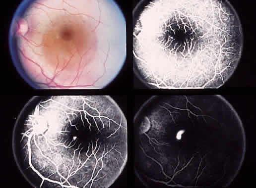
Fig. 3. Color (A) and red-free (B) photgraphs of a fundus with soft drusen and hyperpigmentation. Soft drusen hyperfluoresce during the early phase of angiography (C) and stain in the late phase (D).

Soft drusen are present in 13% to 20% of the population, and the prevalence is age-related.11 Their presence increases the risk for development of RPE abnormalities, geographic atrophy, and SRNV.24,25 For this reason, the presence of soft drusen is sufficient to make a diagnosis of AMD. The Macular Photocoagulation Study (MPS) showed that eyes with soft drusen had a 30% risk of CNV development, whereas eyes without large drusen or pigment abnormalities had only a 10% risk.24

PERIPHERAL DRUSEN

Although drusen are most commonly found in the macular region, they may be present anywhere in the fundus. Peripheral drusen are often surrounded by rings of pigment. This pigmentation may be confluent, linear, or radiating, producing a reticular pattern referred to as senile reticular pigmentary degeneration. There is a strong correlation between peripheral drusen and pigmentation and the macular changes of AMD.26,27

SICK RPE

Also known as degeneration of the RPE or nongeographic atrophy of the RPE, sick RPE is manifested by mottling of the pigment epithelium with hypopigmentation in a stippled pattern. This occurs in 8% to 10% of the population.28 Degeneration of the RPE leads to degeneration of the overlying neurosensory retina. Fluorescein angiography reveals hyperfluorescence in areas of hypopigmentation alternating with blocked fluorescence in areas of pigment clumping. Histopathologically, there are basal linear and basal laminar deposits.29 Eyes with sick RPE are at a higher risk of developing soft drusen, geographic atrophy, and choroidal neovascularization.

RPE HYPERPIGMENTATION

Focal hyperpigmentation of the RPE is more often seen in eyes with soft or large drusen (Fig. 4). Such a finding is correlated with a higher risk of developing geographic atrophy or more soft drusen. Focal hyperpigmentation also confers a higher risk of progression to neovascularization, particularly when large drusen or soft drusen are also present. Patients with choroidal neovascularization in one eye have a 58% to 73% risk of developing CNV in the fellow eye over 5 years if there are more than two large drusen and focal hyperpigmentation in the macula.30

Fig. 4. Focal retinal pigment epithelium hyperpigmentation is noted along with drusen.

PIGMENT EPITHELIAL DETACHMENT

Serous detachment of the RPE is common in AMD. As drusen develop, there is a loosening of the adherence between the RPE basement membrane and the inner collagenous portion of Bruch's membrane, predisposing the patient to the development of a serous pigment epithelial detachment. The RPE and its underlying basal lamina may be elevated by serous fluid, and the overlying sensory retina may be detached as well. There is no practical difference between a serous pigment epithelial detachment and soft drusen formation. Although these detachments develop in a variety of morphologic patterns, they typically appear as sharply demarcated, dome-shaped, round to oval elevations of the RPE (Fig. 5). The surface of the pigment epithelial detachment may be smooth and homogeneous, or it may be associated with overlying pigment clumping or atrophy. The presence of an overlying sensory retinal detachment is usually evidence of underlying SRNV. A shallow sensory retinal detachment that extends beyond the area of the pigment epithelial detachment may result from the breakdown of the physiologic RPE pump or a disruption of the tight junctions between RPE cells, but usually it is the result of underlying SRNV.31,32

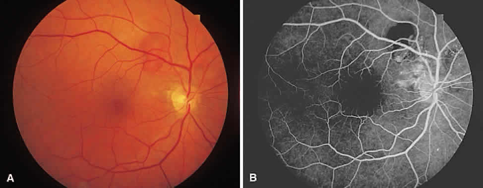
Fig. 5. A. Pigment epithelial detachment in association with soft drusen. B. On angiography, dye fills the detachment but is limited by its margins.

A serous pigment epithelial detachment often causes visual distortion and loss of vision.33 The prognosis for these detachments depends on the underlying disease process.34 If present in younger patients, it may be part of central serous retinopathy and has a good prognosis. If, however, older persons develop serous pigment epithelial detachment, there is the likelihood for the development of underlying SRNV33,35 in one third to one half of patients.32,36 The size of the pigment epithelial detachment is important in determining the likelihood of SRNV development, with smaller detachments less likely to harbor underlying neovascular growth.33,35 Although laser photocoagulation may flatten a detachment, no beneficial effect from laser therapy has been shown in the absence of SRNV.37

Pigment clumping may develop on the dome of the pigment epithelial detachment, or the detachment may spontaneously collapse with associated RPE atrophy.33,38,39 It is also possible that eyes with pigment epithelial detachments that go on to develop SRNV may already harbor unappreciated occult vessels.40,41

Fluorescein angiography of a serous pigment epithelial detachment typically shows gradual and uniform staining of the sub-RPE material, similar to the increased intensity of light seen when the rheostat on a light bulb is turned up. There is, however, a great range of morphologic and angiographic findings with these detachments, and the presence of occult SRNV can be postulated on the basis of these patterns.42 Angiographic findings suggestive of underlying occult SRNV include irregular hyperfluorescence, multiple areas of intense hyperfluo-res-cence (“hot spots”) not associated with overlying window defect, and a notch sign.32,42,43 A notch sign refers to an area of pigment epithelial detachment where the smooth, rounded border is crimped in toward the center of the detachment. This is believed to be caused by traction on the pigment epithelial detachment by an underlying occult SRNV membrane.

The pathogenesis of a pigment epithelial detachment has not been clearly elucidated. It has been postulated that the fluid may be derived from blood vessels growing on the inner surface of Bruch's membrane; however, subpigment epithelial neovascularization is not universal in these detachments.42,44 It has also been suggested that the fluid may come, in part, from the RPE, rather than from the choroid. Fluid is actively transported across the RPE from the sensory retina toward Bruch's membrane. If Bruch's membrane is changed or compromised, perhaps by lipid deposition that renders it hydrophobic, then there would be significant resistance to water flow. The result would be fluid accumulation in the sub-RPE space.45

ATROPHIC MACULAR DEGENERATION

The atrophic form of macular degeneration has been called geographic because the areas of RPE atrophy tend to form well-demarcated borders that do not relate to specific anatomic structures (Fig. 6).3,13,38,46 It has also been called dry macular degeneration, senile choroidal sclerosis, and central areolar choroidal atrophy.13,47,48 Atrophic macular degeneration leads to significant visual loss in almost all cases; patients describe a gradual and subtle blurring of vision that relates to the degree of foveal involvement. A minimum of 175 μm of the retina should be involved to classify a patient as having geographic atrophy.12

Fig. 6. A. Color photograph of geographic atrophy and drusen. B. Angiogram shows window defect type of hyperfluorescence.

Atrophic macular degeneration may evolve in several ways. Areas of RPE atrophy often follow the fading of drusen. Dystrophic calcification may also be seen in soft drusen. Eventually, the overlying RPE atrophies.21 Initially, these areas form discrete patches of atrophy that coalesce with time. The areas initially affected usually involve the perifoveal area and spread around the fovea, preserving central vision. However, in most cases, with time the fovea is involved and central vision is lost.7,39

Macular atrophy may occur in the absence of faded drusen. The RPE atrophy may be preceded by a reticular configuration of pigment clumping. The atrophy tends to expand more rapidly in all other directions than toward the fovea.39 However, although the fovea shows a certain amount of resistance to the expansion of atrophy, it is ultimately involved, and in most cases central vision is lost. Occasionally, a bull's-eye ring of atrophy surrounds the fovea before the center is involved. This foveal sparing may be due to an accumulation of lipofuscin in the posterior pole or possibly to the presence of xanthophyll pigment.7 The progression of atrophy is variable, ranging from 15 to 375 μm a year.39

Geographic atrophy may follow the spontaneous or laser-induced collapse of a serous pigment epithelial detachment, especially when formed by a confluence of soft drusen.7,49,33,36,39,49,50 In areas of geographic atrophy, the outer nuclear layer rests directly on the basal lamina. The outer plexiform layer is thinned and vacuolated, but the inner nuclear layer is less affected. The choroidal capillaries remain patent for a time, but in long-standing RPE atrophy, they end abruptly at the end of the atrophic area. The gradual obliteration of the choriocapillaris that follows RPE atrophy can be seen clinically as slow filling on fluorescein angiography that, in time, progresses to nonfilling.51

In cases of long-standing atrophy, choroidal arteries may appear to be sheathed, prompting the name choroidal sclerosis. Histologic examinations, however, reveal only a fibrous replacement of the arterial media, without thickening of the walls; the lumen remains patent.7 Choroidal atrophy occurs secondary to a reduction in nutritional demands of the outer retina. As the choriocapillaris and middle layer of choroidal vessels are lost, the larger choroidal vessels become more prominent.

Functional visual loss can result from a ring scotoma, despite good Snellen acuity. It takes approximately 10 years from the onset of atrophic change to reach a level of 20/200, although an interval of 5 years has been observed between the time of atrophic foveal encroachment and the loss of fixation.7,52 Each year, 8% of eyes decrease from 20/50 or better to 20/100 or worse.39 There is a great tendency for the areas of atrophy to become bilaterally symmetric with time. Also, SRNV can develop at the border of atrophic RPE, especially if there is a ring of soft drusen around the geographic atrophy. Because the neovascular response requires the presence of degenerating RPE, the SRNV is almost always found at the edge of atrophy rather than in the center of an atrophic area.

SUBRETINAL NEOVASCULARIZATION

The Eye Disease Case-Control Study Group showed that there is an increased risk to progression to SRNV if there is a history of cigarette smoking, elevated serum cholesterol, and multiparity.53 A protective effect of carotenoids was also found. Serum levels of vitamins E and C were not associated with the risk of progression to SRNV.54

SRNV refers to the growth of abnormal new vessels beneath the sensory retina or RPE. Choroidal neovascularization, another term for SRNV, emphasizes the choroidal vascular origin of the new vessels.55 SRNV is an abnormality found in many diseases in which the integrity of the RPE, Bruch's membrane, and choriocapillaris has been compromised.56–59 The effect on vision from SRNV derives from its tendency to leak fluid beneath and into the sensory retina, to bleed, and to create a fibrovascular disciform scar in the macular region.

Patients with AMD often develop SRNV and initially present with blurred or distorted vision, micropsia, or scotoma.60 Clinically, the subretinal neovascular membrane may appear as a dirty-gray discoloration beneath the retina and may be accompanied by an overlying sensory retinal detachment and cystoid edema. A pigmented ring may encircle the membrane, caused either by RPE hyperplasia or pigment ingestion. Often subretinal or sub-RPE hemorrhage is present. In the absence of retinal vascular disease, trauma, or a choroidal tumor, subretinal hemorrhage should be considered to be caused by SRNV until proven otherwise. Rarely, hemorrhage from SRNV may dissect through the retina to create a vitreous hemorrhage. Occasionally, SRNV can be responsible for a turbid sensory retinal or RPE detachment. Turbidity indicates the presence of proteinaceous exudate, fibrin, and blood, all products of abnormally leaking vessels. Subretinal exudate is also a strong indicator of SRNV. If the RPE overlying the SRNV membrane has become thinned, then the arborized vascular structure of the membrane may be seen.61

The presence of a pigment epithelial detachment may also signal the presence of an SRNV membrane, especially if associated with sensory retinal detachment, subretinal exudate or blood, chorioretinal folds, or an RPE tear.16,17,43 Subretinal or sub-RPE blood in the pigment epithelial detachment is also a sign of associated SRNV. The blood and proteinaceous content in a pigment epithelial detachment stains slowly with fluorescein compared with the rapid angiographic staining of a serous pigment epithelial detachment.

Fluorescein angiography is useful in confirming the presence and location of SRNV, which provides information concerning visual prognosis. The early angiogram in classic CNV may reveal a discrete, lacy, arborized plexus of vessels, with a nodular-appearing border (Fig. 7). The edges of the neovascular net are scalloped and irregular. Later in the angiographic study, there is an intense hyperfluorescent fuzziness to the border of the membrane, the result of dye leakage into the subretinal space (Fig. 8). As the study progresses, there is pooling of dye in the subretinal space. The newer, less mature vessels at the edge of the membrane tend to leak more than older vessels. Therefore, the border of the membrane becomes fuzzier and larger, as opposed to the well-defined edge of a serous pigment epithelial detachment. Staining of the fibrous tissue that accompanies all new vessel growth and the pooling of the dye under the pigment epithelial detachment or sensory retina cause the late hyperfluorescent pattern of SRNV. Accumulation of fluorescein dye in cystic spaces in the overlying retina may also contribute to the late hyperfluorescence.

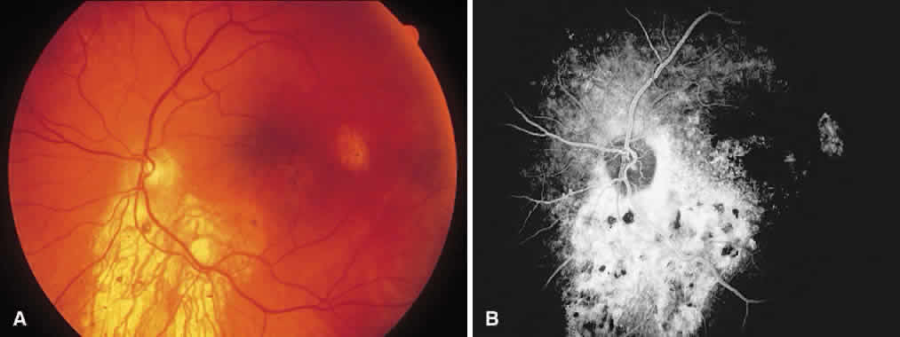
Fig. 7. A. Color photograph shows macular thickening associated with subretinal hemorrhage. B. Full-phase angiogram shows lacy hyperfluorescence.

Fig. 8. Angiogram shows classic subretinal neovascularization with lacy hyperfluorescence and diffuse leakage later from the membrane.

The precise location of SRNV is not always clear. Often the SRNV is obscured by hemorrhage, turbid serous fluid, pigment epithelial detachment, or extensive pigment clumping. Such membranes are referred to as occult SRNV and may be seen when relatively intact RPE overlies thin and subtle SRNV (Fig. 9). Occasionally, clinical signs of SRNV (hemorrhage, exudate, serous detachment) are present, but the SRNV cannot be found by angiography. The visual prognosis is poorer for these eyes than for those with distinct, clearly defined SRNV membranes. In some cases, SRNV hyperfluoresces late in the angiogram but not early. There may be a gradual oozing of fluorescein through multiple pinpoint areas of RPE overlying the SRNV membrane. The slow hyperfluorescence may be related to a decreased flow of blood through the membrane or to a relatively intact RPE.

Fig. 9. A. Color photograph showing subretinal hemorrhage associated with drusen and pigment change. B. Early-phase angiogram shows blocking from subretinal hemorrhage (note overlying retinal vessels) associated with early, diffuse macular hyperfluorescence. The lesion is occult as no classic subretinal neovascularization is noted. C. Late-phase angiogram shows further leakage.

An RPE tear may occur at the junction of attached and detached RPE, where contraction of the SRNV may tear the RPE. The torn edge of RPE retracts and is pulled by the fibrovascular tissue (Fig. 10). In the acute period, a serous detachment of the sensory retina may occur because of the fluid leaking from the exposed choriocapillaris. The subretinal fluid usually resorbs in a few days.43,62,63

Fig. 10. A. Atrophy temporal to the fovea and rolled pigment epithelium centrally, created by a retinal pigment epithelial tear in the left eye. B. Fluorescein angiogram showing intense hyperfluorescence created by the window defect after a retinal pigment epithelial tear. The hypofluorescence corresponds to the area where the pigment epithelium rolled together in accordion fashion.

Indocyanine green angiography is sometimes helpful in cases of occult CNV. Because indocyanine dye is protein-bound, it does not leak from the choriocapillaris as does fluorescein. On stimulation with an infrared source, the dye fluoresces weakly. Well-defined areas of fluorescence are termed hot spots and are assumed to represent CNV.

Feeder vessels can occasionally be seen extending from a natural or laser scar to an area of recurrent CNV (Fig. 11).64 Although they can be seen on fluorescein angiography, high-speed indocyanine green video angiography is often used to increase detection in cases of new subfoveal CNV.65 Feeder vessels can be thought of as having either an umbrella pattern or a racquet pattern.66 In an umbrella pattern, the feeder vessel is oriented in an anteroposterior fashion between the CNV complex and choroidal space. With a racquet pattern, the feeder vessel passes more obliquely from the choroidal space toward the CNV complex.

Fig. 11. A. Atrophic laser scar with hemorrhage and subretinal fluid at the temporal border signals recurrent subretinal neovascularization. B. Early-phase angiogram shows a racquet pattern feeder vessel giving rise to the recurrent choroidal neovascularization complex.

HISTOLOGY

The SRNV arises from the choroid and passes through Bruch's membrane to invade the subpigment epithelial and subsensory retinal space. The vessels may pass through pre-existing breaks in Bruch's membrane or may produce breaks in the membrane.67 However, breaks in Bruch's membrane can occur without any growth of vessels from the choriocapillaris. The SRNV proliferates under the RPE and destroys it. The vessels may also cause hemorrhagic or serous detachment of the RPE, the sensory retina, or both. Cystic changes may develop in the overlying sensory retina.

New vessel growth is accompanied by fibrous tissue, which ultimately becomes the dominant pathologic change and results in a disciform scar involving the choroid, RPE, and sensory retina.59 Active neovascularization may intermittently bleed at the edges of the developing scar. Further, the overlying retinal vessels and the choroidal vessels supplying the scar may anastomose. Also, although found most frequently in the macular region, extramacular disciform lesions may occur and simulate choroidal melanomas.

PATHOGENESIS

The association between new vessel growth and drusen formation has been well described; both processes are related to degeneration of the RPE. Soft drusen represent the breakdown of the hyaline content in hard drusen or the focal accumulation of membrane debris that lies between the RPE basement membrane and the inner collagenous layer of Bruch's membrane. These detachments predispose to pigment epithelial detachment formation and to SRNV. Why SRNV develops is unknown, but evidence suggests that choroidal vessels, in the absence of an effective barrier and inhibitory factors released by normal RPE, may be exposed to mitogenic and chemotoxic retinal factors that stimulate SRNV.57,68,69

TREATMENT

The MPS produced its first result in 1982 and continued with more than two dozen reports. As a prospective study, it helped define acceptable treatment for CNV associated with AMD, ocular histoplasmosis, and idiopathic CNV. The influence of different laser wavelengths used in treatment was also studied.70 Laser treatment can be classified into subfoveal, juxtafoveal (1 to 199 μm from the center of the foveal avascular zone), and extrafoveal types.

EXTRAFOVEAL TREATMENT. The first MPS71 report found that 60% of untreated eyes and only 25% of treated eyes had severe visual loss (six or more lines of vision) in the AMD group. These results led to the termination of this arm of the study. The 3-year results72 found that 62% of untreated eyes and 47% of treated eyes had suffered severe visual loss. The 5-year results were similar to the 3-year results.73 The higher incidence of severe visual loss in the treated group at the 3-year follow-up versus baseline was due to recurrent CNV, which was seen in 54% of eyes. Almost three quarters of recurrences occurred in the first year after treatment. Cigarette smoking was associated with a higher risk of recurrence (85% vs. 51%).

JUXTAFOVEAL TREATMENT. The 5-year data for AMD showed that treated eyes were less likely to have severe visual loss compared with untreated eyes (55% versus 65%) (Fig. 12). Recurrences occurred in 47% after 5 years in the treated group.73 Peripapillary CNV was studied as well (Fig. 13): it was found that 14% of treated and 26% of untreated eyes had severe visual loss.74

Fig. 12. A. Subretinal hemorrhage is noted in the juxtafoveal area. B. Midphase angiogram shows lacy hyperfluorescence. This was treated with laser photocoagulation. C. Two weeks after treatment, color photograph shows early scar formation. D. Persistent choroidal neovascularization is noted on angiography at the temporal border. This requires retreatment.

Fig. 13. A. Neurosensory retinal detachment is noted extending from the optic nerve to the fovea. B. Angiogram shows hyperfluorescence near the temporal border of the nerve from a peripapillary choroidal neovascularization membrane. Note how the margins of the detachment are made evident by the dye. This cushion of fluid protects the neurosensory retina as the choroidal neovascularization membrane is treated with laser photocoagulation. C. Two weeks after laser treatment, a laser scar can be seen adjacent to the nerve. The neurosensory detachment has resolved. D. Posttreatment angiogram shows staining of scar. No detachment is noted.

SUBFOVEAL TREATMENT. Three months after subfoveal treatment, 20% of treated eyes suffered severe visual loss compared with 11% of untreated eyes. After 2 years, 20% of treated eyes still had suffered severe visual loss, but now 37% of untreated eyes had suffered severe visual loss. After 4 years, 23% of treated and 45% of untreated eyes had suffered severe visual loss.

The size of the subfoveal CNV is important in predicting benefit from subfoveal laser treatment. CNV size is grouped as small lesion (1 MPS disc area or less), medium lesion (1 to 2 MPS disc areas), or large lesion (greater than 2 MPS disc areas). Lesion size is compared with preoperative visual acuity:

  • For small lesions, if vision is 20/125 or worse, treated eyes uniformly do better than untreated eyes. If vision is 20/100 or better, treated eyes do worse in the first year and then better than untreated eyes thereafter.
  • For medium lesions, if vision is 20/200 or worse, treated eyes uniformly do better than untreated eyes. If vision is 20/160 or better, treated eyes fare worse than untreated eyes in the first year and then do better thereafter.
  • For large lesions, if vision is 20/200 or worse, treated eyes do only slightly better than untreated eyes. If vision is 20/160 or better, treated eyes do worse for 18 months and there is little difference between treated eyes and untreated eyes thereafter.75

Many retina specialists use these criteria in considering which patient to treat with the laser based on the clinical setting. The patient must understand the goals of treatment and the high risk of persistence (13%) and recurrence (35%) with subsequent need for more laser therapy.76 After the initial treatment, an angiogram is repeated after 2 weeks to evaluate the adequacy of treatment. Subsequent to this visit, patients may be seen at 3-month intervals for the first year and at 6-month intervals for the second year. Angiography is performed if needed.

The choice of laser wavelength for SRNV near the fovea depends on the clinical setting, but theoretically it should include the use of wavelengths not absorbed by yellow xanthophyll pigment (green, yellow, or red). The presence of blood or the degree of pigmentation in the area to be treated also may determine the choice of wavelength (e.g., krypton or dye red [620 to 630 nm] laser will be minimally absorbed by the red hemoglobin in blood). However, the MPS found no significant difference between argon green and krypton red laser.75

Feeder vessels can be treated with laser, limiting the amount of damage done to retinal tissue. Ideally, a wavelength absorbed by hemoglobin (576 nm [dye yellow] or 530 nm [YAG green]) should be used. In a study by Shiraga and associates,65 there was a 70% closure rate, with 30% having recurrent or persistent SRNV. Vision was stabilized or improved in 68%, and good preoperative vision, lack of fibrous scarring, small size of SRNV (less than 2 disc areas), and further distance from the foveal avascular zone were associated with better visual outcomes. Staur-enghi and colleagues66 found that multiple treatments may be needed to close a feeder vessel. The dimensions and number of the feeder vessels can compromise success.

Complications from laser treatment for SRNV associated with AMD include inadequate treatment (because of inadequate intensity or inadequate coverage of the membrane), resulting in persistent or recurrent SRNV. Other complications are RPE tear, epiretinal membrane formation, inadvertent foveal photocoagulation, choroidal vascular obstruction, choroidal folds, hemorrhage caused by overly intense treatment, and nerve fiber layer defects that occur with heavy treatment of SRNV that lies beneath the papillomacular bundle.59,77

Patients with AMD should test their vision daily with the Amsler grid chart. If they notice blurred vision or distortion, they should report immediately to their physician, who should perform a complete macular examination and fluorescein angiogram. If treatment is to be performed, it should be carried out immediately. SRNV can grow up to 40 μg per day, and a treatable membrane that initially is safely away from the foveal center may be beneath the fovea in a short time. Time is of the essence for patients with SRNV secondary to AMD.

Patients with CNV in one eye often ask about the risk to their fellow eye. The MPS30 showed that the following are independent risk factors when found in the fellow eye: five or more drusen (RR = 2.1), focal hyperpigmentation (RR = 2.0), one or more large drusen (more than 63 μm, RR = 1.5), and hypertension (RR = 1.7). The 5-year incidence rates for development of CNV in the fellow eye ranged from 7% (no risk factors) to 87% (all four risk factors).

If patients have untreatable SRNV or bilateral disciform scars, they should be assured that SRNV almost never causes total blindness and that their functional abilities may be improved somewhat with low vision aids. They should be made aware of the range of services available to persons with a visual handicap, including large-print newspapers, books-on-tape, television viewers, and community support services, and they should be encouraged to continue independent, productive, and creative lives.

EXPERIMENTAL TREATMENT MODALITIES LIGHT.

Light in the visible and ultraviolet spectra has been postulated as contributing to macular degeneration.78 Recent studies, however, have failed to confirm a link between photic stress and macular degeneration. These studies include the Chesapeake Bay Waterman Study,79 the Eye Disease Case-Control Study,80 and the Beaver Dam Eye Study.81

NUTRITION. Antioxidants have also been postulated to cause oxidative damage to the RPE, with a consequent increased risk of macular degeneration. The National Health and Nutrition Examination Survey found that people who ate diets rich in fruits and vegetables were less likely to have AMD.82 The Eye Disease Case-Control Study80 found that increased levels of carotenoids were somewhat protective against SRNV in AMD. No benefit was found with increased levels of vitamins C and E or selenium. Zinc is involved in many of the enzymatic processes in the retina, and some believe that supplementation may be protective against AMD. The Eye Disease Case-Control Study, however, failed to find a benefit with higher zinc levels. Currently, the Age-Related Eye Disease Study is being conducted to study the relation between nutrition, diet, and AMD.

LASER TREATMENT OF DRUSEN. Laser treatment of soft drusen has been studied as a means of stabilizing acuity and preventing SRNV. One large study, the Choroidal Neovascularization Prevention Trial, found an increased risk of SRNV in laser-treated eyes that halted the study.83 Olk and colleagues84 used the infrared diode laser at 810 nm to treat patients with drusen. Treatment parameters included light and visible burns and invisible burns. Both treatment types led to reduction in the number of drusen in approximately 65%, but this happened earlier in the visible burn group (12 versus 18 months). Treated eyes did somewhat better in terms of vision compared with nontreated eyes. This difference was noted at 12 months and was still significant at the 2-year follow-up. Importantly, treated eyes had similar rates of visual loss as untreated eyes. The rate of development of CNV was similar between treatment and observation groups at 2 years. A forthcoming trial, the Prophylactic Treatment of AMD Trial, will compare subthreshold treatment of drusen with infrared laser to observation.

PHOTODYNAMIC THERAPY. Photodynamic therapy is a two-step process in which a photosensitizing drug is injected and then irradiated with monochromatic laser light. This results in the creation of a triplet state that combines with oxygen to create free radicals. Tumors and neovascular tissue have an affinity for the photosensitizing drug and are therefore more prone to free radical damage than healthy tissue. The primary damage is to endothelial cells.85,86 It is hoped that with photodynamic therapy, the overlying healthy RPE and retina can be spared, therefore allowing vision to be maintained.

Two photosensitizers are undergoing trials by the Food and Drug Administration: verteporfin, a benzoporphyrin derivative, and purlytin (tin ethyl etiopurpurin, SnET2). Verteporfin has an absorbance peak at 689 nm, purlytin at 664 nm. Verteporfin (Visudyne; Ciba Vision, Duluth, GA) is being evaluated in the Treatment of Age-Related Macular Degeneration with Photodynamic Therapy Investigation (TAP) and the Verteporfin in Photodynamic Therapy study (VIP). The TAP study involves patients with subfoveal, classic CNV less than 5400 μm and visual acuity between 20/40 and 20/200. The VIP study includes cases of subfoveal CNV not included in the TAP study (occult CNV or visual acuity better than 20/40).

The dose of Visudyne to be injected is based on body surface area (6 mg/m2 body surface area). A 30-ml solution of the drug is injected over 10 min-utes. Five minutes after the infusion is complete, the macula is irradiated over 83 seconds with a spot size 1000 μm greater than the greatest size of the CNV complex.87 One-year outcome data for phase 1 and 2 studies were recently released.88 Sixty-one percent of treated eyes had lost fewer than 15 lines of vision compared with 46% of untreated eyes; restated, 15% of treated eyes showed a benefit from Visudyne. Eyes with more classic CNV (more than 50% of the lesion) did better with photodynamic therapy. There was no treatment benefit in lesions whose classic component made up less than 50% of the lesion. Leakage recurred by 12 weeks in all cases, although it was often less pronounced than before verteporfin therapy. Retreatment with Visudyne is effective in treating recurrent leakage, although long-term data are lacking.89

Photodynamic therapy should still be considered experimental because long-term data are not available. Its application in the armamentarium against neovascular AMD will become clear as more data accumulate.

TRANSPUPILLARY THERMOTHERAPY. Transpupillary thermotherapy involves irradiating the macula with an infrared laser at 810 nm. Beam size is adjustable up to 3.0 mm to cover the lesion size. Power is adjusted so that an invisible to barely visible treatment effect is noted. The treatment lasts for 60 seconds. Reichel and colleagues90 conducted a pilot study with 15 patients (16 eyes) who had occult CNV. Visual acuity was 20/400 or better in all patients. Follow-up ranged from 6 to 25 months. Two eyes received one retreatment (13%), and one eye required two retreatments (6%). Three eyes (19%) improved by two or more lines, nine (56%) were stable, and four (25%) lost two or more lines of vision. Fifteen eyes (94%) were judged to have less exudation. A larger trial should help define the use of transpupillary thermotherapy in the treatment of occult AMD.

RADIATION THERAPY. Studies evaluating x-ray irradiation for CNV in AMD have mostly been small.91 X-ray treatment appears to halt progression of CNV but may not cause regression. Many studies have not found a significant difference between radiation doses.92 The Radiation Therapy for Age-Related Macular Degeneration Study studied the effect of eight fractions of 2 Gy external beam radiation compared with sham treatment in a randomized, prospective, double-blind trial involving many centers. They found no benefit at the 1-year point.

Radiation-associated choroidal neovasculopathy was reported by Spaide at the 1998 American Academy of Ophthalmology meeting.93 Twelve of 95 patients (13%) treated with 1000 cGy external beam radiation developed this, compared with 7 of 98 (7%) treated with 2000 cGy. There was significant loss of acuity.

PNEUMATIC DISPLACEMENT OF SUBMACULAR HEMORRHAGE. Dense submacular hemorrhage often results in poor vision, with acuity less than 20/200 in most cases.94 Heriot95 used tissue plasminogen activator (tPA) and C3F8 gas to displace subfoveal blood. The procedure should be performed within 3 weeks of symptoms. A dose of 25 μg tPA in 0.1 ml is injected into the vitreous cavity. A paracentesis is then performed, followed by injection of SF6 or C3F8. After 3 hours, prone positioning is assumed for the next day to 3 days. Alternatively, gas can be injected without tPA and the patient re-evaluated after 48 hours. If displacement is inadequate, tPA can then be injected.96 Displacement of blood can allow earlier diagnosis of the cause of hemorrhage. The underlying pathology may dictate final visual acuity. This technique has not been evaluated in a prospective study.

SUBMACULAR SURGERY. Because laser photocoagulation of subfoveal CNV results in loss of central vision and is associated with a high rate of recurrence, the Submacular Surgery Trials were initiated to study whether removal of subfoveal CNV could alter the known history. This study has several arms that address new subfoveal CNV, subfoveal CNV associated with histoplasmosis or idiopathic, and submacular hemorrhage related to AMD. Long-term follow-up has found that AMD-related CNV treated with submacular surgery can result in better-than-expected stable vision. Nevertheless, damage to the RPE leads to loss of photoreceptors and choriocapillaris that ultimately degrades vision (Fig. 14).97

Fig. 14. A. Large choroidal neovascularization complex is noted with subretinal hemorrhage and fluid. B. The patient underwent submacular surgery with removal of the choroidal neovascularization complex. Note loss of retinal pigment epithelium in area of membrane and hemorrhage. C. Three months after surgery, the choriocapillaris has atrophied and choroidal vessels are better visualized.

MACULAR TRANSLOCATION. Techniques and indications for macular translocation are evolving. Machemer and Steinhors98 initially reported a technique using 360-degree retinotomy with intraoperative removal of CNV and scleral resection. This has evolved to scleral imbrication with limited macular translocation. Postoperative laser photocoagulation is performed to the CNV. Complications are common. There is a 10% to 20% retinal detachment rate, with occasional proliferative vitreoretinopathy. Limited translocation with scleral imbrication results in formation of a retinal fold, which is usually directed inferiorly by detaching the inferior retina. As much as 30% of the time, the fold may go through the fovea.99 As more experience is gained with macular translocation, its place in the treatment of CNV from multiple causes will become more clear.

Back to Top
CONDITIONS ASSOCIATED WITH SUBRETINAL NEOVASCULARIZATION
Any disease process that results in an abnormality at the level of the RPE-Bruch's membrane-choriocapillaris complex can be associated with SRNV and subsequent disciform scarring. Table 1 lists the diseases that have been associated with the development of SRNV. This section includes the entities most commonly complicated by SRNV.


TABLE 1. Conditions Associated With Subretinal Neovascularization

  Hereditary/Congenital
  Best's vitelliform dystrophy
  Adult-onset foveal pigment epithelial dystrophy (adult vitelliform dystrophy)
  Choroideremia
  Coloboma (chorioretinal, retinal)
  Fundus flavimaculatus (Stargardt's disease)
  Osteogenesis imperfecta
  Retinitis pigmentosa
  Dominant drusen
  Optic nerve pits
  Degenerative
  Age-related macular degeneration
  Angioid streaks
  Drusen (optic nerve head)
  Myopia
  Inflammatory/Infections
  Acute multifocal posterior placoid pigment epitheliopathy
  Birdshot choroidopathy
  Histoplasmosis
  Recurrent multifocal choroiditis (punctate inner choroiditis, multifocal choroiditis)
  Rubella
  Fungal chorioretinitis (Candida, Aspergillus, others)
  Sarcoidosis
  Serpiginous (geographic helicoid peripapillary choroiditis)
  Toxoplasmosis
  Vogt-Koyanagi-Harada
  Sympathetic ophthalmia
  Toxocara canis
  Syphilis
  Traumatic
  Intraocular foreign body
  Photocoagulation (endolaser photocoagulation)
  Cryotherapy
  Subretinal fluid drainage (internal, external)
  Scleral perforation
  Tumor
  Choroidal nevus
  Choroidal osteoma
  Combined retinal---retinal pigment epithelium hamartoma
  Choroidal melanoma
  Choroidal hemangioma
  Metastatic carcinoma
  Other
  Chronic retinal detachment
  Neovascularization of the ora serrata
  Idiopathic subretinal neovascularization of the young
  Retinal telangiectasis


HISTOPLASMOSIS

The presumed ocular histoplasmosis syndrome is a major cause of visual impairment in the central and eastern United States, especially in the Ohio and Mississippi River valleys. Histoplasma capsulatum is a dimorphic soil fungus that is acquired through the respiratory tract and usually results in an asymptomatic, self-limited infection, but it can develop into a disseminated infection in immunocompromised persons.100,101 Presumed ocular histoplasmosis syndrome has been clinically linked to H. capsulatum, even though organisms are usually not found in autopsied eyes of patients known to have the disorder.

The triad of ocular findings in histoplasmosis comprises peripheral, punched-out, atrophic choroidal lesions; peripapillary choroidal scars; and macular lesions with or without SRNV. The peripapillary disc scarring consists of choroidal and RPE atrophy, with a line of pigment at the disc margin of the scar, as opposed to some normal eyes with optic nerve crescents that have pigment at the outer border of the scar. The active lesion of histoplasmosis is a small round area of choroiditis that can create a small overlying sensory retinal detachment. This picture can be difficult to differentiate from active SRNV. The areas of active choroiditis are not associated with vitreous cells. The lesions become quiet and create atrophic, and later pigmented, scars called histo spots, which appear symptomatically during the patient's lifetime.

SRNV may be associated with a previous area of histoplasmosis scarring (Fig. 15). The visual prognosis is poorer for eyes with SRNV and a perifoveal choroidal scar in the fellow eye. If such a scar exists, there is a 20% to 25% chance of the development of a neovascular membrane in 5 years; without such scars, there is only about a 4% chance of SRNV developing from the scarring around the disc.102,103 A follow-up evaluation of patients in the MPS showed that choroidal neovascularization is often preceded by an atypical histo spot.104 Macular SRNV, however, has been described in the absence of preceding atrophic scars.105 Prognosis for vision remains good: 81% can be expected to have 20/20 or better acuity after 5 years when the fellow eye has had CNV.104

Fig. 15. A. Histo spots are noted in association with choroidal neovascularization (CNV) membrane (dirty green) and subretinal hemorrhage. B. Corresponding angiogram shows transmitted fluorescence from histo spots, mild peripapillary atrophy, blocked fluorescence from subretinal hemorrhage, and CNV in association with a histo spot. C. After laser treatment to the CNV complex, hemorrhage has resolved and there is early scarring of the CNV membrane. D. Corresponding angiogram shows staining of the scar.

Laser treatment has been shown to be an effective treatment of SRNV associated with presumed ocular histoplasmosis syndrome and located 200 μm or more from the center of the foveal avascular zone.106 In the ocular histoplasmosis section of the MPS, 34% of untreated eyes versus 9% of treated eyes suffered severe visual loss of six or more lines after 18 months of follow-up. Corticosteroids are not believed to be of benefit for the associated SRNV.

Subretinal surgery for the removal of subfoveal CNV has been performed in ocular histoplasmosis. Berger and colleagues107 reported that acuity improved by two lines in 35%, was unchanged in 44%, and worsened in 21%. Recurrence of the subfoveal CNV occurred in 38% and was more common in eyes that had undergone preoperative laser photocoagulation. The median time to recurrence was 5 months. Perfusion of the subfoveal choriocapillaris may have implications for visual improvement after subfoveal surgery.108 If subfoveal membranes have an extrafoveal ingrowth site, the prognosis is better for visual recovery as opposed to a subfoveal or unidentifiable ingrowth site.109 Melberg and colleagues110 studied recurrences and found that recurrent lesions were extrafoveal in 16%, juxtafoveal in 18%, and subfoveal in 66%. These eyes were treated with laser photocoagulation, further subfoveal surgery, or observation. Eyes amenable to laser did better in terms of vision.

The differential diagnosis of presumed ocular histoplasmosis syndrome includes a group of diseases of unknown cause that create multifocal choroidal scarring and are known variously as recurrent multifocal choroiditis, punctate inner choroidopathy, multifocal choroiditis with progressive subretinal fibrosis, and pseudohistoplasmosis.111–114 Many patients who have these diagnoses also have associated vitreous inflammation, and the pattern of scarring changes with time more than in presumed ocular histoplasmosis syndrome.

MYOPIA

Degenerative, or pathologic, myopia is one of the leading causes of blindness in the world.115 The chorioretinal lesions that accompany myopia have been attributed either to progressive axial elongation or to a genetically determined atrophic process independent of anatomic eye wall changes.116–118

The myopic disc is oval, with a long vertical axis, and may appear tilted or oblique; it often has a temporal concentric area of depigmentation, called a temporal crescent. Posterior pole staphyloma may be present early in life, and its incidence increases with age.119 The sensory retina, RPE, and choroid are thinned, and the underlying choroidal vessels are prominent (Fig. 16). Chorioretinal atrophy may be extensive and may appear as round or irregular yellow-white areas with focal deposits of pigment.

Fig. 16. Myopic fundus with temporal crescent and prominent choroidal vessels.

The margins of atrophy are well demarcated. There is a tendency for multiple small areas of focal atrophy to coalesce into large lesions that may involve the fovea and result in visual loss.

Lacquer cracks are linear or stellate ruptures in Bruch's membrane and RPE and are typical of pathologic myopia. They are usually fine, irregular in caliber, yellowish-white, horizontal, branching breaks that occur most often in the macular region (Fig. 17).

Fig. 17. A. Crescent-shaped lacquer crack in a myopic eye. B. Angiogram shows window defect type of hyperfluorescence. C. One year later, the lacquer crack has extended.

The occurrence or extension of a lacquer crack can be associated with a macular hemorrhage that may occur in the absence of SRNV. Because of the close anatomic relationship between Bruch's membrane, the RPE, and the choriocapillaris, a break in Bruch's membrane can result in a small, focal, round subretinal hemorrhage that usually resolves, leaving good central vision.120,121

SRNV may occur in 5% to 10% of the myopic population; with highly myopic eyes, the chances of SRNV may exceed 40%.122 SRNV usually exhibits a small, juxtafoveal gray lesion with a shallow overlying sensory detachment and subretinal hemorrhage (Fig. 18). With time, an unusual pigmentary response, called a Fuchs spot, results in RPE hyperplasia over the SRNV. Progressive atrophy often develops around the Fuchs spot. The SRNV of pathologic myopia does not have the same proliferative potential as the SRNV of AMD. The visual prognosis in pathologic myopic eyes with SRNV is not clearly defined. Some investigators report the visual acuity stabilizing after an acute decrease in vision associated with the SRNV-related subretinal hemorrhage (Forster's spot).

Fig. 18. A. Choroidal neovascularization (CNV) membrane in a myopic eye associated with subretinal hemorrhage. Treatment was deferred. B. Corresponding angiogram shows hyperfluorescence from the CNV membrane and blockage from the hemorrhage. C. Nine months later, the CNV membrane has enlarged and more blood is noted. D. Corresponding angiogram shows the hyperfluorescence from the classic CNV membrane. E. Treatment was performed with a resultant atrophic macular scar.

The benefits of laser treatment for myopic SRNV are not well established. Some studies have shown an initial benefit with laser treatment of juxtafoveal SRNV, but it seems to have questionable long-term advantage.115,120,123 Laser treatment in pathologically myopic eyes carries additional risks, with the precise location of the foveola difficult to determine. Moreover, the laser scar tends to enlarge with time and may extend through the fovea.124–126 Additional, although experimental, treatment modalities include submacular surgery, macular translocation, and photodynamic therapy for CNV associated with high myopia.

ANGIOID STREAKS

Angioid streaks develop from pathologic alterations of the RPE, Bruch's membrane, and the choriocapillaris.125,127 The primary change is a linear, cracklike dehiscence in Bruch's membrane. These cracks, called angioid streaks, radiate from the peripapillary area with irregular or serrated margins throughout the fundus. Angioid refers to the similarity between these reddish-brown cracks and blood vessels (Fig. 19).

Fig. 19. A. Reddish-brown angioid streaks are seen radiating from the nerve. B. Corresponding angiogram shows hyperfluorescence along the course of the angioid streaks.

SRNV may grow through the breaks in Bruch's membrane and result in disciform macular scarring. Vision may be lost because of slowly progressive macular atrophic changes related to the angioid streaks. Patients may also develop a serous pigment epithelial detachment associated with SRNV.

Angioid streaks are found in several conditions, including pseudoxanthoma elasticum (GronbladStrandberg syndrome), osteitis deformans (Paget disease), sickle cell anemia, senile elastosis (aging), and fibrodysplasia (Ehlers-Danlos syndrome).59,127–131

Pseudoxanthoma elasticum is a systemic disease of elastic tissue primarily affecting the eyes, the cardiovascular system, and the skin. Its name derives from the peculiar confluent, yellowish papules (plucked chicken-skin appearance) that develop on the flexural surfaces of the neck and antecubital fossa. Most patients with pseudoxanthoma elasticum have angioid streaks. Other findings in pseudoxanthoma elasticum include peau d'orange pigmentation, caused by the mottled blending of posterior confluent yellow RPE lesions with the normal orange peripheral pigmentation; reticular pigmentary dots, occurring in 10% to 15% of patients with pseudoxanthoma elasticum and perhaps indicating a predisposition for the development of SRNV; punched-out RPE atrophic spots, often accompanied by central pearly white, crystalline lesions; and optic nerve drusen.59,125,127–129,132–134

Paget disease, characterized by irregular thickening, rarefaction, and deformity of bone, results in calcification of Bruch's membrane and angioid streaks (10% to 15%). These patients may develop SRNV and disciform scarring, and some may show the peau d'orange pigmentary discoloration seen in pseudoxanthoma elasticum.59,135,136

SRNV associated with angioid streaks may be treated with laser, which may improve the visual prognosis in some eyes. Other experimental treatments remain unproven.

CHOROIDAL RUPTURE

Traumatic choroidal ruptures may result from blunt ocular trauma (Fig. 20). They appear as crescent-shaped gaps in the RPE-Bruch's membrane-choriocapillaris complex. The rupturing of the Bruch's membrane and choriocapillaris can result in bleeding under the RPE and sensory retina. After the hemorrhage clears, the large curvilinear ruptures can be seen. They often run parallel, or concentric, to the disc and often pass through or near the fovea (Fig. 21). These choroidal ruptures may be associated with the development of SRNV. There is occasionally a history of trauma that leads to subretinal hemorrhage and SRNV. Laser treatment may be effective in treating SRNV that threatens the fovea.137,138

Fig. 20. Dramatic fundus photo of a traumatic macular hole with a large choroidal rupture temporally. Note the pigment disruption.

Fig. 21. A. Concentric choroidal rupture associated with hemorrhage. B. Angiogram shows hyperfluorescence corresponding with the rupture.

Back to Top
EPIRETINAL MEMBRANES
Epiretinal membranes are known by a variety of names, including cellophane maculopathy, surface wrinkling retinopathy, internal limiting membrane contraction, premacular gliosis, and macular pucker.139–142 Most terms refer to clinical descriptions of retinal pathology created by epiretinal membranes of varying degrees of severity and various cell morphologic characteristics. Epiretinal membranes occur with numerous ocular conditions and diseases, including vascular, inflammatory, dystrophic, traumatic, neoplastic, and degenerative conditions. Most epiretinal membranes occur after posterior vitreous detachment (idiopathic epiretinal membranes) or retinal detachment surgery (and should be considered a mild manifestation of the proliferative vitreoretinopathy spectrum of disease).141–145

Most patients with epiretinal membranes are asymptomatic. Those with more severe epiretinal membranes may notice blurred vision, distortion, diplopia, and even profound central visual loss. Most epiretinal membranes remain stable, although approximately 25% of eyes have progressive loss of visual acuity.59,139,140 The thickness of the membrane on optical coherence tomography may correlate with the degree of visual loss.146 Angiography may show vascular leakage and macular edema due to the membrane.

The ophthalmoscopic findings depend on the severity of the epiretinal membrane. A membrane may be present as a glistening, refractile sheen overlying the macula (cellophane maculopathy). More severe epiretinal membranes may wrinkle or pucker the macula. Vascular tortuosity and tethering, foveal ectopia, sensory retinal detachment, punctate hemorrhages, irregular vascular dilation, leakage and macular edema, cotton-wool spots or fluffy areas of whitish inner retina (presumably related to traction-induced axoplasmic stasis), and pseudoholes may also occur. A pseudohole occurs when an epiretinal membrane contracts and draws in the rim of the foveal depression, steepening the slope of the foveal depression. This alters the foveal light reflex, making it appear a darker red. The combination of a darker-red foveola and the steeper slopes of the depression underlying the epiretinal membrane may give the impression of a full-thickness macular hole (i.e., a pseudohole).

Different cell types may account for the formation of epiretinal membranes, although fibrous astrocytes have been implicated most frequently.147–151 Other cell types, including fibrocytes, RPE cells, myofibroblasts, macrophages, inflammatory cells, hyalocytes, and vascular endothelial cells, have also been described as contributing to epiretinal membranes. Myofibrocytes may be a more common cell type in younger patients (aged 30 or younger).152,153

When epiretinal membranes cause significant visual loss, surgical removal is considered. The surgery involves removing the posterior vitreous, elevating the epiretinal membrane edge with a fine instrument, and tangentially peeling the epiretinal membrane from the retina with an intraocular foreign body forceps (Fig. 22). The chances of improved vision with posterior vitrectomy and epiretinal membrane stripping are 70% to 80%.145,154–156 Patients can expect to regain approximately half of the vision lost due to the development of the membrane, with vision improving up to 9 months after surgery; however, most of the visual improvement comes in the first 2 or 3 months after surgery. Even with macular edema, vision can be expected to improve at least a few lines.156 Juvenile idiopathic epiretinal membranes can also be successfully treated with vitrectomy.157

Fig. 22. A. Epiretinal membrane causing distortion of macular vessels. B. Postoperative result showing normalization of retinal structure.

The most common complication of vitrectomy and epiretinal membrane stripping surgery is increased nuclear sclerosis, occurring during the subsequent years in most patients.145,158,159 Many of these patients require cataract surgery within several years of their vitrectomy. The surgery also carries with it the other complications associated with any posterior vitrectomy.

Back to Top
VITREORETINAL TRACTION SYNDROME
The vitreoretinal traction syndrome refers to a partial posterior vitreous detachment with residual adhesion between the attached vitreous and the macula and optic nerve.59,160–167 This traction can produce decreased vision, distortion, diplopia, cystic retinal change, retinal striae, and pseudoswelling at the disc. In the vitreomacular adhesion, the posterior hyaloid face can be observed sweeping down to the elevated foveal rim or perifoveal area and, infrequently, to the central fovea. Sometimes there is a broad area of persistent vitreomacular attachment 2 to 3 mm around the macula. Rarely, there is a complete posterior vitreous detachment with only a thin anteroposterior sheet extending to the center of the macula. The traction produced can create minimal foveal ectopia, sensory macular elevation, an uneven ridge of elevated foveal crest, retinal vascular leakage, and macular edema (Fig. 23). Occasionally, cystic retinal change may be produced in the absence of retinal vascular leakage, presumably related to the traction itself. There is almost always a curvilinear area of opacified hyaloid on the optic nerve margin related to the traction that also creates disc leakage on angiography.

Fig. 23. Full-phase angiogram shows optic nerve leakage and macular edema in a case of vitreomacular traction syndrome. B-scan ultrasonography shows vitreous inserting into the macula with resultant traction.

Spontaneous separation of the posterior vitreous occurs in approximately 11% of cases over 5 years.168 Macular detachment secondary to vitreoretinal traction has also been reported.169 Vitrectomy is indicated if progressive macular degeneration, macular detachment, cystic change, and decreased vision are present. The vitreoretinal anatomy is readily observed at the time of surgery. In some eyes, the gentle traction created by the initial central vitrectomy disengages the vitreous from the macula. In most cases, a tapered extrusion needle, an intraocular foreign body forceps, or a membrane-peeling instrument is needed to tease the posterior hyaloid face from its macular attachment. Histologic studies show that the fibrous astrocyte is the predominant cell type. Myofibrocytes can be present as well and can cause retinal traction.170

When the vitreomacular traction is removed, the retinal vascular leakage usually stops, although the cystic retinal changes may remain unchanged. Macular cystic changes not associated with retinal vascular leakage may also disappear after surgical release of the vitreomacular traction. Visual acuity is seen to improve two lines in up to 75% of cases unless a macular traction detachment is present.169,171,172 Forty percent may achieve better than 20/50 vision.171

Back to Top
CYSTOID MACULAR EDEMA
Cystoid macular edema (CME) is a frequent cause of reduced central vision. It is a nonspecific pathologic response to a variety of ocular conditions and diseases, including many retinal vascular and chorioretinal diseases dealt with in other chapters in these volumes. Essentially, any condition inducing intraocular inflammation, retinal vascular occlusion, or retinal traction may be associated with CME. The most common conditions in which CME is found are shown in Table 2.


TABLE 2. Conditions Associated With Macular Edema

  Retinal Vascular Diseases
  Diabetic retinopathy
  Venous occulsion (Branch vein occlusion, Central retinal vein occlusion)
  Radiation retinal vasculopathy
  Retinal telangiectasis (idiopathic juxtafoveal telangiectasis, Coats disease, Leber's miliary aneurysms)
  Retinal arterial macroaneurysm
  Retinopathy of prematurity
  Retinal Inflammatory Diseases
  Birdshot chorioretinopathy
  Pars planitis
  Sarcoidosis
  Toxoplasmosis
  Posterior scleritis
   Behçet's
  Cytomegalovirus
  Other intraoclar retinitis and infections
  Tumors
  Choroidal hemangioma
  Combined retinal---retinal pigment epithelium hamartoma
  Choroidal melanoma
  Toxic
  Epinephrine
  Nicotinic acid
  Latanoprost (Xalatan)
  Postoperative
  Cataract surgery (Irvine-Gass syndrome)
  Retinal detachment
  Keratoplasty
  Refractive surgery
  Photocoagulation (especially panretinal photocoagulation for proliferative diabetic retinopathy)
  Cryopexy
  Hereditary
  Familial exudative vitreoretinopathy
  Goldmann-Favre syndrome
  Infantile cystoid macular edema
  Juvenile X-linked retinoschisis
  Retinitis pigmentosa
  Optic pit
  Dominant cystoid macular edema
  Other
  Vitreoretinal traction syndrome
  Epiretinal membranes
  Subretinal neovascularization
  Postpartum cystoid macular edema
  Idiopathic cystoid macular edema


CME and visual loss may develop weeks to years after cataract surgery. This postoperative visual decline was first noted after intracapsular surgery in the 1950s; it was later demonstrated by fluorescein angiography to be due to intraretinal cysts.173–176 These cysts result either from edematous, degenerating Müller cells or from expansion of extracellular spaces in the inner plexiform and inner nuclear layers caused by serous exudate.177–179 The Irvine-Gass syndrome, initially referred to as CME after intracapsular cataract extraction, is often used to describe visually significant CME that follows cataract surgery, regardless of extraction technique or intraocular lens implantation (Fig. 24).

Fig. 24. Color (A) and fluorescein angiogram (B) in cystoid macular edema. Classic petaloid edema is seen on angiography. The disc is hyperfluorescent.

Although 50% to 70% of patients who have had an intracapsular cataract develop retinal vascular leakage on fluorescein angiography, few show clinical evidence of cystic change.59,180 Extracapsular cataract extraction is associated with CME much less frequently than is intracapsular cataract extraction.181 Phacoemulsification is associated with CME in less than 0.5% of cases.182 YAG capsulotomy increases this risk to 1.2%.183 A complete posterior vitreous detachment is usually present, and generally there is an inflamed disc on fluorescein angiography. Rarely, there is a vitreomacular attachment accompanying the wet macula. There may be photophobia and conjunctival injection if vitreous strands to the wound are present.

Eyes with vision that decreases to the 20/70 level or worse have roughly a one in three chance of spontaneous improvement.184 Two thirds of patients with intracapsular cataract extraction with CME regain 20/30 or better vision 3 to 12 months after surgery.59 Occasionally, spontaneous visual improvement takes place years after surgery, leaving 0.5% to 2% of patients with uncomplicated intracapsular cataract extraction with permanent visual loss secondary to CME.59 The incidence is roughly the same for extracapsular cataract extraction and posterior lens implant, although it is increased with iris fixation and anterior chamber lenses.185,186 More recently, it has been shown that the thickness of the macula correlates better with visual function than does leakage on angiography or visual acuity.187

The pathogenesis of CME remains uncertain. Depending on the clinical situation, varying theories can be used to understand the pathogenesis. Considerable experimental and clinical evidence implicates prostaglandin-mediated inflammation. Prostaglandins are compounds derived from cell membrane phospholipids, specifically arachidonic acid. Prostaglandins and related compounds increase microvascular permeability, resulting in breakdown of the blood-ocular barrier and CME. In the event of vitreous incarceration to a cataract wound, a mechanical force may be exerted on the vitreomacular interface, leading to vascular decompensation and leakage.

TREATMENT

If CME has been present for 2 or more years, there likely has been irreversible change in the macula.182 Therefore, prompt treatment on recognition of the disorder is warranted. Studies have shown that prophylactic treatment before cataract surgery can be effective in reducing the incidence of angiographic CME and clinically significant macular edema.187 Diclofenac (Voltaren; Ciba Vision) and flurbiprofen (Ocufen) are as effective as prednisolone for prophylaxis.188

The pharmacologic treatment of CME has been directed at blocking prostaglandin activity, either by inhibiting the formation of arachidonic acid with corticosteroids or inhibiting the formation of prostaglandins from arachidonic acid with indomethacin (Indocin), diclofenac, or ketorolac (Acular; Allergan, Irvine, CA). In most cases, topical medication is initiated first.

If CME is refractory to topical medicines, repository steroids should be considered. Methylprednisolone acetate (Depo-Medrol) or triamcinolone acetonide (Kenalog) can be given in the sub-Tenon's space or in the retrobulbar location for a more concentrated and constant dose of anti-inflammatory medicine.189 The usual dose is 40 mg delivered in 1 ml solution. The injection can be repeated after 3 months if there is no improvement. Most specialists stop after a total of three doses if there has been no improvement, because there are no prospective studies establishing the duration and frequency of injections. Caution must be used in eyes known to be sensitive to steroid-induced elevation of the intraocular pressure.

Acetazolamide (Diamox) has been used for CME associated with retinitis pigmentosa. Experimentally, acetazolamide facilitates transport of water from the subretinal space, across the retinal pigment epithelium, into the choroid.190 It has been shown to lead to resolution of CME in postoperative pseudophakes as well.191 Dorzolamide (Trusopt), a topical carbonic anhydrase inhibitor, does not appear to be effective in treating CME associated with retinitis pigmentosa or chronic uveitis.192,193 Another carbonic anhydrase inhibitor, methazolamide (Neptazane), does not appear to be as effective as acetazolamide in CME associated with retinitis pigmentosa.194

In eyes with chronic CME, vitreous incarceration, photophobia, and peaked pupil, vitrectomy may be beneficial.184 In postoperative eyes where there is an intact posterior capsule and an in-the-bag intraocular lens, justification of vitrectomy is less clear. Macular grid photocoagulation has been attempted in uveitis-associated CME, with resolution of the edema and improvement or stabilization of vision in some patients.195

Back to Top
IDIOPATHIC JUXTAFOVEAL RETINAL TELANGIECTASIS
Idiopathic juxtafoveolar retinal telangiectasis (IJRT) represents a group of disorders that have been subclassified based on ophthalmoscopic findings and natural course. Gass and Oyakawa196 originally proposed the classification in 1982, and Gass and Blodi197 revised it in 1993.

Secondary causes of juxtafoveal telangiectasis include retinal venous obstruction, macroaneurysms, diabetes, radiation retinopathy, carotid occlusive disease, sickle retinopathy, retinal hemangiomas, retinal astrocytoma, intraocular inflammation, Eales disease, and tapetoretinal degenerations.197 These should be excluded to classify a patient as having IJRT. It has been suggested that patients with juxtafoveal telangiectasis be tested specifically for diabetes, because patients with early background diabetic retinopathy may have changes very similar to IJRT.

The ophthalmoscopic appearance varies between the subsets of IJRT. Table 3 summarizes the features of the different groups. Group 1A patients are usually male with unilateral involvement. Most are white. The area of involvement is located temporal to the fovea and is often more than 2 disc diameters and straddles the horizontal raphe. There is vascular compromise of the telangiectatic vessels with exudate and retinal edema. Gass believed that this is developmental and in the spectrum of Coats syndrome. Loss of vision is due to exudate into the fovea and macular edema, often with cystoid change. Laser photocoagulation to prevent accumulation of lipid in the fovea is helpful. Spontaneous resolution of the edema with improvement in vision can occur over many years. Close to half of these patients have peripheral telangiectasis.198 Group 1B patients are similar except that the involved area is smaller (Figs. 25 and 26). Laser treatment is effective for macular edema and exudates.


TABLE 3. Idiopathic Juxtafoveolar Retinal Telangiectasis


 Group 1AGroup 1BGroup 2AGroup 2BGroup 3AGroup 3B
Usual age at onsetThird decadeFourth decadeFifth decadeFirst decadeFourth to fifth decadeThird to fourth decade
       
Gender predilectionMale (90%)Male (88%)NoneMale (100%)FemaleMale
Area of involvement2 DD or moreSmall1 DD around foveaSmallEnlargement of FAZ to more than 1500 <gm>mEnlargement of FAZ
LateralityUnilateral (90%)Unilateral (88%)Bilateral (98%)Bilateral (100%)BilateralBilateral
TreatmentConsider photocoagulation earlyConsider photocoagulation earlyConsider foro neovascularizationConsider for neovascularizationNoneNeurologic evaluation
Associated findingsExudates, retinal edemaExudatesFoveal atrophy, right-angled venules, crystalline deposits, SRNVSRNV, telangiectasisCapillary occlusion, minimal exudatesCapillary occlusion, minimal exudates
Familial involvementNoNoYes, in 2%Yes, in 100%YesYes
Extent of visual loss20/40 but may be HMUp to 20/4020/200 or less20/7020/5020/50
CommentsA mild form of Coats syndromeLimited form of 1A with better prognosisMost common form; some develop diabetesOnly two cases reportedPolycythemia, arthritis, and hypoglycemia are associatedAssociated with fibrinoid necrosis of the brain

DD, disc diameter; FAZ, foveal avascular zone; HM, hand motion; SRNV, subretinal neovascularization.
(Based on Gass JDM, Blodi BA: Idiopathic juxtafoveolar retinal telangiectasis. Ophthalmology 100:1536---1546, 1993)


Fig. 25. A. Group 1B idiopathic juxtafoveolar retinal telangiectasia. Circinate hard exudate and edema are noted. B. Angiography shows telangiectatic change in the area of the exudate.

Fig. 26. Macular telangiectasia are seen with associated exudate. The aneurysmal dilation is more in line with Coats syndrome. Peripheral telangiectasia were noted.

Group 2A is the most common form of IJRT (Fig. 27). The patients are older than in group 1, with an equal incidence of men and women. The disease occurs bilaterally. Gass believed that this is probably an acquired disorder. Visual loss occurs from atrophy of the foveal retina rather than exudation or retinal edema. However, leakage does occur and can be shown angiographically.199 This is centered within 1 disc diameter of the fovea. Patients have metamorphopsia or decreased vision.

Fig. 27. Group 2A idiopathic juxtafoveolar retinal telangiectasia. A & B. Both eyes show involvement. A retinal pigment plaque is noted in the right macula, denoting more extensive involvement. Note the surrounding retinal atrophy. C & D. Angiogram shows leakage greater in the right eye than the left. Note the transmitted fluorescence from foveal atrophy.

Gass described five stages in the evolution of this group. The earlier stages are often noted in the less-involved eye of symptomatic patients.

Stage 1 involves staining of the vessel wall due to its thickening. This is often in the deep capillary plexus. Green and associates200 showed histologically that there is thickening of the basement membrane with loss of endothelial cells and pericytes, very similar to diabetic vascular change. These capillaries do not adequately carry on metabolic exchange, leading to derangement in retinal function with eventual loss (stage 2). As the outer capillary bed becomes more involved, right-angled venules from the inner retina form to drain the bed (stage 3). Further metabolic compromise leads to loss of photoreceptor cells and consequently a decline in visual acuity. Gass did not explain the role of the RPE in the nourishment of the photoreceptors in relation to this disease. With loss of retinal tissue, a lamellar macular hole may form (stage 4).197,201 This loss of retinal cells induces intraretinal neovascularization, SRNV, exudation, and hemorrhage. RPE cells can form pigment plaques after having migrated into the retina. Development of neovascularization can lead to rapid loss of vision. In Gass' study, no patient had a neovascular complex that was amenable to treatment, either because loss of vision was due to foveal atrophy or the neovascularization was located below the fovea.

In Gass' study, patients who were treated before the development of neovascular membranes often did worse. There have been two patients who have had subretinal hemorrhage after laser treatment. These were probably due to SRNV that developed after treatment.197,202 Patients who seem to have group 2A lesions have been treated successfully with laser.203 However, a more recent report of 14 patients204 did not show any improvement or stabilization of vision after parafoveal laser treatment. Retinal hemorrhage, vascularized retinal scars, and RPE changes can develop after laser treatment, but this does not seem to affect visual acuity. Consequently, the treating ophthalmologist and the patient must decide on the best course of action. A stereo-angiogram showing leakage, combined with a recent decline in vision, may warrant a trial of treatment. Untreated, vision may worsen, stabilize, or, rarely, improve.

Golden refractile deposits near the inner surface of the retina were seen to occur in the reports by Gass196,197 and Moisseiev and colleagues.205 The number of deposits does not appear to correlate with the degree of visual loss. These may be the footplates of degenerating Müller cells, as Gass suggested, or calcium or cholesterol deposits. Group 2A patients have developed diabetes, and some persons with diabetes have had disease resembling group 2A IJRT. Consequently, a fasting blood sugar may be advisable in these patients.

There are few reports of group 2B IJRT occurring in families and affecting younger patients. This group does not show right-angled venules, retinal deposits, or pigmented plaques but does display SRNV. Both patients in Gass' paper were males who had bilateral disease with visual loss to 20/70.

Group 3A disease has been seen in women. There is telangiectasis with mild exudation. The major finding is capillary occlusion with enlargement of the foveal avascular zone. Visual acuity, though, is better than would be expected and may be maintained for years. The disease is bilateral, and there can be familial involvement. Arthritis, polycythemia vera, and hypoglycemia can be associated.

Interestingly, Mansour and Schachat206 found the foveal avascular zone to be smaller in IJRT; however, they did not use the current classification of IJRT. Because group 2A is the most common form of IJRT, most patients studied probably belonged in this group.

Group 3B patients have an enlarged foveal avascular zone with associated central nervous system vasculopathy. They are often men, and the disease can run in families. Thus, all group 3 patients should be evaluated for systemic diseases.

IJRT probably represents different diseases. Group 1 is more often associated with leakage and is in the spectrum of Coats syndrome. Group 2 patients display some leakage, but ultimately visual loss is from foveal atrophy. Group 3 is associated with changes in the foveal avascular zone. Photocoagulation is useful in group 1 and often group 2 but would not be beneficial, theoretically, in group 3. Treatment spots are usually 100 to 500 μm and applied as light burns in a grid pattern to the areas of leakage outside the foveal avascular zone. The vessels do not need to be treated directly. Neovascular membranes, when treatable, are treated with confluent, more moderate burns.

Back to Top
MACULAR HOLE
Although macular holes may be associated with trauma, myopia, CME, and inflammation, most are idiopathic.207,208 Idiopathic macular holes occur most commonly in the seventh decade of life and in women more frequently than men (2:1),209–216 leading some to suggest a possible hormonal role.211,213,217 Also, a higher-than-expected prevalence of cardiovascular disease in some series has suggested possible vascular factors.214,215 However, a more recent study found only age older than 55, female sex, estrogen use, and increased plasma fibrinogen (greater than 2.95 g/L) to be significant.218 As many as 100,000 people are affected in the United States.219

THEORY

Various theories have been advanced attempting to explain the pathogenesis of macular holes. Historically, patients diagnosed with a macular hole usually had a history of significant ocular trauma. Consequently, trauma was believed to be the cause of macular holes. Cystic degeneration at the fovea was noted by Fuchs and Coats, leading to the theory that trauma or vascular changes could cause cystoid changes in the fovea. The cystic spaces would then coalesce to form a macular hole.219 Involutional macular thinning was proposed by Morgan and Schatz.214 It was believed that choroidal vascular changes led to foveal and RPE changes. Cystic degeneration then caused permanent architectural changes at the fovea and RPE, leading to involutional macular thinning. Vitreous traction at the thin retina would then lead to a macular hole. The vitreous has been implicated in the pathogenesis of macular holes since the early 20th century.219 Initially, vitreous bands were thought to develop, although none were clinically observed. Worst220 described a premacular vitreous bursa which was attached to the macula and could exert anteroposterior traction on the fovea. However, no anteroposterior traction bands were clinically observed that would have led to a macular hole. It is generally believed that detachment of the posterior vitreous is not a causal event in the evolution of a macular hole.219

CLASSIFICATION

Gass221 is credited with the currently accepted classification and theory of macular hole formation. Tangential vitreous traction is thought to be the cornerstone of premacular and macular hole formation.212,222 Tangential traction of the prefoveal cortical vitreous detaches the foveola, creating a yellow spot 100 to 200 μm in diameter. This is a stage 1A hole (Fig. 28). Intraretinal xanthophyll is thought to impart the yellow color. As the fovea elevates, the yellow spot becomes a ring, a stage 1B lesion (Fig. 29). Vision is in the 20/25 to 20/70 range. Fine, radiating retinal striae may also be seen clinically. As the hole progresses, a dehiscence of the photoreceptors occurs at the umbo, creating an occult hole. As the prefoveal vitreous cortex separates, an eccentric hole is revealed. Detachment of the posterior hyaloid may occur, creating a pseudo-operculum. This is termed a stage 2 hole and is defined as being less than 400 μm in diameter. The yellow ring may disappear because traction is relieved with separation of the prefoveal vitreous cortex. Histopathologic studies of opercula show two types. Pseudo-opercula are composed of astrocytes, Müller cells, and internal limiting membrane. True opercula have glial elements as well as avulsed foveal cones.223 A stage 3 hole is reached when the hole enlarges to more than 400 μm in diameter (Fig. 30). This may occur because of myofibroblastic activity near the internal limiting membrane.224 Yellow, nodular deposits are seen on the surface of the RPE in half of stage 3 holes. Nearly all stage 3 holes have a cuff of surrounding neurosensory detachment. If a posterior vitreous detachment occurs (with a Weiss ring), a stage 4 hole is reached. Vision is between 20/70 and 20/400 in stage 3 and 4 holes.

Fig. 28. Stage 1A macular hole with small yellow spot.

Fig. 29. Stage 1B macular hole with yellow ring.

Fig. 30. A. Stage 3 macular hole with cuff of subretinal fluid. B. Angiography reveals transmitted fluorescence at the hole and leakage into the subretinal space corresponding the cuff of fluid.

DIAGNOSIS

Diagnosing a macular hole is best done clinically with either a contact or non-contact lens at the slit lamp. The main considerations in the differential are a fenestrated epiretinal membrane (a pseudohole) and lamellar macular holes. Useful clinical tests include the Watzke-Allen sign and the laser aiming beam test. Both tests are subjective. In the Watzke-Allen test, a narrow beam of light is focused on the retinal defect. The patient is then asked whether the light beam appears to narrow or whether there is a break in the narrow slit beam. In the laser aiming beam test, a 50-μm spot size is used; with a full-thickness macular hole, the spot disappears when placed in the area of the hole.225 Technologies such as scanning laser ophthalmoscopy and optical coherence tomography can elegantly show macular holes but are not widely available and may not add significant new information.219 Lamellar macular holes do not progress to full-thickness macular holes.212 Scanning laser ophthalmoscope microperimetry can differentiate between a lamellar hole and a full-thickness hole by evaluation whether a scotoma is present.226

HISTOLOGY

Histopathologic analysis of eyes with full-thickness macular holes shows oval or round retinal defects with rounded retinal edges and a surrounding cuff of retinal detachment with subretinal fluid. Up to 79% have CME, and 68% have epiretinal membranes.219,227 Surgically, Blain and coworkers228 found that an epiretinal membrane was removed in only 30% of cases. There is a variable amount of photoreceptor disruption in the area of neurosensory detachment. Madreperla and colleagues229 studied a treated macular hole and found the hole to be sealed by Müller cell processes; photoreceptors adjacent to the hole appeared normal. Rosa and associates230 also found that Müller cell proliferation in a postoperative macular hole case had closed the hole.

NATURAL HISTORY AND FELLOW EYES

The evolution of stage 1 macular holes to full-thickness macular holes was studied by the Vitrectomy for Prevention of Macular Hole Study Group.231 The study was designed to compare surgery and observation for stage 1 lesions in patients with full-thickness macular holes in their fellow eyes. Although the study was terminated due to poor recruitment, it was found that 40% of those in the observation group progressed to full-thickness holes over 2 years, whereas 37% progressed in the vitrectomy group (not statistically significant). Another finding was that in eyes with stage 1 holes, visual acuity was predictive of progression to full-thickness holes. Eyes with acuity between 20/50 and 20/80 had a 66% rate of progression; eyes with acuity between 20/25 and 20/40 had a 30% risk of progression.232 Most stage 2 macular holes go on to become stage 3 or 4 full-thickness macular holes irrespective of the presence of a full posterior vitreous detachment.219

The Vitrectomy for Macular Hole Study Group also reported on vitrectomy versus observation for stage 2 macular holes.233,234 Vitrectomy was performed with removal of the posterior hyaloid and fluid-gas exchange. After 1 year, 71% of eyes randomized to observation progressed to full-thickness macular hole, whereas only 20% of eyes assigned to surgery progressed. In eyes that developed full-thickness holes after surgery, the size of the hole was found to be smaller and reading ability was better than in observation eyes that progressed.

The risk to the fellow eye is reported as being between 3% and 22%.189,235 More recently, the 5-year risk for normal fellow eyes was reported at 15.6%.236 Chew and colleagues237 found the rate of development of a new macular hole during follow-up in fellow eyes that were unaffected at baseline was 4.3% for 3 or fewer years of follow-up, 6.5% for 4 to 5 years of follow-up, and 7.1% for 6 or more years of follow-up. Spontaneous regression of the macular hole occurred in 8.6%. If the macula is normal and a posterior vitreous detachment is present, the risk is believed to be closer to 3%. In the presence of a stage 1 lesion and no posterior vitreous detachment, the risk is probably 40%.219

TREATMENT

In 1991, Kelly and Wendel238 reported on vitrectomy with removal of the posterior hyaloid and fluid-gas exchange followed by postoperative face-down positioning to treat full-thickness macular holes. Their initial results were 58% anatomic success. In these successfully repaired eyes, vision improved by two lines or more in 73% (42% overall). In their follow-up study,239 anatomic success was achieved in 73%, with 55% showing visual improvement of two lines or more. They also suggested that holes of less than 6 months' duration have better outcomes. This appears to be confirmed by other studies (Fig. 31).240,241

Fig. 31. A & B. Preoperative photograph shows a stage 3 macular hole with focal hyperfluorescence seen angiographically. C & D. Postoperative photograph shows that the hole is closed and the fovea is flat. Angiogram shows resolution of the hyperfluorescence.

The critical step in surgery for macular holes involves removing the posterior hyaloid face, thereby relieving the traction forces on the hole. If an epiretinal membrane is present, it should be removed to facilitate flattening of the edges of the macular hole. Removal of the internal limiting membrane has recently been advocated by some surgeons.242,243 One study found a 96% closure rate after internal limiting membrane removal versus a 71% closure rate without removal.242

Adjuvants had been advocated to encourage closure of the hole after surgery. Substances have included autologous blood and blood products,244 transforming growth factor β2,245 and thrombin. Transforming growth factor β2 was later found not to be effective,246,247 and most surgeons have moved away from using autologous blood products.248 Tamponade of the break is then created by filling the vitreous cavity with a nonexpansile concentration of gas, either C3F8 or SF6. Strict face-down positioning is maintained for 7 to 14 days by patients with gas tamponade. When this position is maintained, debris may be seen in some cases on the corneal endothelial surface.

Air and silicone oil have also been used. The use of silicone oil requires removal of the oil in approximately 6 weeks. There can be slight progression of nuclear sclerosis. The oil should fill greater than 90% of the vitreous cavity.249

VISUAL OUTCOMES

The Vitrectomy for Treatment of Macular Hole Study Group evaluated surgery versus observation for stage 3 and 4 macular holes.250 Although follow-up was only 6 months, improvement in visual function was noted with macular hole surgery. More than three fourths had progression of nuclear sclerotic cataract, confounding the visual acuity testing. Other studies have noted continual improvement of visual acuity several years after surgery. In a recent study,251 median acuity increased from 20/125 before surgery to 20/50 1 year after. Median acuity was 20/30 3 years after macular hole surgery. The authors found that progression of nuclear sclerotic cataract masks visual improvement in the first year. Patients consequently have good visual recovery after cataract surgery.

COMPLICATIONS

Significant complications have been reported with macular hole surgery. Patients may develop neck and shoulder pain from strict face-down positioning. Use of intraocular gas can cause intraocular pressure elevations in the postoperative period; this can usually be managed medically.252 As with any vitreous surgery, especially with the use of gas, phakic patients will have progression of nuclear sclerosis.

Surgery involves forcible detachment and peeling of the posterior hyaloid face. This can lead to formation of retinal breaks and retinal detachment. It is recommended that an indirect, depressed ophthalmoscopic examination be performed during surgery before fluid-air exchange. Nevertheless, retinal breaks can develop in the postoperative period: the Vitrectomy for Treatment of Macular Hole Study Group reported a rate of 11%.253 Others have reported an incidence of retinal detachment and retinal tears between 1% and 14%.239,254,255 One study found the average time between surgery and retinal detachment was 5.5 weeks.256 There is a higher likelihood that breaks will be located inferiorly.254,256 These detachments can be treated with good anatomic and visual results. Heier and colleagues256 used vitrectomy techniques with scleral buckling to repair the detachments, reasoning that vitrectomy allows removal of all subretinal fluid by endodrainage, thus preventing postoperative accumulation of fluid in the posterior pole, and thereby opening the macular hole.

Visual field defects, some symptomatic, have been noted after uneventful macular hole surgery.257–261 The incidence, when reported, ranges between 7% and 23%. Most commonly, a dense, wedge-shaped temporal scotoma is found on visual field testing. There can be segmental pallor of the optic nerve head along with defects in the nerve fiber layer corresponding to the visual field loss. The exact cause is unclear. Theories include trauma to the optic nerve during inducement of the posterior vitreous detachment or during the fluid-air exchange, compression of the nerve fibers by gas tamponade, or dessication of the nerve fiber layer during prolonged exposure to air before air-gas exchange.

The Vitrectomy for Macular Hole Study Group reported a 33% incidence of RPE alteration.253 Poliner and Tornambe262 evaluated 12 successful macular hole repairs and found angiographic evidence of RPE swelling that resolved over time. However, a mottled fluorescence pattern was still seen. Steroids had no effect. Because the light pipe is held close to the macula during peeling of membranes from around the hole, phototoxicity may result; this can compromise visual recovery in an otherwise successful surgery.

Reopening of macular holes can occur either early in the postoperative period or after the intraocular gas has dissipated. Duker and colleagues263 reported a 4.8% incidence of late reopening. The mean time for reopening was 12.5 months but ranged from 2 to 22 months. Acute loss of vision was reported by patients, and visual recovery can occur with reoperation. The authors also reported that in one patient an epiretinal membrane sealed the hole and improved vision. Johnson and associates264 evaluated in-office air-fluid exchange for early reopening of macular holes 1 to 8 weeks after macular hole surgery. They were able to achieve closure of the hole in 74%, with improvement of vision in all cases. Ohana and Blumenkranz265 evaluated in-office laser treatment to the base of the hole followed by fluid-gas exchange. Yellow wavelength was used with a spot size of 50 to 100 μm. Twelve to 27 spots were placed one burn-width apart at 0.1-second duration and 45 to 100 mW power in a circular pattern to the RPE at the base of the open hole. The end point of the burn was a light-gray color. They were able to achieve an 87% closure rate with one or more in-office procedures. Vision improved in all patients whose holes were closed.

Back to Top
CENTRAL SEROUS RETINOPATHY
Central serous retinopathy is a disorder of unknown cause that typically affects adults aged 20 to 50,59,266–270 although persons in their 80s may also be affected. Although men are affected more often than women,59,267,270,271 there have been several cases of central serous retinopathy occurring in association with pregnancy.272,273 In some of these cases, subsequent pregnancies have been associated with a recurrence of central serous retinopathy.274,275 Central serous retinopathy may be more common in whites, Hispanics, and possibly Asians276 than in blacks.

The initial visual complaint is usually blurred vision, although positive scotoma, micropsia, and metamorphopsia may also be noted.59,267,270,277,278 Visual acuity ranges from 20/20 to 20/200, but it is most commonly 20/40 or better.59,267

The ophthalmoscopic appearance reveals a well-defined, somewhat round, shallow elevation of the sensory retina59,270 (Fig. 32). Usually the serous fluid is clear, although in some cases it may appear turbid and may obscure the underlying choroidal vascular markings.59,270 A light reflex may be seen along the margin of the serous retinal elevation. In some cases, when central serous retinopathy has been present for several weeks, fine yellow subretinal precipitates may be visible on the back surface of the elevated retina.59,267,270 A small detachment of the RPE is frequently detectable in the area of the serous retinal elevation. Usually the RPE detachment is small and appears as a grayish or yellowish, well-circumscribed elevation at the level of the RPE. Sometimes the RPE detachment is outside the area of apparent sensory retinal detachment, or several RPE detachments may be detectable. Occasionally, an RPE detachment without an overlying serous retinal detachment is observed.59 Rarely, the area of the RPE detachment is obscured by a whitish fibrinous subretinal exudate overlying its surface.59

Fig. 32. Color photograph shows serous detachment of the macula. Early in the angiogram, a hyperfluorescent spot is noted nasal to the fovea. This enlarges on the subsequent frames.

In the severe, recurrent form of this disease, the retinal detachment may extend from the macular or optic nerve area to the inferior retinal periphery in a teardrop shape, forming a dependent retinal detachment.279 Chronic fluid accumulation may produce extensive depigmentation and alterations of the RPE (Fig. 33). In some chronic cases, lipid retinal exudate, retinal telangiectasis, pigment migration, and CME may be observed.279 Another unusual variant of central serous retinopathy presents as a bullous sensory retinal detachment with shifting fluid, turbid subretinal fluid, and multiple RPE detachments.280–282

Fig. 33. A. Severe, recurrent central serous retinopathy has resulted in macular pigment alterations along with depigmentation temporally and inferior to the optic nerve. B. Corresponding angiogram highlights the alterations to the retinal pigment epithelium.

In a study of multifocal central serous disease with exudative retinal detachment and subretinal fibrosis, Sharma and colleagues283 found the macula to be involved in the detachment 72% of the time. Corticosteroid therapy was found to be of no benefit, whereas laser therapy resulted in resolution of the exudative retinal detachment in all cases. Improvement of visual acuity by greater than 2 lines was obtained in 69%. A mean of 6.7 leaks was found.

Fluorescein angiography usually demonstrates a small area of early hyperfluorescence and leakage near the margin of the RPE detachment.59,268 Occasionally, the leak rises superiorly like a smokestack and spreads to fill the serous retinal detachment. Convection currents within the serous retinal elevation are believed to influence and cause the superiorly rising leak.284 Most cases (90%), however, do not demonstrate this appearance; they show, instead, a focal area of leakage that spreads in a diffuse manner.268,271 Usually, only one area of leakage occurs, which is most frequently within 1 mm of the central fovea and located superonasally.266,271 The active leak in some cases, however, is outside the macula. Although bilateral involvement occurs in approximately 20% of cases,59,277 careful examination of the fellow eye shows evidence of previous or current disease in one third to two thirds of patients.277 RPE detachments or small areas of RPE disturbances can be demonstrated in such cases.

In severe, recurrent, or chronic cases, alterations and depigmentation of the RPE may alter the angiographic appearance. In such cases, mottled diffuse hyperfluorescence may be seen in the macular area. A distinct smokestack leak is usually not seen; rather, one or more small leaks, or hot spots, or a diffuse ooze is present. In some of these cases, large vertically oriented tracts or gutters of RPE depigmentation are evident, extending from the posterior pole to the inferior periphery.

NATURAL HISTORY

Central serous retinopathy is usually a self-limited disease with a good prognosis for visual recovery.59,267,269,270,285 Spontaneous reattachment of the retina occurs in most cases in 3 to 6 months,265,269,277,285 after which visual acuity improves and continued subjective improvement may be noted for 6 or more months. Despite this improvement, some patients may be aware of a mild persistent visual deficit, metamorphopsia, or micropsia.267,269,277 Some patients with prolonged or recurrent episodes develop some degree of permanent reduction in the visual acuity to 20/200 or less.265,276

Recurrences affect 20% to 50% of patients.59,270,277,285–287 When a recurrence develops, the leakage area is most frequently within 500 μm of the previous leakage area.271 The prognosis for visual recovery is progressively poorer with each recurrence.269–288

PATHOGENESIS

The cause of central serous retinopathy is not known. Similar psychological and personality profiles are noted in many patients with central serous retinopathy. A major life stress, either work-related or personal, is present in many persons shortly before the development of the disorder.278 One study evaluated psychological profiles and found that a type A personality profile, particularly a hard-driven and competitive personality, was much more common in patients with central serous retinopathy than in the control group.289 It has been postulated that higher plasma catecholamine levels in these patients may play a role in formation of the serous detachments. Similar detachments have been produced in monkeys after intravenous injections of epinephrine, suggesting a possible mechanism by which a personality trait may predispose to central serous retinopathy.

Adrenocorticoid and glucocorticoid steroids have been associated with central serous retinopathy. Gass and Little290 reported on three patients who developed central serous retinopathy with bullous retinal detachment after institution of oral steroid therapy. Two of the patients had a history of chronic recurrent retinal detachments before institution of corticosteroid treatment. In one patient, bilateral chronic inferior retinal detachment developed. All three patients had severe permanent visual loss in one or both eyes. Haimovici and colleagues291 reported on six patients who developed central serous retinopathy with inhaled or intranasal steroid therapy. Retinopathy developed between 1 day and 7 years after institution of steroid treatment for asthma, bronchitis, or allergic disorders. Leakage resolved in patients who discontinued the steroids. Three patients continued using steroids; retinopathy resolved in two and improved in the third. Three others stopped steroids, with resolution of central serous retinopathy. Glucocorticoids may lead to central serous retinopathy by increasing catecholamine release. This could lead to pump dysfunction at the RPE or increased permeability of the choroidal vasculature.291 Zamir292 reported on one patient who developed bilateral central serous retinopathy after intramuscular synthetic adrenocorticotrophic hormone (ACTH) treatment for arthritis. The retinopathy resolved 2 months after discontinuation. Again, RPE pump dysfunction or vascular permeability is hypothesized to lead to central serous retinopathy.

The exact physiologic alterations that occur to produce the characteristic RPE leak associated with central serous retinopathy are not clear.293,294 The RPE has a tremendous capacity to remove fluid from the subretinal space.293 For serous retinal detachment to form and be maintained, there must be an alteration in the net balance of fluid flow into the subretinal space. A focal leak in the RPE may produce a serous retinal detachment because of a dysfunction in a larger area of the RPE that prevents adequate removal of this fluid. The serous retinal detachment then increases until enough healthy RPE is exposed to this fluid and begins to pump it into the underlying choriocapillaris. At this point, the broken equilibrium is divided equally between the rate of fluid influx and efflux. How the RPE detachment that is associated with many cases of central serous retinopathy occurs is not clear.

TREATMENT

Laser photocoagulation of the RPE leak is associated with shortening the duration of the serous detachment but has not been shown to be of benefit in terms of final visual acuity or recurrence.266,269,286,287,295–300 Khosla and colleagues301 reported that laser treatment is associated with significant loss and slower recovery of contrast sensitivity.

Laser treatment is not necessary in most situations because of the generally self-limited nature of central serous retinopathy and the good visual prognosis. If a serous retinal detachment has been present 10 to 12 weeks, or if examination reveals retinal degenerative change, laser treatment is considered.288,302,303 At that time, if the location of the leak is 500 μm or more from the center of the fovea, treatment is directed to the leaking area using a 200-μm spot size and enough energy to achieve a light intensity burn.59,288,302 Treatment within 500 μm of the foveal center is believed to carry an increased risk of subsequent SRNV development. After laser treatment, the serous retinal detachment resolves within 1 to 4 weeks.274,296,304 Indirect treatment in the area of the sensory retinal detachment but away from the active leak has been demonstrated to be ineffective.287 There is no evidence to indicate any treatment benefit from corticosteroids,285 and some have suggested that they may exacerbate central serous retinopathy.290,305

Laser treatment for central serous retinopathy that has persisted for 4 months appears to shorten the duration of the serous detachment, to improve final acuity, and to decrease the incidence of recurrence. There may be some benefit of using dye yellow versus argon green in hastening the resolution of subretinal fluid.306

Back to Top
IDIOPATHIC POLYPOIDAL CHOROIDAL VASCULOPATHY
In 1990, Yannuzzi and colleagues307 described a choroidal vasculopathy that led to hemorrhage and exudation in 11 patients. Idiopathic polypoidal choroidal vasculopathy (IPCV) has also been called the posterior uveal bleeding syndrome and multiple recurrent serosanguineous RPE detachments in black women. IPCV typically affects women in their fifth to seventh decades. Darker-skinned persons are affected more than whites. There is no association with features seen in high myopia (lacquer cracks), AMD (soft drusen), or ocular inflammation. IPCV is usually bilateral. There may be an association with hypertension.

Clinically, there are two characteristic findings. A sub-RPE vascular branching network is usually located in the area of the papillomacular bundle but can be isolated below the fovea. Reddish-orange nodules, called choroidal excrescences, are found at the edge of the vascular networks. Recurrent exudation and hemorrhage can occur under the RPE or retina (Fig. 34). Breakthrough bleeding into the vitreous is possible. Over time, the orange masslike lesions flatten with resolution of the leakage. New tubular vessels emanate from the flattened orange lesion.

Fig. 34. A. Characteristic polypoidal lesion is seen under the superior temporal quadrant vein. There is a thin layer of subretinal hemorrhage. B. Angiography shows choroidal excrescences as hyperfluorescent lesions. There is blocked fluorescence from the subretinal hemorrhage.

Fluorescein angiography shows filling of IPCV lesions with the choroidal vasculature. There is blocked fluorescence due to hemorrhage, exudate, or the RPE. IPCV vessels can leak, although not as much as in a choroidal neovascular membrane seen in AMD. The polypoid structures are hyperfluorescent and stain or leak on late frames. Indocyanine green angiography shows that IPCV vessels fill slower than choroidal vessels. The network of abnormal vessels is also larger than suspected clinically. The polypoid structures leak dye slowly.308

Vision is typically better in IPCV than in AMD, and vision can be improved with treatment of the lesions. In some cases, scarring can occur with attendant visual loss. Treatment is carried out to areas of leakage from the choroidal excrescences. Unlike in AMD, the entire complex need not be treated in IPCV.

IPCV can be differentiated from AMD in the following ways. AMD patients are usually white. They usually have typical drusen and RPE changes that are often bilateral. The vessels seen in CNV due to AMD are not clinically visible. There is frequent scarring in AMD.308–311

Back to Top
ACKNOWLEDGMENT
The authors thank Mr. James Watson, CRA, for his help in assembling the illustrations used in this manuscript.
Back to Top
REFERENCES

1. McDonnel JM: Ocular embryology and anatomy. In Ryan SJ (ed): Retina, pp 5–16. St Louis: CV Mosby, 1989

2. Yanoff M: Macular pathology. In Yannuzzi LA, Gitter KA, Schatz H (eds): The Macula: A Comprehensive Text and Atlas, pp 3–13. Baltimore: Williams & Wilkins, 1979

3. Guyer DR, Schachat AP, Green WR: The choroid: Structural considerations. In Ryan SJ (ed): Retina, pp 17–36. St Louis: CV Mosby, 1989

4. Kahn HA, Moorehead HB: Statistics on Blindness in the Model Reporting Area, 1969-1970, Publication No. (NIH) 73–427. Washington DC: US Government Printing Office, 1973

5. Leibowitz HM, Krueger DE, Maunder LR et al: The Framingham Eye Study monograph: An ophthalmological and epidemiological study of cataract, glaucoma, diabetic retinopathy, macular degeneration, and visual acuity in a general population of 2631 adults, 1973-1975. VI. Macular degeneration. Surv Ophthalmol 24(Suppl):428–457, 1980

6. Klein BE, Klein R: Cataracts and macular degeneration in older Americans. Arch Ophthalmol 100:571–573, 1982

7. Sarks SH, Sarks JP: Age-related macular degeneration: Atrophic form. In Ryan SJ (ed): Retina, pp 149–173. St Louis: CV Mosby, 1989

8. Ghafour IM, Allan D, Foulds WS: Common causes of blindness and visual handicap in the west of Scotland. Br J Ophthalmol 67:209–213, 1983

9. Ferris FL III: Senile macular degeneration: Review of epidemiologic features. Am J Epidemiol 118:132–151, 1983

10. Klein R, Rowland ML, Harris MI: Racial/ethnic differences in age-related maculopathy.Third National Health and Nutrition Examination Survey.Ophthalmology 102:371–381, 1995

11. Klein R, Klein BEK, Linton KLP: Prevalence of age-related maculopathy. The Beaver Dam Eye Study. Ophthalmology 99:933–343, 1992

12. Bird AC, Bressler NM, Bressler SB et al: An international classification and grading system for age-related maculopathy and age-related macular degeneration. Surv Ophthalmol 39:367–374, 1995

13. Young RW: Pathophysiology of age-related macular degeneration. Surv Ophthalmol 31:291–306, 1987

14. Eagle RC Jr: Mechanisms of maculopathy. Ophthalmology 91:613–625, 1984

15. Bressler NM, Bressler SB, Fine SL: Age-related macular degeneration. Surv Ophthalmol 32:375–413, 1988

16. Gass JDM: Stereoscopic Atlas of Macular Diseases: Diagnosis and Treatment, pp 46–50, 170–175. 2d ed. St Louis: CV Mosby, 1977

17. Schatz H: Essential Fluorescein Angiography: A Compendium of 100 Classic Cases, pp 42–56. San Anselmo, CA: Pacific Medical Press, 1983

18. Kenyon KR, Maumenee AE, Ryan SJ et al: Diffuse drusen and associated complications. Am J Ophthalmol 100:119–228, 1985

19. Burns RP, Feeney-Burns L: Clinico-morphologic correlations of drusen of Bruch's membrane. Trans Am Ophthalmol Soc 78:206–225, 1980

20. El Baba F, Green WR, Fleischmann J et al: Clinicopathologic correlation of lipidization and detachment of the retinal pigment epithelium. Am J Ophthalmol 101:576–583, 1986

21. Green WR, McDonnell PJ, Yeo JH: Pathologic features of senile macular degeneration. Ophthalmology 92:615–627, 1985

22. Sarks SH: Aging and degeneration in the macular region: A clinico-pathological study. Br J Ophthalmol 60:324–341, 1976

23. Sarks SH: Council lecture: Drusen and their relationship to senile macular degeneration. Aust J Ophthalmol 8:117–130, 1980

24. Bressler SB, Maguire MG, Bressler NM, Fine SL: The Macular Photocoagulation Study Group: Relationship of drusen and abnormalities of the retinal pigment epithelium to the prognosis of neovascular macular degeneration. Arch Ophthalmol 108:1442–1447, 1990

25. Klein R, Klein BEK, Jensen SC et al: The 5-year incidence and progression of age-related maculopathy: The Beaver Dam Eye Study. Ophthalmology 104:7–21, 1997

26. Bastek JV, Siegel EB, Straatsma BR, Foos RY: Chorioretinal juncture: Pigmentary patterns of the peripheral fundus. Ophthalmology 89:1455–1463, 1982

27. Humphrey WT, Carlson RE, Valone JA Jr: Senile reticular pigmentary degeneration. Am J Ophthalmol 98:717–722, 1984

28. Bressler NM, Bressler SB, West SK et al: The grading and prevalence of macular degeneration in Chesapeake Bay watermen. Arch Ophthalmol 107:847–852, 1989

29. Bressler NM, Silva JC, Bressler, SB et al: Clinicopathologic correlation of drusen and retinal pigment epithelial abnormalities in age-related macular degeneration. Retina 14: 130–142, 1994

30. Macular Photocoagulation Study Group: Risk factors for choroidal neovascularization in the second eye of patients with juxtafoveal or subfoveal choroidal neovascularization secondary to age-related macular degeneration. Arch Ophthalmol 115:741–747, 1997

31. Elman MJ, Fine SL: Exudative age-related macular degeneration. In Ryan SJ (ed): Retina, pp 175–200. St Louis: CV Mosby, 1989

32. Elman MJ, Fine SL, Murphy RP et al: The natural history of serous retinal pigment epithelium detachment in patients with age-related macular degeneration. Ophthalmology 93:224–230, 1986

33. Meredith TA, Braley RE, Aaberg TM: Natural history of serous detachments of the retinal pigment epithelium. Am J Ophthalmol 88:643–651, 1979

34. Gass JDM, Norton EW, Justice J Jr: Serous detachment of the retinal pigment epithelium. Trans Am Acad Ophthalmol Otolaryngol 70:990–1015, 1966

35. Lewis ML: Idiopathic serous detachment of the retinal pigment epithelium. Arch Ophthalmol 96:620–624, 1978

36. Poliner LS, Olk RJ, Burgess D, Gordon ME: Natural history of retinal pigment epithelial detachments in age-related macular degeneration. Ophthalmology 93:543–551, 1986

37. Moorfields Macular Study Group: Retinal pigment epithelial detachments in the elderly: A controlled trial of argon laser photocoagulation. Br J Ophthalmol 66:1–16, 1982

38. Blair CJ: Geographic atrophy of the retinal pigment epithelium: A manifestation of senile macular degeneration. Arch Ophthalmol 93:19–25, 1975

39. Schatz H, McDonald HR: Atrophic macular degeneration: Rate of spread of geographic atrophy and visual loss. Ophthalmology 96:1541–1551, 1989

40. Sarks SH: New vessel formation beneath the retinal pigment epithelium in senile eyes. Br J Ophthalmol 57:951–965, 1973

41. Green WR, Key SN III: Senile macular degeneration: A histopathologic study. Trans Am Ophthalmol Soc 75:180–254, 1977

42. Casswell AG, Kohen D, Bird AC: Retinal pigment epithelial detachments in the elderly: Classification and outcome. Br J Ophthalmol 69:397–403, 1985

43. Gass JDM: Serous retinal pigment epithelial detachment with a notch: A sign of occult choroidal neovascularization. Retina 4:205–220, 1984

44. Gass JDM: Pathogenesis of tears of the retinal pigment epithelium. Br J Ophthalmol 68:513–519, 1984

45. Bird AC: Pathogenesis of serous detachment of the retina and pigment epithelium. In Ryan SJ (ed): Retina, pp 99–105. St Louis: CV Mosby, 1989

46. Gass JDM: Drusen and disciform macular detachment and degeneration. Arch Ophthalmol 90:206–217, 1973

47. Klein BA: Some aspects of classification and differential diagnosis of senile macular degeneration. Am J Ophthalmol 58:927–939, 1964

48. Sarks SH: Senile choroidal sclerosis. Br J Ophthalmol 57:98–109, 1973

49. Burns RP, Feeney-Burns L: Clinico-morphologic correlations of drusen of Bruch's membrane. Trans Am Ophthalmol Soc 78:206–225, 1980

50. Bird AC, Marshall J: Retinal pigment epithelial detachments in the elderly. Trans Ophthalmol Soc UK 105(pt 6):674–682, 1986

51. Hyvarinen L, Maumenee AE, George T, Weinstein GW: Fluorescein angiography of the choriocapillaris. Am J Ophthalmol 67:653–666, 1969

52. Maguire P, Vine AK: Geographic atrophy of the retinal pigment epithelium. Am J Ophthalmol 102:621–625, 1986

53. Eye Disease Case-Control Study Group: Risk factors for neovascular age-related macular degeneration. Arch Ophthalmol 110:1701–1708, 1991

54. Eye Disease Case-Control Study Group: Antioxidant status and neovascular age-related macular degeneration. Arch Ophthalmol 111:104–109, 1993

55. Campochiaro PA, Glaser BM: A retina-derived stimulator(s) of retinal pigment epithelial cell and astrocyte proliferation. Exp Eye Res 43:449–457, 1986

56. Gass JDM: Drusen and disciform macular detachment and degeneration. Trans Am Ophthalmol Soc 70:409–436, 1972

57. Ryan SJ: Subretinal neovascularization. In Ryan SJ (ed): Retina, pp 107–125. St Louis: CV Mosby, 1989

58. Schatz H, Yannuzzi LA, Gitter KA: Subretinal neovascularization. In Yannuzzi LA, Gitter KA, Schatz H (eds): The Macula: A Comprehensive Text and Atlas, pp 180–201. Baltimore: Williams & Wilkins, 1979

59. Gass JDM: Stereoscopic Atlas of Macular Diseases: Diagnosis and Treatment. 4th ed. St Louis: CV Mosby, 1997

60. Hyman LG, Lilienfeld AM, Ferris FL III, Fine SL: Senile macular degeneration: A case-control study. Am J Epidemiol 118:213–227, 1983

61. Bressler SB, Bressler NM, Fine SL et al: Natural course of choroidal neovascular membranes within the foveal avascular zone in senile macular degeneration. Am J Ophthalmol 93:157–163, 1982

62. Cantrill HL, Ramsay RC, Knobloch WH: Rips in the pigment epithelium. Arch Ophthalmol 101:1074–1079, 1983

63. Decker WL, Sanborn GE, Ridley M et al: Retinal pigment epithelial tears. Ophthalmology 90:507–512, 1983

64. Macular Photocoagulation Study Group: Subfoveal neovascular lesions in age-related macular degeneration: Guidelines for evaluation and treatment in the Macular Photocoagulation Study. Arch Ophthalmol 109:1242–1257, 1991

65. Shiraga F, Ojima Y, Matsuo T et al: Feeder vessel photocoagulation of subfoveal choroidal neovascularization secondary to age-related macular degeneration. Ophthalmology 105:662–669, 1998

66. Staurenghi G, Orzalesi N, LaCarpia A, Aschero M: Laser treatment of feeder vessels in subfoveal choroidal neovascular membranes: A revisitation using dynamic indocyanine green angiography. Ophthalmology 105:2297–2305, 1998

67. Heriot WJ, Henkind P, Bellhorn RW, Burns MS: Choroidal neovascularization can digest Bruch's membrane: A prior break is not essential. Ophthalmology 91:1603–1608, 1984

68. Campochiaro PA, Jerdon JA, Glaser BM: The extracellular matrix of human retinal pigment epithelial cells in vivo and its synthesis in vitro. Invest Ophthalmol Vis Sci 27:1615–1621, 1986

69. Glaser BM, Campochiaro PA, Davis JL Jr, Jerdan JA: Retinal pigment epithelial cells release inhibitors of neovascularization. Ophthalmology 94:780–784, 1987

70. Berger JW, Maguire M, Fine SL: The Macular Photocoagulation Study. In Kertes PJ, Conway MD (eds): Clinical Trials in Ophthalmology, pp 71–92. Baltimore: Williams & Wilkins, 1998

71. Macular Photocoagulation Study Group: Argon laser photocoagulation for senile macular degeneration: Results of a randomized clinical trial. Arch Ophthalmol 100:912–918, 1982

72. Macular Photocoagulation Study Group: Argon laser photocoagulation for neovascular maculopathy: Three-year results from randomized clinical trials. Arch Ophthalmol 104:694–701, 1986

73. Macular Photocoagulation Study Group: Krypton laser photocoagulation for neovascular lesions of age-related macular degeneration. Results of a randomized trial. Arch Ophthalmol 108:816–824, 1990

74. Macular Photocoagulation Study Group: Subfoveal neovascular lesions in age-related macular degeneration. Guidelines for evaluation and treatment in the Macular Photocoagulation Study. Arch Ophthalmol 109:1242–1257, 1991

75. Macular Photocoagulation Study Group: Visual outcome after laser photocoagulation for subfoveal choroidal neovascularization secondary to age-related macular degeneration. The influence of initial lesion size and initial visual acuity. Arch Ophthalmol 112:480–488, 1994

76. Macular Photocoagulation Study Group: Persistent and recurrent neovascularization after laser photocoagulation for subfoveal choroidal neovascularization of age-related macular degeneration. Arch Ophthalmol 112:489–499, 1994

77. Yeo JH, Marcus S, Murphy RP: Retinal pigment epithelial tears: Patterns and prognosis. Ophthalmology 95:8–13, 1988

78. Young RW: Solar radiation and age-related macular degeneration. Surv Ophthalmol 32:252–269, 1988

79. Taylor HR, West S, Munoz B et al: The long-term effects of visible light on the eye. Arch Ophthalmol 110:99–104, 1992

80. The Eye Disease Case Control Study Group: Risk factors for neovascular age-related macular degeneration. Arch Ophthalmol 110:1701–1708, 1992

81. Cruickshanks KJ, Klein R, Klein BEK: Sunlight and age-related macular degeneration: The Beaver Dam Eye Study. Arch Ophthalmol 111:514–518, 1993

82. Goldberg J, Flowerdew G, Smith E et al: Factors associated with AMD: Analysis of the first NHANES. Am J Epidemiol 128:700–710, 1988

83. Ho A, Javornik N, Maguire M et al: The Choroidal Neovascularization Prevention Trial (CNVPT): Assessment of macular risk factors for CNV through 1 year. Invest Ophthalmol Vis Sci 38(Suppl):17, 1997

84. Olk RJ, Friberg TR, Stickney KL et al: Therapeutic benefits of infrared (810-nm) diode laser macular grid photocoagulation in prophylactic treatment of nonexudative age-related macular degeneration. Ophthalmology 106:2082–2090, 1999

85. Miller J, Walsh A, Kramer M et al: Photodynamic therapy of experimental choroidal neovascularization using lipoprotein-delivered benzoporphyrin. Arch Ophthalmol 113:810–818, 1995

86. Husain D, Miller J, Michaud N et al: Intravenous infusion of liposomal benzoporphyrin derivative for photodynamic therapy of experimental choroidal neovascularization. Arch Ophthalmol 114:978–985, 1996

87. Miller JW, Schmidt-Erfurth U, Sickenberg M et al: Photodynamic therapy with verteporfin for choroidal neovascularization caused by age-related macular degeneration. Results of a single treatment in a phase 1 and 2 study. Arch Ophthalmol 117:1161–1173, 1999

88. TAP Study Group: Photodynamic therapy of subfoveal choroidal neovascularization in age-related macular degeneration with verteporfin. One-year results of 2 randomized clinical trials—TAP report 1. Arch Ophthalmol 117:1329–1345, 1999

89. Schmidt-Erfurth U, Miller JW, Sickenberg M et al: Photodynamic therapy with verteporfin for choroidal neovascularization caused by age-related macular degeneration. Results of retreatments in a phase 1 and 2 study. Arch Ophthalmol 117:1177–1187,1999

90. Reichel E, Berrocal AM, Ip M et al: Transpupillary thermotherapy of occult subfoveal choroidal neovascularization in patients with age-related macular degeneration. Ophthalmology, 106:1908–1914, 1999

91. Ciulla TA, Danis RP, Harris A: Age-related macular degeneration: A review of experimental treatments. Surv Ophthalmol 43:134–146, 1998

92. Spaide RF, Guyer DR, McCormick B et al: External beam radiation therapy for choroidal neovascularization. Ophthalmology 105:24–30, 1998

93. Spaide RF: Current status of radiotherapy for AMD. Presented at AAO Vitreoretinal Update, New Orleans, 1998

94. Avery RL, Fekrat S, Hawkins BS, Bressler NM: Natural history of subfoveal subretinal hemorrhage in age-related macular degeneration. Retina 16:183–189, 1996

95. Heriot WJ: Further experience in management of submacular hemorrhage with intravitreal tPA. Presented at AAO Vitreoretinal Update, San Francisco, 1997

96. Johnson MW: Pneumatic displacement of submacular hemorrhage. Presented at AAO Vitreoretinal Update, Orlando, 1999

97. McDonald HR: Submacular surgery: Where we stand today. Presented at AAO Vitreoretinal Update, Orlando, 1999

98. Machemer R, Steinhors UH: Retinal separation, retinotomy and macular relocation, 1: Experimental studies in rabbit eye. Graefes Arch Clin Exp Ophthalmol 231:629–634, 1993

99. Lewis H: Panel discussion: Complications of macular translocation. Presented at AAO Vitreoretinal Update, Orlando, 1999

100. Holland GN: Endogenous fungal infections of the retina and choroid. In Ryan SJ (ed): Retina, pp 625–636. St Louis: CV Mosby, 1989

101. Patz A, Fine SL: Subretinal neovascularization. In Yannuzzi LA, Gitter KA, Schatz H (eds): The Macula: A Comprehensive Text and Atlas, pp 209–217. Baltimore: Williams & Wilkins, 1979

102. Klein ML, Fine SL, Knox DL, Patz A: Follow-up study in eyes with choroidal neovascularization caused by presumed ocular histoplasmosis. Am J Ophthalmol 83:830–835, 1977

103. Lewis ML, Van Newkirk MR, Gass JDM: Follow-up study of presumed ocular histoplasmosis syndrome. Ophthalmology 87:390–399, 1980

104. Macular Photocoagulation Study Group: Five-year follow-up of fellow eyes of individuals with ocular histoplasmosis and unilateral extrafoveal or juxtafoveal neovascularization. Arch Ophthalmol 114:677–688, 1996

105. Ryan SJ Jr: De novo subretinal neovascularization in the histoplasmosis syndrome. Arch Ophthalmol 94:321–327, 1976

106. Macular Photocoagulation Study Group: Argon laser photocoagulation for ocular histoplasmosis: Results of a randomized clinical trial. Arch Ophthalmol 101:1347–1357, 1983

107. Berger AS, Conway M, Del Priore LV et al: Submacular surgery for subfoveal choroidal neovascular membranes in patients with presumed ocular histoplasmosis. Arch Ophthalmol 115:991–996, 1997

108. Akduman L, Del Priore LV, Desai VN et al: Perfusion of the subfoveal choriocapillaris affects visual recovery after submacular surgery in presumed ocular histoplasmosis syndrome. Am J Ophthalmol 123:90–96, 1997

109. Melberg NS, Thomas MA, Burgess DB: The surgical removal of subfoveal choroidal neovascularization. Ingrowth site as a predictor of visual outcome. Retina 16:190–195, 1996

110. Melberg NS, Thomas MA, Dickinson JD, Valluri S: Managing recurrent neovascularization after subfoveal surgery in presumed ocular histoplasmosis syndrome. Ophthalmology 103:1064–1067, 1996

111. Cantrill HL, Folk JC: Multifocal choroiditis associated with progressive subretinal fibrosis. Am J Ophthalmol 101:170–180, 1986

112. Dreyer RF, Gass JDM: Multifocal choroiditis and panuveitis: A syndrome that mimics ocular histoplasmosis. Arch Ophthalmol 102:1176–1184, 1984

113. Morgan CM, Schatz H: Recurrent multifocal choroiditis. Ophthalmology 93:1138–1147, 1986

114. Watzke RC, Packer AJ, Folk JC et al: Punctate inner choroidopathy. Am J Ophthalmol 98:572–584, 1984

115. Sanborn G, Coscas G: Choroidal neovascular membrane in degenerative myopia. In Ryan SJ (ed): Retina, pp 201–215. St Louis: CV Mosby, 1989

116. Curtin BJ: The Myopias: Basic Science and Clinical Management. Philadelphia: Harper & Row, 1985

117. Blach RK: Degenerative myopia. In Krill AE, Archer DB (eds): Hereditary Retinal and Choroidal Diseases, Vol 2: Clinical Characteristics, pp 911–937. Hagerstown, MD: Harper & Row, 1977

118. Curtin BJ: Physiologic vs. pathologic myopia: Genetics vs. environment. Ophthalmology 86:681–691, 1979

119. Curtin BJ: The posterior staphyloma of pathologic myopia. Trans Am Ophthalmol Soc 75:67–86, 1977

120. Avila MP, Welter JJ, Jalkh AE et al: Natural history of choroidal neovascularization in regenerative myopia. Ophthalmology 91:1573–1581, 1984

121. Klein RM, Curtin BJ: Lacquer crack lesions in pathologic myopia. Am J Ophthalmol 79:386–392, 1975

122. Hotchkiss ML, Fine SL: Pathologic myopia and choroidal neovascularization. Am J Ophthalmol 91:177–183, 1981

123. Jalkh AE, Weiter JJ, Trempe CL et al: Choroidal neovascularization in degenerative myopia: Role of laser photocoagulation. Ophthalmic Surg 18:721–725, 1987

124. Yannuzzi LA, Shakin JL, Milch FA: Complications of laser photocoagulation of subretinal neovascularization secondary to pathologic myopia. In Gitter KA, Schatz H, Yannuzzi LA, McDonald HR (eds): Laser Photocoagulation of Retinal Disease. San Anselmo, CA: Pacific Medical Press, 1988

125. Clarkson JG, Altman RD: Angioid streaks. Surv Ophthalmol 26:235–246, 1982

126. Yannuzzi LA, Shokin JL, Milch FA: Complications of laser photocoagulation of subretinal neovascularization secondary to pathologic myopia. In Gitter KA, Schatz H, Yannuzzi LA, McDonald HR (eds): Laser Photocoagulation of Retinal Disease, pp 219–227. San Anselmo, CA: Pacific Medical Press, 1988

127. Federman JL, Schields JA, Tomer TL: Angioid streaks. II. Fluorescein angiographic features. Arch Ophthalmol 93: 951–962, 1975

128. Gills JP Jr, Paton D: Mottled fundus oculi in pseudoxanthoma elasticum: A report on two siblings. Arch Ophthalmol 73: 792–795, 1965

129. Ferderman JL, Shields JA, Tomer TL, Arnesley WH: Angioid streaks. In Yannuzzi LA, Gitter KA, Schatz H (eds): The Macula: A Comprehensive Text and Atlas, pp 218–231. Baltimore: Williams & Wilkins, 1979

130. Condon PI, Serjeant GR: Ocular findings in homozygous sickle cell anemia in Jamaica. Am J Ophthalmol 73:533–543, 1972

131. Geeraets WJ, Guerry D III: Angioid streaks and sickle-cell disease. Am J Ophthalmol 49:450–470, 1960

132. Deutman AF: Macular dystrophies. In Ryan SJ (ed): Retina, pp 243–298. St Louis: CV Mosby, 1989

133. McDonald HR, Schatz H, Aaberg TM: Reticular-like pigmentary patterns in pseudoxanthoma elasticum. Ophthalmology 95:306–311, 1988

134. Meislik J, Neldner K, Reeve EB, Ellis PP: Atypical drusen in pseudoxanthoma elasticum. Ann Ophthalmol 11:653–656, 1979

135. Gass JDM, Clarkson JG: Angioid streaks and disciform macular detachment in Paget's disease (osteitis deformans). Am J Ophthalmol 75:576–586, 1973

136. Paton D: The Relation of Angioid Streaks to Systemic Disease. Springfield, IL: Charles C Thomas, 1972

137. De Laey JJ: Choroidal neovascularization after traumatic choroidal rupture. In Gitter KA, Schatz H, Yannuzzi LA, McDonald HR (eds): Laser Photocoagulation of Retinal Disease, pp 227–233. San Anselmo, CA: Pacific Medical Press, 1988

138. Hilton GF: Late serosanguineous detachment of the macula after traumatic choroidal rupture. Am J Ophthalmol 79:997–1000, 1975

139. McDonald HR, Aaberg TM: Idiopathic epiretinal membranes. Semin Ophthalmol 1:189–195, 1986

140. McDonald HR, Schatz H: Introduction to epiretinal membranes. In Ryan SJ (ed) Retina, pp 789–795. St Louis: CV Mosby, 1989

141. Fine SL: Idiopathic preretinal macular fibrosis. Int Ophthalmol Clin 17:183–189, 1977

142. Lobes LA Jr, Burton TC: The incidence of macular pucker after retinal detachment surgery. Am J Ophthalmol 85:72–77, 1978

143. Machemer R, Laqua H: Pigment epithelium proliferation in retinal detachment (massive periretinal proliferation). Am J Ophthalmol 80:1–23, 1975

144. Machemer R, Van Horn D, Aaberg TM: Pigment epithelial proliferation in human retinal detachment with massive periretinal proliferation. Am J Ophthalmol 85:181–191, 1978

145. McDonald HR, Verre WP, Aaberg TM: Surgical management of idiopathic epiretinal membranes. Ophthalmology 93:978–983, 1986

146. Wilkins JR, Puliafito CA, Hee MR et al: Characterization of epiretinal membranes using optical coherence tomography. Ophthalmology 103:2142–2151, 1996

147. Bellhorn MB, Friedman AH, Wise GN, Henkind P: Ultrastructure and clinicopathologic correlation of idiopathic preretinal macular fibrosis. Am J Ophthalmol 79:366–373, 1975

148. Foos RY: Vitreoretinal juncture: Epiretinal membranes and vitreous. Invest Ophthalmol Vis Sci 16:416–422, 1977

149. Green WR, Kenyon KR, Michels RG et al: Ultrastructure of epiretinal membranes causing macular pucker after retinal reattachment surgery. Trans Ophthalmol Soc UK 99:65–77, 1979

150. Kampik A, Kenyon KR, Michels RG et al: Epiretinal and vitreous membranes: Comparative study of 56 cases. Arch Ophthalmol 99:1445–1454, 1981

151. Kampik A, Green WR, Michels RG, Nase PK: Ultrastructural features of progressive idiopathic epiretinal membrane removed by vitreous surgery. Am J Ophthalmol 90:797–809, 1980

152. Smiddy WE, Maguire AM, Green WR et al: Idiopathic epiretinal membranes. Ultrastructural characteristics and clinicopathologic correlation. Ophthalmology 96:811–820, 1989

153. Smiddy WE, Michels RG, Gilbert HD, Green WR: Clinicopathologic study of idiopathic macular pucker in children and young adults. Retina 12:232–236, 1992

154. Margherio RR, Cox MS Jr, Trese MT et al: Removal of epimacular membranes. Ophthalmology 92:1075–1083, 1985

155. Trese MT, Chandler DB, Machemer R: Macular pucker. I. Prognostic criteria. Graefes Arch Clin Exp Ophthalmol 221:12–15, 1983

156. Ma SS, Barloon S, Maberley AL, Kestle J: Effect of macular edema on surgical visual outcome in eyes with idiopathic epiretinal membrane. Can J Ophthalmol 31:183–186, 1996

157. Desai UR, Blinder KJ, Dennehy PJ: Vitrectomy and juvenile epiretinal membrane. Ophthalmic Surg Lasers 27:137–139, 1996

158. Michels RG: Vitrectomy for macular pucker. Ophthalmology 91:1384–1388, 1984

159. Rice TA, De Bustros S, Michels RG et al: Prognostic factor in vitrectomy for epiretinal membranes of the macula. Ophthalmology 93:602–610, 1986

160. Schepens CL: Fundus changes caused by alterations of vitreous body. Am J Ophthalmol 39:631–633, 1955

161. Maumenee AE: Further advances in the study of the macula. Arch Ophthalmol 78:151–165, 1967

162. Jaffe NS: Vitreous traction at the posterior pole of the fundus due to alterations in the vitreous posterior. Trans Am Acad Ophthalmol Otolaryngol 71:642–652, 1967

163. Reese AB, Jones IS, Cooper WC: Macular changes secondary to vitreous traction. Am J Ophthalmol 64(Suppl):544–549, 1967

164. Reese AB, Jones IS, Cooper WC: Vitreomacular traction syndrome confirmed histologically. Am J Ophthalmol 69: 975–977, 1970

165. Boniuk M: Cystic macular edema secondary to vitreoretinal traction. Surv Ophthalmol 13:118–121, 1968

166. Smiddy WE, Michels RG, Glaser BM, De Bustros S: Vitrectomy for macular traction caused by incomplete vitreous separation. Arch Ophthalmol 106:624–628, 1988

167. Reese AB, Jones IS, Cooper WC: Vitreomacular traction syndrome confirmed histologically. Am J Ophthalmol 69: 975–977, 1970

168. Hikichi T, Yoshida A, Trempe CL: Course of vitreomacular traction syndrome. Am J Ophthalmol 119:55–61, 1995

169. Melberg NS, Williams DF, Balles MW et al: Vitrectomy for vitreomacular traction syndrome with macular detachment. Retina 15:192–197, 1995

170. Smiddy WE, Green WR, Michels RG, de la Cruz Z: Ultrastructural studies of vitreomacular traction syndrome. Am J Ophthalmol 107:177–185, 1989

171. McDonald HR, Johnson RN, Schatz H: Surgical results in the vitreomacular traction syndrome. Ophthalmology 101:1397–1402, 1994

172. Smiddy WE, Michels RG, Green WR: Morphology, pathology, and surgery of idiopathic vitreoretinal macular disorders. A review. Retina 10:288–196, 1990

173. Ayyala RS, Cruz DA, Margo CE et al: Cystoid macular edema associated with latanoprost in aphakic and pseudophakic eyes. Am J Ophthalmol 126:602–604, 1998

174. Irvine SR: A newly defined vitreous syndrome following cataract surgery, interpreted according to recent concepts of structure of vitreous. Am J Ophthalmol 36:599–619, 1953

175. Gass JDM, Norton EW: Cystoid macular edema and papilledema following cataract extraction: A fluorescein funduscopic and angiographic study. Arch Ophthalmol 76:646–661, 1966

176. Gass JDM, Norton EW: Follow-up study of cystoid macular edema following cataract extraction. Trans Am Acad Ophthalmol Otolaryngol 73:665–682, 1969

177. Gass JDM, Anderson DR, Davis EB: A clinical, fluorescein angiographic, and electron microscopic correlation of cystoid macular edema. Am J Ophthalmol 100:82–86, 1985

178. Fine BS, Brucker AJ: Macular edema and cystoid macular edema. Am J Ophthalmol 92:466–481, 1981

179. Yanoff M, Fine BS, Brucker AJ, Eagle RC Jr: Pathology of human cystoid macular edema. Surv Ophthalmol 28(Suppl):505–511, 1984

180. Hitchings RA, Chisholm IH: Incidence of aphakic macular edema: A prospective study. Br J Ophthalmol 59:444–450, 1975

181. Allen A, Jaffe NS: Intraocular lenses: Cystoid macular edema—preliminary study. Trans Am Acad Ophthalmol Otolaryngol 81:OP133–134, 1976

182. Fung WE, Custis PH: Aphakic and pseudophakic cystoid macular edema. In Ryan SJ (ed): Retina, pp 1797–1810. St Louis: CV Mosby, 1994

183. Steinert RF, Puliafito CA, Kumar SR et al: Cystoid macular edema, retinal detachment, and glucoma after Nd:YAG laser posterior capsulotomy. Am J Ophthalmol 112:373–380, 1991

184. Fung WE: Vitrectomy for chronic aphakic cystoid macular edema: Results of a national, collaborative, prospective, randomized investigation. Ophthalmology 92:1102–1111, 1985

185. Jaffe NS, Clayman HM, Jaffe MS: Cystoid macular edema after intracapsular and extracapsular cataract extraction with and without an intraocular lens. Ophthalmology 89:25–29, 1982

186. Stark WJ Jr, Maumenee AE, Fagadau W et al: Cystoid macular edema in pseudophakia. Surv Ophthalmol 28(Suppl):442–451, 1984

187. Nussenblatt RB, Kaufman SC, Palestine AG et al: Macular thickening and visual acuity: Measurement in patients with cystoid macular edema. Ophthalmology 94:1134–1139, 1987

188. Brown RM, Roberts CW: Preoperative and postoperative use of nonsteroidal antiinflammatory drugs in cataract surgery. Insight 21:13–16, 1996

189. Thach AB, Dugel PU, Flindall RJ et al: A comparison of retrobulbar versus sub-Tenon's corticosteroid therapy for cystoid macular edema refractory to topical medications. Ophthalmology 104:2003–2008, 1997

190. Marmor MF, Maack T: Enhancement of retinal adhesion and subretinal fluid absorption by acetazolamide. Invest Ophthalmol Vis Sci 23:121–124, 1982

191. Tripathi RC, Fekrat S, Tripathi BJ et al: A direct correlation of the resolution of pseudophakic cystoid macular edema with acetazolamide therapy. Ann Ophthalmol 23:127–129, 1991

192. Grover S, Fishman GA, Fiscella RG, Adelman AE: Efficacy of dorzolamide hydrochloride in the management of chronic cystoid macular edema in patients with retinitis pigmentosa. Retina 17:222–231, 1997

193. Whitcup SM, Csaky KG, Podgor MJ et al: A randomized, masked, cross-over trial of acetazolamide for cystoid macular edema in patients with uveitis. Ophthalmology 103: 1054–1062, 1996

194. Fishman GA, Gilbert LD, Anderson RJ et al: Effect of methazolamide on chronic macular edema in patients with retinitis pigmentosa. Ophthalmology 101:687–693, 1994

195. Suttorp-Schulten MS, Feron E, Postema F et al: Macular grid laser photocoagulation in uveitis. Br J Ophthalmol 79:821–824, 1995

196. Gass JD, Oyakawa RT: Idiopathic juxtafoveolar retinal telangiectasis. Arch Ophthalmol 100:769–780, 1982

197. Gass JDM, Blodi BA: Idiopathic juxtafoveolar retinal telangiectasis: Update of classification and follow-up study. Ophthalmology 100:1536–1546, 1993

198. Casswell AG, Chaine G, Rush P, Bird AC: Paramacular telangiectasis. Trans Ophthalmol Soc UK 105:683–692, 1986

199. Patel B, Duvall J, Tullo AB: Lamellar macular hole associated with idiopathic juxtafoveolar telangiectasia. Br J Ophthalmol 72:550–551, 1988

200. Green WR, Quigley HA, De la Cruz Z et al: Parafoveal retinal telangiectasis. Light and electron microscopy studies. Trans Ophthalm Soc UK 100:162–170, 1980

201. Patel U, Gupta SC: Wyburn-Mason syndrome. Neuroradiology 31:544–546, 1990

202. Friedman SM, Mames RN, Stewart MW: Subretinal hemorrhage after grid laser photocoagulation for idiopathic juxtafoveolar retinal telangiectasis. Ophthalm Surg 24:551–553, 1993

203. Hutton WL, Snyder WB, Fuller D et al: Focal parafoveal retinal telangiectasis. Arch Ophthalmol 96:1362–1367, 1978

204. Patel B, Duvall J, Tullo AB: Lamellar macular hole associated with idiopathic juxtafoveolar telangiectasia. Br J Ophthalmol 72:550–551, 1988

205. Moisseiev J, Lewis H, Bartov E et al: Superficial retinal refractile deposits in juxtafoveal telangiectasis. Am J Ophthalmol 109:604–605, 1990

206. Mansour AM, Schachat A: Foveal avascular zone in idiopathic juxtafoveolar telangiectasia. Ophthalmologica 207:9–12, 1993

207. Duke-EIder S, Dobree JH: System of Ophthalmology, Vol 10, Diseases of the Retina, p 545. St Louis: CV Mosby, 1967

208. Lister W: Holes in the retina and their clinical significance. Br J Ophthalmol 8:1–20, 1924

209. Aaberg TM, Blair CJ, Gass JDM: Macular holes. Am J Ophthalmol 69:555–562, 1970

210. Avila MP, Jalkh AE, Murakami K et al: Biomicroscopic study of the vitreous in macular breaks. Ophthalmology 90:1277–1283, 1983

211. James M, Feman SS: Macular holes. Graefes Arch Clin Exp Ophthalmol 215:59–63, 1980

212. Johnson RN, Gass JDM: Idiopathic macular holes: Observations, stages of formation, and implications for surgical intervention. Ophthalmology 95:917–924, 1988

213. McDonnell PJ, Fine SL, Hillis AI: Clinical features of idiopathic macular cysts and holes. Am J Ophthalmol 93:777–786, 1982

214. Morgan CM, Schatz H: Idiopathic macular holes. Am J Ophthalmol 99:437–444, 1985

215. Morgan CM, Schatz H: Involutional macular thinning: A pre-macular hole condition. Ophthalmology 93:153–161, 1986

216. Gass JDM: Macular dysfunction caused by vitreous and vitreoretinal interface abnormalities: Vitreous traction maculopathies. In Gass JDM: Stereoscopic Atlas of Macular Diseases: Diagnosis and Treatment, pp 676–693. 3d ed. St Louis: CV Mosby, 1987

217. Evans JR, Schwartz SD, McHugh JD et al: Systemic risk factors for idiopathic macular holes: A case-control study. Eye 12:256–259, 1998

218. Eye Disease Case-Control Study Group: Risk factors for idiopathic macular holes. Am J Ophthalmol 118:754–761, 1994

219. Ho AC, Guyer DR, Fine SL: Macular hole. Surv Ophthalmol 42:393–416, 1998

220. Worst JGF: Cisternal systems of the fully developed vitreous body in the young adult. Trans Ophthalmol Soc UK 97:550–554, 1977

221. Gass JD: Reappraisal of biomicroscopic classification of stages of development of a macular hole. Am J Ophthalmol 119:752–759, 1995

222. Gass JDM: Idiopathic senile macular hole: Its early stages and pathogenesis. Arch Ophthalmol 106:629–639, 1988

223. Ezra E, Munro PM, Charteris DG et al: Macular hole opercula. Ultrastructural features and clinicopathological correlation. Arch Ophthalmol 115:1381–1387, 1997

224. Yooh H, Brooks HL Jr, Capone A Jr et al: Ultrastructural features of tissue removed during idiopathic macular hole surgery. Am J Ophthalmol 122:67–75, 1996

225. Martinez J, Smiddy WE, Kim J, Gass JD: Differentiating macular holes from macular pseudoholes. Am J Ophthalmol 117:762–767, 1994

226. Tsujikawa M, Ohji M, Fujikado T et al: Differentiating full-thickness macular holes from impending macular holes and macular pseudoholes. Br J Ophthalmol 81:117–122, 1997

227. Frangieh GT, Green WR, Engel HM: A histopathologic study of macular cysts and holes. Retina 1:311–336, 1981

228. Blain P, Paques M, Massin P et al: Epiretinal membranes surrounding idiopathic macular holes. Retina 18:316–321, 1998

229. Madreperla SA, Geiger GL, Funata M et al: Clinicopathologic correlation of a macular hole treated by cortical vitreous peeling and gas tamponade. Ophthalmology 101:682–686, 1994

230. Rosa RH Jr, Glaser BM, de la Cruz Z et al: Clinicopathologic correlation of an untreated macular hole and a macular hole treated by vitrectomy, transforming growth factor-beta-2, and gas tamponade. Am J Ophthalmol 122:853–863, 1996

231. de Bustros S: Vitrectomy for prevention of macular holes. Results of a randomized multicenter clinical trial. Vitrectomy for Prevention of Macular Hole Study Group. Ophthalmology 101:1055–1059, 1994

232. Kokame GT, de Bustros S: Visual acuity as a prognostic indicator in stage I macular holes. Vitrectomy for Prevention of Macular Hole Study Group. Am J Ophthalmol 120:112–114, 1995

233. Kim JW, Freeman WR, Azen SP et al: Prospective randomized trial of vitrectomy or observation for stage 2 macular holes. Vitrectomy for Macular Hole Study Group. Am J Ophthalmol 121:605–614, 1996

234. Kim JW, Freeman WR, el-Haig W et al: Baseline characteristics, natural history, and risk factors to progression in eyes with stage 2 macular holes. Results from a prospective randomized clinical trial. Vitrectomy for Macular Hole Study Group. Ophthalmology 102:1818–1828, 1995

235. Akiba J, Kakehashi A, Arzabe CW et al: Fellow eyes in idiopathic macular hole cases. Ophthalmic Surg 23:594–597, 1992

236. Ezra E, Wells JA, Gray RH et al: Incidence of idiopathic full-thickness macular holes in fellow eyes. A 5-year prospective natural history study. Ophthalmology 105:353–359, 1998

237. Chew EY, Sperduto RD, Hiller R et al: Clinical course of macular holes: The Eye Disease Case-Control Study. Arch Ophthalmol 117:242–246, 1999

238. Kelly NE, Wendel RT: Vitreous surgery for idiopathic macular holes: Results of a pilot study. Arch Ophthalmol 109:654–659, 1991

239. Wendel RT, Patel AC, Kelly NE et al: Vitreous surgery for macular holes. Ophthalmology 100:1671–1676, 1993

240. Ryan EH Jr, Gilbert HD: Results of surgical treatment of recent-onset full-thickness idiopathic macular holes. Arch Ophthalmol 112:1545–1553, 1994

241. Willis AW, Garcia-Cosio JF: Macular hole surgery. Comparison of longstanding versus recent macular holes. Ophthalmology 103:1811–1814, 1996

242. Olsen TW, Sternberg P Jr, Capone A Jr et al: Macular hole surgery using thrombin-activated fibrinogen and selective removal of the internal limiting membrane. Retina 18:322–329, 1998

243. Park DW, Lee JH, Min WK: The use of internal limiting membrane maculorrhexis in treatment of idiopathic macular holes. Korean J Ophthalmol 12:92–97, 1998

244. Gaudric A, Massin P, Paques M et al: Autologous platelet concentrate for the treatment of full-thickness macular holes. Graefes Arch Clin Exp Ophthalmol 233:549–554, 1995

245. Glaser BM, Michels RG, Kupperman BD et al: Transforming growth factor beta 2 for the treatment of full-thickness macular holes: A prospective randomized study. Ophthalmology 99:1162–1172, 1992

246. Thompson JT, Sjaarda RN, Glaser BM et al: Increased intraocular pressure after macular hole surgery. Am J Ophthalmol 121:615–622, 1996

247. Thompson JT, Smiddy WE, Williams GA et al: Comparison of recombinant transforming growth factor-beta-2 and placebo as an adjunctive agent for macular hole surgery. Ophthalmology 105:700–706, 1998

248. Smiddy WE, Pimentel S, Williams GA: Macular hole surgery without using adjunctive additives. Ophthalmic Surg Lasers 28:713–717, 1997

249. Goldbaum MH, McCuen BW 2nd, Hanneken AM et al: Silicone oil tamponade to seal macular holes without position restrictions. Ophthalmology 105:2140–2148, 1998

250. Freeman WR, Azen SP, Kim JW et al: Vitrectomy for the treatment of full-thickness stage 3 or 4 macular holes: Results of a multicentered randomized clinical trial. Vitrectomy for Treatment of Macular Hole Study Group. Arch Ophthalmol 115:11–21, 1997

251. Leonard RE II, Smiddy WE, Flynn HW Jr, Feuer W: Long-term visual outcomes in patients with successful macular hole surgery. Ophthalmology 104:1648–1652, 1997

252. Chen CJ: Glaucoma after macular hole surgery. Ophthalmology 105:94–100, 1998

253. Banker AS, Freeman WR, Kim JW et al: Vision-threatening complications of surgery for full-thickness macular holes. Vitrectomy for Macular Hole Study Group. Ophthalmology 104:1442–1452, 1997

254. Sjaarda RN, Glaser BM, Thompson JT et al: Distribution of iatrogenic retinal breaks in macular hole surgery. Ophthalmology 102:1387–1392, 1995

255. Park SS, Marcus DM, Duker JS et al: Posterior segment complications after vitrectomy for macular hole. Ophthalmology 102:1071–1076, 1995

256. Heier JS, Topping TM, Frederick AR Jr et al: Visual and surgical outcomes of retinal detachment following macular hole repair. Retina 19:110–115, 1999

257. Hutton WL, Fuller DG, Snyder WB et al: Visual field defects after macular hole surgery. A new finding. Ophthalmology 103:2152–2158, 1996

258. Boldt HC, Munden PM, Folk JC et al: Visual field defects after macular hole surgery. Am J Ophthalmol 122:371–381, 1996

259. Pendergast SD, McCuen BW 2d: Visual field loss after macular hole surgery. Ophthalmology 103:1069–1077, 1996

260. Paques M, Massin P, Santiago PY et al: Visual field loss after vitrectomy for full-thickness macular holes. Am J Ophthalmol 124:88–94, 1997

261. Yan H, Dhurjon L, Chow DR et al: Visual field defect after pars plana vitrectomy. Ophthalmology 105:1612–1616, 1998

262. Poliner LS, Tornambe PE: Retinal pigment epitheliopathy after macular hole surgery. Ophthalmology 99:1671–1677, 1992

263. Duker JS, Wendel R, Patel AC et al: Late re-opening of macular holes after initially successful treatment with vitreous surgery. Ophthalmology 101:1373–1378, 1994

264. Johnson RN, McDonald HR, Schatz H, Ai E: Outpatient postoperative fluid-gas exchange after early failed vitrectomy surgery for macular hole. Ophthalmology 104:2009–2013, 1997

265. Ohana E, Blumenkranz MS: Treatment of reopened macular hole after vitrectomy by laser and outpatient fluid-gas exchange. Ophthalmology 105:1398–1403, 1998

266. Annesley WH Jr: Augsburger JJ, Shakin JL: Ten-year follow-up of photocoagulated central serous choroidopathy. Trans Am Ophthalmol Soc 79:335–346, 1981

267. Burton TC: Central serous retinopathy. In Blodi FC (ed): Current Concepts in Ophthalmology, Vol 3, pp 1–28. St Louis: CV Mosby, 1972

268. De Lacy JJ: Central serous chorioretinopathy: To treat or not to treat. Doc Ophthalmol 61:367–372, 1986

269. Lyons DE: Conservative management of central serous retinopathy. Trans Ophthalmol Soc UK 97:214–216, 1977

270. Straatsma BR, Allen RA, Pettit TH: Central serous retinopathy. Transactions of the Pacific Coast Ophthalmologic Society Annual Meeting 47:107–127, 1966

271. Spitznas M, Huke J: Number, shape, and topography of leakage points in acute type I central serous retinopathy. Graefes Arch Clin Exp Ophthalmol 225:437–440, 1987

272. Bedrossian RH: Central serous retinopathy and pregnancy [letter]. Am J Ophthalmol 78:152, 1974

273. Eastenberg DM, Ober RR: Central serous choroidopathy in pregnancy. Arch Ophthalmol 101:1055–1058, 1983

274. Chumbley LC, Frank RN: Central serous retinopathy and pregnancy. Am J Ophthalmol 7:158–160, 1974

275. Cruysberg JR, Deutmann AF: Visual disturbances during pregnancy caused by central serous choroidopathy. Br J Ophthalmol 66:240–241, 1982

276. Yannuzzi LA, Gitter KA, Schatz H (eds): The Macula: A Comprehensive Text and Atlas, pp 145–165. Baltimore: Williams & Wilkins, 1979

277. Klein ML, Van Buskirk M, Friedman E et al: Experience with nontreatment of central serous choroidopathy. Arch Ophthalmol 91:247–250, 1974

278. Gelber GS, Schatz H: Loss of vision due to central serous chorioretinopathy following psychological stress. Am J Psychiatry 144:46–50, 1987

279. Yannuzzi LA, Shakin JL, Fisher YL, Altomonte MA: Peripheral retinal detachments and retinal pigment epithelial atrophic tracts secondary to central serous pigment epitheliopathy. Ophthalmology 91:1554–1572, 1984

280. Benson WE, Shields JA, Annesley WH Jr: Tasman W: Idiopathic central serous chorioretinopathy with bullous retinal detachment. Ann Ophthalmol 12:920–924, 1980

281. Gass JDM: An unusual manifestation of idiopathic central serous choroidopathy. Am J Ophthalmol 75:810–821, 1973

282. Mazzuca DE, Benson WE: Central serous retinopathy: Variants. Surv Ophthalmol 31:170–174, 1986

283. Sharma T, Badrinath SS, Gopal L et al: Subretinal fibrosis and nonrhegmatogenous retinal detachment associated with multifocal central serous chorioretinopathy. Retina 18:23–29, 1998

284. Shimizu K, Tobari I: Central serous retinopathy dynamics of subretinal fluid. Mod Probl Ophthalmol 9:152–157, 1971

285. Nanjiani M: Long-term follow-up of central serous retinopathy. Trans Ophthalmol Soc UK 97:656–661, 1977

286. Dellaporta A: Central serous retinopathy. Trans Am Ophthalmol Soc 74:144–153, 1977

287. Robertson DM, Ilstrup D: Direct, indirect, and sham laser photocoagulation in the management of central serous chorioretinopathy. Am J Ophthalmol 95:457–466, 1983

288. Robertson DM: Argon laser photocoagulation treatment in central serous chorioretinopathy. Ophthalmology 93: 972–974, 1986

289. Yannuzzi LA: Type A behavior and central serous chorioretinopathy. Retina 7:111–131, 1987

290. Gass JD, Little H: Bilateral bullous exudative retinal detachment complicating idiopathic central serous chorioretinopathy during systemic corticosteroid therapy. Ophthalmology 102:737–747, 1995

291. Haimovici R, Gragoudas ES, Duker JS et al: Central serous chorioretinopathy associated with inhaled or intranasal corticosteroids. Ophthalmology 104:1653–1660, 1997

292. Zamir E: Central serous retinopathy associated with adrenocorticotrophic hormone therapy. A case report and a hypothesis. Graefes Arch Clin Exp Ophthalmol 235:339–344, 1997

293. Marmor MF: New hypothesis on the pathogenesis and treatment of serous retinal detachment. Graefes Arch Clin Exp Ophthalmol 226:548–552, 1988

294. Spitznas M: Pathogenesis of central serous retinopathy: A new working hypothesis. Graefes Arch Clin Exp Ophthalmol 224:321–324, 1986

295. Ficker L, Vafidis G, While A, Leaver P: Long-term follow-up of a prospective trial of argon laser photocoagulation in the treatment of central serous retinopathy. Br J Ophthalmol 72:829–834, 1988

296. Gass JDM: Photocoagulation treatment of idiopathic central serous choroidopathy. Trans Am Acad Ophthalmol Otolaryngol 83(pt 1):456–467, 1977

297. Landers MB III, Shaw HE Jr: Anderson WB Jr: Sinyai AJ: Argon laser treatment of central serous chorioretinopathy. Ann Ophthalmol 9:1567–1572, 1977

298. Leaver P, Williams C: Argon laser photocoagulation in the treatment of central serous retinopathy. Br J Ophthalmol 63:674–677, 1979

299. Watzke RC, Burton TC, Leaverton PE: Ruby laser photocoagulation therapy of central serous retinopathy. I. A controlled clinical study. II. Factors affecting prognosis. Trans Am Acad Ophthalmol Otolaryngol 78:205–211, 1974

300. Watzke RC, Burton TC, Woolson RF: Direct and indirect laser photocoagulation of central serous choroidopathy. Am J Ophthalmol 88:914–918, 1979

301. Khosla PK, Rana SS, Tewari HK et al: Evaluation of visual function following argon laser photocoagulation in central serous retinopathy. Ophthalmic Surg Lasers 28:693–697, 1997

302. Novak MA, Singerman LJ, Rice TA: Krypton and argon laser photocoagulation for central serous chorioretinopathy. Retina 7:162–169, 1987

303. Slusher MM: Krypton red laser photocoagulation in selected cases of central serous chorioretinopathy. Retina 6:81–84, 1986

304. Theodossiadis G, Tongos D: Treatment of central serous retinopathy: A comparative study with and without light coagulation. Ophthalmologica 169:416–431, 1974

305. Wakakura M, Ishikawa S: Central serous chorioretinopathy complicating systemic corticosteroid treatment. Br J Ophthalmol 68:329–331, 1984

306. Burumcek E, Mudun A, Karacorlu S, Arslan MO: Laser photocoagulation for persistent central serous retinopathy: Results of long-term follow-up. Ophthalmology 104:616–622, 1997

307. Yannuzzi LA, Sorenson J, Spaide RF, Lipson B: Idiopathic polypoidal choroidal vasculopathy. Retina 10:1–8, 1990

308. Spaide RF, Yannuzzi LA, Slakter JS et al: Indocyanine green videoangiography of idiopathic polypoidal choroidal vasculopathy. Retina 15:100–110, 1995

309. Yannuzzi LA, Ciardella A, Spaide RF et al: The expanding clinical spectrum of idiopathic polypoidal choroidal vasculopathy. Arch Ophthalmol 115:478–485, 1997

310. Moorthy RS, Lyon AT, Rabb MF et al: Idiopathic polypoidal choroidal vasculopathy of the macula. Ophthalmology 105:1380–1385, 1998

311. Yannuzzi LA, Wong DWK, Sforzolini BS et al: Polypoidal choroidal vasculopathy and neovascularized age-related macular degeneration. Arch Ophthalmol 117:1503–1510, 1999

Back to Top