Chapter 48
The Optic Nerve in Glaucoma
DOUGLAS R. ANDERSON
Main Menu   Table Of Contents

Search

THE NORMAL RETINA AND OPTIC NERVE
PRESSURE-INDUCED DAMAGE
VISUAL EFFECTS
FUNDUSCOPIC FINDINGS
CLINICAL EXAMINATION OF THE OPTIC NERVE
REFERENCES

THE NORMAL RETINA AND OPTIC NERVE
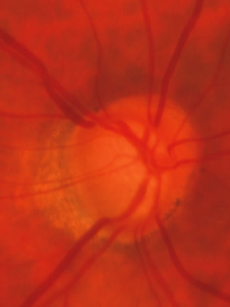
The approximately 1 to 1.5 million axons that form the optic nerve arise from the retinal ganglion cells and course toward the optic disc in a well-known pattern (Fig. 1). From the nasal retina the fibers take a straight course toward the disc. Axons originating temporal to the fovea arc around the macula to enter the upper and lower poles of the optic nerve head. The macula fibers pass directly to the temporal quadrant of the disc in the papillomacular bundle.

Fig. 1. Fundus photograph shows the normal pattern of the retinal nerve fiber layer. (Courtesy of P. Juhani Airaksinen, MD).

The axons maintain an orderly grouping in the retina as bundles partitioned by sheets of Müller's cells.1–3 A sheen reflected from the many bundles taking parallel courses gives the retina a striated appearance4 that is most prominent where the nerve fiber layer is thickest near the disc, especially in the arcuate bundles arriving at the poles of the disc from the temporal arcades.5,6 The attentive observer can see the striae of light reflected from these bundles and recognize their partial or total absence when bundles of axons disappear in glaucomatous or other forms of optic atrophy.4,7–10 The backscatter of light is determined by the cylindric nature and size of the light-scattering structures, possibly the axonal microtubules. They produce reflection with directional, spectral, and polarization (retardation) properties that have been studied and quantified, providing a basis for measuring the integrity of the retinal nerve fiber layer.11–14 The manner of layering of axons within intraretinal bundles2,3,6,15 may not be exactly the same in all primate species. However, a basic orderliness is present in the retina and seems to be maintained in the chiasm and beyond to the lateral geniculate body.16–19 Discrete scotomas and other defects caused by damage to nerve fiber bundles are produced by localized insults in the retinal nerve fiber layer or in the optic nerve head. Diffuse pressure on the posterior optic nerve by a mass lesion typically produces a central scotoma or diffuse visual dysfunction, but occasional instances of more localized damage in this region produce well-localized nerve fiber bundle defects, attesting to the existence of orderly bundles in this region also.12–15 On arriving at the lateral geniculate body, the axons are still organized and synapse in an orderly array.

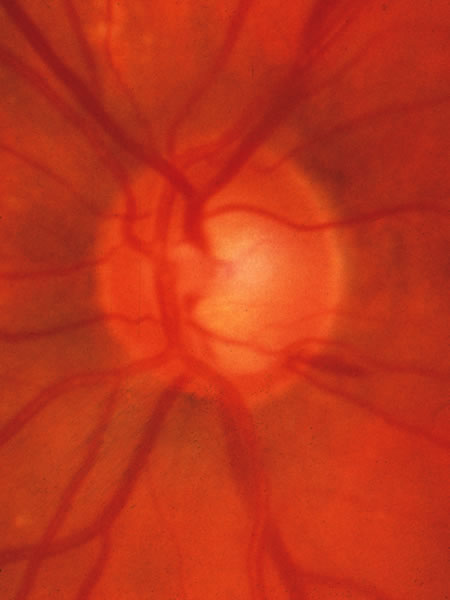
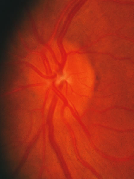
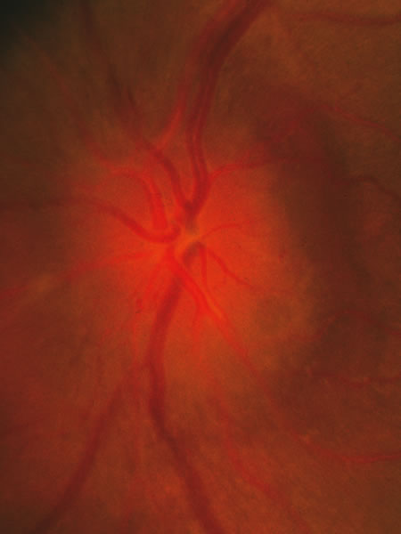
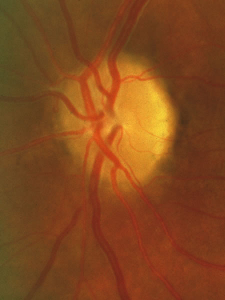
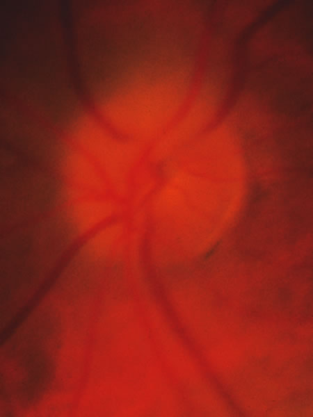
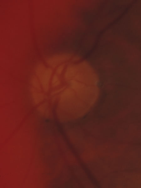
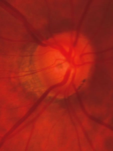
In the retinal nerve fiber layer, axons converge from every direction toward the optic disc and turn to enter the optic nerve through an opening in the outer retina, the choroid, and the sclera. The features and anatomic variation of the normal optic nerve head, or optic disc, are illustrated in Figures 2, 3, 4, 5, 6, 7, 8, 9, 10, 11, 12, 13, 14, 15, and 16. A physiologic excavation (cup) results if the chorioscleral canal is larger than required for the approximately 1 to 1.5 million axons and the supporting glial cells and blood vessels. The size of the excavation depends on how ample is the size of the chorioscleral canal.20,21 In discs where the chorioscleral canal matches the number of axons, the chorioscleral canal itself is typically somewhat taller than it is wide (Figs. 2 and 3). However, because the number of nerve fibers entering the upper and lower poles of the disc is greater than in the temporal and nasal sectors, the boundary of the physiologic cup is more or less circular.22 When the disc is large, it may be nearly circular but the cup likewise nearly circular. However, except in discs of anomalous shape, the width of the rim of neuroretinal tissue is noted in normal, nonglaucomatous optic discs to be greatest in the inferior meridian followed by the superior meridian, and narrowest in the temporal quadrant.23

Fig. 2. Normal optic nerve configuration. Notice that the height (H) of the disc is greater than the width (W). The width of the neuroretinal tissue (A) is also greater in the vertical meridian than in the nasal and temporal meridians (B); thus, the physiologic cup is round.

Fig. 3. Normal disc untilted, taller than it is wide. The neuroretinal tissue in the upper and lower sectors is more abundant in these sectors than others, so the central cup is round. A narrow white line marks the disc boundary and represents the lip of sclera that in humans almost universally separates the choroid from the optic disc tissue around the entire circumference.

Fig. 4. Influence of the tilt of the scleral canal on the slope of the canal. An outward slope of the chorioscleral canal (A) corresponds to a steep wall of the cup (A′). A perpendicular wall of chorioscleral canal (B) corresponds to a sloping wall of the cup (B′).

Fig. 5. In most eyes, a flange of sclera marks the boundary of the disc, separating the choroid from the disc tissue, the appearance of which is illustrated in Figures 2 and 6, as well as others throughout the chapter.

Fig. 6. Normal optic nerve head of moderate size with modest sized physiologic cup. Of note again is the white rim of sclera around the whole disc, seen most easily on the temporal side where the nerve fiber tissue crossing the disc margin is thinner and obscures the scleral rim less. There is a small peripapillary crescent (zone) of thin retinal pigment epithelium on the temporal side of the disc, as diagrammed in Figure 7.

Fig. 7. Peripapillary crescent caused by misalignment of the several layers of the retina, choroid, and sclera. The choroid is not covered completely by the choriocapillaris and retinal pigment epithelium (RPE).

Fig. 8. Tilted disc, with vessel trunk beneath a nasal ledge, a sloping disc surface on the temporal side, and a peripapillary crescent (β zone) the edge of which envelops the upper and lower poles. Although difficult to judge at times in tilted discs, the neuroretinal tissue is thinned at the poles of the disc in a manner typical of glaucoma.

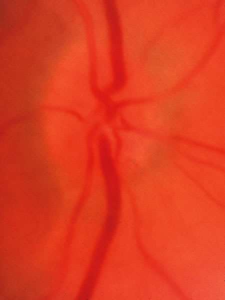
Fig. 9. Normal small disc with no cup because it is filled with neuroretinal tissue, which consists of the axons leaving the eye to form the optic nerve, along with the astroglia and the capillary bed.

Fig. 10. Physiologic cup of approximately one third the disc diameter, resulting from a chorioscleral canal slightly larger than required to accommodate the exiting axons along with the astroglia and capillaries. The scleral lip is again present around the entire circumference, even though not conspicuous where the pink nerve fiber layer is thick enough to obscure it. There is no peripapillary zone of thin retinal pigment epithelium or choroid to form a crescent or halo.

Fig. 11. Normal large disc with a large cup. The disc margin is nearly circular, and the cup has a slight horizontal oval shape, because the neuroretinal tissue is more abundant in the inferior and superior sectors than in the nasal and temporal sectors.
Fig. 12. Another large cup in a large disc. There is no location where the tissue is absent with the cup reaching the rim of the disc. The most typical sequence of the bulk of neuroretinal tissue, from greatest to least, is inferior, superior, nasal, temporal (ISNT).
Fig. 13. Large disc that had a large physiologic cup, on which is superimposed glaucomatous tissue loss. The glaucomatous damage can be recognized by the fact that in the upper and lower sectors the tissue is thinner than nasally and temporally. In particular, careful inspection shows that at the inferior pole in a narrow region where a small venous tributary crosses the margin at the 6:30 position, the cup reaches just about to the disc margin, which is the inner edge of the white stripe that represents the scleral lip. The scleral lip sometimes has a sufficiently pink hue that is mistaken to be remaining neuroretinal tissue in a location where in fact there are not remaining axons. The ongoing glaucomatous process was signified by occasional splinter hemorrhages in this patient. Because the physiologic cup was large initially, the visual loss is strikingly moderate because despite the large cup, only a modest proportion of the original neuroretinal tissue has been lost.
Fig. 14. Normal disc with a sloping surface of the disc surface (which is the same as a sloping margin of the cup wall). If tissue loss begins in the lower temporal sector of a disc, as is typical of glaucoma, it may be difficult to recognize the beginning of glaucomatous damage with backward displacement of the surface of “saucerization.” It would be particularly difficult in this disc, because the tissue is a homogeneous color with few vessles by which to judge the location of the tissue surface.

Fig. 15. Disc without glaucomatous disease, with a tilted exit of the optic nerve. The center of the optic nerve with the vascular trunk is markedly displaced toward the upper nasal edge. The bottom of the cup is represented as a small pale area just temporal to the place where the major retinal vessels penetrate the lamina cribrosa. The lower temporal disc surface, which in a sense is the lateral and temporal wall of the cup, is a broad sloped area of neuro-retinal tissue, somewhat pale because it is spread out over the scleral support of the sloped exit canal. This is another example of a normal disc within which early glaucomatous damage in the lower temporal sector might be difficult to recognize.

Fig. 16. Normal disc with moderate-sized cup. The vascular trunk is at the upper nasal edge of the cup, and the lower temporal sector has a sloped neuroretinal surface. This example has the fundamental anatomic features that make it difficult to recognize early glaucomatous loss in the lower temporal sector, but not to the unusually exaggerated degree of the previous two figures.

The shape of the physiologic cup is affected by the obliquity of the wall of the canal through the choroid and sclera. The wall of the cup is steep where the wall of the canal is angled outward.24 Where the canal has a wall perpendicular to the ocular coats the cup has a sloping wall (Fig. 4). The slope of the cup may vary from one sector to another, and there is considerable individual variation in the size and shape of the chorioscleral canal and therefore of the optic cup. In many eyes, the optic nerve exits through a canal the passes nasally and upward into the orbit, so that the upper nasal neuroretinal rim is bounded by a steep wall when viewed from the ophthalmoscopic viewpoint, while the lower temporal wall is of the cup is sloped, so that ophthalmoscopically the cup wall is, in essence, being viewed from within the cup rather than from above. In other discs, particularly those with a large cup, the walls are perpendicular to the scleral wall in all meridians. There is also typically no evident slope of the exit canal in eyes with only a small dimple of a cup, resulting from a near-perfect match between the size of the chorioscleral canal and the amount of neuroretinal tissue (axons and glia) that comprise the optic disc. The normal anatomic variations were noted and classified by Eschnig,25 and the glaucomatous changes to be discussed below are superimposed on the anatomic character present natively.

The canal through which the optic nerve exits from the eye traverses several layers of peripapillary tissue: the outer retina, the pigment epithelium, Bruch's membrane, the choriocapillaris, the outer choroid, and the sclera. In nearly all human eyes, in all or some meridians, a flange of sclera extends as far forward as the pigment epithelium, forming something of a border for the canal,25,26 and this can be seen ophthalmoscopically as a scleral rim around the disc (Figs. 5, 6, and 7). With or without such an obvious flange, the several chorioretinal layers may not be perfectly aligned with one another. Perhaps most typically, as the optic nerve exits obliquely in an upper nasal direction, the lower temporal sector of the disc viewed ophthalmoscopically is seen to have a crescent of misaligned edges of the several chorioretinal layers, such that the outer choroid and sclera may be left uncovered by the choriocapillaris and pigment epithelium (Fig. 8). Because of the tilt, the scleral flange is not seen as a narrow band; instead, by ophthalmoscopy its inner surface is viewed obliquely as it merges with the optic nerve sheath. In some myopic eyes, the crescent is most extensive on the temporal side, and in other eyes the crescent may be directly inferior, or for that matter less frequently in almost any position. In yet other variations the crescent encompasses much or all of the entire circumference of the disc, with misalignment or retraction of the various chorioretinal layers. When these eyes have glaucoma and the thin or retracted choroid surrounds the disc entirely, the area of thin choroid and absent retinal pigment epithelium has been termed a glaucomatous halo. The deformed anatomy associated with the tilted exit canal or the peripapillary crescent may mark the sector of the disc that is most susceptible to injury from elevated intraocular pressure27; for example, the lower temporal sector of the disc is most frequently the first sector affected.

Back to Top
PRESSURE-INDUCED DAMAGE
In the past, some believed that the damage to the optic disc in glaucoma occurred as the result of some sort of local sclerosis. Elevated intraocular pressure was not thought to be the cause, but simply to be an accompanying manifestation of a sclerotic process, that affected both the trabecular meshwork and the optic disc. Seeming evidence for this idea was a dissociation between the degree of cupping and the degree of pressure elevation,28–30 which is now attributed to individual variation in susceptibility to damage from intraocular pressure.31

A causative role of intraocular pressure is evident from cases of secondary glaucoma and from the fact that nerve damage is produced by elevated intraocular pressure in glaucoma produced experimentally in animals. Experienced clinicians have long agreed that progressive damage to the disc is halted when satisfactory lowering of intraocular pressure is achieved,32 and several recent clinical trials have now also shown more formally a cause-and-effect relationship between the level of intraocular pressure and damage.33–35 However, considerable individual variation is evident. Some individuals have intraocular pressure well above the normal range (ocular hypertension) for a long time without developing the anatomic and functional changes typical of glaucoma. Others, meanwhile, undergo progressive harm while the intraocular pressure is in the statistically normal range (normal-tension glaucoma), and the rate of harm is reduced with the intraocular pressure is lowered. Something makes the some optic nerves more susceptible to pressure-induced injury, and the gradation of susceptibility extends down into the normal range.

Factors that determine the susceptibility of the optic nerve to pressure-induced injury must exist and vary in amount to explain the individual variation in susceptibility to pressure-induced injury. Although the occurrence of damage does correlate with the level of intraocular pressure (and with its correlate, the resistance to aqueous humor outflow), the correlation with age is equally strong.28,29 This indicates that some of the susceptibility factors are age-related and that they are as important as the intraocular pressure in determining the visual loss in glaucoma. That some of the factors are genetic is evident from relationships to family history and race. Genetic makeup may impact not only the tendency to elevated intraocular pressure, but also susceptibility to harm that depends on the level of intraocular pressure. Observed relationships of particular susceptibility to female gender, migraine and other manifestations of vascular dysregulation, and clinical correlates or manifestations of vascular occlusive disease suggest the types of physiologic processes that may be altered by altered by the level of intraocular pressure.

Details of the pathophysiologic mechanism by which intraocular pressure produces optic nerve damage, exactly what the participating causative factors are, and how they interact together remain largely unknown.36 However, some clues are emerging. In some cases, inability to regulate blood flow in the optic nerve (dysregulation or inadequate autoregulation, which may be associated with migraine, with vascular overreaction to cold or stress, or with low blood pressure) is postulated by many37,38 to limit the ability of blood vessels in the optic nerve head to maintain sufficient blood flow when the intraocular pressure rises and challenges the circulation by raising venous pressure. It has been shown that individuals vary in their ability to regulate blood flow in the optic nerve,39 and potential mechanisms for local blood flow control by optic nerve capillaries have been studied in the laboratory.40 In other cases, vascular occlusive disease seems to play a role. If ischemia is a major cause for optic nerve damage in glaucoma, the different clinical presentation and appearance of a glaucomatous disc from that of a major acute vascular occlusive event (acute anterior ischemic optic neuropathy) remains a puzzle.

The are other events in the pathogenic pathways. Pressure seems to affect the astroglia, which express inducible nitric oxide synthase when the pressure is high,41 which on one hand might enhance vasodilation, but can also cause cytotoxic levels of nitric oxide. Axonal transport is impaired in the region of the lamina cribrosa, perhaps as a result of local ischemia,42 which may be reversed in time.43 With more serious or prolonged ischemia, damage to the axon membrane and entry of calcium may stimulate calcium-dependent proteases with irreversible damage to the axon segment would be expected. Any of these local events may prohibit transport of trophic factors, in particular nerve growth factor, to the retinal ganglion cells. In turn, after a time of inadequate arrival of growth factors, apoptosis is triggered with eventual death of the ganglion cell and its entire axon.44 Once triggered, apoptosis may occur even if the axon ischemia and blockage was reversed. It may be speculated that individual variation in the degree and duration of ischemia, free radical damage, and time of growth factor depravation before apoptosis is triggered might account for individual variation in susceptibility and in the rate of glaucomatous damage. Release of glutamate by dying ganglion cells has been postulated to add to the damage through excitotoxicity of adjacent retinal ganglion cells.45 While an interesting possibility, it remains uncertain whether excitotoxicity plays a major role in the pathogenic process.

Especially in cases in which the intraocular pressure is not particularly high, the collapse and stretching of the lamina cribrosa and other connective tissue in the region remains unexplained but is the histologic correlate to the clinically different appearance of the glaucomatous disc from other forms of optic atrophy (simple loss of axons). Recent evidence46 that a thin cornea places an eye at higher risk of damage when carrying an elevated intraocular pressure has suggested to some that the character of connective tissue in these individuals affects vulnerability, but there remains debate about whether the thin cornea simply produces an erroneous pressure measurement that masks the magnitude of the elevation of intraocular pressure.

Individuals vary in the rate of damage and in the level of intraocular pressure that is harmful, but perhaps also in the details of which of several root causes might participate or dominate in an individual, and which consequential events in the pathogenic sequence might be influenced by the intraocular pressure. Young individuals with secondary glaucoma and substantially elevated intraocular pressure develop cupping that is in part a diffuse stretching of the supporting tissues, and the elastic stretching may be reversible, while axon loss is not reversible. Older individuals with normal or slightly elevated intraocular pressure may develop shallower cupping with pallor thought to be related to chronic ischemia, which may involve all sectors of the disc or be quite localized. Cases may vary further depending on whether an occlusive disease or regulatory dysfunction participates in the postulated ischemic component, with the level of intraocular pressure possibly being more relevant in nonocclusive cases.47 There is more to learn about the entire pathogenic process, how to identify the dominant participating pathogenic processes in a given individual, and the pathways that are common to all cases that produce the typical clinical features of chronic glaucoma. Such information can be used in part to assess individual risk. Moreover, each of the contributing causes and participating pathophysiologic pathways involved in all cases or in particular identified individuals may be amenable to treatment that will improve the visual outcome.

We wait for the day when we will know more about the pathogenic process, know what the various susceptibility factors are, are able to measure them for the purpose of predicting how much pressure the eyes of a given person can tolerate, and can attack the susceptibility factors therapeutically. Until then, we know that a particular person is highly susceptible to future glaucomatous damage mainly when we observe that the pathogenic process is active or that the damage has begun,31 for example noting splinter optic disc hemorrhages or early glaucomatous anatomic or psychophysical (visual field) abnormality. The level of intraocular pressure is a weak predictor, but remains the focus of attention mainly because it is the only risk factor participating directly in the pathogenic mechanism that we know both how to measure and to treat.

The susceptibility factors seem in large part to be in the constitution of the person, perhaps the result of systemic influences (e.g., vascular status) or the fact that the two eyes of a person possess similar anatomic and physiologic traits on a genetic basis. Thus, if one eye has suffered damage from elevated intraocular pressure, the other eye is likely soon to follow if it too has or develops a pressure elevation.48 The two eyes may not always be damaged to the same degree. When one eye carries a somewhat higher pressure, it is usually the more damaged eye.49,50 Occasionally, a unilateral carotid artery obstruction, an anatomical difference between the two eyes (e.g., marked anisometropia) or some unidentified factor seems to make one eye more susceptible than the other, even with symmetric intraocular pressures.

It appears that the susceptibility factors are absent, or present only to a low degree, in the majority of persons, especially the young. Therefore, many people can carry a moderately elevated intraocular pressure for many years without damage, but there is a limit, and few eyes will tolerate a pressure of 50 or 60 mm Hg for very long. Approximately half of eyes have suffered harm when discovered to have an intraocular pressure of 35 mm Hg.30 It can be presumed that susceptibility factors are stronger in persons who suffer damage from mildly abnormal pressures (in the low and middle 20s) or in the normal range than in the majority of eyes, which typically tolerate such modest pressures easily for a long time. A few persons are exceptionally susceptible to damage and they suffer damage from modest pressure elevations, or in the high teens (low-tension glaucoma, or more accurately, normal-tension glaucoma). Damage while the pressure is in the low teens occurs even more rarely.

Although it is sometimes thought surprising that a pressure in the normal range can be damaging, it is not so surprising if it is kept in mind that 18 mm Hg is, as an absolute pressure level, nearly 80% of 23 mm Hg, which is readily accepted as potentially damaging to the susceptible person. If susceptibility factors lower the resistance of the disc to the point that 23 mm Hg is damaging, it is not a much further step to make the disc sensitive to 18 mm Hg. This reasoning suggests that it is arbitrary to consider low-tension glaucoma as being different than ordinary chronic open-angle glaucoma. The distinction that is made in traditional diagnostic classification results from fallaciously equating abnormal (i.e., a pressure level that is infrequent) with unsafe (i.e., a pressure that is harmful).

Indeed, there is growing sentiment that the basic nature of the disease is the same when the intraocular pressure is normal as when it is abnormally high, so that primary open angle glaucoma should no longer be distinguished from normal-tension glaucoma. While it is important to clarify our thinking by considering these diseases to have the same underlying mechanisms, there may be cases of glaucomatous damage that occurs without the intraocular pressure playing a part (and these would mainly have normal intraocular pressures), and in any event, cases that occur with normal intraocular pressure must have particularly active participation of the susceptibility factors. Noting that a case if glaucoma has a normal intraocular pressure guides the clinician to recognize that these susceptibility factors are more dominant in this patient than the intraocular pressure and one day may guide that clinician to focus attention on treatment directed at these factors in these cases and on the intraocular pressure in those cases with particularly elevation intraocular pressure.

In typical cases of chronic glaucoma, the damage occurs over a prolonged period of time. After the most susceptible axons are already damaged, it may be speculated that damage to other, previously unaffected axons may occur as a result of one or more of the following: (1) age causes a reduction in resistance to damage so that the next most susceptible axons become susceptible to the pressure that is being carried; (2) weakening of the disc by partial cupping increases the susceptibility of the remaining axons; (3) intraocular pressure progressively rises or has higher peaks of pressure as glaucoma continues; (4) damage depends not only on the pressure level but also on the time of exposure to the pressure (thus, the damage occurs as a cumulative effect of the force exacted by the intraocular pressure); or simply (5) somewhat arbitrarily a certain number of axons (or proportion of remaining axons) suffer during numerous small episodes, with cumulative damage.

Sometimes, however, in susceptible individuals glaucomatous cupping of the disc can proceed rapidly, within a month or two, even with moderate pressure elevation (25 to 30 mm Hg) if the pressure rose to this new level rather suddenly and exceeds the susceptibility level of all the axons all at once. This situation is encountered with a sudden (but perhaps mild) pressure rise in secondary glaucomas. Many cases may suffer no harm from a sudden pressure rise to 35 mm Hg from trauma or uveitis, for example, but a few eyes will suffer rapid, nearly total cupping in just a week or two with lesser elevation of pressure, say to the mid or upper 20s. Therefore, cases with recent onset of pressure rise must be monitored closely until it is clear whether this eye is being damaged or not.

In some cases of chronic glaucoma the cupping and field loss are recognized to progress in a series of small episodes a year or two apart, perhaps as a result of episodic upward swings of intraocular pressure or episodic swings of the constitutional factors that decrease the tolerance level. Even in cases that seem to be a continuous slow loss of axons, there may be smaller more frequent episodes that affect a few axons or bundles every week or two, but are each too small to be perceived as separate events. A single dramatic episode of transient vulnerability is sometimes blamed for nerve damage and field loss in patients who have had a hemodynamic crisis. Patients with such a history are less likely than others to progress51 if they have returned to good health and episodes of ill health are not recurrent. Progression may be accompanied by the occurrence of splinter hemorrhages that appear transiently on the optic disc.52–57 Although observed only occasionally, such hemorrhages may occur between ophthalmoscopic examinations in the majority of untreated patients and in patients whose glaucoma is progressing despite treatment. They are seen most often in cases of normal-tension glaucoma, perhaps because they most likely have an abundance of susceptibility factors with intraocular pressure that has not been lowered sufficiently to halt damage.

Often one region of the disc is affected earlier and more severely than another. When axon loss occurs more predominantly in certain bundles, cupping characteristically extends toward the disc rim in the sector that has lost neural tissue. If the preferential loss is in the most typical location (in the upper and lower sectors, most characteristically just temporal to the inferior pole), the cup expands vertically,22 and may form a notch at the disc rim, for example, at the inferior the pole of the optic disc. In such typical cases, the loss of axons and ganglion cells predominates in the corresponding arcuate regions of the temporal retina.

In contrast, when axon loss is evenly diffuse, the cup expands concentrically,58,59 and there is an accompanying generalized depression of the field. In the middle of the spectrum, where the majority of cases lie, there may be some widespread diffuse loss of axons but more severely near the pole of the disc. Thus, as the cup extends and deepens vertically, there is also temporal and nasal unfolding of the cup.

Back to Top
VISUAL EFFECTS
Loss of axons naturally affects the visual function in the regions of the retina from which the axons arose. Color sense,60–63 contrast sensitivity,63–66 and acuity67,68 are among the functions lost, in addition to the differential intensity visual threshold typically measured in classic visual-field testing.69–75 A certain proportion of nerve fibers must be lost in the affected retinal region before the diminished visual function is recognizably reduced from the normal range, from the previously documented visual status, or from the threshold in the surrounding unaffected (or less affected) region.76 For example, a 50% loss of axons in a bundle serving a given retinal region may produce a 0.5 log-unit (5 dB) reduction in visual sensitivity. If one or several adjacent bundles lose 50% or more of their axons, a scotoma results in which visual sensation in the corresponding region of the field will be recognizably reduced compared to that in the surrounding regions. A localized scotoma may be recognized because of major loss of axons in a confined region while no axons are lost in 95% of the disc area. However, if the axon loss is more diffuse, the resulting mild widespread depression may remain for quite a while within the range of normal visual sensation. Such diffuse axon loss is more difficult to recognize as an acquired reduction of visual sensation,58,63,66,71,77–79 unless comparison is made either with a previously documented baseline in that person or with the opposite, less affected eye. Fortunately, bilaterally equal diffuse loss of nerve fibers with bilaterally equal cupping and loss of visual field is infrequent. Even when axon loss is diffuse, one region is usually more affected than another and one eye more than the other. Therefore, glaucomatous nerve damage and visual-field loss can usually be recognized at an acceptably early stage by fundus examination and automated perimetry by80: (1) characteristic localized cupping and field loss, (2) by asymmetry of field in the upper and lower positions, (3) by asymmetry of cupping or field loss between the two eyes that is in keeping with asymmetry of intraocular pressure, or (4) by a change from a previous baseline disc photograph or baseline threshold perimetry. The conclusion that an acquired or asymmetric generalized depression of visual field is glaucomatous must be in keeping with other findings suggestive of glaucoma and out of keeping with any other causes of visual depression that might be present, such as amblyopia, anisometropia, alteration in media clarity, or indeed simply deviant ocular anatomy or function that may accompany high refractive error.
Back to Top
FUNDUSCOPIC FINDINGS
When evaluating the optic disc clinically, it is helpful to have a mental image of the underlying tissue changes. These are twofold: altered configuration of the lamina cribrosa and other connective tissue, and loss of neuroretinal tissue.

Connective tissue changes consist of elastic stretching caused by the force of the intraocular pressure and of connective tissue disruption with collapse of the layered sheets of the lamina cribrosa. Reversibility of cupping sometimes seen when the intraocular pressure is lowered if there remains an elastic element to the stretching backward bowing of the lamina cribrosa and outward stretching of the scleral border of the optic nerve head, especially in the young. This reversibility should not be construed as axon recovery or regeneration. Much of the connective tissue disruption and collapse is irreversible and produces a permanent component to the excavation, in addition to the loss of axons and perhaps of some astroglia. It seems that the change in the lamina cribrosa and other connective tissue that accounts for the fact that glaucomatous atrophy of the optic disc differs in clinical and histologic appearance from that of other optic atrophies. When there is considerable backward bowing of the lamina cribrosa without much axon loss, the disc may be severely cupped with surprisingly little visual effect detected on clinical examination.

Adding to the excavation caused by backward bowing and collapse of the lamina cribrosa is the loss of axons, as well as reduction or displacement of the astroglia and microvasculature within the optic nerve head. This neuroretinal tissue diminishes in the rim that surrounds any physiologic excavation that may have been present. As the supporting tissue recedes and the tissue bulk diminishes, the surface of the disc and the surface of its cup collapse. The surface of the neuroretinal rim recedes backward, but may not be conspicuous because the adjacent inner retinal surface also recedes backward as the nerve fiber layer of the retina also becomes thin. In patients in whom backward movement of the disc surface is the earliest sign recognized, the disc has sometimes been described as “saucerized.”81

Easier to see but difficult to describe and quantify is the excavation, or cup, in the center of the optic disc. It is a three-dimensional cavity that can be appreciated in stereoscopic photographs, binocular views clinically, and more modern imaging methods. Although three-dimensional aspects can be quantified, clinicians struggle to describe, quantify, or communicate the qualities of the cup in two-dimensional terms, such as having a diameter (cup/disc ratio) or area in a two-dimensional monocular ophthalmoscopic or photographic view, or a two-dimensional shape, such as round or oval. The reference plane is often by necessity perpendicular to the line of sight of the photograph or biomicroscopic view, although some make an effort to describe the cup/disc ratio or shape in a plane perpendicular to the axis of the nerve as it exits. Conventionally, the cup diameter is sometimes recorded at the plane of the retinal surface but as more sophisticated technology arose, other useful reference planes have been defined, and there is no universal standard or definition of how the size and shape of a cup should be recorded and communicated.

While waiting for standardized ways to quantify the disc configuration, clinicians do make an effort to describe and record the extent of the excavation. There is considerable variation in the appearance of glaucomatous cupping (Figs. 17, 18, 19, 20, 21, 22, 23, 24, 25, 26, 27, 28, 29, 30, 31, and 32), sometimes depending on the physiologic configuration before the onset of glaucomatous disease. The lateral expansion of the cup is easier to appreciate than the backward recession of the surface, and it is sensible to gauge progression of glaucomatous cupping as an increase in the cup diameter. Where the wall of the cup is steep, a cup margin is easy to delineate and consensus is easy to reach about enlargement of the cup when tissue is being lost is such a sector. However, in any sector(s) in which the cup has a sloping surface, it may be difficult or meaningless to define a cup margin with two-dimensional parameters, making it impossible to quantify in a uniform manner what is meant by a cup radius or cup/disc ratio for that meridian.82 One can conceive of the reference plane either as concentric with the ocular coats or as perpendicular to the axis of the exiting optic nerve, perhaps achieving reasonably reproducible assignment of a cup/disc ratio within a group that has reached a pragmatic consensus in its terminology.83

Fig. 17. Tilted disc with vessel trunk penetrating the lamina cribrosa in the upper nasal region and a sloped temporal margin. Moreover, the disc is large, and a large physiologic cup would be expected. Some thinning of the tissue in the sloped inferior sector may be suspected, but the neuroretinal rim at the superior pole of the disc is distinctly thinner than the rim in the nasal and other sectors (violating the inferior, superior, nasal, temporal [ISNT] rule). Finally, the white base of the cup, contrasted with the adjacent pinker tissue of the neuroretinal rim, is taller than it is wide; this is virtually never seen in normal discs.

Fig. 18. Baseline photograph of normal disc.
Fig. 19. Same disc as Figure 18 after 5 years of elevated intraocullar pressure. The horizontally oriented venous tributary that crosses the disc margin in the 1:00 position has less tissue between it and the cup than in Figure 18, showing the beginning of glaucomatous tissue loss. Even without the baseline photograph, a suspicion of early glaucoma may be recognized by the fact that the neuroretinal tissue in the upper sector is not thicker than the rim in the nasal sector the way it should be.

Fig. 20. Glaucomatous disc in which the thinner rim of tissue in the superior sector is more evident than in the previous example. As the initial physiologic cup be imagined in a disc of this size to have been one third to one half the disc diameter, the percentage of the total rim that has been lost is not great, and in keeping with this, the visual field loss consists of mild depression of the inferior hemifield with a detectable nasal step and three relative scotomas in the inferior arcuate region.

Fig. 21. Glaucomatous disc with cupping to the inferior margin of the disc. The tangle of vessels help define the shape of the sharp demarcation of the cup at the inferior pole.

Fig. 22. Glaucomatous disc with cupping nearly to the inferior margin of the disc. The vessels help define the excavated shape of the disc surface at the inferior pole. The downward expansion of the cup is also evident from the oval region of pallor that extends downward from the central cup that surrounds the place where the vascular trunk penetrates the lamina cribrosa.

Fig. 23. Disc with a localized and conspicuous loss of neuroretinal tissue in the inferior sector of the disc, a configuration than might be described as a notch.

Fig. 24. Glaucomatous cupping to the inferior rim with undermining of the rim temporal to the vein, producing a shadow that might be described as a pit-like notch. The use of the term notch has no clinical or pathophysiologic implications but is purely descriptive. Adjacent to the disc inferiorly is a small beta-zone crescent. Notice that the disc is tilted with vascular trunk disappearing through the lamina cribrosa near the superior pole, while the inferior sector, at least temporally, probably had a sloped surface before it became excavated.

Fig. 25. Glaucomatous disc with excavation fairly localized to the superior sector. In this case the vessels penetrate the lamina cribrosa in the inferior half of the disc. The deviation of the small vessel near the upper pole marks the surface configuration. The upward expansion of the pale base of the cup nearly to the disc margin is a distinctive sign that certainly signifies abnormality, but can be missed by indirect ophthalmoscopy if the strong lighting imparts a pink color to the region and the subtle small vessel deviation is not noticed.

Fig. 26. Glaucomatous disc. The excavation inferiorly may be difficult to recognize without a good stereoscopic view with a slit beam and biomicroscopy. The thinning superiorly is subtle. The inferior temporal surface is expected to have a sloped surface in a tilted disc with a temporal crescent, and glaucomatous excavation in this region requires close inspection, as well as experience and judgment. Especially by indirect ophthalmoscopy it may appear to have a good pink color, and the presence of excavation in the sloped region may easily go unrecognized.

Fig. 27. Glaucomatous disc with a crescent that nearly surrounds the entire disc circumference. The vessel configuration suggests thinning of tissue at the upper and lower disc sectors, and the pale base of the cup is taller than it is wide. With stereoscopic view the cup has a shape in which one can imagine a vertically oriented American football could be nestled. There may not be much visual field change, because an ample amount of tissue remains, but these signs suggesting a vertically oval configuration of the excavation should not be overlooked.

Fig. 28. Glaucomatous disc with cupping to the nasal side. A thin remnant of neuroretinal tissue remains just inside the white stripe of the scleral lip in the lower nasal region. Under some lighting conditions the adjacent β zone of atrophy may be mistaken for intact neuroretinal tissue. The field loss extending temporally from the blind spot may be overlook if perimetry covers only the central 24 degrees and the significance of one or two abnormal spots temporal to the blind spot is dismissed as artifact.

Fig. 29. Large disc and expected large physiologic cup, which has, however, superimposed glaucomatous damage with loss at the superior rim, where the cup reaches the disc margin in one location. Thinner tissue in the superior sector than elsewhere is distinctly abnormal, as is having the cup extend to the rim of the disc in any location.

Fig. 30. Total cupping of the disc caused by use of topical corticosteroid. Severe cupping can occur without accompanying atrophy of peripapillary tissue, although in some cases, progressive cupping is accompanied by progressive atrophy of adjacent retinal pigment epithelium and choroid.

Fig. 31. Advanced glaucomatous cup with complete loss of tissue at the upper and lower poles of the disc, a splinter hemorrhage nasally, and a glaucomatous halo of atrophic choroid and retinal pigment epithelium. The disc itself is separated from the peripapillary β zone by a conspicuous white stripe of the scleral lip.

Fig. 32. An obviously glaucomatous disc with particular loss of tissue in the inferior and superior sectors. A splinter hemorrhage among the axons crossing the disc margin is present at the 4:30 meridian. Typical glaucomatous hemorrhages are elongated and usually extend from the disc tissue across the disc margin somewhat into retina. They may overlie an adjacent peripapillary zone of choroidal or pigment epithelial atrophy, but usually have at least one end touching the disc margin. Nonglaucomatous hemorrhages are usually obvious in context of diabetic retinopathy, retinal vein occlusion, papillitis, etc.

To be clear, the purpose of noting the quantified or qualitative features of the disc in clinical practice is twofold with reference to glaucoma: to recognize whether the disc is damaged and to recognize whether there has been further damage since a previous examination. For the first purpose it is important to be familiar with the variations in appearance of normal optic discs (Figs. 2, 3, 4, 5, 6, 7, 8, 9, 10, 11, 12, 13, 14, 15, and 16), and how each of these may change as damage occurs (Figs. 17, 18, 19, 20, 21, 22, 23, 24, 25, 26, 27, 28, 29, 30, 31, and 32). With experience, presence of damage may be recognized by a single examination. In the next paragraphs the features to note will be described, but there is room for improvement in the ability to recognize abnormality accurately and efficiently in clinical practice. An advantage of automated image analysis is that it is less dependent on the experience and expertise of the individual clinician. For the second purpose (determining change), a means to record the state of the disc on the baseline occasion is needed. Drawings are perhaps used by most clinicians, but depend on the observational method used (direct ophthalmoscopy, binocular indirect ophthalmoscopy, biomicroscopy) as well as the talent of the artist-clinician to represent a three dimensional configuration on a two-dimensional drawing. High-magnification stereoscopic photographs are often a better record of the disc's state, but too often after years pass, progression is still difficult to judge if a different camera is used, exposure, magnification, and degree of stereopsis is not the same, and the development or removal of cataract affects the image. Sometimes, too, in regions of the disc without vessels or surface texture, the tissue surface may not be easily appreciated even in stereoscopic photographs. Images created by other methods (for example scanning ophthalmoscopy) are sometimes subject to the same lack of standardization or reproducibility.

Both recognition of the presence of damage and the recognition of change are particularly problematic in one frequent disc configuration, that with a somewhat tilted exit of the nerve upward and nasally (Figs. 8 and 14, 15, and 16). In these discs, the cup surface slopes in the lower temporal quadrant, which is often also devoid of major vessels to help localize the surface. In the ophthalmoscopic view, this surface simply recedes away from the observer and loss of tissue is difficult to identify, as the tissue simply becomes thinner and paler. Only in the more advanced stages might the cupping be recognized by the formation of a distinct notch at the inferior pole at one edge of the lower temporal sector. It is particularly unfortunate that this region of the disc is so typically the earliest and most involved, corresponding to the upper hemifield visual field abnormality. Moreover, in this sector it is difficult to evaluate the thickness of the layer of retinal nerve fibers crossing the disc margin.82 These problematic features are most difficult in the disc of with high axial myopia, because the sclera simply curves backward to merge with vaginal sheaths of the optic nerve, the choroid gradually thins within the β zone as it approaches the nerve exit, and it becomes arbitrary to designate any location as the margin of the disc itself.

Fortunately, the difficulties and limitations just emphasized are problematic only in some cases, both in describing and quantifying the features of a normal and abnormal optic disc, but we can nonetheless make useful notations of the characteristics of the optic disc configuration. Efforts to observe and record these features are in most cases very useful for the diagnosis of glaucoma and for monitoring its progress.

The area (or diameter) of pallor may be described separately from the size of the cup.84 As a descriptive matter, the region of pallor represents the base of the cup, and its diameter is judged there, while the cup diameter is conventionally judged at or near the inner retinal surface. It is not typical of glaucoma for the tissue of the neuroretinal rim to be pale in glaucoma, but for the tissue to be absent in the affected part of the neuroretinal rim. If there is any rim tissue present with a sector of pallor, nonglaucomatous atrophy must be suspected (except at times as a residual of an acute attack of glaucoma).

Glaucomatous loss of axons and thinning of the neuroretinal tissue may be diffuse or localized. If the axons are lost diffusely, the excavation may enlarge concentrically,58,85,86 retaining its round outline, and may be difficult to distinguish from a physiologic configuration.87 This is completely analogous to the difficulty in recognizing diffuse axon loss by visual field testing. The pathologic nature of the enlarged excavation can be recognized if it can be compared to the previous status of the disc58 (hence the importance of careful documentation of the optic disc appearance in all patients with elevated intraocular pressure) or if the degree of enlargement is different in the two eyes59 (acquired enlargement may be asymmetric but the physiologic cups are nearly always the same in the two eyes,88–96 unless there is anisometropia or other congenital asymmetry). Sometimes the experienced observer can pinpoint the physiologic nature of the large excavation by recognizing that the diameter of the scleral canal is unusually large in comparison with the diameter of the emerging central retinal vessels (Figs. 11 to 13). Large, round scleral canals, as opposed to smaller canals that are taller than they are wide, seem particularly common in black patients, and in such discs the branches of the central retinal vessels may dive through the pink neuroretinal rim rather than pass along its surface into the large physiologic cup. In the absence of such clues, and in any event if the intraocular pressure is elevated, an excavation larger than 0.6 cup-to-disc ratio is usually considered suspicious and should observed carefully until time proves that it is not enlarging.

Typically, before the cupping proceeds very far, preferential loss of neuroretinal tissue becomes evident, usually near the poles of the disc. The damage can be recognized either by noting that the rim is thinner than it should be compared to other sectors, or by the fact that the cup has become distinctly oval in the vertical direction. When the disc (and cup) is large enough to judge the breadth of the rim, a normal disc is has the broadest rim in the inferior meridian, followed in order by the superior, nasal, and temporal rim23 (Figs. 11 and 12). If the rim is not thickest at the two poles of the disc and thinnest temporally, glaucomatous damage should be strongly suspected (Fig. 13). The neuroretinal tissue becomes thinner in the affected sector such that the cup expands toward the lower temporal or upper temporal margins of the disc, assuming a vertically oval outline.22,97 Recognition of thinning of the neuroretinal rim near the vertical poles of the disc, with corresponding enlargement of the cup in a vertical direction, is the single most effective way to recognize the presence of glaucomatous damage.98,99 Depending on the configuration of the disc, the loss of tissue in the vertical sections may be most easily recognized as thinning of the rim, by a vertical extension of the cup or by a vertical shape to the region of pallor representing the floor of the cup. Except when the disc is obviously anomalous, any vertical tendency of these sorts deserves attention. For example, when the disc itself is small, and prior to the onset of glaucoma was completely full of neuroretinal tissue (essentially no cup except for a central dimple), the onset of glaucomatous tissue loss can be deceivingly small and unapparent.100 Therefore, in a noticeably small disc, any cup is suspect, but particularly if the cup is recognized to be a thin vertical slit with a vertically oriented pale base, it should not be passed over as nonglaucomatous. Such cases often seem to have field loss or retinal nerve fiber loss become evident before the glaucomatous nature of the cupping is recognized. The strong preference for early involvement of the vertical sectors notwithstanding, the localized loss of tissue may sometimes occur in any sector of the disc, even nasally (associated with a temporal wedge field defect), and a cup with extension localized toward any point on the disc circumference is decidedly abnormal. Localized tissue damage is typically associated with a nerve fiber bundle defect in the appropriate portion of the visual field. In myopic eyes with the disc tilted and a crescent over the temporal sector, thinning of the temporal rim can be glaucomatous, and the field defects are more often closer to the point of fixation than in other eyes. Evaluation of discs in high axial myopia is often made even more difficult because of an enlarged anomalous disc with an irregular shape embedded in a staphyloma, making the edge of the disc difficult to define, and loss of nerve fibers nearly impossible to recognize with certainty.

These classic appearances of glaucomatous cupping at various stages and in discs of various anatomic configurations has been described.22,85,86,97,101–112 Representative examples are shown in Figures 13 and 19 to 32. Glaucomatous cupping is recognized not only on the basis of the illustrated typical preferential loss of tissue at the poles of the disc but importantly by also looking for asymmetry in the cupping of the optic discs of the two eyes (which are normally the same88–96) or a change from the previous status (hence the importance of documenting carefully the disc status of all glaucoma suspects, preferably with photographs). Especially without the advantage of prior photographs, the degree of damage must take into account the size of the disc,113 and clues about its preglaucomatous configuration, paying more attention to any cupping in small discs100 and recognizing that large cups are expected physiologically in large discs. In nearly all cases of nonglaucomatous optic atrophy, the affected tissue turns pale and the loss of mass is inconspicuous. Rare exceptions occur in moderately large discs with cylindric cups in which glaucoma-like thinning of the rim is noticeable after arteritic anterior ischemic optic neuropathy114,115 (nonarteritic forms tend to occur in small discs) and very rarely in other optic atrophies.

Misalignment and irregularity of the retinal pigment epithelium, choriocapillaris, choroid, and sclera at the disc margin (or partial atrophy of some of these tissue layers) around part or all of the disc circumference gives the appearance of a peripapillary crescent or halo.27,116–128 A crescent is often seen in the sector of the disc that shows the most profound cupping.27,124,126 These regions of disrupted or thin peripapillary tissue have been descriptively divided into α regions of clumped or irregular retinal pigment epithelium and a β region, closer to the disc, in which the choroidal and pigment epithelial tissue are thin with a pale appearance. At the very edge of the disc is typically a thin white rim usually perceptible around the entire circumference, representing a flange of sclera that separates the disc from the choroid in humans. Advanced cupping in older people with low grade elevation of intraocular pressure is also seen in the company of a prominent peripapillary halo (surrounding the entire disc instead of only a portion of the circumference) with β-type thinning of choroidal tissue, but the association is not universal. One form of cupping in normal-tension glaucoma is rather shallow cupping with a prominent halo of thin pigment epithelium and pale choroidal tissue. Another characteristic form in normal-tension glaucoma is a narrow localized crescent at the inferior pole associated with a discrete notch in the neuroretinal rim in which a splinter hemorrhage is sometimes seen.

It is not clear how many of these crescents and halos represent congenital preexisting misalignment of the layers, how many are age-related changes that may occur before the glaucoma develops, and how many are atrophy of the peripapillary tissue that accompanies the insult to the disc. Certainly examples of each can be found. The congenital variety has been postulated to mark a sector of the disc that later is more vulnerable to glaucomatous damage, accounting for field defects close to fixation in cases of temporal myopic crescents and inferior notches of the disc. In keeping with this, it has been postulated that individuals without any peripapillary crescent may be less susceptible to damaging effects from moderate ocular hypertension.129,130 Enlargement of the β zone has been seen with age in nonglaucomatous patients, but is more frequent in those with glaucoma associated with elevated pressure, often older people with mild elevation of intraocular pressure.121 Younger individuals with more marked elevation of intraocular pressure (often secondary glaucoma), however, typically develop deep glaucomatous cupping with loss of all nerve fibers, but no development or enlargement of the peripapillary tissue disturbance.131,132

Transient splinter hemorrhages at the disc margin52–57 (Figs. 31 and 32) can be seen to occur in most cases of untreated glaucoma if observations are made often enough.56 When they are seen in treated cases, it usually signifies that cupping and field loss are progressing,53,54 and sometimes the hemorrhage is seen at the same time that a new field defect appears. Because hemorrhages are easy to overlook and transient, fundus examinations must be both careful and frequent if these hemorrhages are to be detected.

Along with loss of inner retinal nerve fiber layer thickness and disc tissue, the retinal arteries become narrower, presumably because of reduced metabolic need, and the narrowing is often most marked near the disc.133 This narrowing is nonspecific for glaucoma but occurs with all forms of optic atrophy.

Given the anatomic variation in physiologic configuration of the optic nerve head, the varying levels of intraocular pressure that may develop, the individual variation in the level of intraocular pressure that is harmful, and the varying age at which glaucoma may appear, there is a variety of appearances of glaucomatous discs. In younger patients, often with secondary glaucoma and considerable pressure elevation, reversible elastic expansion of support tissue produces a symmetric expansion of the physiologic cup (deep round cup surrounded by a donut-like rim of neuroretinal tissue), which may later develop a diffuse loss of the neuroretinal rim until there is total cupping, or a localized loss to produce a notching the rim that later also becomes thinner as well. In such cases, there is no accompanying atrophy of the peripapillary choroid and retinal pigment epithelium. In somewhat older patients with more moderate idiopathic (primary) pressure elevation, the cupping is shallower and less likely to show general reversible enlargement of the cup before localized excavation is evident. A tilted disc with a sloping surface of the inferotemporal neuroretinal rim and an adjacent crescent is a common physiologic configuration (or more directly temporal in somewhat myopic eyes), and these sectors seem most often to suffer the earliest and most severe damage. These varieties represent the most common patients, and hence the most typical groups, as represented in the figures in this chapter. In some, typically older, patients the excavation is shallow with little or no elevation of intraocular pressure. Peripapillary zones of depigmented or absent pigment epithelium (as a crescent or halo) are more common in older patients who develop damage with little or no pressure elevation, as are splinter hemorrhages. Despite these generalizations, there is considerable variation in the combination and overlap of these features, so that it is difficult to separate the varieties of cupping into distinct groups.134

In infantile glaucoma, as the entire globe enlarges, a concentric enlargement of the cup results when the scleral canal is stretched along with the rest of the globe. The lamina cribrosa of the glaucomatous infant eye may also be elastically bulged backwards. This element of the acquired cupping in infants is not the result of axonal loss. Relaxation of the stretched tissue when the intraocular pressure is relieved may account for the reversibility of cupping often observed in these infant eyes104,135–145 (and occasionally observed to a lesser degree in other glaucomatous eyes in later decades of life146). The cupping caused by scleral stretching may not be completely reversible because with remodeling of collagen in stretched tissues still undergoing development and growth, some of the enlargement of the eye (and of the scleral canal) is permanent. Thus, after successful pressure control, the eye in infantile glaucoma may remain myopic with a large cup without any axon loss. Of course the pressure can also damage the axons, often preferentially at the poles of the disc as in adult glaucoma. This element of cupping is not reversible and is accompanied by visual loss.

In acute angle-closure glaucoma (Fig. 33), the optic disc may swell,147–149 perhaps as a result of ischemia, but because of corneal edema and preoccupation with the angle status, it is not often seen. Even after an attack lasting several days, there is often no visual loss; the disc may remain normal appearing or may develop some degree of pallor resembling that of nonglaucomatous optic atrophy.150–152 Excavation of the disc does not seem to result from an acute attack but may result from persistent residual pressure elevation after the attack. If excavation is already present at the time that a patient presents with symptomatic angle closure, the cupping is evidence that for some time an asymptomatic, perhaps gradual, rise of intraocular pressure elevation preceded the abrupt onset of symptoms.

Fig. 33. Swollen disc tissue during an acute attack of angle closure glaucoma.

Usually all other forms of optic nerve disease (and inner retinal disease) produce simple atropy of the disc without cupping (Figs. 34 to 36), but occasional cases of cupping have been seen with anterior ischemic optic neuropathy, particularly due to giant cell arteritis (Figs. 37 and 38).

Fig. 34. Photograph of a normal left eye at a time when the right eye had an attack of anterior ischemic optic neuropathy.

Fig. 35. Same eye as Figure 34 during a subsequent attack of ischemic optic neuropathy.

Fig. 36. Same eye as in Figure 34 one year later, with atrophy but no excavation.

Fig. 37. Swollen optic nerve superiorly during anterior ischemic optic neuropathy caused by giant cell arteritis.

Fig. 38. Same eye after resolution of the attack, with excavation extending to the superior rim of the disc, mimicking the appearance of glaucomatous cupping.

Back to Top
CLINICAL EXAMINATION OF THE OPTIC NERVE
For glaucoma detection, an eye examination needs to include an examination of the optic disc. This supplements tonometry for glaucoma detection by revealing cases of low-tension glaucoma and patients with variable pressure who happen to have a normal pressure at the time of a routine eye examination. Visual-field examination is appropriate when the pressure is elevated (even if the disc appears healthy) or when the disc is suspiciously abnormal (even if the pressure is normal).

On the basis of family history, the intraocular pressure, the disc configuration, or the visual field, a patient may be diagnosed as having glaucoma, may be suspected possibly as having early glaucoma, or may be judged at risk of developing glaucoma. In any of these situations, it is wise to record the status of the discs with careful drawings or preferably (Figs. 18, 19, 39, and 40), if available, stereoscopic disc photographs. In addition, it is wise to record a carefully quantified visual-field examination (kinetic perimetry with several isopters or automated perimetry with a thresholding strategy) as a baseline.

Fig. 39. Glaucomatous disc photographed in 1992.

Fig. 40. Same eye photographed in 1998, after progression of the cupping and visual field loss. Continued thinning of the neuroretinal rim is evident in nearly every part of the circumference and the course of the veins across the inferior border of the disc show the deeper excavation. This progression would not likely be evident if only disc drawings were used to monitor the disc status. Stereoscopic fundus photographs of other forms of imaging are discussed in Chapter 48A are needed.

Visual acuity and intraocular pressure are measured on every visit thereafter, the optic disc may be examined without necessarily dilating the pupil on every visit, but also with a dilated pupil at regular intervals. Frequent disc examination will increase the chance of observing transient disc hemorrhages in patients in whom glaucoma is progressing.56 If the patient's course seems stable and if the intraocular pressure is not different than usual, the optic disc may be examined through a dilated pupil on approximately every third or fourth visit. A visual-field examination may be conducted during one of the intervening visits, leaving one intervening visit for a refraction repeat gonioscopy, or whatever else might be required for the patient's care.

An office visit every 3 months has been traditional for patients with glaucoma and for patient suspicious for glaucoma who are thought to be stable but is not always appropriate.153 In patients whose stable course has been well documented for a year or two, the interval can be lengthened to 4 months, and in selected patients to 6 months or even longer. On the other hand, patients about whom there is reason for concern (e.g., the pressure is not substantially lowered from the pretherapeutic level, a regular visit shows suspicious change in the disc or field, or a new therapy has been instituted) may need not only to be seen more frequently for a while, but may also require disc and field examinations on every visit until it is either evident they are progressing or evident that they are stable.

Clinical examination of the optic disc is best done through a dilated pupil with the magnified stereoscopic view of slit-lamp biomicroscopy. The angled slit beam reveals the contour of the disc's surface, but the slit is helpful even when placed directly in line with the biomicroscope. As it intersects the surface, a thin, well-focused beam produces a discrete line, which the examiner views with stereopsis. The Hruby (approximately 55 diopter) lens is convenient for routine use and usually will give an adequate view, but a funduscopic contact lens (approximately 64 diopters) should be used in persons whose discs are not seen clearly with the Hruby lens. Apart from the additional inconvenience, a disadvantage of using the contact funduscopic lens routinely on all patients is that the anesthetic eye drops and gonioscopic solution render the cornea less suitable for obtaining a high-quality fundus photograph. The use of a +78 diopter or +90 diopter indirect lens in conjunction with a slit-lamp biomicroscope is becoming increasingly popular. It must be kept in mind that with less magnification (with a stronger power lens) appreciation of depth is reduced because axial magnification (depth) relates to the square of transverse magnification (diameter of the disc in the image). Stereoscopic color disc photographs (taken best at twice the usual magnification) lack the advantage of having a slit to highlight the surface in areas of homogeneous texture and are thus not a substitute for biomicroscopic examination. However, good stereoscopic fundus photographs are best for recording the status of the disc for the sake of future comparison.

The monocular magnified view of direct (or monocular indirect) ophthalmoscopy is the next best choice for viewing the disc and sometimes the only method that can be used with an undilatable pupil. Although the fundus as a whole should always be examined with a binocular indirect ophthalmoscope during a complete eye examination, this instrument is the least suitable for judging glaucomatous excavation of the disc. The strong illumination seems to render a pink color even to an atrophic disc, and with the usual +20 diopter or +30 diopter lens, the stereopsis and magnification without the biomicroscope are not sufficient for the examiner to appreciate saucerized or sometimes even frankly excavated discs.

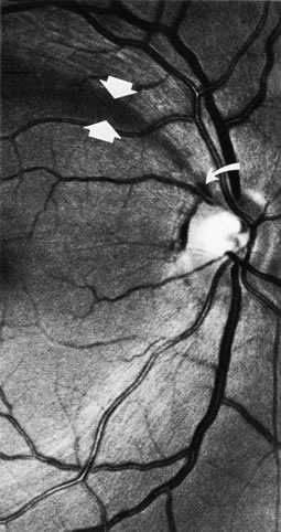
When bundles of nerve fibers drop out, their absence can be noted in the retinal sheen produced by the retinal nerve fiber layer.4,8–10,85 Such defects are easiest to see in the thickest portions of the nerve fiber layer, namely, close to the disc and especially in the arcuate bundles approaching the poles of the disc.5,6 The loss of nerve fibers can be recognized ophthalmoscopically but, as shown in Figure 41, is demonstrated most beautifully in wide-angle fundus photographs taken with blue or green filters.10,154 It is difficult to see if the background is lightly pigmented or if imperfect media produce an imperfect view of the fundus. Some observers are more skilled than others in recognizing nerve-fiber layer disease.

Fig. 41. Retinal nerve fiber layer in glaucoma. A curved wedge (between the broad arrows) represents the loss of nerve fiber bundles corresponding to a sector of the disc marked by a splinter hemorrhage (small curved arrow). (Airaksinen PJ. Mustonen E. Alanko HI: Optic disc hemorrhages precede retinal nerve fiber layer defects in ocular hypertension. Acta Ophthalmol 59:627, 1981.)

Fluorescein angiography has shown areas of slow filling or nonfilling in damaged segments of the disc,155–159 evidence that the microvasculature is lost along with the other elements of the neuroretinal tissue. Fluorescein angiography has not been used for routine clinical evaluation, but in cases of uncertainty about the existence of pressure-induced disc damage, the demonstration of a filling defect could potentially be of value in recognizing neuroretinal tissue loss in discs with very sloped margins, such as in high axial myopia.158

Emerging technologies to record and quantify the optic nerve configuration and the retinal nerve fiber layer are presently used in some offices. The techniques are undergoing improvement. Criteria for abnormality and for progression of damage are being validated. These are described in Chapter 48A.

Back to Top
REFERENCES

1. Radius RL, Anderson DR: The histology of retinal nerve fiber layer bundles and bundle defects. Arch Ophthalmol 97:948, 1979

2. Radius RL, Anderson DR: The course of axons through the retina and optic nerve head. Arch Ophthalmol 97:1154, 1979

3. Minckler DS: The organization of nerve fiber bundles in the primate optic nerve head. Arch Ophthalmol 98:1630, 1980

4. Hoyt WF, Frisén L, Newman NM: Funduscopy of nerve fiber layer defects in glaucoma. Invest Ophthalmol 12:814, 1973

5. Radius RL: Thickness of the retinal nerve fiber layer in primate eyes. Arch Ophthalmol 98:1625, 1980

6. Quigley HA, Addicks EM: Quantitative studies of retinal nerve fiber layer defects. Arch Ophthalmol 100:807, 1982

7. Hoyt WF, Schlicke B, Eckelhoff RJ: Funduscopic appearance of a nerve-fiber-bundle defect. Br J Ophthalmol 56:577, 1972

8. Sommer A, Miller NR, Pollack I, et al: The nerve fiber layer in the diagnosis of glaucoma. Arch Ophthalmol 95:2149, 1977

9. Quigley HA, Miller NR, George T: Clinical evaluation of nerve fiber layer atrophy as an indicator of glaucomatous optic nerve damage. Arch Ophthalmol 98:1564, 1980

10. Airaksinen PJ, Nieminen H, Mustonen E: Retinal nerve fibre layer photography with a wide angle fundus camera. Acta Ophthalmol (Copenh) 60:362, 1982

11. Huang XR, Knighton RW: Linear birefringence of the retinal nerve fiber layer measured in vitro with a multispectral imaging micropolarimeter. J Biomed Opt 7:199, 2002

12. Weinreb RN, Dreher AW, Coleman A, et al: Histopathologic validation of Fourier-ellipsometry measurements of retinal nerve fiber layer thickness. Arch Ophthalmol 108:557, 1990

13. Knighton RW, Huang XR: Directional and spectral reflectance of the rat retinal nerve fiber layer. Invest Ophthalmol Vis Sci 40:639, 1999

14. Knighton RW, Huang X, Zhou Q: Microtubule contribution to the reflectance of the retinal nerve fiber layer. Invest Ophthalmol Vis Sci 39:189, 1998

15. Ogden TE: The nerve-fiber layer of the primate retina: Aan autoradiographic study. Invest Ophthalmol 13:95, 1974

16. Harrington DO: Differential diagnosis of the arcuate scotoma. Invest Ophthalmol 8:96, 1969

17. Hoyt WF, Luis O: Visual fiber anatomy in the infrageniculate pathway of the primate. Arch Ophthalmol 68:94, 1962

18. Hoyt WF, Luis O: The primate chiasm. Arch Ophthalmol 70:69, 1963

19. Hoyt WF: Anatomic considerations of arcuate scotomas associated with lesions of the optic nerve and chiasm: A nauta axon degeneration study in the monkey. Bull Johns Hopkins Hosp 111:57, 1962

20. Teal PK, Morin JD, McCulloch C: Assessment of the normal disc. Trans Am Ophthalmol Soc 70:164, 1972

21. Bengtsson B: The variation and covariation of cup and disc diameters. Acta Ophthalmol (Copenh) 54:804, 1976

22. Kirsch RE, Anderson DR: Clinical recognition of glaucomatous cupping. Am J Ophthalmol 75:442, 1973

23. Jonas JB, Gusek GC, Nnaumann GOH: Die parapapilläre Region in Normal- und Glaukomaugen I. Planimetrische Werte von 312 Glaukom- und 125 Normalaugen. Klin Monastbl Augenheilkd 193:52, 1988

24. Guist G: Coincident Ophthalmoscopy and Histology of the Optic Nerve. Vienna, Guist, 1934

25. Elschnig A: Das Colobom am Sehnerveneintritte und der Conus nach unten. Albrecht von Graefes Arch Ophthalmol 51:391, 1900

26. Anderson DR, Hoyt WF: Ultrastructure of intraorbital portion of human and monkey optic nerve. Arch Ophthalmol 82:506, 1969

27. Anderson DR: Correlation of the peripapillary anatomy with the disc damage and full abnormalities in glaucoma. Doc Ophthalmol Proc Ser 35:1, 1983

28. Armaly MF, Krueger DE, Maunder L, et al: Biostatistical analysis of the collaborative glaucoma study: I. Summary report of the risk factors for glaucomatous visual-field defects. Arch Ophthalmol 98:2163, 1980

29. Armaly MF: Lessons to be learned from the collaborative glaucoma study. Surv Ophthalmol 25:139, 1980

30. Anderson DR: The management of elevated intraocular pressure with normal optic discs and visual fields: I. Therapeutic approach based on high risk factors. Surv Ophthalmol 21:479, 1977

31. Anderson DR: Glaucoma: The damage caused by pressure. Am J Ophthalmol 108:485, 1989

32. Kronfeld PC, McGarry HI: Five year follow-up of glaucoma. JAMA 136:957, 1948

33. Heijl A, Leske MC, Bengtsson B, et al: Reduction of intraocular pressure and glaucoma progression. Arch Ophthalmol 120:1268, 2000

34. Anderson DR, Drance SM, Schulzer M: Writing Committee for Collaborative Normal-tension Glaucoma Study Group. The effectiveness of intraocular pressure reduction in the treatment of normal-tension glaucoma. Am J Ophthalmol 126:498, 1998

35. Kass MA, Heuer DK, Higginbotham EJ, et al: The Ocular Hypertension Study: A randomized trial determines that topical ocular hypotensive medication delays or prevents the onset of primary open-angle glaucoma. Arch Ophthalmol 120:701, 2002

36. Anderson DR, Quigley HA: The optic nerve. In Moses RA, Hart WM Jr (eds): Adler's Physiology of the Eye, 9th ed., pp. 616–640. St. Louis, CV Mosby, 1992

37. Anderson DR: Glaucoma, capillaries, and pericytes. 1. Blood flow regulation. Ophthalmologica 210:257, 1996

38. Flammer J, Orgul S, Costa VP, et al: The impact of ocular blood flow in glaucoma. Prog Retin Eye Res 21:359, 2002

39. Pillunat LE, Anderson DR, Knighton RW, et al: Autoregulation in human optic nerve head circulation in response to increased intraocular pressure. Exp Eye Res 64:737, 1997

40. Matsugi T, Chen Q, Anderson DR: Suppression of CO2-induced relaxation of bovine retinal pericytes by Angiotensin II. Invest Ophthalmol Visual Sci 38:652, 1997

41. Liu B, Neufeld AH: Nitric oxide synthase-2 in human optic nerve head astrocytes induced by elevated pressure in vitro. Arch Ophthalmol 119:2405, 2001

42. Anderson DR, Hendrickson A: Effect of intraocular pressure on rapid axoplasmic transport in monkey optic nerve. Invest Ophthalmol Vis Sci 13:771, 1974

43. Radius RL, Anderson DR: Reversibility of optic nerve damage in primate eyes subjected to intraocular pressure above systolic blood pressure. Br J Ophthalmol 65:661, 1981

44. Kerrigan LA, Zack DJ, Quigley HA, et al: TUNEL-positive ganglion cells in human primary open-angle glaucoma. Arch Ophthalmol 115:1031, 1997

45. Vorwerk CK, Gorla MS, Dreyer EB: An experimental basis for implicating excitotoxicity in glaucomatous optic neuropathy. Surv Ophthalmol 43(Suppl 1):S142, 1999

46. Gordon MO, Beiser JA, Brandt JD, et al: The ocular hypertension treatment study: Baseline factors that predict the onset of primary open-angle glaucoma. Arch Ophthalmol 120:714, 2000

47. Schulzer M, Drance SM, Carter CJ, et al: Biostatistical evidence for two distinct populations with chronic open angle glaucoma. Br J Ophthalmol 74:196, 1990

48. Kass MA, Kolker AE, Becker B: Prognostic factors in glaucomatous visual field loss. Arch Ophthalmol 94:1274, 1976

49. Cartwright MJ, Anderson DR: Correlation of asymmetric damage with asymmetric intraocular pressure in normal-tension glaucoma (low-tension glaucoma). Arch Ophthalmol 106:898, 1988

50. Crichton A, Drance SM, Douglas GR, et al: Unequal intraocular pressure and its relation to asymmetric visual field defects in low-tension glaucoma. Ophthalmology 96:1312, 1989

51. Drance SM, Morgan RW, Sweeney VP: Shock-induced optic neuropathy: Cause of nonprogressive glaucoma. N Engl J Med 288:392, 1973

52. Drance SM, Begg IS: Sector haemorrhage: A probable acute ischemic disc change in chronic simple glaucoma. Can J Ophthalmol 5:137, 1970

53. Drance SM, Fairclough M, Butler DM, et al: The importance of disc hemorrhage in the prognosis of chronic open angle glaucoma. Arch Ophthalmol 95:226, 1977

54. Susanna R, Drance SM, Douglas GR: Disc hemorrhages in patients with elevated intraocular pressure: Occurrence with and without field changes. Arch Ophthalmol 97:284, 1979

55. Airaksinen PJ, Mustonen E, Alanko HI: Optic disc hemorrhages: Analysis of stereophotographs and clinical data of 112 patients. Arch Ophthalmol 99:1795, 1981

56. Bengtsson B, Holmin C, Krakau CET: Disc haemorrhage and glaucoma. Acta Ophthalmol (Copenh) 59:1, 1981

57. Airaksinen PJ, Mustonen E, Alanko HI: Optic disc haemorrhages precede retinal nerve fibre layer defects in ocular hypertension. Acta Ophthalmol (Copenh) 59:627, 1981

58. Pederson JE, Anderson DR: The mode of progressive disc cupping in ocular hypertension and glaucoma. Arch Ophthalmol 98:490, 1980

59. Anderson DR: What happens to the optic disc and retina in glaucoma? Ophthalmology 90:766, 1983

60. Drance SM, Lakowski R, Schulzer M, et al: Acquired color vision changes in glaucoma: Use of 100-hue test and Pickford anomaloscope as predictors of glaucomatous field change. Arch Ophthalmol 99:829, 1981

61. Austin DJ: Acquired colour vision defects in patients suffering from chronic simple glaucoma. Trans Ophthalmol Soc UK 94:880, 1974

62. Poinoosawmy D, Nagasubramanian S, Gloster J: Colour vision in patients with chronic simple glaucoma and ocular hypertension. Br J Ophthalmol 64:852, 1980

63. Motolko M, Drance SM, Douglas GR: The early psychophysical disturbances in chronic open-angle glaucoma: A study of visual functions with asymmetric disc cupping. Arch Ophthalmol 100:1632, 1982

64. Atkin A, Bodis-Wollner I, Wolkstein M, et al: Abnormalities of central contrast sensitivity in glaucoma. Am J Ophthalmol 88:205, 1979.

65. Atkin A, Wolkstein M, Bodis-Wollner I, et al: Interocular comparison of contrast sensitivities in glaucoma patients and suspects. Br J Ophthalmol 64:858, 1980

66. Stamper RL, Hsu-Winges C, Sopher M: Arden contrast sensitivity testing in glaucoma. Arch Ophthalmol 100:947, 1982

67. Phelps CD, Remijan PW, Blondeau P: Acuity perimetry. Doc Ophthalmol Proc Ser 26:111, 1981

68. Anctil JL, Anderson DR: Early foveal involvement and generalized depression of the visual field in glaucoma. Arch Ophthalmol 102:363, 1984

69. Morin JD: Changes in the visual fields in glaucoma: Static and kinetic perimetry in 2,000 patients. Trans Am Ophthalmol Soc 77:622, 1979

70. Drance SM: The glaucomatous visual field. Br J Ophthalmol 56:186, 1972

71. Aulhorn E, Harms H: Early visual field defects in glaucoma. In Leydhecker W (ed): Glaucoma: Tutzing Symposium, 1966, p. 151. Basel, S Karger, 1967

72. Harrington DO: The Bjerrum scotoma. Am J Ophthalmol 59:646, 1965

73. Drance SM, Fairclough M, Thomas B, et al: The early visual field defect in glaucoma and the significance of nasal steps. Doc Ophthalmol Proc Ser 19:119, 1979

74. Brais P, Drance SM: The temporal field in chronic simple glaucoma. Arch Ophthalmol 88:518, 1972

75. Werner EB, Beraskow J: Temporal visual field defects in glaucoma. Can J Ophthalmol 15:13, 1980

76. Quigley HA, Addicks EM, Green WR: Optic nerve damage in human glaucoma: III. Quantitative correlation of nerve fiber loss and visual field defect in glaucoma, ischemic neuropathy, papilledema, and toxic neuropathy. Arch Ophthalmol 100:135, 1982

77. Armaly ME: Ocular pressure and visual fields: A ten-year follow-up study. Arch Ophthalmol 81:25, 1969

78. Lichter PR, Standardi CL: Early glaucomatous visual field defects and their significance to clinical ophthalmology. Doc Ophthalmol Proc Ser 19:111, 1979

79. Hart WM Jr , Yablonski M, Kass MA, et al: Quantitative visual field and optic disc correlates early in glaucoma. Arch Ophthalmol 96:2209, 1978

80. Anderson DR: Automated Static Perimetry. St. Louis, Mosby Year Book, 1992

81. Chandler PA, Grant WM: Lectures on Glaucoma, 2nd ed, p. 57. Philadelphia, Lea & Febiger, 1979

82. Sommer A, Pollack I, Maumenee AE: Optic disc parameters and onset of glaucomatous field loss: I. Methods and progressive changes in disc morphology. Arch Ophthalmol 97:1444, 1979

83. Feuer WJ, Parrish RK II, Schiffman JC, et al: The Ocular Hypertension Treatment Study: Reproducibility of cup/disk ratio measurements over time at an optic disc reading center. Am J Ophthalmol 133:19, 2002

84. Schwartz B: Cupping and pallor of the optic disc. Arch Ophthalmol 89:272, 1973

85. Iwata K: Retinal nerve fiber layer, optic cupping, and visual field changes in glaucoma. In Bellows JG (ed): Glaucoma: Contemporary International Concepts, p. 139. New York, Masson, 1979

86. Shiose Y, Ohmi Y, Kawase Y, et al: Glaucoma and the optic disc: I. Studies on cup and pallor in the optic disc. Jpn J Clin Ophthalmol 32:51, 1978

87. Chan WC, Morin JD, McCulloch C: Optic disc observations in glaucoma. Can J Ophthalmol 11:134, 1976

88. Armaly MF: Optic cup in normal and glaucomatous eyes. Invest Ophthalmol 9:425, 1970

89. Armaly MF: The correlation between appearance of the optic cup and visual function. Trans Am Acad Ophthalmol Otolaryngol 73:898, 1969

90. Fishman RS: Optic disc asymmetry: A sign of ocular hypertension. Arch Ophthalmol 84:590, 1970

91. Pickard R: Variations in the size of the physiological cup and their relation to glaucoma. Proc R Soc Med 14:31, 1921

92. Snydacker D: The normal optic disc: Ophthalmoscopic and photographic studies. Am J Ophthalmol 58:958, 1964

93. Armaly MF: Genetic determination of cup/disc ratio of the optic nerve. Arch Ophthalmol 78:35, 1967

94. Richardson KT: Optic cup symmetry in normal newborn infants. Invest Ophthalmol 7:137, 1968

95. Schwartz B, Reinstein NM, Lieberman DM: Pallor of the optic disc: Quantitative photographic evaluation. Arch Ophthalmol 89:278, 1973

96. Colenbrander MC: Measurement of the excavation. Ophthalmologica 139:491, 1960

97. Anderson DR: Clinical evaluation of the glaucomatous fundus. In: Symposium on Glaucoma: Transactions of the New Orleans Academy of Ophthalmology, p. 95. St. Louis, CV Mosby, 1975

98. Gundersen KG, Heijl A, Bengtsson B. Optic nerve head sector analysis recognizes glaucoma most effectively around disc poles. Acta Ophthalmol Scand 77:13, 1999

99. Gundersen KG, Heijl A, Bengtsson B: Comparability of three-dimensional optic disc imaging with different techniques; a study with confocal scanning laser tomography and raster tomography. Acta Ophthalmol Scand 78:9, 2000

100. Jonas JB, Fernandez MD, Naumann GOH: Glaucomatous optic nerve atrophy in small discs with low cup-to-disc ratios. Ophthalmology 97:1211, 1990

101. Kronfeld PC: The optic nerve. In: Symposium on Glaucoma: Transactions of the New Orleans Academy of Ophthalmology, p. 62. St. Louis, CV Mosby, 1967

102. Begg IS, Drance SM, Goldman H: Fluorescein angiography in the evaluation of focal circulatory ischaemia of the optic nervehead in relation to the arcuate scotoma in glaucoma. Can J Ophthalmol 7:68, 1972

103. Read RM, Spaeth GL: The practical clinical appraisal of the optic disc in glaucoma: The natural history of cup progression and some specific disc-field correlations. Trans Am Acad Ophthalmol Otolaryngol 78:OP-255, 1974

104. Phelps CD: Recognition of glaucomatous cupping. In Blodi FC (ed): Current Concepts in Ophthalmology, Vol 4, p. 72. St. Louis, CV Mosby, 1974

105. Hitchings RA, Spaeth GL: The optic disc in glaucoma: I. Classification. Br J Ophthalmol 60:778, 1976

106. Radius RL, Maumenee AE, Green WR: Pit-like changes of the optic nerve head in open-angle glaucoma. Br J Ophthalmol 62:389, 1978

107. Spaeth GL: Morphological damage of the optic nerve. In Heilmann K, Richardson KT (eds): Glaucoma: Conceptions of a Disease; Pathogenesis, Diagnosis, Therapy, p. 138. Philadelphia, WB Saunders, 1997.

108. Herschler J, Osher RH: Baring of the circumlinear vessel: An early sign of optic nerve damage. Arch Ophthalmol 98:865, 1980

109. Jonas JB, Fernandez MC, Sturmer J: Pattern of glaucomatous neuroretinal rim loss. Ophthalmology 100:63, 1993

110. Jonas JB, Nguyen NX, Naumann GO: Non-quantitative morphologic features in normal and glaucomatous optic discs. Acta Ophthalmol 67(4):361, 1989

111. Jonas JB, Gusek GC, Naumann GOH: Optic disc morphometry in chronic primary open-angle glaucoma. I. Morphometric intrapapillary characteristics. Graefes Arch Clin Exp Ophthalmol 226:522, 1988

112. Jonas JB, Gusek GC, Naumann GOH: Optic disc morphometry in chronic primary open-angle glaucoma. II. Correlations of the intrapapillary parameters to visual field indices. Graefes Arch Clin Exp Ophthalmol 226:531, 1988

113. Bayer A, Harasymowycz , Henderer JD, et al: Validity of a new disk grading scale for extimating glaucomatous damage: Correlation with visual field damage. Am J Ophthalmol 133:758–763, 2002

114. Quigley H, Anderson DR: Cupping of the optic disc in ischemic optic neuropathy. Trans Amer Acad Ophthalmol Otolaryngol 83:755–762, 1977

115. Hayrey SS, Jonas JB: Optic disc morphology after arteritic ischemic optic neuropathy. Ophthalmology 108:1586, 2001

116. Primrose J: Clinical review of glaucomatous discs. In Cant JS (ed): The Optic Nerve; Proceedings of the Second William Mackenzie Memorial Symposium, Glasgow, September 1971, p. 311. London, Henry Kimpton, 1972

117. Primrose J: Early signs of the glaucomatous disc. Br J Ophthalmol 55:820, 1971

118. Primrose J: The incidence of the peripapillary halo glaucomatosus. Trans Ophthalmol Soc UK 89:585, 1969

119. Wilensky JT, Koller AE: Peripapillary changes in glaucoma. Am J Ophthalmol 81:341, 1976

120. Laatikainen L: Fluorescein angiographic studies of the peripapillary and perilimbal regions in simple, capsular and low-tension glaucoma. Acta Ophthalmol Suppl 111, 1971

121. Rockwood EJ, Anderson DR: Acquired peripapillary changes and progression in glaucoma. Graefes Arch Clin Exp Ophthalmol 226:510, 1988

122. Buus DR, Anderson DR: Peripapillary crescents and halos in normal-tension glaucoma and ocular hypertension. Ophthalmology 96:16, 1989

123. Jonas JB, Xu L: Parapapillary chorioretinal atrophy in normal-pressure glaucoma. Am J Ophthalmol 115:501, 1993

124. Jonas JB, Fernandez MC, Naumann GO: Glaucomatous parapapillary atrophy. Occurrence and correlations. Arch Ophthalmol 110(2):214, 1992

125. Jonas JB, Gusek GC, Naumann GOH: Die parapapilläre region in normal- and glaukomaugen. I. Planimetrische werte von 312 glaukom- und 125 normalaugen. Klin Monatsbl Augenheilkd 193:52, 1988

126. Jonas JB, Naumann GO: Parapapillary chorioretinal atrophy in normal and glaucoma eyes. II. Correlations. Invest Ophthalmol Vis Sci 30:919, 1989

127. Jonas JB, Nguyen NX, Gusek GC, Naumann GO: Parapapillary chorioretinal atrophy in normal and glaucoma eyes. I. Morphometric data. Invest Ophthalmol Vis Sci 30:908, 1989

128. Jonas JB, Xu L: Parapapillary chorioretinal atrophy in normal-pressure glaucoma. Am J Ophthalmol 115:501, 1993

129. Kasner O, Feuer WJ, Anderson DR: Possibly reduced prevalence of peripapillary crescents in ocular hypertension. Can J Ophthalmol 24):211, 1989

130. Tezel G, Kolker AE, Wax MB, et al: Parapapillary chorioretinal atrophy in patients with ocular hypertension. I. An evaluation as a predictive factor for the development of glaucomatous damage. Arch Ophthalmol 115:1503, 1997

131. Nevarez J, Rockwood EJ, Anderson DR: The configuration of peripapillary tissue in unilateral glaucoma. Arch Ophthalmol 106:901, 1988.

132. Jonas JB, Grundler AE: Optic disc morphology in juvenile primary open-angle glaucoma. Graefe's Arch Clin Exp Ophthalmol 234:750, 1995

133. Rader J, Feuer WJ, Anderson, DR: Peripapillary vasoconstriction in the glaucomas and the anterior ischemic optic neuropathies. Am J Ophthal 117:72, 1994

134. Nicolela M, Drance SM: Various glaucomatous optic disc appearances and their clinical correlations. Ophthalmology 103:640, 1996

135. Thompson AH: Physiological and glaucoma cups. Trans Ophthalmol Soc UK 40:334, 1920

136. Lister A: The prognosis in congenital glaucoma. Trans Ophthalmol Soc UK 86:5, 1966

137. Chandler PA, Grant WM: Lectures on Glaucoma, p. 327. Philadelphia, Lea & Febiger, 1965

138. Hetherington J: Discussion of paper by Richardson. Invest Ophthalmol 7:140, 1968

139. Shaffer RN, Hetherington J Jr : The glaucomatous disc in infants. A suggested hypothesis for disc cupping. Trans Am Acad Ophthalmol Otolaryngol 73:929, 1969

140. Hetherington JJr , Shaffer RN, Hoskins HDJr : The disc in congenital glaucoma. In Etienne R, Paterson GD (eds): International Glaucoma Symposium, Albi, France, 1974, p. 127. Marseille, Diffusion Generate de Librairie, 1975

141. Neumann E, Hyams SW: Intermittent glaucomatous excavation. Arch Ophthalmol 90:64, 1973

142. Kessing SV, Gregersen E: The distended disc in early stages of congenital glaucoma. Acta Ophthalmol 55:431, 1977

143. Quigley HA: The pathogenesis of reversible cupping in congenital glaucoma. Am J Ophthalmol 84:358, 1977

144. Spaeth GL: Appearances of the optic disc in glaucoma: A pathogenic classification. In Symposium on Glaucoma: Transactions of the New Orleans Academy of Ophthalmology, p. 114. St. Louis, CV Mosby, 1981

145. Quigley HA: Childhood glaucoma. Results with trabeculotomy and study of reversible cupping. Ophthalmology 89:219, 1982

146. Pederson JE, Herschler J: Reversal of glaucomatous cupping in adults. Arch Ophthalmol 100:426, 1982

147. Zimmerman LE, deVenecia G, Hamasaki DI: Pathology of the optic nerve in experimental acute glaucoma. Invest Ophthalmol 6:109, 1967

148. Zimmerman LE: Discussion. In Symposium on Glaucoma: Transactions of the New Orleans Academy of Ophthalmology, p. 192. St. Louis, CV Mosby, 1967

149. Kronfeld PC: Glaucoma and the optic nerve: A historical review. Surv Ophthalmol 19:154, 1974

150. Douglas GR, Drance SM, Schulzer M: The visual field and nerve head following acute angle closure glaucoma. Can J Ophthalmol 9:404, 1974

151. Douglas GR, Drance SM, Schulzer M: The visual field and nerve head in angle-closure glaucoma: A comparison of the effects of acute and chronic angle closure. Arch Ophthalmol 93:409, 1975

152. Radius RL, Maumenee AE: Visual field changes following acute elevation of intraocular pressure. Trans Am Acad Ophthalmol Otolaryngol 83:OP-61, 1977

153. Hodapp E, Parrish RK, Anderson DR: Clinical Decisions in Glaucoma. St. Louis, CV Mosby, 1993

154. Behrendt T, Wilson LA: Spectral reflectance photography of the retina. Am J Ophthalmol 59:1079, 1965

155. Spaeth GL: Fluorescein angiography: Its contributions towards understanding the mechanisms of visual loss in glaucoma. Trans Am Ophthalmol Soc 73:491, 1975

156. Fishbein SL, Schwartz B: Optic disc in glaucoma: Topography and extent of fluorescein filling defects. Arch Ophthalmol 95:1975, 1977

157. Schwartz B, Rieser JC, Fishbein SL: Fluorescein angiographic defects of the optic disc in glaucoma. Arch Ophthalmol 95:1961, 1977

158. Talusan E, Schwartz B: Specificity of fluorescein angio-graphic defects of the optic disc in glaucoma. Arch Ophthalmol 95:2166, 1977

159. Talusan ED, Schwartz B, Wilcox LMJr : Fluorescein angiography of the optic disc: A longitudinal follow-up study. Arch Ophthalmol 98:1579, 1980

Back to Top