Chapter 10 Functional Morphology of the Trabecular Meshwork ELKE LÜTJEN-DRECOLL and JOHANNES W. ROHEN Table Of Contents |
ANATOMY MORPHOLOGY OF THE TRABECULAR LAMELLAE CONNECTIONS OF THE TRABECULAR MESHWORK WITH THE CILIARY MUSCLE TRABECULAR CELLS TRABECULAR MESHWORK IN GLAUCOMATOUS EYES REFERENCES |
ANATOMY |
The chamber angle is formed by the iris root, the connective tissue in
front of the ciliary body (ciliary body band), and the trabecular meshwork (TM) up
to Schwalbe's line (Fig. 1). In the human, Schlemm's canal is situated within the sulcus sclerae. Posteriorly
the sclera protrudes inward by forming the wide, wedge-like
scleral spur where the anterior ciliary muscle tips end and most
of the TM begins (so-called corneoscleral portion). The inner part
of the TM is fixed to the connective tissue in front of the ciliary muscle
and to the iris root and is continuous posteriorly with the uvea (so-called
uveal part of the TM). Internal to the uveal meshwork, radially
oriented tissue strands are seen that run from the iris root up to
the cornea or to the TM. These interconnected strands represent remnants
of the pectinate ligament, which is well developed in most mammalian
eyes (iridial meshwork). Adjacent to Schlemm's canal a loosely
arranged meshwork is formed, which is rich in cells and extracellular
material and is termed the cribriform layer (juxtacanalicular tissue, or
endothelial meshwork). Because Schlemm's canal is smaller in
the anteroposterior direction than the entire distance from the scleral
spur to Schwalbe's line, one can differentiate between a filtering
and a nonfiltering portion of the TM. TRABECULAR MESHWORK AND SITE OF AQUEOUS OUTFLOW RESISTANCE Aqueous humor leaves the anterior chamber mainly by way of Schlemm's canal after having passed through the TM. From Schlemm's canal, the fluid is drained into 25 to 35 collector channels that are connected to the intrascleral and episcleral venous plexus.1,2 Some fluid also penetrates into the ciliary body, leaving the eye through the choroid and sclera (so-called uveoscleral flow) (Fig. 2).3,4 To maintain an intraocular pressure (IOP) of around 17 mm Hg against a venous pressure of 7 to 9 mm Hg, the outflow tissues must provide the necessary resistance. There has been a protracted debate as to the exact anatomical location of the outflow resistance. From experimental studies on enucleated human and monkey eyes it can be concluded that most of the resistance is located internally to Schlemm's canal in the TM, presumably in the cribriform or subendothelial region of Schlemm's canal.5,6 Scanning electron micrographs of serial layers through the entire meshwork and tangential sections (parallel to the inner wall of Schlemm's canal) reveal that the form of the trabecular lamellae and of the intertrabecular spaces changes markedly from the inner to the outer portions of the meshwork. The innermost iridial meshwork consists mostly of long, radial, interconnecting strands forming an irregularly arranged network with rather large openings (Figs. 3 and 4). In the uveal portion, flat sheets are evident that are still relatively irregularly arranged but frequently interconnect with each other. The holes within the uveal meshwork are somewhat smaller than in the iridial meshwork but still show diameters varying between 10 and 30 μm. Toward Schlemm's canal the corneoscleral meshwork is more expanded and forms broad lamellae that run predominantly in an equatorial direction (Fig. 5). The trabecular lamellae interconnect with each other and are regularly arranged. The intertrabecular spaces appear now as elongated rhomboid-like pores, the long diameter of which averages 10 to 20 μm.7 |
MORPHOLOGY OF THE TRABECULAR LAMELLAE | |
The simple trabecular lamellae or beams show more or less the same general
architecture. Each lamella possesses a central core of densely packed
collagen fibers running predominantly in an equatorial direction (Figs. 6 and 7). The trabecular lamellae are normally completely covered by trabecular
cells that rest on a basement membrane. Often the trabecular cells bridge
the intertrabecular spaces and therefore serve as a covering for
two adjacent lamellae. The cells also have cytoplasmic extensions that
are connected with similar processes of cells situated on neighboring
lamellae. Cellular interconnections and cytoplasmic bridges establish
a three-dimensional cellular network. Adjacent trabecular cells are
firmly connected to each other by desmosomes. Gap junctions allowing tonal
coupling between the trabecular cells are also present.8 However, tight junctions are lacking and as a consequence tracers such
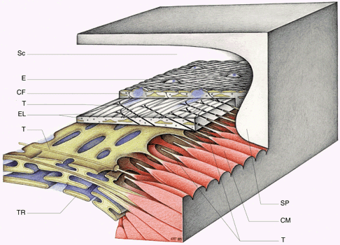
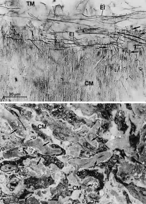
as cationic ferritin freely penetrate into the central core of the trabeculae (Fig. 8).9 The central core of the trabecular lamellae contains numerous collagen and elastic fibers embedded in a homogeneous ground substance rich in hyaluronan and proteoglycans.10–12 The elastic fiber net of the corneoscleral meshwork is continuous posteriorly with that of the scleral spur and anteriorly with that of the sulcus sclerae. In the uveal meshwork the elastic fibers are continuous posteriorly with the elastic fibers that are located within the connective tissue between the ciliary muscle tips and in front of the circular muscle portion.13 The elastic fibers differ in structure and composition from most of the elastic fibers in the body so that they were termed elastic-like fibers.14 Ultrastructurally, they reveal an electron-dense core sparsely intermingled with electron-lucent material(Figs. 9 and 10). These fibers are surrounded by a more or less homogeneous sheath. Treatment with pancreatic elastase (see Fig. 10)14 and immunohistochemical studies using antibodies against α-elastin and tropoelastin15 revealed that only the relatively small electron-lucent area in the central core of the elastic-like fibers contains elastin and its soluble precursor tropoelastin. These components are embedded in electron-dense material of yet unknown nature. The sheaths consist mainly of elastic microfibrils and collagenous fibrils, embedded in pro-teoglycans that can be digested by chondroitinases.14 In infant eyes, the sheath is very small, whereas in adult eyes the sheath is well developed and becomes continuously thicker with increasing age.10,14,15 In the adult the fibrous material of the sheath consists mainly of the so-called curly collagen (lattice collagen or long-spacing collagen). This banded material presumably contains type VI collagen16 (Fig. 11). The collagenous fibers found in the central core of the corneoscleral lamellae show the normal periodicity of 64 to 68 nm, and clusters of long-spacing collagen have also been found in these regions. According to immunohistochemical studies of Murphy and coworkers,17 there is a pronounced staining for type III and type I collagen in the trabecular lamellae. The basement membranes of the lamellae that stain for type IV collagen and laminin are continuous anteriorly with the basement membrane of the corneal endothelium.10,17–20 CRIBRIFORM LAYER AND AQUEOUS OUTFLOW RESISTANCE In contrast to the corneoscleral meshwork, the cribriform layer does not show a regular organization of trabecular lamellae and beams, but the cells are distributed within the extracellular material. The material consists of an irregularly arranged network of fine fibrils, ground substance, and an elastic-like fiber system. In histologic sections oriented in a tangential plane, the fibrous network appears to be a mesh surrounding many holes or openings of different sizes (Fig. 12). This was the reason the term cribriform meshwork was introduced.21 Serial tangential sections showed in the electron microscopic dimension that the fiber network belongs to a complicated system that connects the inner wall endothelium with the trabecular lamellae. The elastic-like fibers of the cribriform layer form a regularly arranged network of interlacing fibers that are continuous with the elastic-like fibers of the adjacent corneoscleral lamellae (Fig. 13). Toward the inner wall endothelium, bundles of fine fibrils separate from the elastic-like fiber sheaths and connect the subendothelial network of elastic-like fibers either with the endothelium of Schlemm's canal or with the subendothelial basement membrane material. Therefore, these fibrils were called “connecting fibrils” (Fig. 14).22 In the adult eye, the connecting fibrils are embedded in a homogeneous ground substance that is digestible with chondroitinases.14
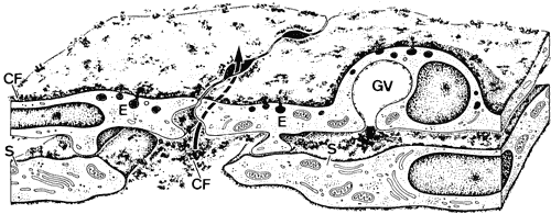
Electron microscopic investigations reveal thatthere are optically empty spaces between the connecting fibrils. In an experimental study on monkey eyes, we found that the area of such “empty spaces” in the cribriform region is positively correlated with outflow resistance values measured by anterior chamber perfusion.23 We therefore assumed that these spaces may represent preferential pathways for aqueous outflow through the cribriform layer. Calculations about the pressure gradient across the cribriform and subendothelial layers lead to the statement that if the spaces were really empty, the extracellular material and cells in this region might provide only insignificant resistance to aqueous outflow.24,25 If, however, the preferential pathways are coated with a 0.35-μm-thick glycosaminoglycan layer, the pressure gradient would rise to the physiologically observed values. It is possible that hyaluronan, glycosaminoglycan, or proteoglycan gel, lost during the embedding procedure, also contributes to formation of outflow resistance.26 This hypothesis was recently supported by experimental studies in the living monkey where in fact 80% to 90% of outflow resistance was shown to be located in front of Schlemm's canal, probably within the cribriform layer of the TM.6 INNER WALL OF SCHLEMM'S CANAL The endothelial lining of the canal consists of a complete monolayer of flat endothelial cells that, like other vascular endothelial cells, stain for factor VIII and plasminogen.27 Unlike other vascular endothelium, the endothelial cells of Schlemm's canal do not rest on a complete basement membrane. A basement membrane is formed initially during embryonic development, but it gradually disappears as aqueous circulation starts.28,29 In its final form Schlemm's canal is more comparable with a lymphatic than a blood vessel. Lymphatic vessels do not usually possess basement membranes. Their endothelial lining is perfused from outward to inward and often connected with elongated cytoplasmic processes of adjacent pericytes. As in lymphatics, the endothelial cells of the inner wall of Schlemm's canal also develop cytoplasmic processes that interdigitate with similar processes of underlying cells of a second row underneath the canal endothelium (Fig. 15). The subendothelial cell layer is not complete and consists of elongated, star-like cells oriented predominantly in a radial anteroposteriordirection. In contrast, the endothelial cells ofSchlemm's canal, measuring about 160 μm in length and covering an area of 408 μm2 run mostly in an equatorial direction.30 The cellular and fibrillar connections with the cribriform layer may stabilize the inner wall during IOP variations and changes in the perfusion rate. The double-layered structure of the inner wall is often seen to be elevated and protrudes into the lumen of the canal when IOP is particularly high at the time of tissue fixation. If IOP is low, the two cell layers are pressed together from the luminal side, thus presumably preventing a reflux of blood into the TM and anterior chamber.31,32 If IOP is slightly elevated, only the endothelial cells bulge into the lumen of the canal, thus forming large vacuoles, usually called giant vacuoles. Since Holmberg33 first described these “giant vacuoles” within the inner wall endothelium of Schlemm's canal, the discussion about their functional significance has never been resolved.34 Many of the giant vacuoles communicate with the subendothelial space by way of an often large opening, but very few communicate with the lumen of Schlemm's canal. If there is a communication with the canal, it is usually by a small pore on the canal side of the cell.35 When serial sections are employed, it is possible to demonstrate that some “vacuoles” have openings on the inner and outer sides, thus forming transcellular microchannels. Tripathi34,36 proposed that the endothelial cells lining Schlemm's canal have the ability to transfer aqueous humor in bulk by a cyclical pressure-dependent process that starts initially as an invagination from the trabecular side and proceeds to the formation of a transcellular channel by a perforation in the outer membrane of the vacuole. This hypothesis has, however, not yet been verified. On the other hand, it is now generally accepted that both the frequency and size of giant vacuoles are proportional to the pressure in the anterior chamber at the time of fixation.37 Nearly 20,000 pores have been counted at the luminal side of the inner wall endothelium in human eyes. Therefore, as calculations showed, only 5% to 10% of the entire resistance can be localized within the endothelial lining of human eyes.35,38 This, again, points to the assumption that the subendothelial layers play the major role in forming and possibly regulating outflow resistance. Because occasionally ground substance or particles are found within the giant vacuoles, the transcellular channel system may provide a mechanism by which extracellular material can be “cleared” or “washed out” from he cribriform layer. Inomata and associates39 have shown in cynomolgus monkeys that particles suspended in gelatin solution pass through the transcellular microchannels of the endothelial lining, as do erythrocytes if perfused through the anterior chamber. The endothelial cells of Schlemm's canal are bound together by maculae adherentes and tight junctions.8,37 However, these tight junctions do not form zonulae occludentes, but in places the adjacent cell membranes are separated, forming meandering pathways between interdigitating cell processes. These pathways are often open toward both the cribriform layer and the lumen of the canal (Fig. 16). Anterior chamber perfusion with cationized ferritin in rhesus monkeys has shown that these particles predominantly pass through the paracellular routes.8,9 Often these routes are locally expanded, so that in sagittal sections “vacuole-like” structures are formed.9 According to our observations, macrophages and leukocytes mostly pass through the paracellular routes to leave the eye rather than through the transcellular channels (Fig. 17). |
CONNECTIONS OF THE TRABECULAR MESHWORK WITH THE CILIARY MUSCLE |
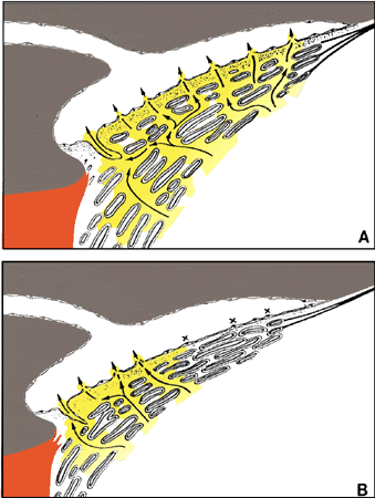
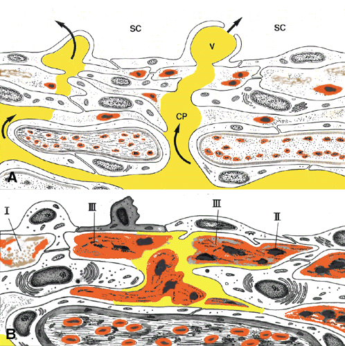
SCLERAL SPUR It has been known for a long time that pilocarpine reduces IOP. However, not before Barany studied the effect of miotics in monkeys has a rationale for the pilocarpine effect been developed.40,41 CILIARY MUSCLE TENDONS How could ciliary muscle contraction affect the outflow pathways in the TM? As shown by Rohen and coworkers, the anterior ciliary muscle tendons are closely connected with the fiber network of the TM.21,42 There are three different types of tendons by which the anterior ciliary muscle tips are connected with the TM or the corneosclera. Type I tendons derive from the outermost longitudinal muscle bundles and enter the sclera or the scleral spur to fix the muscle to the external tunica of the eyeball (Fig. 18). Type II tendons pass the scleral spur to anchor within the TM. They consist of elastic-like fibers that bend into the outer part of the TM and finally join the subendothelial network of elastic-like fibers described earlier. Type III tendons are of collagenous nature. They represent broad, elongated bands that penetrate the TM and insert within the corneal stroma.21,42 The tendons represent the main fixation of the entire ciliary muscle system to the external tunica of the eyeball and therefore seem to be important in the accommodation mechanism. The tendons may also help to expand the system of trabecular lamellae, so that the intertrabecular spaces remain open or enlarge if the ciliary muscle moves forward and inward. Regarding the outflow resistance, this would have little effect in normal eyes. The main effect on aqueous outflow resistance seems to result from actions of the elastic-like type I and II tendons. Because the type I tendons connect the outermost ciliary muscle fiber bundles to the scleral spur, muscle contraction leads to a backward movement of the scleral spur followed by a change in the form of the outflow pathways (Fig. 19).21,42 Inward movements of the type II tendons during muscle contraction have a similar effect. It seems that normally parts of the cribriform layer (including their preferential aqueous pathways) are collapsed so that only a portion of the filtration area is functional. After ciliary muscle contraction the cribriform elastic-like fiber network is pulled inwardly and the connecting fibrils are straightened so that the entire cribriform layer expands. In addition, the lumen of Schlemm's canal is enlarged so that finally the filtering area increases and outflow resistance decreases (Fig. 20). |
TRABECULAR CELLS |
CONTRACTILITY AND INNERVATION Recent experimental and morphologic data indicate that changes in ouflow facility might be induced not only by ciliary muscle contraction but also by shape changes of the trabecular cells themselves. Trabecular meshwork cells express contractile elements such as smooth muscle α-actin and myosin (Fig. 21).43–46 In vitro the cells contract in response to different mediators, including acetylcholine.47 In addition, the presence of muscarinic receptors has been described in human TM cells.48 In contrast, nitrovasodilators induce relaxation of TM cells in vitro.49 In perfused anterior eye segments, substances that contract trabecular cells decrease outflow facility, whereas substances that induce relaxation increase outflow facility.50 How can this effect, which is opposite to that of ciliary muscle contraction and relaxation, be explained? If the cells bridging adjacent trabecular beams contract, this would cause a decrease in width of the intertrabecular spaces (see Figs. 6 and 20), whereas aqueous flow would widen the spaces if the cells are relaxed. The effect of TM contraction on the three-dimensional structure of the meshwork would then resemble that shown for ciliary muscle relaxation, whereas relaxation of the TM would cause a similar effect as that induced by ciliary muscle contraction. The mechanism by which contraction or relaxation of TM cells is mediated in vivo is not known. It is tempting to speculate that the numerous nerve terminals found in the TM might be involved in this outflow regulatory effect of the TM (Fig. 22).51–54 In human and monkey TM, abundant nitrergic nerve terminals were found.54 The terminals are most numerous in the cribriform layer, in which the cells are not only connected to each other and to the cribriform elastic network but also to the inner wall endothelium itself. NO-induced relaxation of these cells would therefore influence the outflow pathways most effectively. In human eyes, there are also abundant cholinergic nerve terminals in the TM. In elderly human eyes, the number of TM cells staining for α-sm-actin decreases, but there are always positively stained cells in the posterior portion of the TM in the scleral spur and in single cells of the cribriform layer.46 The distribution of cholinergic nerve fibers in elderly human eyes was essentially the same as that seen for α-sm-actin staining.54 If cholinergic stimulation simultaneously would cause ciliary muscle and TM cell contraction, the net effect of this stimulation would be an increase in outflow facility because the force of the ciliary muscle contraction would be much stronger than that of the TM contraction. In this case, contraction of TM cells might be required to produce a concurrent stiffening of the TM to avoid disruption of TMby the pull of the muscle tendons during contrac-tion. Numerous nerve terminals staining with antibodies against substance P are in intimate contact with the elastic fibers of the TM and the cribriform region.54 Because the TM elastic fibers are important stress-bearing components, this arrangement of nerve terminals may allow sensing of trabecular stretch, changed by ciliary muscle contraction or relaxation or by IOP changes. They might therefore represent part of the afferent pathway, the nitrergic and cholinergic terminals part of the efferent pathway responsible for relaxation or contraction of TM. In human eyes, a similar innervation has also been found in the scleral spur.55,56 SYNTHETIC CAPACITY Outflow resistance is, however, not only influenced by mechanical factors but also depends on the biologic activity of the trabecular cells. Important activities include synthesis or lysis of extracellular material and phagocytosis. Anterior chamber perfusion always results in a washout of extracellular material that has to be constantly replaced to maintain the necessary outflow resistance. The ability of cultured trabecular cells to synthesize different components of extracellular material has been shown by several investigators.57–59 Using 36S-sulfate and 14C-glycosamine to label glycosaminoglycans, it was shown that monolayer cultures of human trabecular cells are capable of synthesizing cellular and extracellular glycosaminoglycans. All monolayer cultures studied so far produce hyaluronan, heparan sulfate, chondroitin sulfate, and dermatan sulfate glycosamino-glycans in various quantities. An increase in glycosaminoglycan synthesis was noted after addition of hyaluronan to the medium, which indicated the existence of a positive feedback mechanism in the meshwork.60 If in cases of transient hypotonia a reflux of blood into Schlemm's canal and eventually into the TM occurs, a blockage of outflow pathways by fibrin clots might result. It has already been shown by Pandolfi that the TM possesses to some extent a fibrinolytic activity.61 Cultured human TM cells are capable of synthesizing large amounts of tissue plasminogen activator. This was found to be more than in vascular endothelium. However, in contrast to vascular cells, the trabecular cells produce comparatively little inhibitor, indicating that fibrinolysis is more important than clotting within the TM.62 PHAGOCYTIC CAPACITY The TM has a great capacity for self-cleaning, mainly owing to phagocytic processes of trabecular cells. It was first shown by Rohen and van der Zypen63 that trabecular cells are capable of phagocytosing foreign particles or tracer material perfused through the anterior chamber. In the TM of older persons, usually a number of trabecular cells are found that contain pigment granules. Pigment deliberation always takes place from the pigmented epithelial layers of the ciliary body or iris during life. The granules reach the outflow channels with aqueous circulation and are then phagocytosed by trabecular cells. Experimentally introduced particles such as colloidal gold, horseradish peroxidase, vital stains, and even erythrocytes or cellular debris are immediately engulfed by trabecular cells of monkey or cat eyes and thus eliminated from the aqueous circulation.63–67 Trabecular cells contain among other organelles lysosomes, so that the phagocytosed material can be digested within the phagolysosomes. There is no clear-cut evidence that trabecular cells in vivo detach after phagocytosis from the trabecular beams and leave the TM. In the cat eye, trabecular cells leave the eye after phagocytic challenge, but this cell loss had recovered by the end of 150 days.67 However, in human eyes many of the trabecular cells, particularly those containing pigmented granules, stay for a long time within the meshwork.68 Using different macrophage antibodies, we found a positive staining only of single cells.69 Most of the trabecular cells remained unstained, indicating that these cells presumably do not present antigens. Some of the stained cells are probably macrophages, which move through the intertrabecular spaces. They usually leave the TM by way of Schlemm's canal. We often found macrophages passing theparacellular routes of the inner wall endothelium and squeezing through the intercellular spaces into the lumen of the canal (see Fig. 17). The origin of these cells is unknown. They probably derive from the ciliary body or the iris. CELLULARITY AND REGENERATION Although human meshwork cells can proliferate in culture, their capacity for replication in situ appears limited. Defined countings of cell number have shown that there is a steady decline in cells throughout life. A linear decrease in cellularity (nuclei per solid tissue area) and a decrease in absolute cell number (nuclei per histologic section) has been found by Alvarado and associates70 that amounts to 0.56% per year at a constant rate. At 20 years of age the estimated cell number for the whole meshwork was calculated by Grierson and Howes71 as 763,000. The number decreased by 80 years to 403,000, with a loss rate of 6,000 per year (Fig. 23).72 Interestingly, the rate of nuclear loss was found to differ in different regions of the meshwork. The decrease in cell nuclei is least in the cribriform layer and greatest in the central region of the TM. Nevertheless, the loss of cells appears relatively small in comparison to the many injuries and “cleaning processes” that take place during the entire life span. The question arises whether there is any evidence for regenerative processes in the meshwork. In normal sections through primate eyes, mitotic figures have never been observed. Trabecular cells do not reveal a great regenerative capacity in vitro. In organ cultures particularly, the uveal cells degeneraterather quickly, whereas the cribriform layer cells often survive and proliferate. In vivo, after experimentally induced damage of the TM with loss of cells, clusters of large, cuboidal cells forming elongated cell strands were seen in monkey eyes.73,74 The cell groups are situated mainly in the anteriormost portion of the TM or the transition zone to the cornea. Similar signs of all proliferation have also been observed after laser trabeculoplasty in human eyes. Laser trabeculoplasty can stimulate trabecular cell replication and activation. Autoradiographic studies with H3-thymidine in organ cultures of human autopsy eyes revealed a fourfold increase in cell division 2 days after laser trabeculoplasty.75 Nearly 60% of this cell division was localized to the anterior, nonfiltering region of the TM. SCHWALBE'S LINE CELLS The cells distributed within the transition area between cornea and meshwork often contain a great amount of endoplasmic reticulum and a large num-ber of mitochondria as well as many electron-dense granules. Raviola76 has called these cells “Schwalbe's line cells.” She believed they were active secretory elements that produce some kind of surfactant. Stone and colleagues77 later found that these cells also stain for neuron-specific enolase. We found that staining for the enzyme hyaluronan synthase is prominent in Schwalbe's line cells.78 We must assume that in this region there is a specific cell population whose functional significance is still unknown. OUTFLOW CHANNELS OF SCHLEMM'S CANAL After passing through the TM, aqueous humor enters Schlemm's canal and then the collector channels. Schlemm's canal is not a uniform vessel but is often divided into different parts by bridges or septa.79 The septa cross the lumen of the canal mostly in an oblique direction. They are often fixed to the outer wall of the canal at places where the collector channels begin. Thus the septa, which often form twisted or spiral bands, can be considered to be guiding structures for aqueous humor toward the openings of the collector channels (Figs. 24 and 25). Some bridges do not cross the entire lumen of the canal but remain part of the outer wall, adjacent to a collector channel entrance. The number and form of septa and bridges vary to large extent. There are also regional differences with regard to form, number, and development of these structures. The structure of the outer wall of Schlemm's canal differs very much from that of the inner wall. The endothelial lining is single-layered, with a well-developed basement membrane. The cells do not possess transcellular microchannels. The adjacent stroma consists of collagenous and elastic-like fibers intermingled with fibroblasts. The 25 to 35 collector channels drain the aqueous humor toward the intrascleral or episcleral venous plexus.1,2,79 Two different types of collector channels can be distinguished (Fig. 26). Direct channels run directly toward the episcleral plexus without any connections with the intrascleral vessels. If these channels contain aqueous humor, they can be identified in the limbal region as “aqueous veins.”80 In the human, normally only four to six direct channels exist. The indirect collector channels, which are more numerous nasally than temporally, join the intrascleral venous plexus in various distances from Schlemm's canal. A collector channel is distinguished by a thin wall that consists of endothelium, basement membrane, and a discontinuous row of pericytes. It is separated from the sclera by loosely arranged connective tissue, which occasionally contains pigmented cells, lymphocytes, and macrophages. |
TRABECULAR MESHWORK IN GLAUCOMATOUS EYES |
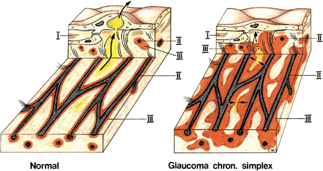
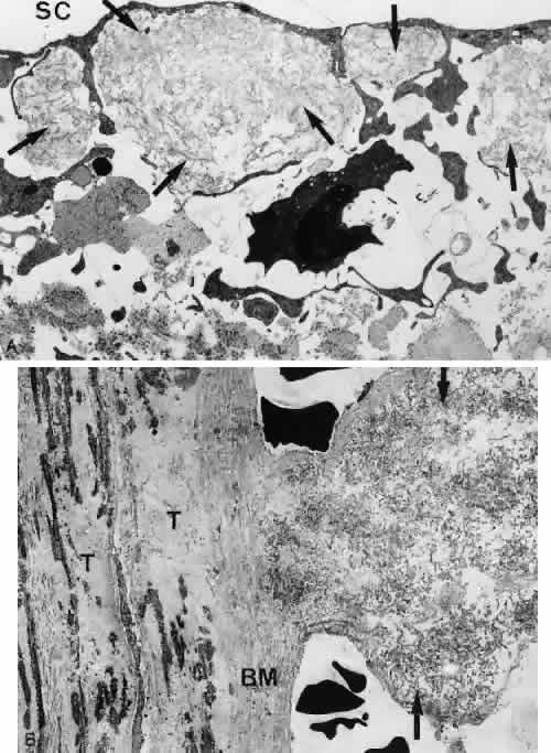
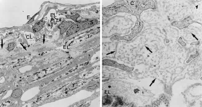
POAG In trabeculectomy specimens derived from later stages of primary open-angle glaucoma (POAG),Rohen and Witmer81 found excessive amounts of extracellular material within the cribriform layer of the TM underneath the endothelial lining of Schlemm's canal, which was called “plaque material” (Figs. 27 and 28). Three different types of plaques were initially distinguished. Type I plaques are mostly situated immediately underneath Schlemm's canal and appear relatively homogeneous or granular in structure. The amount of type I plaques seems to decrease rather than increase with aging.82 Type II plaques appear in sagittal sections as electron-dense dots. In tangential sections, however, it turns out that they represent cross sections of the central core of the elastic-like fibers (Figs. 27, 28, 29). The diameter of the electron-dense core of the elastic-like fibers (type II plaques) located within the cribriform layer does not change significantly with age or with POAG. Type III plaques consist of electron-dense material containing fine, banded fibrils. When analyzing tangential sections, we can see that these plaques derive from the elastic-like fiber sheaths. These plaques are defined therefore as “sheath-derived” plaques.14,22,83 Because these plaques develop predominantly within the elastic-like fiber network underneath the inner wall of the canal, they often become confluent, thus forming wide plates. A quantitative evaluation of the area taken by the sheath-derived plaque material in normal and glaucomatous eyes has shown that there is a continuous increase of this material with increasing age and that the area of plaque material in cases of POAG is significantly higher than in normal eyes of members of the same age groups (Fig. 30).83 The augmentation of the sheath-derived plaque material in cases of POAG might become the cause of outflow resistance elevation if a greater percentage of outflow channels in the cribriform layer and the inner wall endothelium is blocked or closed off by this material (Fig. 31).83–86 The composition and nature of the glaucomatous plaque material are still unknown. In trabeculectomy specimens of glaucomatous eyes, fine fibrils adhering to the subendothelial elastic-like fiber network were identified as parts of the sheath mate-rial dissolved by enzyme digestion.14 These fibrilsmight provide a base for the deposition of additionalmaterial, so that the holes within the cribriformelastic-like network become progressively smaller.In the end stage of POAG, broad interlacing plates beneath the inner wall endothelium are formed (Fig. 29). Sheath-derived plaques have also been found in the outer wall of Schlemm's canal (see Fig. 27). The amount of this material was found to be greater in glaucomatous than in age-matched normal eyes, but this increase was less than in the inner wall.83 In addition, typical sheath-derived plaques were observed within the intramuscular connective tissue of the anterior ciliary muscle tips,13 in regions where elastic-like fibers are present (Fig. 32). Because these fibers also come in contact with aqueous humor by the uveoscleral flow, the same factors that induce the pathologic changes in the TM may also be responsible for the plaque formation in the anterior portion of the ciliary muscle. In donor eyes with POAG in different stages of the disease, a significant correlation was found between areas of plaques beneath the inner wall of Schlemm's canal and nerve fiber loss, determined in cross sections through the postlaminar optic nerve.86 This correlation either indicates that the factors inducing plaque formation in the outflow pathways are also involved in the pathogenesis of the glaucomatous optic neuropathy, or that a sequence of events occurs (namely, plaque formation induces an IOP increase that then causes degeneration of optic nerve fibers). In the trabecular cells of a number of glaucomatous eyes, an increased expression of the stress protein αB-crystallin was observed.87 In normal eyes this protein is expressed mainly in cribriform cells,88,89 whereas in several glaucomatous eyes expression of this protein was also found in trabecular cells covering the beams of the corneoscleral and uveal meshwork. These changes were seen not only in eyes with POAG but also in eyes with pseudoexfoliation glaucoma, presumably indicating that stress to the cells (e.g., through elevated IOP) could be causative for the overexpression of αB-crystallin in TM cells of glaucomatous eyes. In the TM cells of glaucomatous eyes, we also found an increased expression of the MYOC/TIGR gene product.87 The functional significance of this protein is not yet known. Defects of the MYOC/TIGR gene have been detected in several patients with early onset of glaucoma (juvenile glaucoma), but in patients with POAG only 3% had MYOC/TIGR gene defects. It is therefore possible that MYOC/TIGR protein expression in trabecular cells of glaucomatous eyes is a secondary phenomenon. In glaucomatous eyes, a significant decline in cellularity of the TM compared with old eyes of the same age groups was found. The cell loss in the TM takes place in a gradient-like manner. Alvarado and coworkers90 described that in the nonglaucomatous and glaucomatous meshwork, the cellularity curves are parallel to each other. These authors suggested there is a congenital basis for the disease and that the patient presumed to have glaucoma might be born with a decreased number of trabecular cells. A progressive cell loss may also explain trabecular fusion, because without a continuous cell cover, adhesions between denuded portions of adjacent trabecular beams can develop. Grierson found a linear age-related increase in the frequency of fusions between trabecular beams, particularly in the inner portions of the meshwork.85 In trabeculectomy specimens of advancedPOAG, we often found an extreme “hyalinization” of the uveal and corneoscleral lamellae. The basement membranes were enormously thickened (Fig. 33), revealing a great number of clusters of long-spacing collagen. Thus, the basement membranes show a “shagreen-like” pattern. Often the basement membrane thickening begins at the inner surface of the trabecular lamellae, suggesting there is a factor that reaches the meshwork from the anterior chamber side. The nature of this factor is not yet known. PEXG In cases of pseudoexfoliation glaucoma, the intertrabecular spaces and the cribriform layer often contain clusters of flake-like material consisting of randomly arranged short fine fibrils (Fig. 34).91–94 Two types of fibrils have been distinguished: the thinner type A fibrils, with a diameter of around 15 to20 nm and a periodicity of about 50 nm, and the thicker type B fibrils (diameter 30–45 nm), which show no periodicity. The nature and composition of the pseudoexfoliation fibrils are still not known, but staining for glycoproteins,95 microfibrils of elastic tissues, and amyloid P96–99 has been demonstrated. In eyes with pseudoexfoliation syndrome, Schlötzer-Schrehardt and Naumann95 found a significant correlation between the amount of pseudoexfoliative material located in the subendothelial region of Schlemm's canal with IOP, indicating that the material in fact is responsible for an increase in outflow resistance. Our data support these findings. In addition, we found that the amount of pseudoexfoliative material was significantly correlated with the loss of axons in the optic nerve and with IOP,68 indicating that an IOP increase is one of the factors causative for the glaucomatous optic nerve neuropathia in these eyes. PIGMENTARY GLAUCOMA In cases of pigmentary glaucoma, many trabecular endothelial cells and cells of the cribriform layer were found filled with pigment granules.100,101 It is not yet known whether pigmentary glaucoma is caused by pigment obstruction (mechanically) or by disturbances of trabecular cell functions induced by pigment granule phagocytosis (biologically). As a rule the inner wall endothelium is not pigmented.According to Johnson,102 the pigmentation of the cribriform layer is less than that of the uveal meshwork. In trabeculectomy specimens, most pigment was phagocytosed, but some free granules were also seen in the intertrabecular spaces or the pathways of the cribriform layer. In other respects, the structure of the TM appeared relatively normal.100,101 JUVENILE GLAUCOMA In cases with juvenile glaucoma, the TM showed changes similar to those seen in eyes with steroid-induced glaucoma: namely, fibrils arranged in afingerprint-like pattern and increased amounts of fibrillar material beneath the inner wall of Schlemm's canal.103 However, the amount of sheath-derived plaques, increased in eyes with POAG, was also elevated in eyes with juvenile glaucoma. CORTICO-STEROID GLAUCOMA The mechanism of pressure elevation in corticosteroid-induced glaucoma is also not understood. In trabeculectomy specimens and in donor eyes treated with corticosteroids for various time periods,106 accumulation of extracellular material distinct from the sheath-derived plaques typical for POAG and from pseudoexfoliative material was observed.104–106 Directly beneath the inner wall endothelium, an accumulation of densely packed fine fibrils was seen. At places these fibrils separated the connecting fibrils from the inner wall endothelium (Fig. 35).106 In the cribriform meshwork, fingerprint-like arranged material resembling basement membranes was found.104,106 The pathogenesis of the morphologic changes seen in the different kinds of glaucoma is not yet clarified. The observed differences in morphology support the hypothesis that glaucoma is a multifactorial disease. Further studies are necessary to find the specific factors that induce these characteristic morphologic changes. |