Chapter 43
Anatomy of the Ciliary Body and Outflow Pathways
THOMAS F. FREDDO and HAIYAN GONG
Main Menu   Table Of Contents

Search

THE CILIARY BODY AND AQUEOUS PRODUCTION
THE TRABECULAR MESHWORK AND CONVENTIONAL AQUEOUS OUTFLOW
SUMMARY
ACKNOWLEDGMENTS
REFERENCES

An understanding of the glaucomas begins with an understanding of the anatomy of the structures related to the production and drainage of aqueous humor, particularly the ciliary body and the trabecular meshwork. The ciliary body is the sole source of aqueous production. The trabecular meshwork is part of the principal, pressure-dependent pathway for aqueous outflow and harbors the requisite resistance to this flow needed to maintain normal intraocular pressure. Regulation of this flow is dependent, in part, on the anatomic connections between the smooth muscle of the ciliary body and the trabecular meshwork into which its tendons extend.

More extensive treatises on the ultrastructure of the ciliary body and trabecular meshwork appear elsewhere.1,2 The following is intended as an introduction and overview for the practicing clinician and clinician-in-training.

Back to Top
THE CILIARY BODY AND AQUEOUS PRODUCTION

THE CILIARY BODY

The ciliary body performs three principal functions: (1) It produces and secretes aqueous humor into the posterior chamber of the eye; (2) it contains the smooth muscle that acts on the crystalline lens, via the zonular fibers, to shift the focus of the eye from far to near (accommodation); and (3) by extension of certain smooth muscle fibers and their tendons into the adjacent trabecular meshwork, it can assist in the drainage of aqueous humor from the eye. There is also some evidence supporting a role for the ciliary body in the production and slow turnover of certain molecular components of the vitreous humor.3

The ciliary body extends posteriorly from the root of the iris to the ora serrata. In the adult eye, the distance from the surgical limbus to the ora serrata is approximately 7 mm on the temporal side of the globe and 6 mm on the nasal side. The ciliary body is grossly subdivided into two portions, the pars plicata and the pars plana.

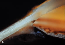
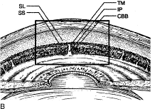
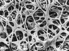
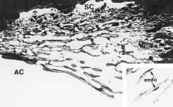
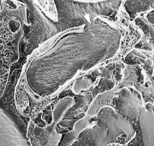
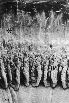
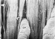
The pars plicata exhibits 70 to 80 corrugated, fin-like ridges on its inner surface that are arranged around the circumference of the crystalline lens (Fig. 1). Each individual ridge is referred to as a ciliary process, and these are divided into categories of major and minor processes based on their relative height. Major processes predominate, and they extend into the posterior chamber of the eye approximately 1 mm. Minor processes are about one third as high. In the scanning electron micrograph shown in Figure 2 these structures and their relationships are clearly discernible. It is important to appreciate, however, that the zonular fibers supporting the crystalline lens have been removed from the surface of this specimen. These fibers form a continuous carpet over the inner surface of the pars plana and are then channeled into the valleys between adjacent processes in the pars plicata on their way to the lens capsule. As such, in vivo, the valleys between adjacent ciliary processes are largely filled with zonules leaving only the crest of each process exposed to the posterior chamber (Fig. 3).

Fig. 1. Macrophotograph of the crystalline lens and surrounding ciliary body. The fin-like ridges representing the ciliary processes of the pars plana region are evident, surrounded by the smooth-surfaced pars plana.

Fig. 2. Scanning electron micrograph of the inner surface of the iris and ciliary body with the lens and zonules removed. The pars plicata region is distinguished from the more posterior pars plana region (asterisk) by the presence of major (M) and minor (m) ciliary processes. (From Morrison JC, Van Buskirk EM, Freddo T. Anatomy, microcirculation and ultrastructure of the ciliary body. In: Ritch R, Shields MB, Krupin T (eds). The Glaucomas. St Louis: CV Mosby, 1989.)

Fig. 3. High magnification scanning electron micrograph showing the confluence of zonular fibers leaving the pars plana (top) channeling into the valleys between adjacent ciliary processes. CP, Ciliary processes.

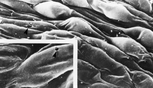
The pars plana, as its name implies, exhibits a flat inner surface. This portion of the ciliary body is much thinner and less vascularized than the pars plicata, making it the safest point of entry into the eye for vitreoretinal surgical procedures.

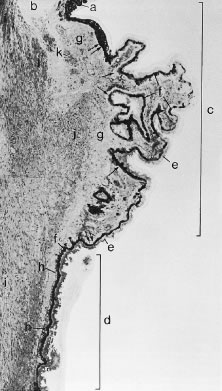
In meridional sections, the ciliary body appears as shown in Figure 4. The entire surface of the ciliary body, facing the inside of the eye, is lined by a bilayered epithelium. These two layers are named for their relative content of melanin pigment. The layer closest to the posterior chamber is devoid of pigment and is called the nonpigmented ciliary epithelium. The layer closest to the ciliary body stroma is called the pigmented ciliary epithelium for its melanin content. The nonpigmented ciliary epithelium is continuous with the posterior pigmented epithelium of the iris anteriorly and with the neurosensory retina posteriorly at the ora serrata. The pigmented ciliary epithelium continues anteriorly as the anterior myoepithelium of the iris (which includes the iris sphincter muscle) and posteriorly as the pigmented epithelium of the retina. The double layer of epithelium covering the ciliary body actually represents two simple epithelia that are joined at their apical surfaces following the invagination of the optic cup during embryogenesis. As a result, the basement membrane of the nonpigmented ciliary epithelium faces the posterior chamber and that of the pigmented ciliary epithelium joins both epithelial layers to the ciliary body stroma.

Fig. 4. Light micrograph of ciliary body of a 50-year-old man. The iris root (a) and the anterior chamber angle (b) are seen at the top. The pars plicata (c) and a portion of the pars plana (d) are included within brackets; they are lined by the nonpigmented (e) and pigmented (f) ciliary epithelium. The basement membrane of the pigmented ciliary epithelium increases (h) in thickness with age. The ciliary body stroma (g) and the three portions of the ciliary muscle (i, longitudinal; j, radial; k, circular bundle) are also shown. (From Hogan M, Alvarado J, Wedell J: Ciliary body and posterior chamber. In: Histology of the Human Eye. Philadelphia: WB Saunders, 1971, p 270.)

The ciliary body stroma is composed of a loose connective matrix that supports the nerves and blood vessels that course within it. This connective tissue matrix extends into the core of each ciliary process. The ciliary body stroma is directly continuous with the stroma of the iris anteriorly and with the choroidal stroma posteriorly. Because the anterior surface of the iris has no epithelial covering, substances released into the ciliary body stroma have access to the anterior chamber by diffusing from the ciliary body stroma to the iris surface.4,5 Similarly, fluid entering the ciliary body stroma from the anterior chamber can reach the choroid by traveling along the uveoscleral outflow pathway described later. Between the loose connective tissue stroma and the inner surface of the sclera is a complex smooth muscle called the ciliary muscle, also described later.

PRODUCTION OF AQUEOUS HUMOR

To provide anatomic correlations for the physiology of aqueous humor formation, it is most convenient to describe the process in two steps: (1) elaboration of a plasma filtrate from which aqueous humor is derived, and (2) formation of aqueous humor from this filtrate. Although these steps are not independent, the first is related primarily to the ciliary body microvasculature and the second to the ciliary epithelium.

Ciliary Body Microvasculature

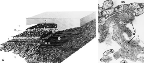
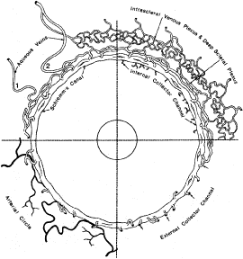
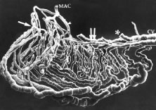
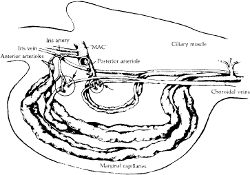
When viewed in cross section, each ciliary process contains a fibrovascular core that is continuous with the ciliary body stroma and covered by the bilayered ciliary epithelium (Fig. 5). The arterioles that serve the ciliary body stroma, like those serving the iris, arise from the discontinuous major circle of the iris.6 Each major process is served by a set of anterior and posterior arterioles7 (Figs. 6 and 7). The anterior arterioles supply the large diameter capillaries, near the crests of the processes, whereas the posterior arterioles supply the smaller caliber capillaries deep within each process. The direction of blood flow in these systems is from anterior to posterior, toward a network of choroidal veins. The blood from the entire ciliary body ultimately leaves the eye via the vortex veins.

Fig. 5. Light micrograph of ciliary processes (asterisks) shows each process contains a fibrovascular core with large diameter capillaries (arrow). NPE, Nonpigmented ciliary epithelium; PE, pigmented ciliary epithelium. (Lütjen-Drecoll E: Functional morphology of the ciliary epithelium. In: Lütjen-Drecoll E (ed): Basic Aspects of Glaucoma Research. Stuttgart, Germany: FK Schattauer, 1982, p 79.)

Fig. 6. A cast of the vasculature within a single ciliary process. The major circle of the iris (MAC) gives rise to an anterior (arrow) and posterior (arrowhead) arteriole. Double arrow denotes choroidal vein (CV). Drainage from ciliary muscle to choroidal veins has been removed at asterisk. (From Morrison JC, Van Buskirk EM, Freddo T. Anatomy, microcirculation and ultrastructure of the ciliary body. In: Ritch R, Shields MB, Krupin T (eds). The Glaucomas. St Louis: CV Mosby, 1989.)

Fig. 7. Diagrammatic representation of Fig. 6 demonstrating the system of anterior and posterior arterioles that enter each ciliary process, ultimately draining into a set of choroidal veins. (From Morrison JC, Van Buskirk EM, Freddo T. Anatomy, microcirculation and ultrastructure of the ciliary body. In: Ritch R, Shields MB, Krupin T (eds). The Glaucomas. St Louis: CV Mosby, 1989.)

Blood flow in the ciliary body is most likely regionalized and under autonomic control. Scanning electron microscopic studies have revealed the presence of localized constrictions in casts of afferent arterioles in the ciliary body microvasculature that may reflect the presence of a sphincter-like system for controlling blood flow.7–10 Through these mechanisms, alterations in blood flow can influence aqueous humor production by increasing or decreasing the amount of filtrate made available to the ciliary epithelium.

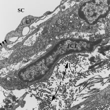
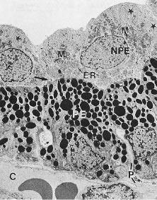
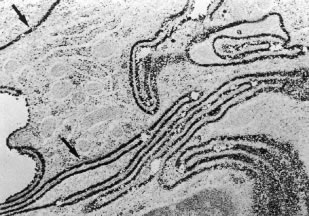
The capillaries derived from the posterior arterioles that serve the ciliary muscle are nonfenestrated and do not leak plasma proteins under normal conditions (Fig. 8). By contrast, the capillaries derived from the anterior arteriole, which pass within the stromal core of each ciliary process, lack tight junctions and are lined by fenestrated endothelial cells (Fig. 9). Using tracers for plasma protein leakage such as horseradish peroxidase (HRP), the capillaries of the ciliary body stroma are seen to be very permeable to macromolecules, as well as to ions and fluid (Figs. 8 and 10 ). As such, these vessels are limited in their capacity to serve as a selective permeability barrier. That function is one of several reserved for the ciliary epithelium.

Fig. 8. A 200-μm thick section of the anterior uvea of a monkey eye, reacted for the demonstration of intravenously injected horseradish peroxidase (HRP). The ciliary body stroma is filled with black, HRP reaction product that has leaked from its fenestrated vessels. The ciliary muscle shows relatively little staining resulting from the nonfenestrated vessels that serve it. The dark coloration of the iris is not HRP but the normal dark pigmentation imparted by the pigmented epithelial layers on its posterior surface.

Fig. 9. Transmission electron micrograph demonstrates the appearance of capillaries in the ciliary body stroma. Numerous fenestrations (arrowheads) are evident along the circumference of the vessel wall. (From Morrison JC, Van Buskirk EM, Freddo T. Anatomy, microcirculation and ultrastructure of the ciliary body. In: Ritch R, Shields MB, Krupin T (eds). The Glaucomas. St Louis: CV Mosby, 1989.)

Fig. 10. Transmission electron micrograph demonstrates that granular horseradish peroxidase (HRP) reaction product leaks through fenestrations (arrowheads) into the surrounding ciliary body stroma. (From Morrison JC, Van Buskirk EM, Freddo T. Anatomy, microcirculation and ultrastructure of the ciliary body. In: Ritch R, Shields MB, Krupin T (eds). The Glaucomas. St Louis: CV Mosby, 1989.)

The Ciliary Epithelium

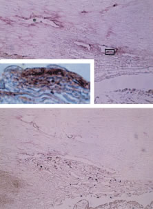
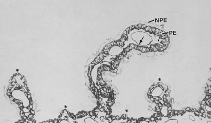
As noted earlier, the two cell layers that constitute the ciliary epithelium are named for their relative content of melanin pigment. The layer closest to the ciliary body stroma is the pigmented ciliary epithelium and that closest to the posterior chamber of the eye is the nonpigmented ciliary epithelium (Fig. 11). The morphology of the ciliary epithelium varies along the surface of the ciliary body, in accord with the different demands placed on it in various locations.1 From this analysis, it appears that it is primarily the epithelia at the tips of the ciliary processes that are involved in the production of aqueous humor. In these areas, immunoelectron microscopic studies have clearly documented both Na-K-ATPase activity (Fig. 12) and carbonic anhydrase activity11,12 (Fig. 13). Both are known to be central to the production of aqueous humor and one of them, carbonic anhydrase, is regularly targeted for pharmacologic inhibition to reduce intraocular pressure in glaucoma.13

Fig. 11. Electron micrograph of the ciliary epithelium (cynomolgus monkey, pars plicata). The posterior chamber is at the top and the ciliary body stroma, with its fenestrated capillaries (C), is at the bottom. Extensive basolateral infoldings of the nonpigmented ciliary epithelial cells (NPE) are evident (stars). The apical surfaces of both layers face one another (arrow). PE, Pigmented ciliary epithelium; M, mitochondria; ER, endoplasmic reticulum; P, paracellular space. (Lütjen-Drecoll E: Functional morphology of the ciliary epithelium. In: Lütjen-Drecoll E (ed): Basic Aspects of Glaucoma Research. Stuttgart, Germany: FK Schattauer, 1982, p. 79.)

Fig. 12. Ultrahistochemistry of the ciliary epithelium stained for the enzyme Na-K-ATPase. Note basolateral infoldings of the nonpigmented ciliary epithelium show intense reaction product for the enzyme (arrows). (From Flügel C, Lütjen-Drecoll E: Presence and distribution of Na+/K+-ATPase in the ciliary epithelium of the rabbit. Histochemistry 88:613, 1988; Tamm ER, Lütjen-Drecoll E: Ciliary body. Microsc Res Tec 33:390, 1996. Reprinted by permission of Wiley-Liss, a subsidiary of John Wiley and Sons, Inc.)

Fig. 13. Electron micrograph of the ciliary epithelium of the albino rabbit after staining for carbonic anhydrase. The reaction product is seen predominantly at the membrane infoldings (in) of the nonpigmented epithelium (NPE) and pigmented epithelium (PE), as well as on the apical cell membranes (Ap). Cytoplasmic staining is found only in the PE. (Lütjen-Drecoll E, Loennerholm G: Carbonic anhydrase distribution in the rabbit eye by light and electron microscopy. Invest Ophthalmol Vis Sci 21:782, 1981. Copyright held by The Association for Research in Vision and Ophthalmology.)

THE BLOOD-AQUEOUS BARRIER IN THE CILIARY BODY

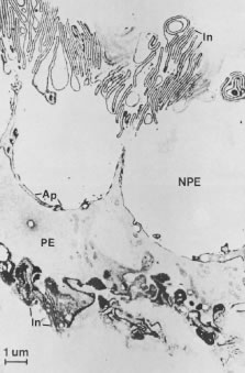
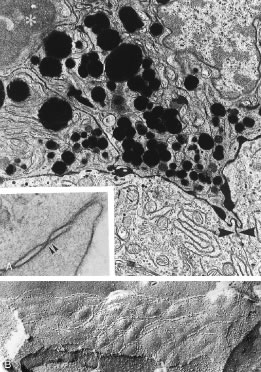
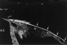
Aqueous humor is a clear nutrient fluid, derived from plasma, that provides for the metabolic needs of the avascular tissues of the eye. Although free amino acid levels in aqueous humor are nearly equivalent to those found in plasma,14 the protein content of aqueous humor in the anterior chamber is less than 1% of that found in plasma.15 Proteins create turbidity and turbidity scatters light, which degrades the optical efficiency of the eye. To prevent this and to ensure that potential antigens in the bloodstream are prevented from reaching the anterior and posterior chambers of the eye and the lens, vitreous, and retina, a selective barrier must be positioned between the bloodstream and the aqueous humor. As noted earlier, the microvasculature of the ciliary body stroma is composed of fenestrated capillaries that readily leak fluids, ions, and plasma proteins to provide the reservoir from which the ciliary epithelium secretes aqueous humor. To ensure that the protein-laden fluid in the ciliary body stroma and the composition of the aqueous humor remain different, a barrier to macromolecular diffusion is interposed between the ciliary body stroma and the posterior chamber by the ciliary epithelium.16 As noted earlier (see Figs. 8 to 10), following injection of HRP in monkeys, this protein readily leaves the lumen of the fenestrated capillaries of the ciliary body stroma.17 The protein diffuses to the ciliary epithelium and easily moves between adjacent pigmented ciliary epithelial cells, filling the intercellular cleft18 (Fig. 14A). On reaching the interface between the juxtaposed apical surfaces of the pigmented and nonpigmented layers, the HRP is again able to freely permeate the intercellular cleft between these cell layers. But when the HRP attempts to diffuse along the intercellular cleft between adjacent nonpigmented ciliary epithelial cells, its progress toward the aqueous humor in the posterior chamber is blocked by an apicolateral junctional complex composed of a zonula occludens (tight junction), a zonula adherens, and desmosome19,20 (see Fig. 14A). In sections, the tight junction is seen as a series of apparent fusion points between the membranes of adjacent nonpigmented ciliary epithelial cells (see Fig. 14,Inset). Using the technique of freeze-fracture, however, allows an en face view of the tight junctions, revealing that these fusion points are actually cross sections of a complex system of branching and anastomosing protein strands that surround the entire circumference of each cell.19 This web of strands spans the intercellular cleft and is mirrored on the adjacent cell, which blocks the intercellular cleft around the entire circumference of each cell, to limit diffusion of macromolecules (see Fig. 14B). Disruption of this junctional complex has been shown to occur in experimental anterior uveitis, resulting in increased amounts of protein in the aqueous humor.21 This extra protein scatters the light of the slit lamp and is described clinically as flare.

Fig. 14. A. Black horseradish peroxidase (HRP) reaction product fills the ciliary body stroma (asterisk) and blackens the intercellular cleft between adjacent pigmented ciliary epithelial cells, as well as the cleft between the apical surfaces of the pigmented and nonpigmented layers. Further diffusion of HRP toward the posterior chamber is blocked by tight junctions between adjacent nonpigmented epithelial cells (arrowheads). Inset. Higher magnification demonstrates the points of fusion (arrows) between membranes of adjacent nonpigmented ciliary epithelial cells that constitute the tight junction. B. Freeze-fracture electron micrograph demonstrates the branching and anastomosing tight junctional strands of a zonula occludens between adjacent nonpigmented ciliary epithelial cells. (From Morrison JC, Van Buskirk EM, Freddo T. Anatomy, microcirculation and ultrastructure of the ciliary body. In: Ritch R, Shields MB, Krupin T (eds). The Glaucomas. St Louis: CV Mosby, 1989.)

Although not involved directly in the barrier properties of the ciliary epithelium, another intercellular junction, the gap junction, has been shown to play an important role in aqueous production. Gap junctions are known to provide a means for metabolic and electrotonic coupling of cells. In the ciliary epithelium, cells of both layers are interconnected by these junctions and they provide a low resistance pathway for the passage of current and ions between cells, allowing them to function as a syncytium in the coordinated secretion of aqueous humor.22,23 When the ciliary body is inflamed, it has been shown that gap junctions largely disappear, contributing to a reduction in aqueous secretion and to the often lower intraocular pressure that accompanies inflammation of the anterior uvea.21 Attempts to exploit gap junction uncoupling as a means of reducing aqueous production in glaucoma have yet to be done.

THE CILIARY MUSCLE

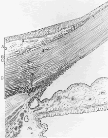
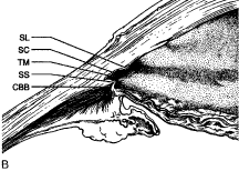
Most of the added thickness of the pars plicata region of the ciliary body results from its layers of smooth muscle. The ciliary muscle is innervated by parasympathetic fibers that originate in the Edinger-Westphal nucleus, leaving the midbrain with cranial nerve (CN) III to be conveyed from its nerve branch to the inferior oblique and then to the ciliary ganglion. Here these fibers synapse, and postganglionic fibers are conveyed to the eye and, therefore, to the ciliary body. Classically three layers of smooth muscle are described as shown in Figure 15. The smooth muscle cells that constitute the outermost of these three layers run in an anterior-posterior direction, and, thus, in meridional sections of the eye, these fibers are cut in longitudinal section. As such, this layer is called the longitudinal bundle of the ciliary muscle (Brucke's muscle). These fibers originate in stellate prongs, called epichordal stars that anchor into the inner surface of the sclera in the posterior portion of the pars plana (see Fig. 15). From this origin, the longitudinal bundle extends anteriorly to insert on the posterior surface of the scleral spur, a projection of scleral tissue that extends inward from the scleral surface like a shelf at the posterior limit of the trabecular meshwork. As described later, contraction of this portion of the ciliary muscle appears to pull on the scleral spur. Doing so results in improved outflow of aqueous humor through the trabecular meshwork. This is the presumed beneficial effect underlying the use of miotics in the treatment of glaucoma.

Fig. 15. Drawing of the ciliary body showing the layers of the ciliary muscle as seen from two perspectives—a step-wise dissection beginning from just beneath the sclera and as seen in meridional sections. The longitudinal bundle (1) is seen originating from epichoroidal stars (f) that attach the muscle to the inner surface of the sclera and continue forward to the scleral spur (d). Its tendons continue beyond into the trabecular meshwork (a). Beneath the longitudinal bundle, the bifurcating fibers (g) of the radial bundle (2) are seen, and, finally, the circular bundle (3) is evident. Schlemm's canal (b) and the external collector channels (c) are also shown. (With permission from: Hogan M, Alvarado J, and Wedell J: Ciliary body and posterior chamber. In: Histology of the Human Eye. Philadelphia: WB Saunders, 1971, p. 270.)

The middle layer of smooth muscle is called the radial bundle, and the innermost layer represents a confined band of smooth muscle cells that encircle the ciliary body. In meridional sections of the eye, these fibers are seen in cross section and are called the circular bundle (Muller's muscle, no relationship to the smooth muscle of the same name found within the eyelid). The circular bundle appears to play less of a role in facilitating aqueous outflow than in the process of accommodation.

These classical separations of anatomic and physiologic function are exaggerations of the way in which this complex muscle actually works. Intricate structural analyses of the various portions of this muscle have revealed that they are not separate and distinct.1

THE INTERSTITIUM OF THE CILIARY MUSCLE AND UVEOSCLERAL OUTFLOW

Only a few years ago a discussion of the ciliary body would not have made specific mention of the connective tissue interstices of the ciliary muscle. However, recent evidence suggests that the interstices of this muscle represent a part of an increasing important alternate pathway for the drainage of aqueous humor called the uveoscleral pathway. Unlike the pathway for aqueous outflow through the trabecular meshwork, discussed later, flow through the uveoscleral pathway is thought generally to be pressure independent.24,25 This pathway conveys aqueous humor from the anterior chamber into the portion of the ciliary muscle that is directly accessible from the anterior chamber angle. The fluid appears to then flow within the connective tissue fascicles that interweave with the smooth muscle fibers of the ciliary muscle. Ultimately, the fluid reaches the inner surface of the sclera. Although not an obvious feature, the ciliary body is separated from the sclera (over most of its length) by a potential space, the supraciliary space, which is continuous posteriorly with the suprachoroid. Having only minimal physical connections to the sclera, except at its anterior and posterior margins, the ciliary body is held against the outer wall of the eye primarily by the intraocular pressure. Aqueous moves posteriorly within the potential space of the suprachoroid as shown in Figure 16, leaving the eye by either diffusing through the sclera (uveoscleral) or by diffusing through the scleral emissaria for the vortex veins (uveovortex) flow.

Fig. 16. Light micrograph demonstrating the distribution of fluoresceinated dextran after intracameral perfusion into the anterior chamber (AC). The tracer has moved posteriorly into the angle tissues (a) and through the ciliary muscle (b) to finally reach the supraciliary and suprachoroidal space (arrows). Tracer has also freely permeated the iris stroma (c). (From Tripathi R. Uveoscleral drainage of aqueous humor. Exp Eye Res 1977;25(Suppl):305.)

Recently, certain prostanoid derivatives have been shown to be able to augment uveoscleral outflow, and they are proving to be a valuable addition to the glaucoma armamentarium.26,27 Several morphologic studies have suggested that the mechanism of action of these drugs may be through a partial dissolution of the ground substance in the interstices of the ciliary muscle that facilitates movement of fluid along this pathway.28

Back to Top
THE TRABECULAR MESHWORK AND CONVENTIONAL AQUEOUS OUTFLOW
Aqueous humor, secreted into the posterior chamber of the eye, enters the anterior chamber through the pupil. Because the pupillary margin rests on the anterior surface of the lens, its varying anatomic relationship with the lens can prompt a relative obstruction of aqueous flow through the pupillary aperture called relative pupillary block. Flow through the pupil can also be obstructed by inflammatory adhesions of the pupil to the lens, called posterior synechiae.

Assuming none of these complications are present, aqueous passes through the pupil and circulates within the anterior chamber of the eye, driven by the convection current created by the temperature difference between the warm iris and the cooler cornea. Rising posteriorly, and falling anteriorly, aqueous drains through the trabecular meshwork in the iridocorneal angle.

As mentioned earlier, 5% to 10% of total aqueous outflow leaves the normal eye via the uveoscleral pathway.24,25 But most of the aqueous humor drains from the anterior chamber into the venous system of the episclera, via a system of progressively smaller channels within the trabecular meshwork and finally into a circumferentially oriented vessel called Schlemm's canal. From this canal, circuitous channels weave toward the surface of the sclera, ultimately joining the episcleral vasculature. Flow through this system is passive, down a pressure gradient, and, thus, any event that elevates episcleral venous pressure (e.g., carotid-cavernous sinus fistula) will result in elevation of intraocular pressure to a level that again exceeds episcleral pressure in order for aqueous outflow to resume.

THE TRABECULAR MESHWORK

The trabecular meshwork is a triangular, wedge-shaped band of tissue encircling and bridging the anterior chamber angle (Fig. 17). The apex of the triangle is attached to the peripheral edge of Descemet's membrane of the cornea that is called Schwalbe's line or ring. Expanding as it moves posteriorly from this point of origin, the trabecular meshwork attaches to the sclera and to the respective stromas of the ciliary body and the peripheral iris. Indeed, thin strands of pigmented tissue often rise from the peripheral iris to meet the trabecular meshwork. These are called iris processes.

Fig. 17. Drawing of the limbus to illustrate the structures evident by light microscopy. The limbal conjunctiva (A) is formed by an epithelium (1) and an areolar connective tissue stroma (2). Tenon's capsule (B) forms an ill-defined connective tissue layer of the episclera (C). The limbal corneosclera occupies area D. a, conjunctival vessels; b, corneal arcades; single arrow, terminus of Bowman's layer. The triangular tissue wedge of the trabecular meshwork (I) originates at Schwalbe's line (double arrows) and broadens posteriorly to become continuous with the root of the iris and the ciliary body. An iris process (k) extends from the iris surface to the trabecular meshwork. Entering the posterior margin of the trabecular meshwork is the scleral spur (f), to which tendons of the longitudinal bundle of the ciliary muscle (g) are attached. Aqueous passing through the trabecular meshwork enters the canal of Schlemm (h), deep scleral plexus (e), intrascleral plexus (d), and finally the episcleral vessels (c). (From Hogan M, Alvarado J, Weddell J. Histology of the Human Eye. Philadelphia: WB Saunders, 1971.)

Projecting into the base of this triangle is a shelf-like lip of sclera called the scleral spur. An imaginary line drawn from the scleral spur to Schwalbe's line separates the trabecular meshwork into two major parts. The portion of the trabecular meshwork closer to the anterior chamber than this imaginary line is called the uveal meshwork, because it extends from Schwalbe's line to the stromas of the ciliary body and iris. The meshwork just external to the imaginary line also extends from Schwalbe's line but connects to the projecting shelf of sclera. This portion of the meshwork is called the corneoscleral meshwork.

The smooth muscle fibers of the longitudinal bundle of the ciliary muscle are attached to the posterior surface of the scleral spur. As mentioned earlier, when these fibers contract, they pull the lip of the shelf posteriorly and, in doing so, separate the layers of the deeper meshwork attached to the anterior surface of the spur. Opening the meshwork in this fashion appears to facilitate aqueous drainage and is the basis for the use of miotics in increasing aqueous outflow to reduce intraocular pressure in glaucoma.

Those involved in the clinical management of the glaucomas must be able to appreciate the anatomy of the angle as viewed from two perspectives—the view obtained from meridional sections and the view obtained gonioscopically. These two views are compared in Figures 18 and 19. Figure 18 shows a macrophotograph of the angle in a meridional view, whereas Figure 19 shows an en face view of the angle of a monkey eye with Schlemm's canal filled with blood to show its relative position. Figure 19 shows the angle from the same perspective as obtained by gonioscopy. One can clearly see the light reflection from Schwalbe's line and, below it, the trabecular meshwork overlying the blood-filled Schlemm's canal. Below Schlemm's canal the lighter coloration given by the scleral spur is evident and finally, below the scleral spur, the very dark coloration given by the pigment in the ciliary body stroma is seen. This lowest layer of the meshwork, seen from the gonioscopic perspective, is referred to as the ciliary body band.

Fig. 18. Macroscopic photograph (a) and corresponding sketch (b) identifying structures visible in a meridional section of the normal anterior chamber angle. The anterior chamber is artificially deepened because of posterior sagging of the iris following removal of the crystalline lens. The heavily pigmented region corresponds to the posterior or “filtering” meshwork. SL, Schwalbe's line; SC, Schlemm's canal; TM, trabecular meshwork; SS, scleral spur; CBB, ciliary body band. (From Freddo T: Ocular anatomy and physiology related to aqueous production and outflow. In: Fingeret M, Lewis T (eds). Primary Care of the Glaucomas. 2nd ed. New York: McGraw-Hill, 2001.)

Fig. 19. Macroscopic photograph (a) of angle structures viewed from the perspective shown in the corresponding sketch (b). Schlemm's canal is filled with blood in this specimen, demonstrating its relationship to the other angle structures. An iris process is also shown. (From Freddo T: Ocular anatomy and physiology related to aqueous production and outflow. In: Fingeret M, Lewis T (eds). Primary Care of the Glaucomas. 2nd ed. New York: McGraw Hill, 2001.)

For comparison, these same structures are shown in a goniophotograph of a normal open angle in Figure 20. A series of alternating dark and light bands is evident corresponding to the areas shown in Figure 21. The uppermost dark band is Schwalbe's line, which is commonly decorated with various amounts of pigment even in the normal eye. The lighter line below that represents the anterior or “nonfiltering” meshwork. This portion of the meshwork has no Schlemm's canal behind it. As such, the amount of flow in this portion of the meshwork is small, and the amount of pigment phagocytosed by the trabecular cells in this region is small as well. Below this lighter line is a darker line corresponding to the posterior or “filtering” meshwork. This is the portion of the meshwork leading most directly to Schlemm's canal. Here the flow is greater, and, thus, the amount of pigment phagocytosed by the trabecular endothelial cells in this area is greater. Below this dark line is another lighter line corresponding to the scleral spur. Finally, below that, the lowest dark line corresponds to the ciliary body band. The reader is urged to compare the meridional and gonioscopic views of these angle structures until the relationships between them are fully appreciated.

Fig. 20. Goniophotograph of a normal open angle representing a view analogous to that shown at higher magnification in Fig. 19. Five alternating dark and light bands are evident in the angle. The uppermost dark band corresponds to Schwalbe's line. The uppermost light band corresponds to the anterior or “nonfiltering” meshwork. The next dark band corresponds to the posterior or “filtering” meshwork. The next light band corresponds to the scleral spur. The final dark band, just above the peripheral iris, corresponds to the ciliary body band. (Photograph courtesy Rodney Gutner, O.D.) (From Freddo T: Ocular anatomy and physiology related to aqueous production and outflow. In: Fingeret M, Lewis T (eds). Primary Care of the Glaucomas. 2nd ed. New York: McGraw-Hill, 2001.)

Fig. 21. Drawing of the aqueous outflow apparatus and adjacent tissues shown from two perspectives—a gonioscopic view and the view obtained from meridional sections. The positions of the dark bands correspond to the positions of the pigmented bands seen in the goniophotograph in Fig. 20. The uppermost dark band covers Schwalbe's line (g) and the transition of the corneal endothelium and the trabecular endothelium (j). The uppermost light band corresponds to the anterior or “nonfiltering” meshwork. Note that this portion of the meshwork does not lead to Schlemm's canal (a). The next dark band corresponds to the posterior or “filtering” meshwork that does lead to Schlemm's canal. The next light band corresponds to the scleral spur (d), and the lowest dark band corresponds to the “ciliary body band.” CB, Ciliary body; i, longitudinal bundle of the ciliary muscle; h, iris process. (From Hogan M, Alvarado J, Weddell J: Ciliary body and posterior chamber. In: Histology of the Human Eye. Philadelphia: WB Saunders, 1971, p 137.)

The Uveal and Corneoscleral Meshwork

The uveal meshwork is composed of branching and anastomosing cord-like beams (Fig. 22). Thecores of these beams are composed of collagen and elastin that are enveloped in a continuous wrapping of thin endothelial cells that are capable of removing particulates from the flow pathways by phagocytosis29 (Fig. 23). Phagocytosis would seem to be at some long-term cost to the meshwork. As in the corneal endothelium, mitotic rates are inadequate to fully replenish this population, resulting in an age-related loss of endothelial cells.30 Whether this progressive loss contributes to the development of glaucoma remains uncertain.31,32

Fig. 22. Scanning electron micrograph demonstrates the appearance of the cord-like trabecular beams of the uveal face of the trabecular meshwork as seen from the anterior chamber. (From Freddo TF, Patterson MM, Scott DR et al. Invest Ophthalmol Vis Sci 25:278, 1984. Copyright held by The Association for Research in Vision and Ophthalmology.)

Fig. 23. Light micrograph of the trabecular meshwork. In this section, Schlemm's canal (SC) is divided into two parallel channels. Outlines of giant vacuoles can be seen protruding into the lumen of the canal (asterisks). AC, anterior chamber. Inset. Higher magnification view of trabecular beams demonstrating the central core of connective tissue surrounded by slender trabecular endothelial cells. (From Freddo T: Ocular anatomy and physiology related to aqueous production and outflow. In: Fingeret M, Lewis T (eds). Primary Care of the Glaucomas. 2nd ed. New York: McGraw-Hill, 2001.)

Clearly in the uveal meshwork there are ample open spaces for aqueous flow. As one progresses deeper into the meshwork, however, the spaces become progressively smaller (see Fig. 23). As such, in the corneoscleral meshwork, the appearance is more aptly described as perforated sheets. The basic structure of the corneoscleral meshwork is similar to that seen in the uveal meshwork. A central core of collagen and elastic fibers is enveloped in a thin endothelial wrapping.

The Juxtacanalicular Region

The portion of the trabecular meshwork between the corneoscleral beams and the inner wall of Schlemm's canal has a fundamentally different structure. Instead of connective tissue beams confined within endothelial wrappings, the juxtacanalicular (JCT) region is an open connective tissue matrix in which fibroblast-like cells, rather than endothelial cells, are found (Fig. 24). In addition, detailed ultrastructural studies have documented that tendons from the longitudinal bundle of the ciliary muscle extend into the meshwork, culminating in a system of elastic fibers that connect to the inner wall of Schlemm's canal, called the cribriform plexus33 (Fig. 25A and B). It is within this connective tissue matrix of the JCT region that most of the resistance to aqueous outflow is held to reside, but the actual source of this resistance has remained elusive.34

Fig. 24. Transmission electron micrograph demonstrating the appearance of the connective tissue matrix and cells in the juxtacanalicular region (JCT). The matrix is composed of collagen (c) and elastin and the cells are devoid of a basal lamina. The JCT cells extend mushroom-like cell processes (arrowheads) that attach to endothelial cells (E) of the inner wall of Schlemm's canal (SC). (From Freddo T: Ocular anatomy and physiology related to aqueous production and outflow. In: Fingeret M, Lewis T (eds). Primary Care of the Glaucomas. 2nd ed. New York: McGraw-Hill, 2001.)

Fig. 25. A. Anterior ciliary muscle tendons (T) and their connections with the trabecular meshwork. Tendons from the longitudinal bundle of the ciliary muscle (CM) extend to the scleral spur (SP), into the outermost corneoscleral trabeculae, and into the juxtacanalicular region contributing to the cribriform plexus. Connecting fibrils (CF) extend from the plexus toward the endothelial cells (E) lining the inner wall of Schlemm's canal (Sc). B. Immunoelectron micrograph shows a single connecting fibril (c) attaching to the endothelium (E) of the inner wall of Schlemm's canal (SC). Scattered small black dots represent colloidal gold staining for elastin, confirming that these connecting fibrils contain this protein. (A from Rohen JW. Why is intraocular pressure elevated in chronic simple glaucoma? Anatomical considerations. Ophthalmology 90:758, 1983. B Reproduced with permission of Swets and Zeitlinger Publishers and from Gong H, Trinkaus-Randall V, Freddo T: Ultrastructural immunocytochemical localization of elastin in normal human trabecular meshwork. Curr Eye Res 8:1071, 1989.)

For years, it was assumed that the apparently open spaces of the trabecular meshwork were filled completely with a glycosaminoglycan gel, composed principally of hyaluronic acid.35 The natural extension of this argument was that an age-related increase in this material occurred, increasing the resistance further and resulting in the elevated pressure that characterizes primary open-angle glaucoma (POAG). In the past few years, however, it has been possible to histochemically localize hyaluronan, and it is now clear that most of the open spaces in the uveal and corneoscleral regions of the trabecular meshwork are not filled with a gel (Fig. 26A). Some hyaluronan is found in the JCT region, but it appears that the amount decreases with age (see Fig. 26B) and decreases even further in POAG, raising doubts regarding the long-held theory explaining the increased resistance in POAG.36–38

Fig. 26. Light micrographs immunohistochemically demonstrate the distribution and relative amounts of hyaluronan in the human trabecular meshwork. A. Normal 47-year-old. Red staining for hyaluronan is confined primarily to the juxtacanalicular (JCT) region (rectangle shown at higher magnification in inset) and in the walls of the collector channels (asterisk). B. Less staining is evident in the meshwork of a normal 66-year-old.

SCHLEMM'S CANAL

From the JCT region, aqueous humor must traverse the continuous endothelial lining of Schlemm's canal to enter its lumen. How aqueous humor traverses the endothelium of Schlemm's canal remains one of the enigmatic problems of ocular anatomy and physiology. The wall of the Schlemm's canal closest to the anterior chamber, called the inner wall of Schlemm's canal, manifests unusual, large vacuolar structures called giant vacuoles (Fig. 27). These vacuoles appear to be pressure dependent and are not found unless the inner wall is fixed under conditions of flow.39 Although opinion is not unanimous, the most popular theory is that aqueous traverses the inner wall of Schlemm's canal though tiny openings induced to form within the walls of these vacuoles, called pores40 (Figs. 27 and 28 ). The process appears to be pressure dependent but energy independent.

Fig. 27. Using the technique of quick-freeze/deep-etch, the complexity of the matrix in the juxtacanalicular region is evident (JCT). A giant vacuole is seen (GV) with a pore (arrowhead) opening into the lumen of Schlemm's canal (SC). Specimen from an 81-year-old normal donor eye perfusion fixed at 45 mm Hg intraocular pressure (IOP).

Fig. 28. Scanning electron micrograph showing the lumenal surface of the inner wall of Schlemm's canal. Mounds represent giant vacuoles, a small number of which manifest pores (arrowhead). A pore (arrowhead) is seen at higher magnification in the inset. (From: Gong H, Tripathi RC, Tripathi BJ. Morphology of the aqueous outflow pathway. Microsc Res Tech 33:3336, 1996. Reprinted by permission of Wiley-Liss, Inc., a subsidiary of John Wiley and Sons, Inc.)

THE FLOW PATHWAYS BEYOND SCHLEMM'S CANAL

Approximately 30 external collector channels lead from the outer wall of Schlemm's canal toward the surface of the sclera. External collector channels have been shown to exhibit smooth muscle actin implying that these vessels could constrict and in doing so regulate outflow.41 But, the contribution of this portion of the outflow pathway to overall resistance is presumed to be small. Grant removed the entire trabecular meshwork and opened the anterior chamber to Schlemm's canal. Under these circumstances, 75% of the resistance to outflow was removed.42

From the external collector channels aqueous passes into a tortuous system of passages called the deep scleral plexus that lead in turn to the deep scleral veins and finally to the episcleral veins (Fig. 29). Through this tortuous route the aqueous and blood are mixed. But a smaller number of unique vessels called aqueous veins (of Ascher) bypass this tortuous pathway and connect directly to the episcleral veins.43 Although their significance remains unknown, these unique vessels are readily identified clinically near the limbus because the aqueous humor and blood within them have yet to mix. In these vessels a central clear stream of aqueous is bordered on either side by slower streams of blood.

Fig. 29. Diagrammatic representation of the distal portion of the aqueous outflow pathways from Schlemm's canal. External collector channels (lower right), deep and intrascleral plexuses (upper right), and aqueous veins (1 and 2) (upper left). (From Hogan M, Alvarado J and Weddell J. Histology of the Human Eye. Philadelphia: WB Saunders, 1971.)

Back to Top
SUMMARY
A thorough understanding of the anatomic interrelationships between the ciliary body and the aqueous outflow pathways is an imperative first step toward an understanding of the glaucomas. Our knowledge of even these most fundamental cornerstones changes regularly and should not be assumed to remain static as other information about the glaucomas and their management change.
Back to Top
ACKNOWLEDGMENTS
The editors and authors gratefully acknowledge the work of the previous authors, Randy H. Kardon, M.D., and Thomas A. Weingeist, M.D. Some of their material has been incorporated into this revision. Original work of the authors, presented in chapter, was supported by NEI Grants # EY-04567 and EY-09699 and by unrestricted grants to the Department of Ophthalmology at Boston University from Research to Prevent Blindness, Inc. and The Massachusetts Lions Eye Research Fund, Inc. In all of this work, the expert technical assistance of Rozanne Richman, M.S., is gratefully acknowledged.
Back to Top
REFERENCES

1. Tamm E, Lütjen-Drecoll E: Ciliary body. Microsc Res Tech 33:390, 1996

2. Gong H, Tripathi RC, Tripathi BJ: Morphology of the aqueous outflow pathway. Microsc Res Tech 33:336, 1996

3. Haddad A, De Almeida JC, Laicine EM et al: The origin of the intrinsic glycoproteins of the rabbit vitreous body: An immunohistochemical and autoradiographic study. Exp Eye Res 50:555, 1990

4. Freddo TF, Bartels SP, Barsotti MF et al: The source of proteins in the aqueous humor of the normal rabbit. Invest Ophthalmol Vis Sci 31:125, 1990

5. Barsotti MF, Bartels SP, Freddo TF et al: The source of protein in the aqueous humor of the normal monkey eye. Invest Ophthalmol Vis Sci 33:581, 1992

6. Morrison JC, Van Buskirk EM: Anterior collateral circulation in the primate eye. Ophthalmology 90:707, 1983

7. Morrison JC, Van Buskirk EM: Ciliary process microvasculature of the primate eye. Am J Ophthalmol 97:372, 1984

8. Funk R, Rohen JW: SEM studies of the functional morphology of the ciliary process vasculature in the cynomolgous monkey: Reactions after application of epinephrine. Exp Eye Res 47:653, 1988

9. Funk R, Rohen JW: Scanning electron microscopic study on the vasculature of the human anterior eye segment, especially with respect to the ciliary processes. Exp Eye Res 51:651, 1990

10. Funk R, Rohen JC: Functional morphology of the vasculature in the anterior eye segment. In: Lütjen-Drecoll E, Rohen JW (eds). Basic Aspects of Glaucoma Research II. Stuttgart, Germany: Schattauer, 1990, pp 7–171

11. Flügel C, Lütjen-Drecoll E: Presence and distribution of Na+/K+-ATPase in the ciliary epithelium of the rabbit. Histochemistry 88:613, 1988

12. Lütjen-Drecoll E, Lönnerholm G, Eichorn M: Carbonic anhydrase distribution in the human and monkey eye by light and electron microscopy. Graefes Arch Clin Exp Ophthalmol 220:285, 1983

13. Lippa EA, Carlson LE, Ehinger B et al: Dose response and duration of action of dorzolamide, a topical carbonic anhydrase inhibitor. Arch Ophthalmol 110:495, 1992

14. Reddy DVN, Rosenberg C, Kinsey VE: Steady-state distribution of free amino acids in the aqueous humors, vitreous body, and plasma of the rabbit. Exp Eye Res 1:175, 1961

15. Riley MV: The chemistry of aqueous humor. In: Anderson RE (ed). Biochemistry of the Eye. San Francisco: American Academy of Ophthalmology, 1983

16. Freddo T: Shifting the paradigm of the blood-aqueous barrier. Exp Eye Res 73:581, 2001

17. Vegge T: An epithelial blood-aqueous barrier to horseradish peroxidase in the ciliary processes of the vervet monkey. Z Zellforsch 114:309, 1971

18. Smith RS, Rudt LA: Ultrastructural studies of the blood-aqueous barrier 2. The barrier to horseradish peroxidase in primates. Am J Ophthalmol 76:937, 1973

19. Raviola G, Raviola E: Intercellular junctions in the ciliary epithelium. Invest Ophthalmol Vis Sci 17:958, 1978

20. Hirsch M, Montcourrier P, Arguillere P et al: The structure of tight junctions in the ciliary epithelium. Curr Eye Res 4:493, 1995

21. Freddo T: Intercellular junctions of the ciliary epithelium in anterior uveitis. Invest Ophthalmol Vis Sci 28:320, 1987

22. Green K, Bountra C, Georgiou C et al: An electrophysiological study of rabbit ciliary epithelium. Invest Ophthalmol Vis Sci 26:371, 1985

23. Edelman JL, Sachs G, Adorante JS: Ion transport asymmetry and functional coupling in bovine pigmented and nonpigmented ciliary epithelial cells. Am J Physiol 266:C1210, 1994

24. Bill A: Uveoscleral drainage of aqueous humor in human eyes. Exp Eye Res 12:275, 1971

25. Pederson JE, Toris CJ: Uveoscleral outflow: diffusion or flow? Invest Ophthalmol Vis Sci 28:1022, 1987

26. Bito LZ, Baroody RA, Miranda OC: Eicosanoids as a new class of ocular hypotensive agents. 1. The apparent therapeutic advantages of derived prostaglandins of the A and B as compared with the primary prostaglandins of the E, F and D type. Exp Eye Res 44:825, 1987

27. Nilsson SFE, Stjernschantz J, Bill A: PGF increases uveoscleral outflow. Invest Ophthalmol Vis Sci 28(Suppl):284, 1987

28. Lütjen-Drecoll E, Tamm E: Morphological study of the anterior segment of cynomolgus monkey eyes following treatment with prostaglandin F. Exp Eye Res 47:761, 1988.

29. Rohen JW, van der Zypen E: The phagocytic activity of the trabecular meshwork endothelium: An electron microscope study of the vervet (Cercopithecus æthiops). Graefes Arch Clin Exp Ophthalmol 175:143, 1968

30. Alvarado J, Murphy C, Polansky J et al: Age-related changes in trabecular meshwork cellularity. Invest Ophthalmol Vis Sci 21:714, 1981

31. Alvarado J, Murphy C, Juster R: Trabecular meshwork cellularity in primary open angle glaucoma and non-glaucomatous normals. Ophthalmology 91:564, 1984

32. Grierson I, Howes RC: Age-related depletion of the cell population in the human trabecular meshwork. Eye 1:204, 1987

33. Rohen JW, Futa R, Lütjen-Drecoll E: The fine structure of the cribriform meshwork in normal and glaucomatous eyes as seen in tangential sections. Invest Ophthalmol Vis Sci 21:574, 1981

34. Meapea O, Bill A: Pressures in the juxtacanalicular tissue and Schlemm's canal in monkeys. Exp Eye Res 54:879, 1992

35. Barany EH, Woodin AM: Hyaluronic acid and hyaluronidase in the aqueous humor and the angle of the anterior chamber. Acta Physiol Scand 33:257, 1955

36. Knepper PA, Farbman AI, Tesler AG: Aqueous outflow pathway glycosaminoglycans. Exp Eye Res 32:265, 1981

37. Gong H, Freddo TF: Hyaluronic acid in the normal and glaucomatous human outflow pathway. Invest Ophthalmol Vis Sci 35(Suppl):2083, 1994

38. Knepper PA, Goossens W, Hvizd M et al: Glycosaminoglycans of the human trabecular meshwork in primary open-angle glaucoma. Invest Ophthalmol Vis Sci 37:1360, 1996

39. Grierson I, Lee WR: Pressure-induced changes in the ultrastructure of the endothelium lining Schlemm's canal. Am J Ophthalmol 80:863, 1975

40. Ethier CR, Coloma FM, Sit AJ et al: Two pore types in the inner wall endothelium of Schlemm's canal. Invest Ophthalmol Vis Sci 39:2041, 1998

41. de Kater AW, Spurr-Michaud SJ, Gipson IK: Localization of smooth muscle myosin containing cells in the aqueous outflow pathway. Invest Ophthalmol Vis Sci 31:347, 1990

42. Ellingsen BA, Grant WM: Influence of intraocular pressure and trabeculotomy on aqueous outflow in enucleated monkey eyes. Invest Ophthal 10:705, 1971

43. Ascher KW: Aqueous veins: physiologic importance of visible elimination of intraocular fluid. Am J Ophthalmol 25:1174, 1942

Back to Top