Chapter 113C Fluorescein Angiography in Retinal Vascular Diseases JOSHUA D. STEIN , GARY C. BROWN and ALLEN C. HO Table Of Contents |
RETINAL ARTERIAL OBSTRUCTIVE DISEASE RETINAL VENOUS OBSTRUCTIVE DISEASE OTHER RETINAL VASCULAR ABNORMALITIES REFERENCES |
This section describes the fluorescein angiographic features of retinal arterial obstructive disease, retinal venous obstructive disease, and other abnormalities of the retinal vasculature. |
RETINAL ARTERIAL OBSTRUCTIVE DISEASE | ||||||||||
The retinal vasculature can be affected by obstructive disease within the
large arteries, such as the carotids, the ophthalmic artery, the central
retinal artery, and the smaller retinal arteries. Starting with
the larger vessels and progressing to the smaller ones, these disturbances
are illustrated in the following discussion. OCULAR ISCHEMIC SYNDROME The ocular ischemic syndrome is the conglomeration of ocular symptoms and signs attributable to severe carotid artery obstructive disease.1 Rarely, it can be caused by obstruction of the innominate artery or chronic ophthalmic artery obstruction. Fluorescein angiographic signs of the ocular ischemic syndrome are listed in Table 1.
Table 1. Fluorescein Angiographic Signs of the Ocular Ischemic Syndrome
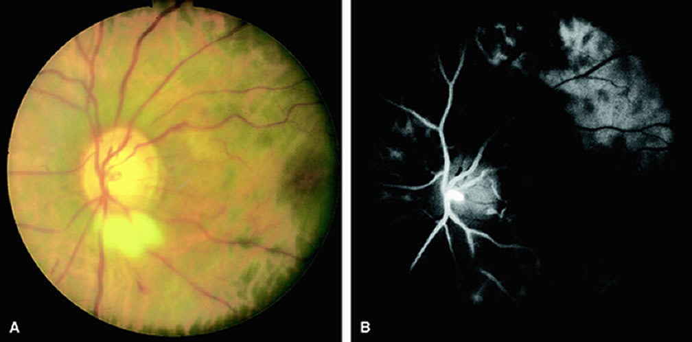
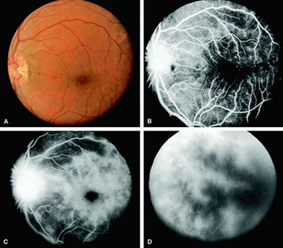
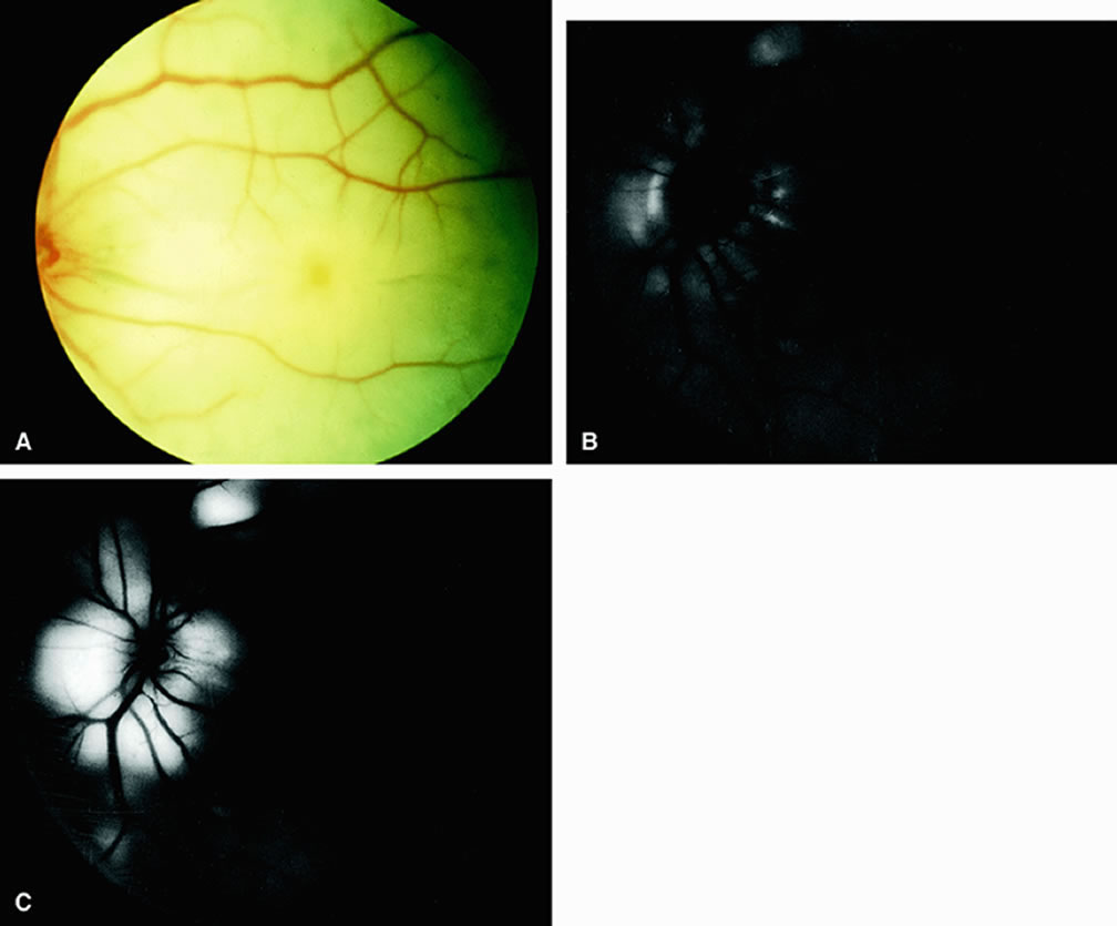
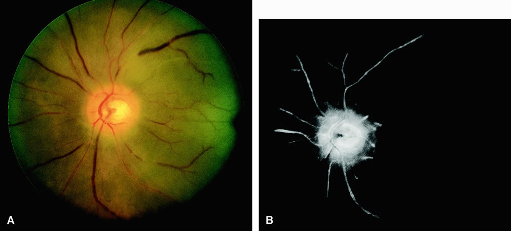
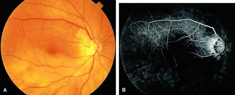
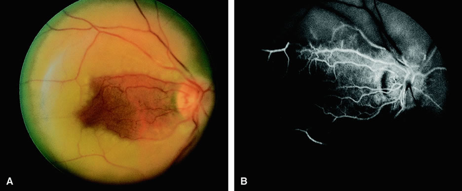
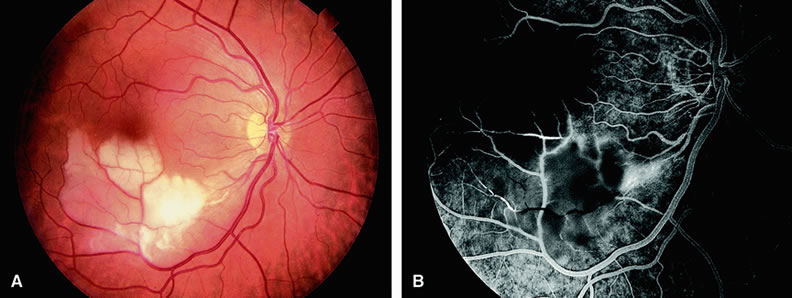
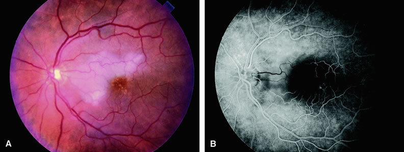
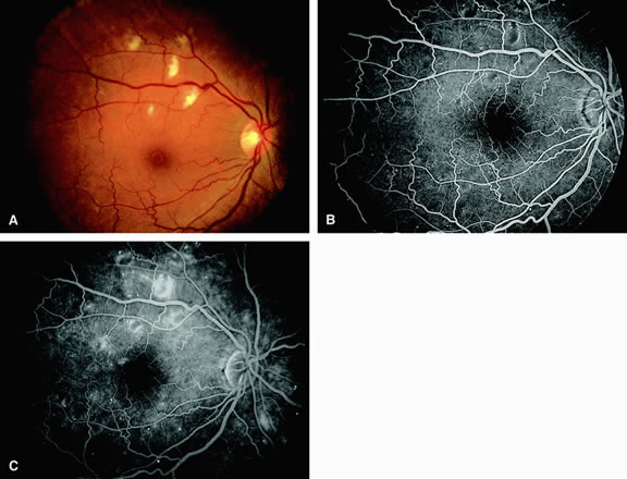
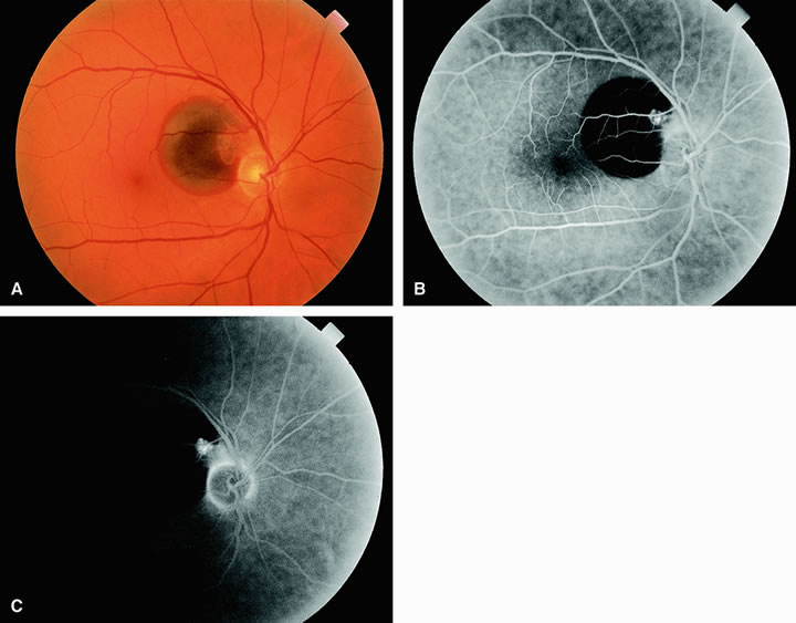
Normally, the choriocapillaris is completely filled with fluorescein dye within 5 seconds after the first appearance of dye within it. In eyes with the ocular ischemic syndrome, this filling can be delayed in extreme cases for 1 minute or longer. The posterior choroid is supplied by the temporal and nasal posterior ciliary arteries.2 In some normal eyes, but particularly in eyes with the ocular ischemic syndrome, delayed, asymmetric filling of the areas supplied by these vessels can be seen (Fig. 1). Filling of the retinal arteries is often also delayed in eyes with the ocular ischemic syndrome, and this manifests as a delayed arm-to-retina circulation time. Although the retinal arteries usually start to fill within 15 seconds after an antecubital intravenous injection of sodium fluorescein dye, this time range varies according to several factors, including the site of injection, the rate of injection, and body circulation. A visible leading edge of dye (see Fig. 1) within a retinal artery is almost always abnormal after an intravenous injection and indicates diminished flow. In the Retina Vascular Unit at Wills Eye Hospital, Philadelphia, the upper-normal limit for retinal arteriovenous transit time (time from the first appearance of dye in the temporal retinal arteries of the arcades to the time when the corresponding veins are completely filled) is considered to be 10 to 11 seconds. The retinal arteriovenous transit time is usually prolonged in eyes with the ocular ischemic syndrome; in fact, this prolongation of time is the most common fluorescein angiographic feature in eyes with the ocular ischemic syndrome. Leakage of fluorescein dye from the retinal vessels, particularly the arteries, occurs in 85% of eyes with the ocular ischemic syndrome (Fig. 2). Presumably, hypoxia and subsequent endothelial cell damage cause this leakage of dye. Leakage of fluorescein dye from the retinal vessels can be seen in the posterior pole and periphery. This hyperpermeability, combined with leakage of serum from microaneurysmal abnormalities, appears to account for the macular edema observed in some eyes with the ocular ischemic syndrome (see Fig. 2). Leakage of fluorescein dye from neovascularization of the disc is seen in approximately one third of eyes with the ocular ischemic syndrome (see Fig. 2). Hyperfluorescence resulting from leaking neovascularization of the retina is less common. Retinal capillary nonperfusion is visible on fluorescein angiography in some cases. Ischemic optic neuropathy is rarely observed. Iris neovascularization is found in approximately two thirds of cases at the time the diagnosis is made. Fluorescein angiography is helpful in differentiating the ocular ischemic syndrome from conditions that can mimic it, including central retinal artery obstruction, mild central retinal vein obstruction, and diabetic retinopathy. Of these conditions, only the ocular ischemic syndrome has delayed choroidal filling present. Moreover, late staining of the retinal arteries is unusual with the other conditions. An increased arteriovenous transit time is usually present in eyes with the ocular ischemic syndrome but can also be seen in eyes with central retinal artery or vein obstruction and eyes with diabetic retinopathy and nonperfusion of the retinal capillary bed. OPHTHALMIC ARTERY OBSTRUCTION Clinically, acute ophthalmic artery obstructions differ from acute central retinal artery obstructions in that persons with the former often have no light perception and the retinal whitening appears more intense on examination.3 A cherry-red spot is often absent in eyes with acute ophthalmic artery obstruction, but its presence does not rule out the diagnosis (Fig. 3A). Electroretinography often reveals diminished amplitudes of both b- and a-waves that are caused by inner and outer retinal ischemia, respectively.3 In contrast, with a central retinal artery obstruction alone, the a-wave amplitude is usually normal and the b-wave amplitude is often diminished because of inner retinal ischemia.4 Fluorescein angiography of eyes with acute ophthalmic artery obstruction shows delayed filling of the retinal vessels and usually the choroidal vessels as well (see Fig. 3B and C). Focal, pinpoint areas of staining resulting from leakage of dye at the level of the retinal pigment epithelium can be seen in some instances. Diffuse staining is also occasionally observed. Prominent staining of the retinal vessels is usually absent with acute ophthalmic artery obstruction, although it can be seen with chronic ophthalmic artery obstruction. RETINAL ARTERY OBSTRUCTION With acute central retinal artery obstruction, filling of the choroid is usually normal. Filling of the retinal arteries is often delayed, and in severe cases, a leading edge of dye can be seen (Fig. 4). A delay in retinal arteriovenous transit time is often noted.5 Box-carring or segmentation of the dye column can be seen in both the retinal arteries and veins when the obstruction is marked. In some cases, the flow appears normal because reperfusion of the blocked artery can occur fairly rapidly.6 Intraretinal leakage of dye in the late phases of the study, in a pattern consistent with macular edema, is generally not seen in eyes with acute central retinal artery obstruction. Fluorescein angiography can help identify eyes with acute central retinal artery obstruction in instances when the retinal whitening is subtle and the diagnosis is in question. Approximately 10% of eyes with acute central retinal artery obstruction have a cilioretinal artery that supplies the retina in the papillomacular bundle and extends into the foveola.7 In more than 80% of these eyes, the visual acuity eventually improves to 20/50 or better. Fluorescein angiography typically shows earlier filling within the patent cilioretinal artery and the veins draining the area that it supplies compared with the filling of the remainder of the retina, which is supplied by the central retinal artery (Figs. 5 and 6). Fluorescein angiography of eyes with marked, acute branch retinal artery obstruction reveals a lack of filling of the retinal capillary bed within the distribution of the involved vessel. Retinal veins that normally drain the damaged area also demonstrate a delay in filling. With severe blockage, retrograde filling can be seen in the distal aspect of an obstructed branch retinal artery (Fig. 7). Cilioretinal artery obstruction is similar to branch retinal artery obstruction except that the former vessel usually emanates from the edge of the optic disc (Fig. 8). Cilioretinal artery obstructions can be seen as isolated fundus abnormalities or in association with central retinal vein obstruction or acute anterior ischemic optic neuropathy.8 Cotton-wool spots are small areas of superficial retinal whitening that usually develop secondary to obstruction of axoplasmic flow caused by areas of focal retinal ischemia.9 Fluorescein angiography in these cases usually demonstrates relative hypofluorescence in the early and middle phase of the study (Fig. 9). Late staining of the cotton-wool spot can occur. As shown in Table 2, the differential diagnosis of cotton-wool spots is extensive; however, cotton-wool spots are most commonly observed in the setting of diabetic retinopathy, hypertensive retinopathy, collagen vascular disease, or hematologic abnormalities.
Table 2. Abnormalities Associated with Cotton-Wool Spots in the Fundus
|
RETINAL VENOUS OBSTRUCTIVE DISEASE |
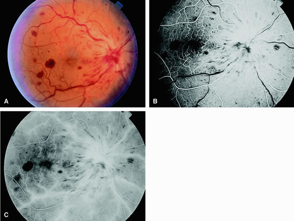
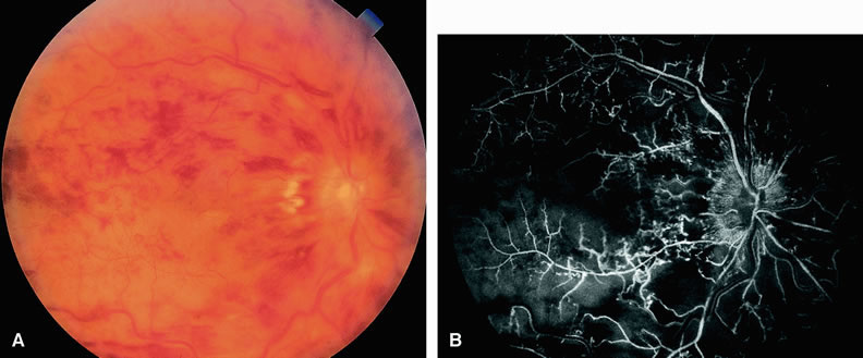
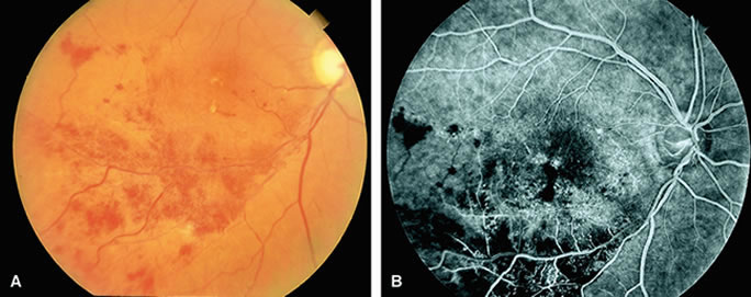
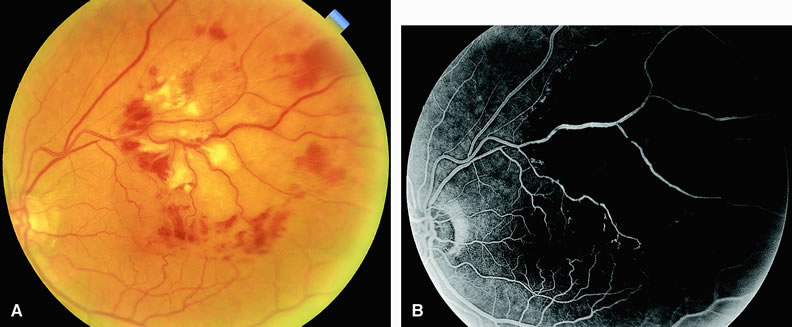
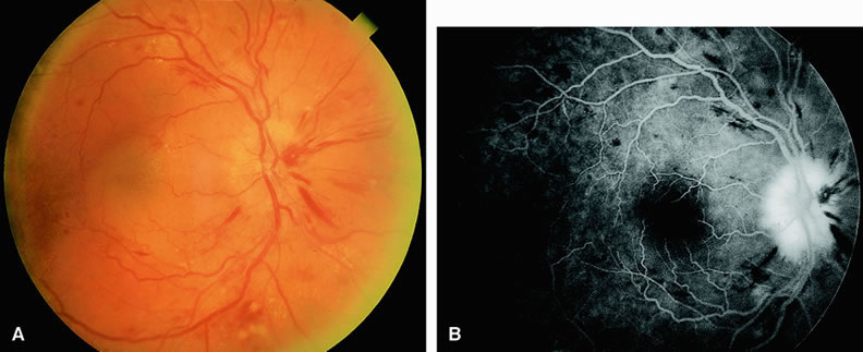
CENTRAL RETINAL VEIN OBSTRUCTION In eyes with central retinal vein obstruction, a thrombus is usually found in the obstructed vessel at or near the lamina cribrosa. Ophthalmoscopic signs of retinal vein obstruction include dilated tortuous veins, intraretinal hemorrhage, and ischemic retinal edema.10 Cotton-wool spots and a swollen optic disc are also often seen. Other less common signs of central retinal vein obstruction include vitreous hemorrhage and exudative retinal detachment. Several classifications have been employed to define central retinal vein obstructions. Central retinal vein obstructions can be divided into ischemic and nonischemic variants, depending in part on whether large areas of retinal capillary nonperfusion are present or absent. This classification is clinically useful because data from the Central Retinal Vein Occlusion Study Group11 and others12 suggest that laser panretinal photocoagulation can result in a regression of neovascularization of the iris, thereby preventing neovascular glaucoma. Compared with eyes that have the ischemic type, eyes with nonischemic central retinal vein obstruction typically have a visual acuity of 20/200 or better, relatively few cotton-wool spots, and less marked retinal hemorrhage.13 Fluorescein angiography usually shows an increased retinal arteriovenous transit time secondary to delayed retinal venous filling. Delayed retinal arterial filling can also be present. The retinal capillary bed is generally well perfused (Fig. 10). The retinal veins appear as a hypofluorescent silhouette against the bright choroid. There is also variable staining of the retinal veins and variable leakage on the angiogram. Approximately 20% of nonischemic central retinal vein occlusions will progress to ischemic central retinal vein occlusions. Ischemic central retinal vein obstructions generally reduce a patient's visual acuity to 20/200 or worse, most commonly to the counting-fingers or hand-motions range. Numerous cotton-wool spots and severe four-quadrant retinal hemorrhaging are often seen. Fluorescein angiography reveals confluent regions of retinal capillary nonperfusion (Fig. 11). Views of the posterior pole, as well as those of the four quadrants, may be necessary to demonstrate the capillary dropout. Magargal and associates12 quantitated the amount of retinal capillary nonperfusion in a posterior-pole 30-degree view in eyes with central retinal vein obstruction and calculated an ischemic index (area of retinal capillary nonperfusion/total area within the posterior 30-degree view). They found that when the index was 80% or greater, approximately 45% of eyes eventually developed rubeosis iridis and neovascular glaucoma. In some eyes with central retinal vein occlusion, determining the degree of retinal capillary nonperfusion with fluorescein angiography is difficult. Worsening visual acuity and progressive intraretinal bleeding indicate that an eye may be progressing toward a more ischemic state.14 In eyes with central retinal vein obstruction and a visual acuity of 20/200 or worse, performing fluorescein angiography at the initial visit can be useful to evaluate perfusion of the retinal capillary bed. The presence of a large amount of intraretinal blood in the posterior pole is not necessarily a contraindication to performing the study, particularly because these eyes are probably more prone to ischemia. The retinal blood generally diminishes rapidly anterior to the posterior pole, facilitating angiographic evaluation of the peripheral retina. BRANCH RETINAL VEIN OBSTRUCTION Branch retinal vein obstruction usually occurs at the intersection of a branch retinal artery with a vein. In general, the artery overlies the vein, and the two vessels share a common adventitial sheath at the crossing. Retinal hemorrhages are present along with cotton-wool spots (Figs. 12 and 13). These hemorrhages usually are reabsorbed over a period of months. Numerous microvascular abnormalities generally remain, and macular edema may be persistent. The Branch Retinal Vein Occlusion Study Group15 demonstrated that eyes with branch retinal vein obstruction and a visual acuity of 20/40 or worse may benefit from grid laser therapy to reduce macular edema. This study group recommended waiting at least 3 months after the onset of the obstruction to permit clearing of the retinal blood before performing fluorescein angiography. If the study shows that macular edema is responsible for the visual loss, grid laser therapy to the affected region within the vascular arcades can be considered. In cases in which foveal nonperfusion is responsible for the decrease in vision, laser therapy has not been shown to improve vision. Eyes with branch retinal vein obstruction and retinal capillary nonperfusion can develop neovascularization of the retina and/or disc. New vessels on the retina or optic disc intensely leak fluorescein dye, whereas larger collateral vessels on the optic disc or in the retina usually do not; thus, fluorescein angiography is helpful in differentiating between neovascularization and collateral vessels. Proper identification of posterior-segment neovascularization in eyes with branch retinal vein obstruction is important because scatter laser treatment in such cases has been shown to reduce vitreous hemorrhage in these eyes.16 In addition, fluorescein angiography can be used to identify the areas of retinal capillary nonperfusion requiring therapy. Scatter sector laser photocoagulation should be delayed until the onset of retinal neovascularization. |
OTHER RETINAL VASCULAR ABNORMALITIES | ||||||||||||||||||||||||
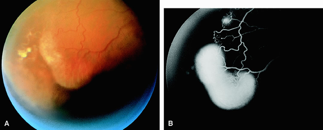
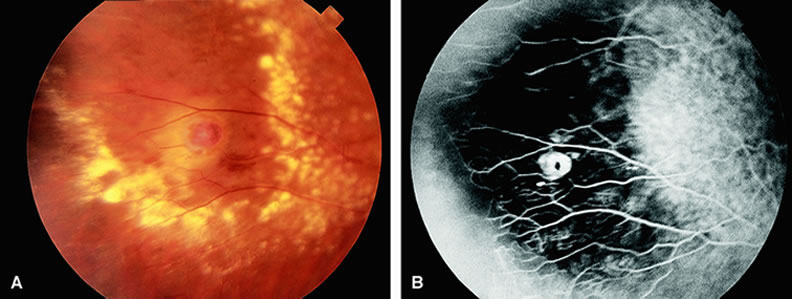
PERIPHERAL PROLIFERATIVE RETINOPATHIES The most common cause of peripheral proliferative retinopathy is sickling hemoglobinopathies. They account for approximately one half of cases.17 Among patients with sickling hemoglobinopathy and peripheral proliferative retinopathy, hemoglobin SC is the most common variant. Other causes of peripheral proliferative retinopathy include retinal vein branch obstruction, diabetes mellitus, sarcoidosis, intravenous drug abuse, and Eales' disease.17 On fluorescein angiography, the posterior pole and the optic nerve often appear normal. Peripheral retinal capillary nonperfusion is seen in most eyes with peripheral extraretinal neovascularization.17,18 Shutdown of the larger peripheral retinal vessels is also often observed. Peripheral retinal neovascularization can assume several configurations, although the most common variant is that of the “sea fan” characteristic of sickle-cell retinopathy18 (Fig. 14). The angiogram demonstrates leakage of dye into the vitreous from these sites of neovascularization. In eyes in which scatter panretinal photocoagulation is being considered to control peripheral neovascularization, fluorescein angiography helps identify the regions of retinal capillary nonperfusion that require treatment. COATS' DISEASE Coats' disease19 is a retinal vascular abnormality of unknown etiology characterized by retinal telangiectatic formations in association with intraretinal and subretinal hard exudates (Fig. 15). Focal areas of dilation and narrowing of the larger retinal vessels are often seen. Fluorescein angiography20 demonstrates hyperfluorescence of the telangiectatic vessels early in the study along with hypofluorescence of proteinaceous exudates and mild hyperfluorescence of subretinal fluid. Enlargement of the retinal capillary bed and widened spaces between these small vessels is characteristic of Coats' disease. Retinal capillary nonperfusion is often seen, but associated retinal neovascularization is rare. Leakage of dye can occur from both the larger and smaller vessel abnormalities. RETINAL ARTERIAL MACROANEURYSM A retinal arterial macroaneurysm is an area of focal dilation, and often outpouching, along the course of an artery. First described by Robertson2 in 1973, the abnormality is often associated with systemic arterial hypertension and is found in older people, more commonly in women. More than one retinal arterial macroaneurysm may be present. Macroaneurysms most often occur in the superotemporal or the inferotemporal retinal arterial arcades, often after the second or third arteriolar branching. Local ocular complications include bleeding and the extravasation of serum and lipid into the surrounding retina.22 Blood from leaking macroaneurysms can extend into the vitreous cavity, retina, and/or subretinal space (Fig. 16). Fluorescein angiography in eyes with an acquired retinal arterial macroaneurysm generally reveals relatively early and late hyperfluorescence of the abnormality. The hyperfluorescence usually increases as the study progresses. There is often narrowing of the vessels proximal and distal to the macroaneurysm. The retinal capillary bed surrounding the macroaneurysm is often dilated, and leakage from local telangiectatic and microaneurysmal abnormalities is often present. Identification of coexistent leaking vessels is important if laser therapy is being considered. The macroaneurysm is often obscured by an overlying hemorrhage. Occasionally, the macroaneurysm can thrombose and lead to obstruction of the retinal artery distal to it. SYSTEMIC ARTERIAL HYPERTENSION Chronic systemic arterial hypertension manifests in the fundus with focal and diffuse retinal arterial narrowing; retinal hemorrhages; cotton-wool spots; and in more severe or acute cases, lipid exudate23 (Fig. 17). The lipid exudates may be deposited in a radial pattern surrounding the macula, forming the so-called macular star. In extreme cases (malignant hypertension), optic disc swelling is also present. Small yellow spots at the level of the retinal pigment epithelium resulting from underlying damage of the choriocapillaris can also be seen. Fluorescein angiography of eyes with acute hypertensive changes shows irregular-appearing constricted arterioles and focal areas of hypofluorescence resulting from retinal capillary nonperfusion in areas corresponding to cotton-wool spots. Telangiectatic retinal vascular abnormalities and microaneurysms often surround the cotton-wool spots and leak dye into the retina as the study progresses. In some instances, large areas of retinal capillary nonperfusion are present. The optic disc is usually hyperfluorescent in eyes with nerve-head swelling and malignant hypertension (see Fig. 17). Focal areas of hyperfluorescence resulting from leakage at the level of the choriocapillaris can also be seen. These areas eventually resolve, leaving pigmented and depigmented foci (Elschnig spots) at the retinal pigment epithelial level.24 Despite findings on fluorescein angiography, ophthalmoscopic evaluation is more important in making the diagnosis. Fluorescein angiography is also not needed for treatment (that is, controlling blood pressure). RADIATION RETINOPATHY The ophthalmic community has long appreciated that radiation is an effective therapeutic option for many intraorbital, periorbital, and intracranial neoplasms. In 1933 in the first reported case of radiation retinopathy, Stallard25 described the potentially harmful effects of radiation on the retina. At that time, clinicians used large doses of radiation, and little effort was made to limit radiation exposure of nonpathologic tissue. In recent years, technologic advances have enabled clinicians to administer radiation in lower doses by using delivery systems that limit exposure of radiation to normal tissue. Although these new approaches have helped reduce the incidence of radiation retinopathy, cases of radiation retinopathy continue to be reported. Radiation retinopathy slowly progresses and usually develops over months after exposure to ionizing radiation. External beam irradiation, 60Co plaque irradiation, and brachytherapy can all cause radiation retinopathy.26 The minimum amount of radiation capable of inducing retinopathy is controversial. Investigators have reported that as little as 1100 cGy can result in retinal damage.27 Amoaku and Archer28 recommend regularly screening all patients who have received at least 3000 cGy of radiation to the eye and closely observing all patients who have been exposed to 5000 cGy or more of radiation to the eye. Brown and colleagues26 reviewed 32 cases of radiation-induced retinopathy. In this group, 20 eyes had been exposed to 60Co plaque irradiation and 16 eyes had been exposed to external beam radiation. As shown in Table 3, hard exudates, microaneurysms, and intraretinal hemorrhages are the most common ophthalmoscopic manifestations of radiation-induced retinopathy. Brown and associates observed a discrepancy in the percentages of eyes developing hard exudates between the two modalities of radiation exposure.
Table 3. Incidence of Retinopathic Signs in 36 Eyes with Radiation Retinopathy
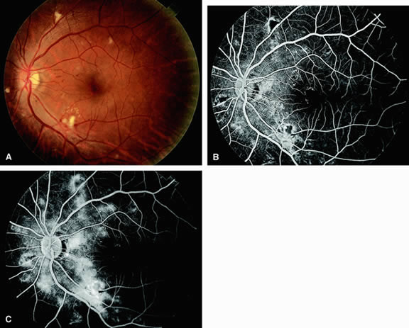
The fluorescein angiographic features of radiation retinopathy include capillary dilatation andclosure, formation of microaneurysms, and telangiectasia (Fig. 18). The dilated, tortuous fusiform capillaries develop vascular incompetence, and leakage of fluorescein can be observed as the retinopathy progresses. End-stage radiation retinopathy is characterized by generalized capillary disorganization resulting in retinal ischemia and cystoid macular edema. |