Chapter 6 Surgical and Nonsurgical Trauma J. DOUGLAS CAMERON Table Of Contents |
NORMAL OCULAR WOUND HEALING HISTOLOGY OF OCULAR SURGICAL PROCEDURES COMPLICATIONS OF SURGICAL TRAUMA NONSURGICAL TRAUMA REFERENCES |
NORMAL OCULAR WOUND HEALING |
Successful wound healing in the eye is a precise, ordered series of events
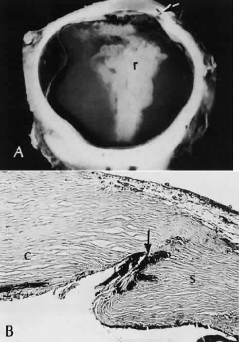
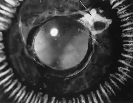
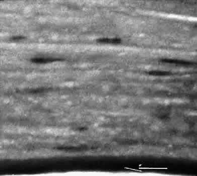
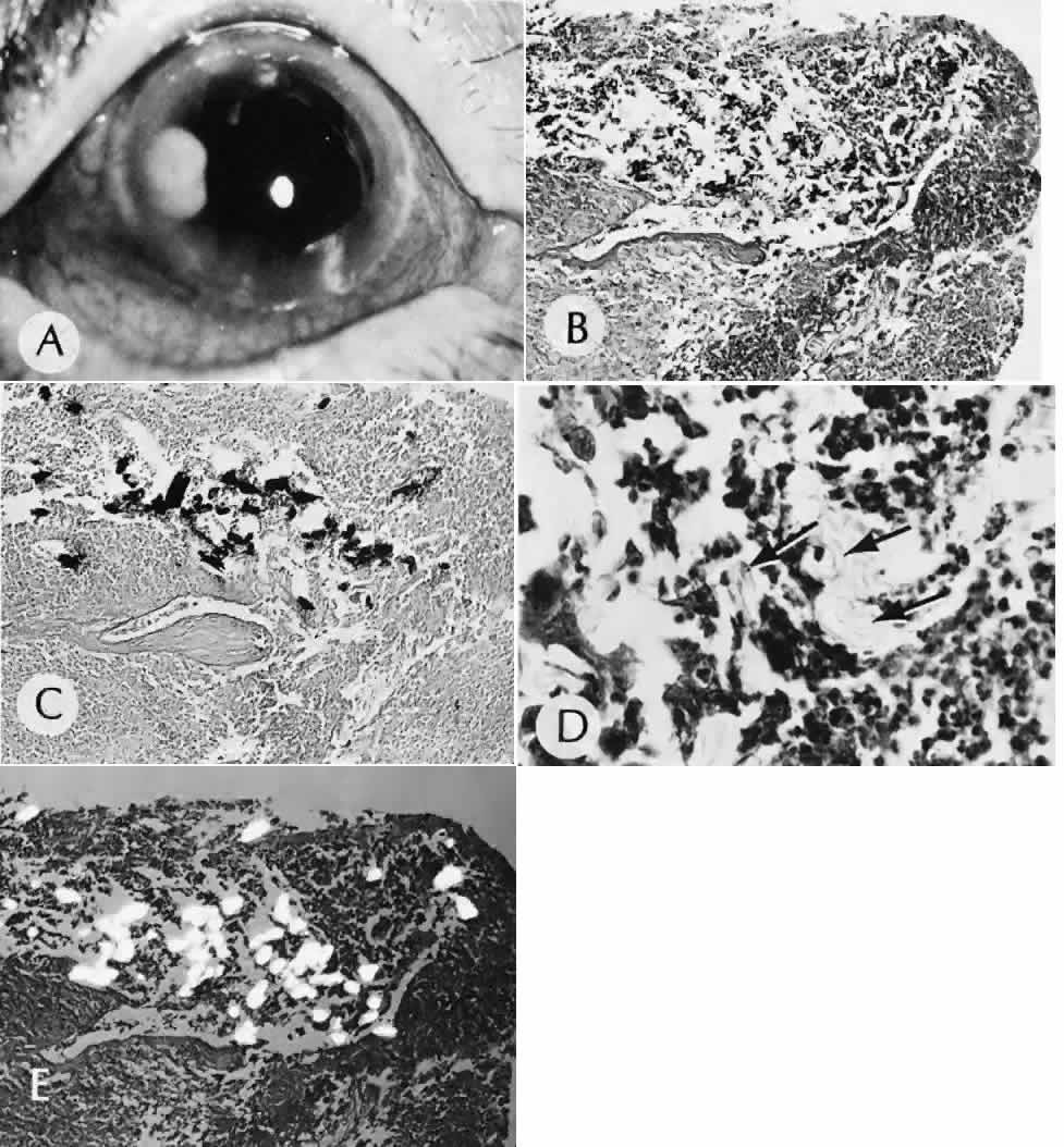
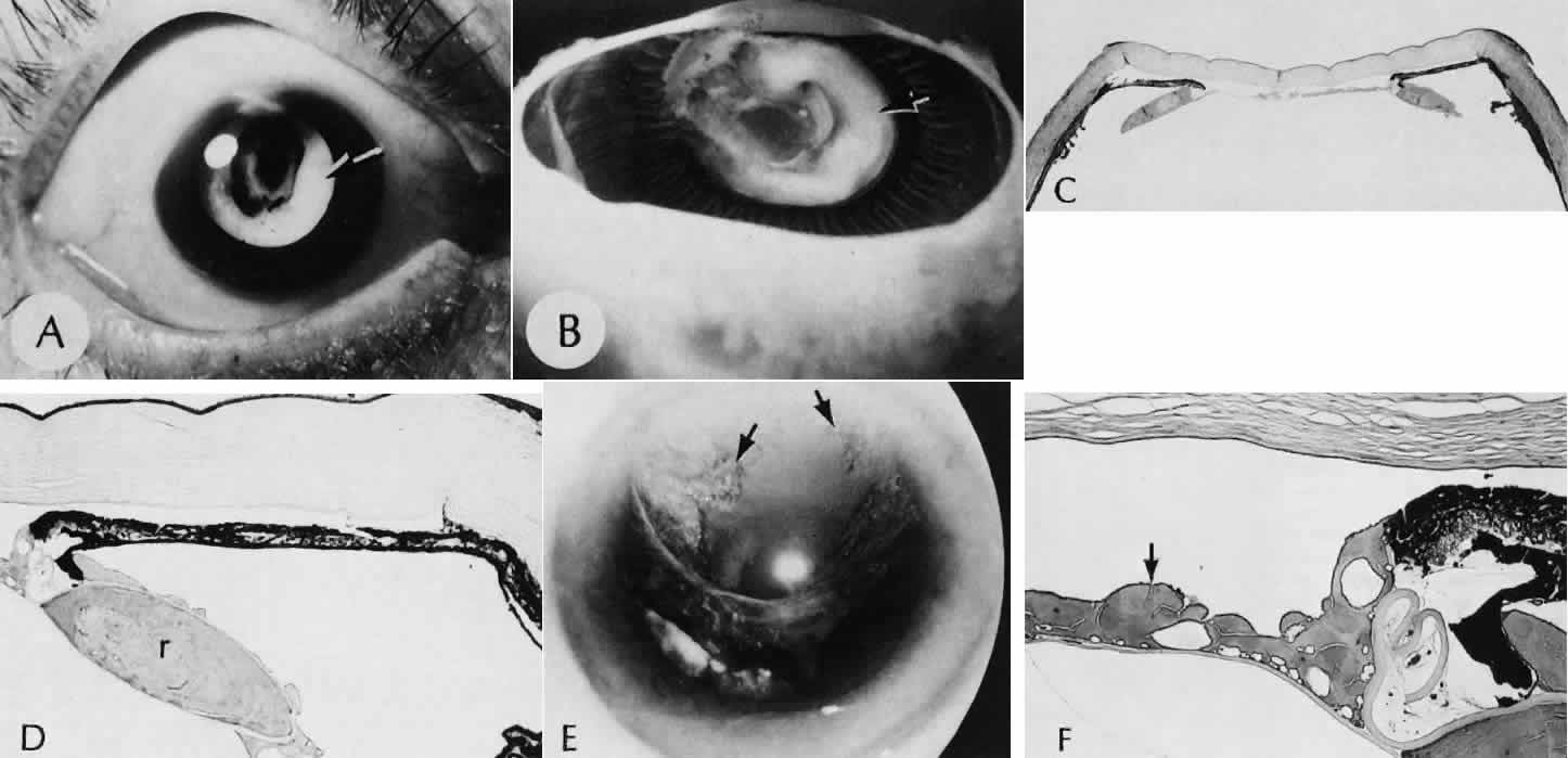
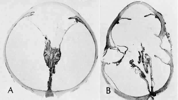
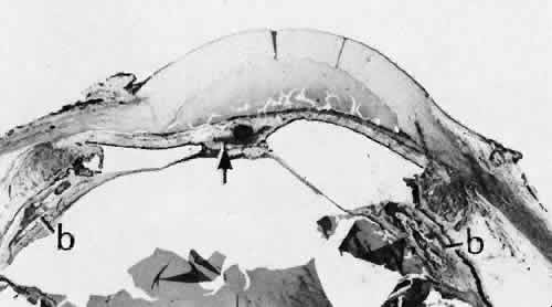
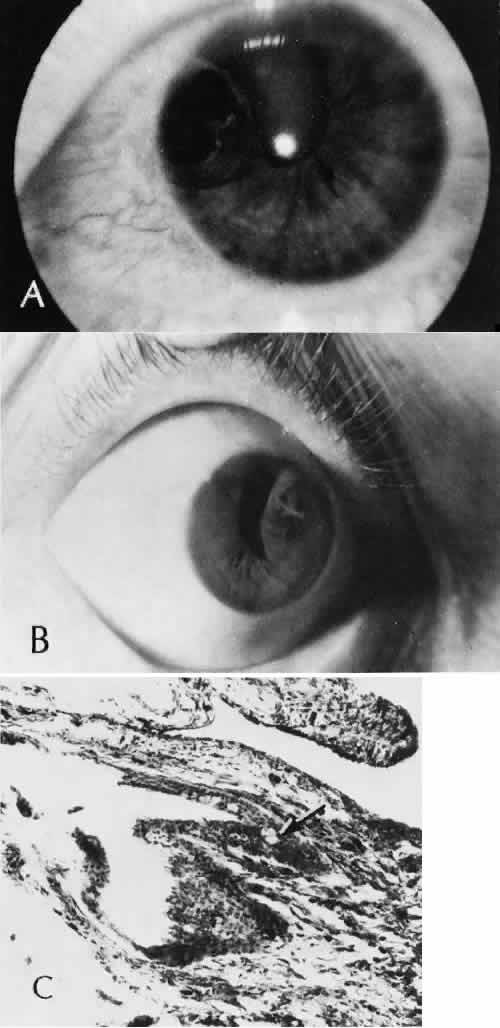
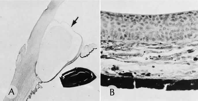
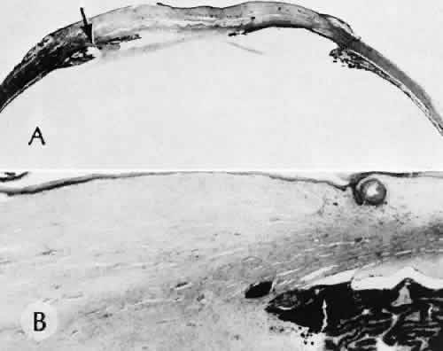
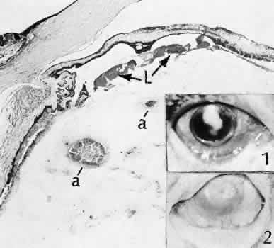
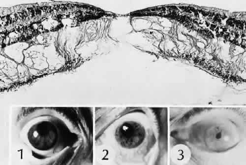
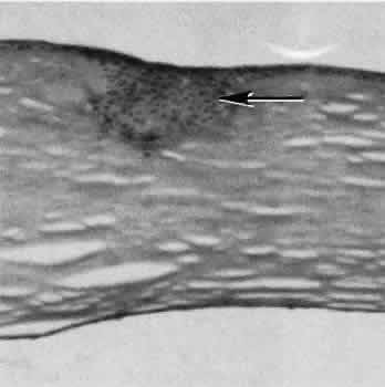
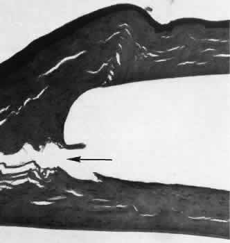
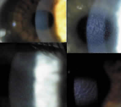
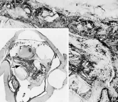
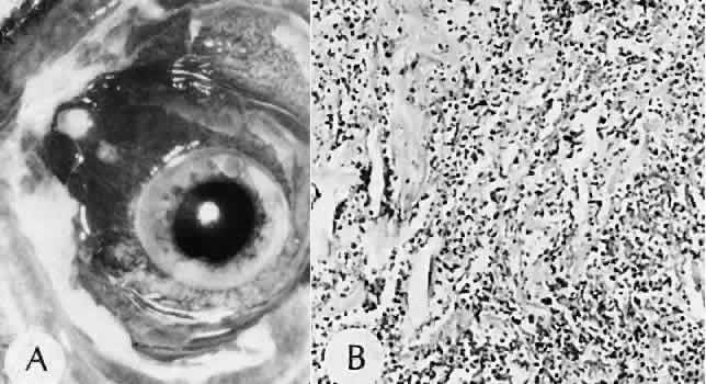
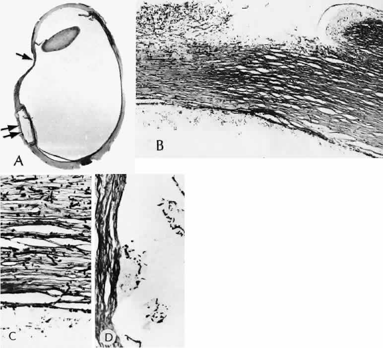
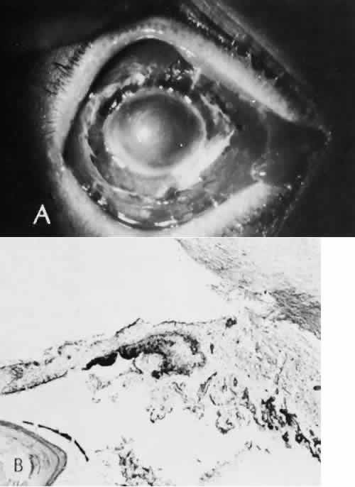
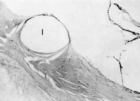
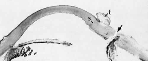
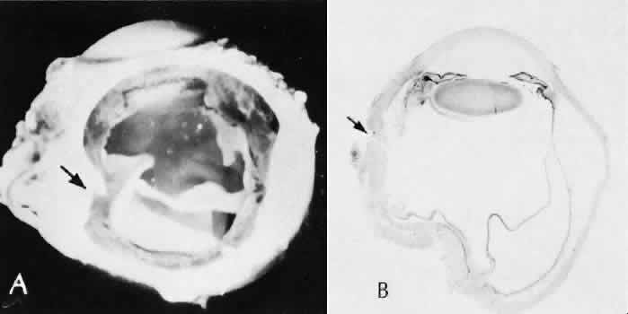
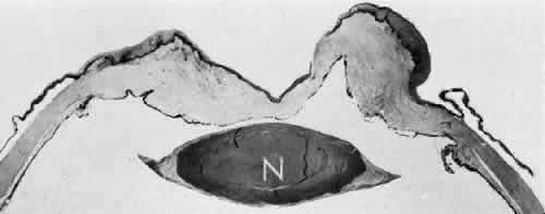
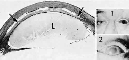
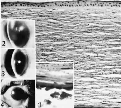
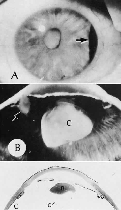
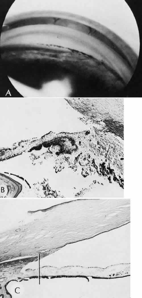
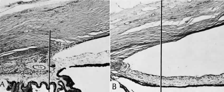
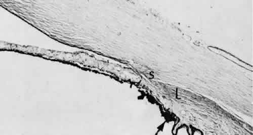
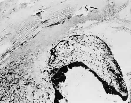
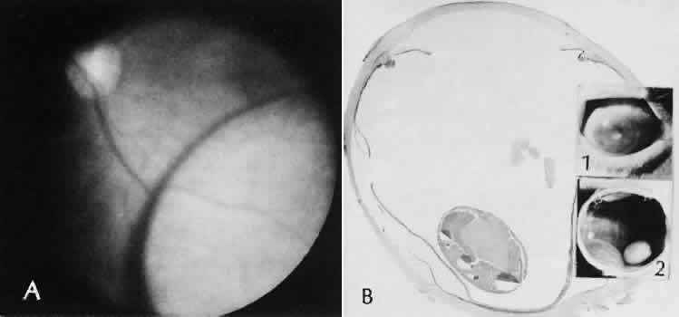
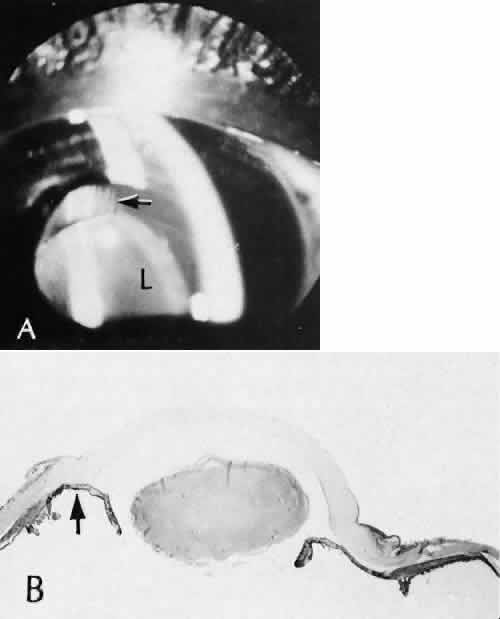
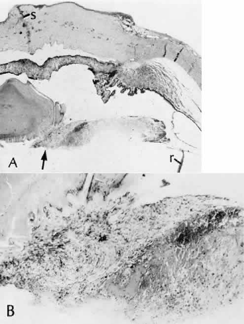
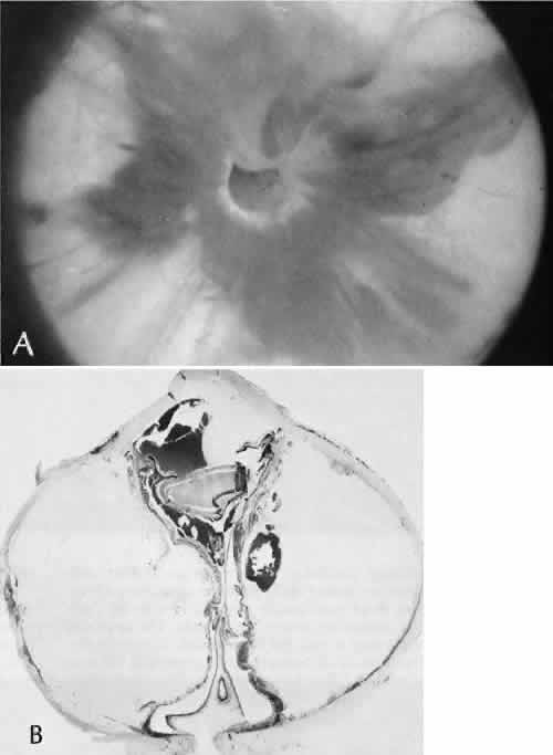
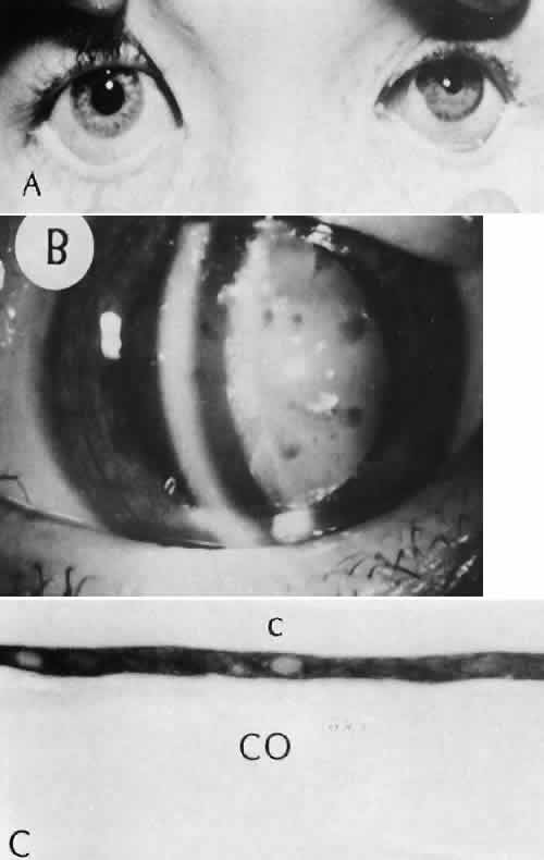
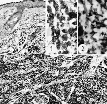
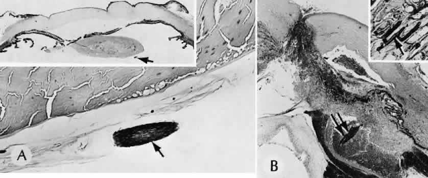
that results in reestablishment of functional ocular anatomy.1–3 The goal of therapeutic intervention in the setting of ocular trauma is to promote the repair process and to allow it to proceed as rapidly as possible and heal as completely as possible without compromising ocular function.4 CELLULAR AND EXTRACELLULAR COMPONENTS The four basic cell types found in wound healing are fibroblasts, vascular endothelial cells, inflammatory cells, and epithelial cells. Specialized cell types that participate in ocular wound healing include corneal endothelial cells, retinal pigment epithelial cells, and Müller cells of the retina. Specific cell populations enter an area of injury under the influence of complex biochemical and biophysical processes involving proteins of the extracellular matrix and the coagulation system. Intracellular proteins, such as tubulin and actin, andmyosin filaments function in cell locomotion. Extracellular matrix proteins, such as fibronectin, laminin, and type IV collagen, orient and regulate cell migration and adhesion.5 Coagulation proteins, such as von Willebrand's factor and plasminogen, alsofunction in regulating the cellular events of healing. Vascular endothelial cells are stimulated to leave their resting state and digest basement membrane, proliferate, migrate, and eventually differentiate under the direction of multiple angiogenic growth factors.6 A wound usually is filled first by a fibrin coagulum. Neutrophils, lymphocytes, and histiocytes (macrophages) enter the wound through fibrin scaffolding to clear necrotic debris and confine toxic or foreign substances. Granulation tissue often is the first type of reparative tissue in wound healing (Fig. 1). Although its composition varies somewhat, small-caliber vascular channels in a delicate collagenous stroma infiltrated by acute and chronic inflammatory cells generally characterize it. This amorphous tissue serves as a template for more definitive repair. Metaplasia is the transformation of a cell from one adult cell phenotype to a second cell phenotype. In advanced wound healing, fibroblasts acquire intracytoplasmic characteristics of smooth muscle cells (myofibroblasts).7 The myofibroblasts are able to contract and bring wound edges together. Vascular endothelial cells proliferate and migrate into the wound from preexisting adjacent vessels. The new “vessel” migrates initially as a solid bulb of endothelial cells. This solid cord of cells will canalize and differentiate into mature arterioles, venules, and capillaries. Fibroblasts grow into the wound in a radial manner but will eventually reorient and secrete collagen along lines of established tissue tension. Epithelial cells are found covering surfaces of tissue. Two distinct types of epithelial cells cover the ocular surface: corneal epithelial cells and conjunctival epithelial cells. Cell replacement is accomplished by stem cells located at the limbus for the corneal epithelium8,9 and at the mucocutaneous junction and possibly throughout the conjunctiva for the conjunctival epithelium.10 Migration and proliferation of surrounding healthy epithelial cells heal surface discontinuity. Apoptosis is a biochemical process leading to programmed cell death. Cell death in apoptosis results from intracellular messages. In necrosis, cell death results from toxic external factors (e.g., hyperosmolality). Apoptosis allows for elimination of entire populations of cells without tissue damage or an inflammatory response.11 Elimination of certain cells is advantageous in embryology (e.g., when scaffolding structures are no longer necessary as with the primary vitreous). In certain neoplastic conditions, however, defects in apoptosis may lead to disadvantageous accumulation of cells. Apoptosis also seems to be highly influential in all types of inflammation, including wound healing. In wound healing, apoptosis may function to control the type and degree of tissue response.12 In the anterior cornea, keratocytes have been observed to undergo apoptosis in response to wounding of the cornea.13,14 Abnormalities of apoptosis may be responsible for such conditions as keloid formation in the skin15 and keratoconus in the cornea.16 Wound healing of highly specialized tissues of the eye has several unique features. CORNEAL HEALING The healing of the cornea is unique relative to other soft tissues, because it lacks blood vessels and because it is lined anteriorly and posteriorly by layers of epithelial-like cells. Epithelial cells of the corneacan produce essential wound healing factors normally produced by platelets.17 Architectural repair is accomplished at the level of the corneal stroma. Abrasions are injuries generally involving only the superficial layer (epithelium) of the cornea (Fig. 2). Abrasions are commonly caused by mechanical injuries or anoxia resulting from contact lens overwear. Some or all of the layers of the surface epithelium are lost, but Bowman's membrane remains intact. After a delay of approximately 1 hour, uninjured epithelial cells at the margin of the wound loosen their intercellular and basal attachments and migrate en masse toward the injured area.18 The shape of the wound margin and the biochemical characteristics of the exposed tissue influence the direction and extent of the migration.19 If the entire corneal epithelium is abraded, migrating epithelial cells derived from the limbal stem cells are able to cover the defect completely within 48 to 72 hours. The epithelium will be much thinner than normal until mitotic division reestablishes normal thickness. Re-formation of epithelial basement membrane may not be detectable for as long as 6 weeks after injury.20 Clinically, the healed wound is transparent. Small defects of Bowman's membrane (Fig. 3) do not heal by fibrous proliferation of the stroma. The defect is filled by proliferating epithelial cells (epithelial facet formation) that reestablish the surface continuity of the cornea. The facet may be seen clinically as a focal, well-demarcated, superficial corneal opacity. A corneal foreign body causes this lesion most often. Penetrating corneal injuries involving at least one third of the stroma (Fig. 4) are covered initially by proliferating surface epithelium that may extend for a considerable distance into the stroma. The extent of corneal epithelial migration is controlled at least in part by contact inhibition of migration once contact with healthy corneal endothelial cells is established.21 Bowman's membrane has elastic properties that tend to pull the anterior margins of the wound apart. Exposure of the relatively dehydrated corneal stroma to tears and blood will cause swelling of the corneal stroma. This swelling of the stroma tends to close the wound. As the stroma heals, new collagen is produced by keratocytes or transformed monocytes. Epithelial cells will regress toward the surface. The repair collagen is different from the native collagen in size and in orientation. The healed wound is densely opaque and obvious clinically, but it may be detected histologically only by the break in Bowman's membrane. Full-thickness wounds of the cornea are associated with retraction of Descemet's membrane and separation of the posterior aspect of the wound (Figs. 5 and 6). Secondary aqueous, a proteinaceous coagulum, may seal the wound posteriorly until healthy surrounding endothelial cells can spread and migrate into the injured area. Mitotic division of the endothelial cells possibly plays an important role in younger patients but probably is not clinically significant in adults. Ultimately, migrated endothelial cells will produce a new Descemet's membrane (Fig. 7).22–25 The portions of Descemet's membrane displaced into the stroma are not resorbed, but remain as the histologic marker of the site of injury. The anterior cellular surface repair of migration and mitosis of epithelial cells is the same as that described for more superficial corneal wounds. The important exception is that with full-thickness injuries, there is a risk of surface epithelial cells migrating along the posterior surface of the cornea or the anterior surface of the iris to the trabecular meshwork. The displaced epithelial cells will cause scarring of the trabecular meshwork and secondary open-angle glaucoma. CONJUNCTIVAL HEALING Conjunctival healing differs from corneal healing because of the presence of blood vessels and a lymphatic system. The conjunctival epithelium heals by migration and mitosis.26 Conjunctival stem cells are thought to originate at the mucocutaneous junction and migrate to the fornix.27 Goblet cells are produced by epithelial progenitors and appear once conjunctival epithelial cell continuity has been reestablished.28,29 The presence of vascular tissue in the substantial propria of the conjunctiva allows for the formation of granulation tissue and scar tissue, as found in soft tissues elsewhere. SCLERAL HEALING The sclera itself does not participate directly in wound healing. Partial-thickness injuries are healed by formation of granulation tissue from the epi-scleral tissue in external wounds or from uveal tissue in internal wounds (Fig. 8). Full-thickness defects of the sclera heal by granulation tissue originating in the episcleral tissue and uveal tract. Mitomycin is used in filtering procedures specifically to prevent the formation of granulation tissue and promote establishment of an aqueous fistula.30 SURGICAL LIMBUS HEALING Currently, there is a trend for the site of the cataract wound to shift from the limbus to the peripheral cornea31 to reduce induced astigmatism32 and to shorten the natural history of cataract wound healing.33 The healing of the wound is similar to that found in central corneal incisions. Healing at the site of a limbal surgical incision involves a combination of the features of repair of the cornea, conjunctiva, and sclera (see Fig. 5). Conjunctival epithelium will heal over an area of granulation tissue originating from the substantia propria of the conjunctiva and the episclera. The granulation tissue seals the wound if the wound edges are well apposed. The remainder of the healing process is similar to the healing of an external scleral wound. Granulation tissue is not formed in the internal portion of the wound because the uveal tract is not involved. The internal wound is healed by endothelial migration and reformation of Descemet's membrane in a manner outlined above for full-thickness corneal wounds.34,35 Because of the lack of blood vessels, the rate of wound healing is slow relative to that of the skin. Whereas an injury to the skin may have regained its tensile strength in 7 to 10 days, injuries to the surgical limbus may require as long as 12 months to regain a stable tensile strength. The tissues of the wound remain structurally weaker than the surrounding uninjured tissue (Fig. 9). The healing of a clear corneal cataract incision proceeds as described above. UVEAL HEALING Wound healing of the posterior uveal tract follows the general principles for healing of vascularized tissue. The iris, however, heals differently. When the iris wound is perpendicular to its circumferential ridges, such as in a typical peripheral iridectomy or iridotomy, the cut edges pull apart. Granulation tissue does not form to close iridectomy incisions probably because of the inhibitory effect of the aqueous flowing through the opening of these small incisions. Iridotomies created by argon laser (Fig. 10) may be anatomically closed by apparent migration of iris pigment epithelium.36,37 LENS HEALING Proliferation and fibrous metaplasia of the lens capsular epithelium (Fig. 11) may close small rents through the lens capsule. After maturation of the fibrous tissue, the most superficial epithelial cells will form a new lens capsule. Most wounds to the lens, small and large, result in cataracts. Small wounds, however, may result in tiny focal opacities.38–39 Lens epithelial cells undergo fibromyoblastictransformation when stimulated by injury. The transformed cells are able to produce type I and type III collagen and glycosaminoglycans.40 This is the fundamental process resulting in opacification of the posterior lens capsule after extracapsular cataract extraction or phacoemulsification.41 Apoptosis (programmed cell death) also plays a role in the formation of secondary cataracts.42 RETINAL HEALING Wound healing of the neurosensory retina follows the principles of wound healing. There is an initial removal of all necrotic tissue by phagocytosis followed by proliferation of cells to form a chorioretinal bond. The healing, however, is modified in most instances by the lack of participation by the blood vessels.43–45 Astrocytes from the neurosensory retinaproliferate from the peripheral viable tissue into the wound and downward into the area of the subretinal space (Fig. 12). Retinal pigment epithelial cells from the peripheral viable tissue undergo fibrous metaplasia and proliferate upward into the area of the subretinal space. When the two proliferating cell types unite, a tight chorioretinal bond is formed. Increased retinal adhesiveness has been estimated to be 140% of the normal degree of adhesiveness 2 weeks after photocoagulation.46 Retinal holes may heal spontaneously if supported by an intact vitreous base or if located adjacent to the retinal pigment epithelium (Fig. 13).47,48 For the retinal pigment epithelium to differentiate, it must be in contact with overlying retinal pigment epithelium.49 Choriocapillaris repair from a photocoagulation wound of the retina appears to proceed in a manner similar to that of repair of capillary thrombosis in other tissues.50 The vascular repair process may not reproduce the lobular architecture of the native choriocapillaris.51 The choriocapillaris may regenerate in areas of restored retinal pigment epithelium.52 Proliferative vitreoretinopathy is an expression of abnormal retinal wound healing.53 Interruption of Bruch's membrane may allow the ingrowth of fibrovascular tissue from the choroid into the subretinal space, resulting in a modified granulation wound healing process and subretinal scarring.54 |
HISTOLOGY OF OCULAR SURGICAL PROCEDURES |
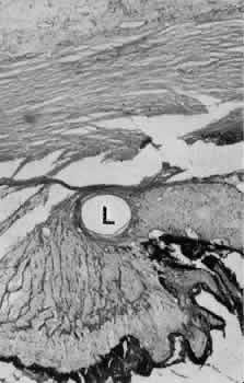
CATARACT EXTRACTION The cataract incision is made into the anterior chamber in such a way as not to injure the trabecular meshwork or unduly injure the cornea. The traditional cataract wound extends from the episcleral tissue posterior to the surgical limbus (defined by the insertion of the conjunctiva and Tenon's capsule) through the corneoscleral tissue to enter the anterior chamber by perforating Descemet's membrane anterior to Schwalbe's line. Clear corneal cataract incision involves only the peripheral cornea. Special circumstances may occur in which a more posterior or anterior route of incision may be desirable.55,56 The most reliable histologic landmark of a healed cataract incision (Fig. 14) is a hiatus of peripheral Descemet's membrane. The cut edges of Desce-met's membrane will curl inward toward the sclera.Endothelial cells may be absent or may have produced a new, thin, periodic acid-Schiff-positive Descemet's membrane over exposed corneal stroma. Occasionally, a fibrous plaque is present at the level of Descemet's membrane. This plaque is another example of fibrous metaplasia, in this instance of the corneal endothelial cells. The corneal stromal portion of the wound may be difficult to identify histologically, except in the early stages of healing. Clues to the position of the intrastromal tract include residual suture material, malorientation of collagen bundles, vascularization along the route of the incision, or incarceration of pigment, lens capsular remnants, or fragments of Descemet's membrane. The superficial portion of the wound tends to heal most completely. Breaks in Bowman's membrane will be present at the site of suture tracts and clear corneal cataract incisions. Also, areas of epithelial cell inclusion may be present in the substantia propria of the conjunctiva at the conjunctival incision. The capsulotomy incision is anterior to the insertion of the zonules through a relatively thick area of the anterior capsule. Most of the lens epithelial cells are removed with the anterior capsule, although some cells may remain in the region of the equator of the lens (Fig. 15). The residual lens capsule is thinnest at its posterior pole. The anterior capsular flap often adheres to the posterior capsule, encasing any residual cortical material and lens epithelial cells. Lens epithelial cells may grow to form large cells, called bladder cells, or may undergo fibrous metaplasia to form a collagenous plaque. Lens cortex outside the lens capsule has an amorphous appearance and is present only in the earliest postoperative specimens. Peripheral iridectomies are peripheral to the iris sphincter in the midportion of the iris rather than at the less accessible iris base. Complete or sector iridectomies include the iris sphincter but do not extend to the iris base. The edges of an iridectomy may show some rounding of contours, but granulation tissue does not form and iris pigment epithelial proliferation does not generally occur. The iris pigment epithelium may be absent for a considerable distance from an iridectomy produced by laser energy. Intraocular lens material (polymethylmethacrylate) often dissolves during tissue processing. Intraocular lenses containing metal components must be removed before embedding because metal will cause extensive artifacts and damage to microtome knives. Often the only clues to the presence of an intraocular lens are subtle compression changes of the iris or ciliary sulcus. Occasionally, residual synthetic material can be identified by polarized light. Anterior chamber lenses may cause a fibrous reaction in the anterior chamber angle (Fig. 16). Occasionally, collagenous tissue will completely encircle a lens (Fig. 17). Iris-supported lenses are associated with the loss of central iris pigment epithelium or residual nylon suture in the posterior chamber. Posterior chamber lenses are the most difficult to identify unless surrounded by lens remnants, in which case a negative image of the optic or loops can be seen. Sulcus-fixed lenses will show focal areas of iris pigment epithelial depigmentation or iris stromal erosion. The lens loop may displace the peripheral iris to come in contact with the trabecular meshwork. PENETRATING KERATOPLASTY Penetrating keratoplasty is a confusing term. The reference point of the incision is the globe; therefore, when a full-thickness graft is performed, the incision penetrates the globe but perforates the cornea. The concave-convex specimen usually is 7.0 to 8.5 mm in diameter and translucent because of fixation. Gross orientation of the specimen is important if pertinent changes are to be represented in the histologic section. Occasionally, as in keratoconus, the graft is placed axially and causes the pathologic area of thinning to be in an eccentric position in the graft. The area of the cone can be detected by observing the shadow of the specimen cast by a strong light. Descemet's membrane will not curl as extensively toward the stroma, as seen with in vivo wounds. Occasionally, Descemet's membrane is lost during tissue preparation because the membrane is easily sheared free of the corneal stroma. With repeat penetrating keratoplasty operations, the original corneal incision may or may not be represented in the specimen because of variations of size and position of the second graft procedure. Penetrating keratoplasty wounds in enucleation specimens can be identified by the changes in Descemet's membrane (Fig. 18). Because the donor tissue is often from a younger person, Descemet's membrane of the graft is thinner than the peripheral membrane of the host. Occasionally, redundant Descemet's membrane from the host intentionally will be left behind in this region by the surgeon. Often small areas of retrocorneal fibrous plaque mark the posterior area of the wound, even in cases of clinically uncomplicated wound healing. In time, the stromal portion of the wound may be undetectable histologically although some malorientation of the collagen lamellae often is present. Bowman's membrane does not reform as a distinct membranelike structure. Needle tracks or suture material may be seen in the tissue adjacent to the wound. Occasionally, epithelial cells may extend for a considerable distance along these suture tracks. REFRACTIVE SURGICAL PROCEDURES Laser in situ keratomileusis (LASIK) has become the predominant surgical procedure for the correction of refractive error, particularly myopia.57 An estimated 1.5 million LASIK procedures were performed in 2000.58 The advantages over previous laser procedures, particularly photorefractive keratectomy, include less postoperative pain, faster return of visual function, less regression of refractive effect, and less central corneal haze.58 The LASIK procedure provides access to the central corneal stroma with a mechanical oscillating steel microtome to create a lamellar flap. The microtome is stabilized to the anterior globe by a suction device that can raise the intraocular pressure to levels between 80 and 360 mm Hg, depending on the type of microtome used.59 The hinge of the flapmay be placed horizontally or vertically. An excimer laser removes a calculated amount of corneal stroma according to an algorithm determined for the type and amount of refractive error. The central corneal epithelium is not disturbed. The anterior corneal flap is repositioned without sutures. Central corneal thickness has become a critical fac-tor in determining the amount of tissue that can be safely removed to protect corneal endothelial cells. In most cases, the safety zone for the posterior cor-neal stroma has been established at 250 microns.60–63This zone is necessary to reduce the risk of endothelial cell damage and the risk of postoperative iatrogenic corneal ectasia.61 Several studies have shown a marked variation of normal central corneal thick-ness in a range of 472 to 651 m.64,65 The central corneal thickness is independent of axial length, age, sex, horizontal corneal diameter, and refractive error.64 Preoperative pachymetry is therefore mandatory. With several microkeratomes, scanning electron microscopy may show a fine undulating contour up to 0.2 mm at the border of the lamellar bed and flap. This feature is formed as chatter lines parallel to the cutting edge of the microkeratome blade (Fig. 19). The frequency of the chatter may relate to the nonlinear pass of the microkeratome. Chatter is absent in rotary keratomes and keratomes using a high oscillation frequency.66–68 The refractive outcome of all refractive procedures depends on the wound healing response of the corneal epithelium and stroma. The wound healing response in LASIK is found primarily at the region of epithelial transection at the edge of the flap. In animal models, epithelial reaction and production of type IV collagen is seen at the most peripheral edge of the lamellar flap. Gelatinase B, which is important in basement membrane remodeling, was localized to the basement membrane zone and superficial stroma.69 There is minimal to no inflammatory infiltrate along the margins of the lamellar bed. However, disorganization of the extracellular matrix extending to a depth of 5 μ from the surgical margin has been observed for as long as 9 months after surgery. The disorganization of the extracellular matrix suggests that the wound healing process is incomplete for a long period after surgery.70 Keratocytes in the region of the edge of the flap can be strongly stimulated by epithelial cell cytokines in part because of the localized absence of Bowman'smembrane. The potential for anterior stromal haze in the setting of LASIK is limited to the flap border, whereas in photorefractive keratectomy, the haze is most likely to occur centrally.71,72 In LASIK, there is a combination of intact corneal epithelium not producing cytokines and an intact Bowman's membrane limiting the diffusion of cytokines to influence underlying keratocytes.73,74 With LASIK, however, there appears to be increased interface reflectivity associated with thin lamellar flaps. There also appears to be a loss of keratocytes in the most anterior flap stroma in the period between 6 months and 2 years after surgery.75 Ophthalmic and maxillary branches of the trigeminal nerve supply the cornea. Most of the corneal stromal nerve supply is within the anterior two thirds of the stroma. Corneal sensation is essential for the maintenance of normal corneal physiology. The LASIK procedure transects nearly all corneal nerves except those located in the hinge of the flap. Corneal sensitivity is reduced 1 to 2 weeks after the LASIK procedure. The highest sensitivity is greatest at the hinge. Sensation has been noted to return during the next 6 months.76 Return of tensile strength to the lamellar incision of the corneal stroma is limited after LASIK. In an animal model, Maurice estimated that the wound healing process reestablished only 50% of the native tensile strength of the stroma.77 This lack of intrastromal adhesion is expressed in a rate of flap dislodgement estimated to be between 1% to 2% of LASIK procedures.78,79 GLAUCOMA SURGICAL PROCEDURES Laser trabeculoplasty causes distortion, shrinkage, and scarring of the collagenous beams of the trabecular meshwork. No perforation occurs into the area of Schlemm's canal. Cyclocryotherapy, cyclodiathermy (Fig. 20), and therapeutic ultrasonography apply energy directly to the pars plicata and cause lysis of the ciliary muscle and occlusion of the vascular supply, leading to extensive necrosis and scarring. Pressure lowering is accomplished by a reduction of aqueous production. Heat applied to the sclera may cause necrosis and localized scleral thinning. Cold applied to the sclera, unless extreme, does not cause any clinical or histologic changes. Cyclodialysis (Fig. 21) creates a cleft between the sclera and the longitudinal muscle of the ciliary body. Aqueous passes directly from the anterior chamber into the suprachoroidal space. Histologically, the longitudinal muscle of the ciliary body is disinserted from the scleral spur. Diaphanous tissue often is present in the suprachoroidal space along the route of filtration. The presence of this tissue differentiates the surgical site from artifactual disinsertion of the ciliary body, which is a common artifact of ocular tissue preparation. Many surgical variations are or have been used to create a fistulous tract between the anterior chamber and the episcleral tissue to allow continuous passage of aqueous from the eye. In most cases, the incision is placed at the surgical limbus or more posteriorly in the sclera. The primary variations center on ways to prevent the fistula from closing. Some older procedures simply used a large (2-mm) trephine hole, which was less likely to close than a smaller opening. Cautery placed at the exposed edges of the sclera causes retraction of the wound edge to maintain the fistula. In trabeculectomy, a half-thickness window of posterior peripheral cornea is excised to create a large internal orifice for a fistulous track under a protective scleral flap. Iridencleisis intentionally includes incarceration of iris into the wound to prevent complete scleral healing. Multiple types of foreign material, from horsehair to gold to silicone, in solid or tubular forms, have been fashioned into setons to maintain the opening to the anterior chamber. Some varieties of the setons route aqueous to the equator of the eye, where it is resorbed by the surrounding tissue. Most recently, various agents have been used to control the rate and extent of fibrous proliferation in a surgical fistula. Currently, the most common agent is mitomycin-C, although 5 fluorouracil has been used in the past.80,81 The exact mechanism of action is not known, but limitation of proliferation and migration of cells involved in the wound healing process appears to play a substantial role.82,83 Trabeculectomy specimens usually consist of a tiny portion of pigmented and translucent tissue. The translucent tissue, representing the posterior peripheral cornea, should be oriented perpendicular to the limbus to show Descemet's membrane and any possible trabecular meshwork. Occasionally, clinically unsuspected epithelial ingrowth or neovascular channels may be found (Fig. 22). The neovascular channels can be differentiated from native vessels by their thin adventitial sheaths. RETINAL REATTACHMENT Evidence of previous retinal reattachment surgery is most commonly shown by synthetic bands and other surgical devices on the surface of an enucleated globe. Occasionally, the episcleral fibrous tissue reaction completely encases these materials in a fibrous cloak. The bands and other materials are easily identified after sectioning the globe. The internal signs of surgery include indentation of the sclera and regions of uveal and retinal pigment epithelial depigmentation caused by cryotherapy. The retina may or may not be attached. The synthetic bands are partially soluble in organic solvents used in tissue dehydration for paraffin embedding. The negative image of the material can be seen, usually in the equatorial region, surrounded by a variable degree of fibrous tissue and focal collections of chronic nongranulomatous inflammatory cells (Fig. 23). Occasionally, a foreign body granulomatous reaction is present near degenerating suture. Often the uveal tract in the area of surgery is completely atrophic, as is the overlying retina. Frequently, the retinal pigment epithelium has become depigmented and areas of fibrous metaplasia may be present. Glial proliferation and chorioretinal adhesion may be seen in areas of cryotherapy or diathermy.84,85 Laser photocoagulation of the retina at therapeutic energy levels results in a chorioretinal adhesion between proliferating Müller cells and retinal pigment epithelial cells or between the glial component and the denuded inner surface of Bruch's membrane. The internal limiting membrane of the retina, the basement membrane of the retinal blood vessels, and Bruch's membrane remain intact. Numerous focal areas of choriocapillaris defects remain in the area of photocoagulation. |
COMPLICATIONS OF SURGICAL TRAUMA |
All ocular surgical procedures are associated with potential complications. A
complication is a deviation from the natural course of the expected
tissue response after a surgical procedure and, in the case of ocular
surgery, usually results in a compromise of ocular function. Complications
may arise from the surgical procedure itself or be the result
of an aberration of the wound healing process. All surgeons will encounter
surgical complications. Eyes afflicted with severe or multiple
disease processes are more likely to develop complications. Early recognition
and a thorough understanding of these deviations from normal allow
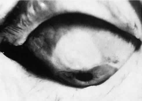
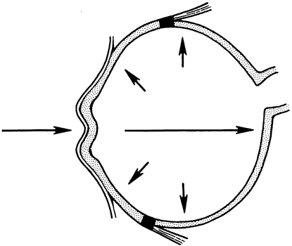
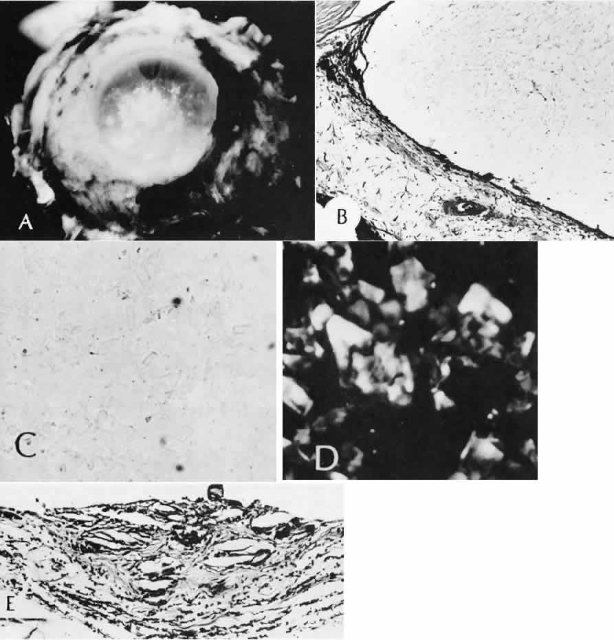
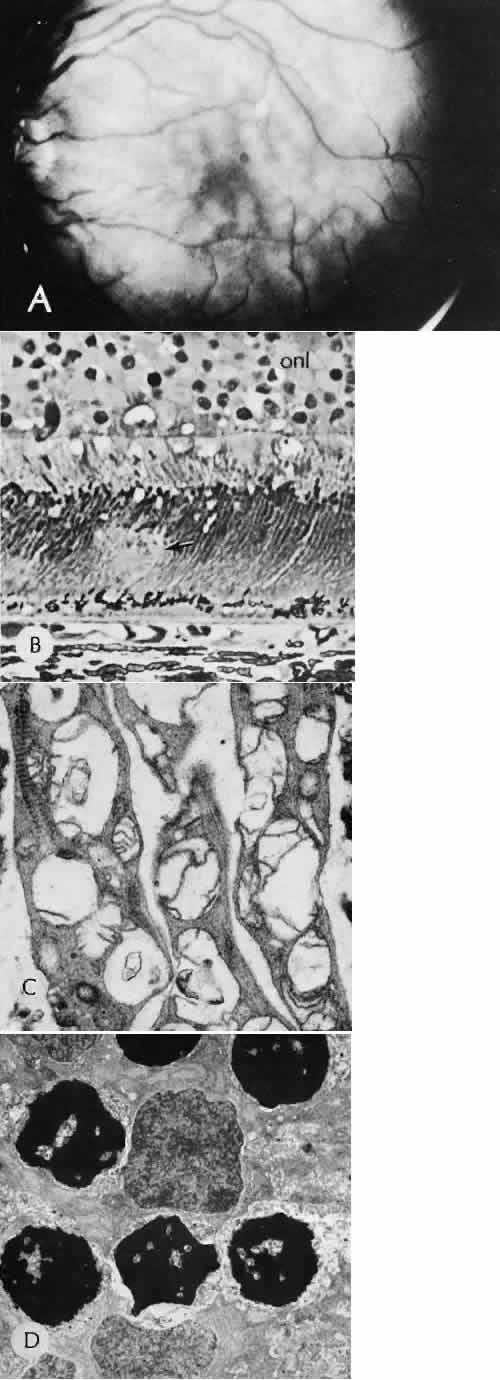
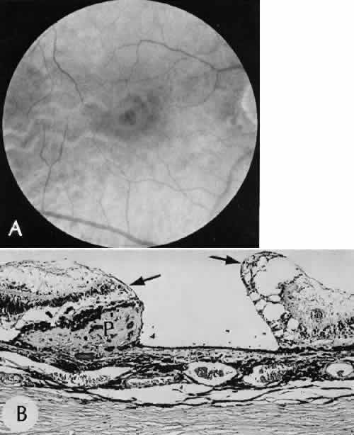
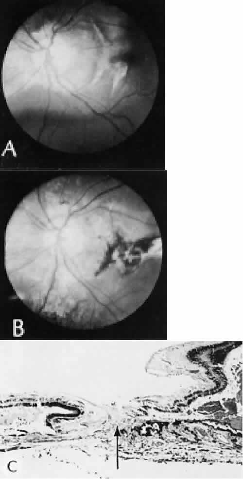
for more effective therapeutic intervention.86 Many complications originate from so-called surgical confusion (Fig. 24). Most arise from a lack of knowledge, a lack of judgment, or problems in perception. At times, lack of attention to detail in preoperative planning may lead to a drug reaction, inadequate anesthesia, misplacement of incisions and sutures, or a patient who is unable to control a cough reflex. What is recognized histologically as the immediate cause of the problem may well have its origin in improper planning of the procedure. CATARACT EXTRACTION Intraoperative Complications Complications occurring from the time the patient enters the operating room until the patient leaves the operating room are considered intraoperative.87,88 Misplacement of an anterior segment incision too far anteriorly into the cornea may create an unacceptable degree of scarring or astigmatism, whereas misplacement too far posteriorly (Fig. 25) may cause the incision to enter the ciliary body instead of the anterior chamber, an especially serious event in glaucoma filtering procedures. Misplacement oflimbal sutures (Fig. 26) may cause anterior wound gaping if placed too far posteriorly or posterior wound gaping if placed too far anteriorly. A deep suture may enter the anterior chamber and lead to wicking and a flat anterior chamber postoperatively. A suture placed at different depths in the two sides of the wound results in faulty apposition of the wound edges. Descemet's membrane is only loosely adherent to the posterior stroma and may be stripped into the anterior chamber at the time of the corneal incision placement (Fig. 27) or injection of sodium hyaluronate. Splitting off of Descemet's membrane from the posterior cornea can lead to postoperative intractable corneal edema.89,90 Intraoperative anterior chamber hemorrhage may result from an inadvertent iridodialysis or from the iridectomy wound. The site of hemorrhage is usually found along the scleral side of the cut edge of the wound. This type of hemorrhage rarely leads to serious clinical problems.91 Rupture of the posterior lens capsule (Fig. 28) increases the risk of vitreous loss and limits options for the implantation of an intraocular lens. Retention of lens capsular tissue or vitreous in the wound (Fig. 29) will significantly delay the wound healing process. Vitreous loss leads to an increased incidence of iris prolapse, bullous keratopathy, epithelial downgrowth, stromal overgrowth, wound infection and endophthalmitis, updrawn or misshapen pupil, vitreous bands, postoperative flat anterior chamber, secondary glaucoma, retinal detachment, cystoid macular edema, optic disc edema, vitreousopacities, vitreous hemorrhage, and other sight-threatening consequences.92 Damage to the endothelium during insertion or positioning of the lens was one of the most common problems with intraocular lens implantation until the widespread use of intraoperative viscoelastics. The endothelial cell damage may manifest on the first postoperative day as corneal edema. The course of the edema may wax and wane and ultimately result in pseudophakic bullous keratopathy months or years after surgery. Detachment of Descemet's membrane, inadvertent iridodialysis, cyclodialysis, or rupture of the posterior capsule also may occur during lens insertion and positioning. Expulsive choroidal hemorrhage (Fig. 30) is a rare catastrophic complication often resulting in total loss of the eye.93 The site of hemorrhage is probably a sclerotic choroidal arteriole where the vessel crosses the suprachoroidal space from the scleral canal. The sudden hypotension after surgical penetration of the globe causes a bending and then a rupture of the arteriole.94 Although most hemorrhages are massive and immediate, they occasionally are delayed, and some may not occur for days or weeks after surgery. Delayed choroidal hemorrhage may occur at the time of corneoscleral suture removal,95 because of clinically unapparent wound dehiscence or as a result of perforation of a corneal ulcer. Histologically, massive choroidal hemorrhagic detachment is associated with a retinal detachment. The retina and choroid may herniate through the scleral wound. A ruptured ciliary artery may be found in the suprachoroidal space. Postoperative Complications Postoperative complications may arise from the time the patient leaves the operating room until approximately 6 weeks after surgery.96 A flat anterior chamber is characterized by anterior displacement of the iris to near or in actual contact with the posterior surface of the cornea. The most common cause is leakage of aqueous along one of the suture tracks. Prolonged decompression of the anterior chamber increases the risk of synechiae formation and intractable secondary closed-angle glaucoma. Corneal endothelial damage may result in bullous keratopathy. Choroidal edema (choroidal hydrops or detachment) (Fig. 31) may be associated with a flat anterior chamber and may potentiate the condition. The choroidal edema will slow or stop aqueous production by the ciliary body, further delaying reformation of the anterior chamber. The histologic characteristics of choroidal edema consist of spreading of the choroidal tissue in a fanlike configuration and eosinophilic fluid filling the intervening spaces. The edema fluid may be lost in processing, leaving multiple apparently empty spaces. Iris or lens capsular incarceration into the wound and extending to the conjunctival space (Figs. 32 and 33) may act as a wick through which aqueous can escape, causing a flat anterior chamber. Histologically, iris, which is frequently recognized only by the presence of melanocytes, is seen in the limbal scar, in the limbal episclera, or in both areas. Vitreous wick syndrome consists of vitreous incarceration in the wound, where the vitreous may extend beyond the wound to the subconjunctival space or occasionally to the ocular surface.97 Anchoring of the vitreous anteriorly in this manner may lead to extensive intraocular inflammation even in the absence of infection. The vitreous may act as a conduitfor bacteria, leading to bacterial endophthalmitis.98–100 Pupillary block glaucoma results from isolation of the posterior chamber from the anterior chamber. Occasionally, even in the presence of a clinically patent peripheral iridectomy or iridotomy, the aqueous cannot exit into the anterior chamber. The block results from contact of formed vitreous, lens remnants, or intraocular lens material with the posterior surface of the iris.101,102 The area of contact may include the iridectomy or iridotomy site. Prolonged contact with any of these tissues or materials will cause posterior synechiae formation. After complete posterior synechiae formation, peripheral anterior synechiae develop. Pupillary block glaucoma (closed-angle glaucoma) may not be clinically evident until aqueous production returns to normal levels after surgery. The intraocular lens may be a stimulus for ongoing uveitis, hyphema, and glaucoma (UGH syndrome) (Fig. 34) through multiple factors.103,104 Although lens manufacturing has improved since the early days of fabrication, retained polishing compounds or burrs and irregular surfaces of the lens or loops may cause a low-grade, sterile endophthalmitis.105 Most intraocular lens materials, with the exception of polymethylmethacrylate, have been shown to stimulate some degree of inflammation, which in turn may cause biodegradation of the lens material.106 Several epidemics of lenses contaminated with fungus have been reported. Intermittent movement or continuous pressure of the intraocular lens material against ocular tissue often will result in focal necrosis, tissue disruption, or microhyphema, which may lead to fibrous scarring. This is particularly important when the tissues of the anterior chamber angle are in contact with an anterior chamber lens. Posterior lens loops may erode the iris pigment epithelium and give rise to a type of pigmentary dispersion syndrome (Fig. 35).107,108 Lenses directly supported by the iris have caused lacerations of the iris because of continuous compression by metallic loops (see Fig. 35).109 Iris-supported intraocular lenses are associated with a high incidence of dislocation and subsequent damage to the eye. Corneal endothelial decompensation (Fig. 36) may lead to bullous keratopathy at almost any time in the postoperative period, depending on the state of health of the endothelium at the time of surgery and the extent and nature of injury to the endothelial cells.110,111 Corneal edema may present early in the postoperative period when the cornea has been compromised by advanced Fuchs corneal dystrophy. If the endothelial cells are healthy and the intraoperative trauma is minimal, the signs of decompensation may not appear for decades. Endothelial damage may be the result of mechanical contact with surgical instruments or an intraocular lens, a toxic reaction to solutions used intraoperatively, poor tissue handling techniques, or prolonged and intensive postoperative inflammation.112 Histologically, the corneal epithelium is generally thinned. There may be areas of intraepithelial basement membrane and cyst formation, resulting from malorientation of the squamous epithelial cells during recovery from multiple episodes of bullous separation from Bowman's membrane (Fig. 37). Bullous separation of the epithelial cells varies in extent but is almost always present. Focal areas of interruption of Bowman's membrane and anterior corneal stroma scarring will mark rupture of bullae and subsequent ulceration. In chronic cases, extensive degenerative pannus formation is often seen. The corneal stroma may show a decreased density of keratocyte nuclei. A decrease in or absence of the artifactual clefts of the corneal stromal lamellae suggests edema of the stroma. Descemet's membrane may be focally or diffusely thickened with preexisting Fuchs corneal dystrophy (Fig. 38). In most cases, Descemet's membrane is normal in character and thickness. Often the corneal endothelial density is reduced to the point that only an occasional endothelial cell nucleus can be seen. Infectious bacterial endophthalmitis (Figs. 39 and 40) usually presents early in the postoperative period.113,114 Pseudomonal and streptococcal organisms often cause rapid and total destruction of the eye. Organisms of especially low virulence, such as Staphylococcus epidermidis and Priopionibacterium acnes, may not present for months and may be misinterpreted as sterile endophthalmitis caused by a toxic reaction to intraocular lens materials or phacoanaphylactic endophthalmitis targeted to lens cortical remnants.115–117 Infectious endophthalmitispresenting months after surgery may be caused by a fungal infection or by bacteria of low virulence as pointed out above. Delayed Complications Delayed complications are those that occur more than 6 weeks after surgery. These complications include corneal endothelial decompensation and delayed infectious endophthalmitis, as discussed previously. Elschnig's pearls (Fig. 41) result from aberrant attempts by lens cells attached to the lens capsule to form new lens “fibers.” Histologically, large, clear lens cells (“bladder cells”) are present behind the iris, in the pupillary space, or in both areas. Soemmerring's ring cataract results from the loss of anterior and posterior cortex and nucleus but with retention of equatorial cortex. Apposition of the central portions of the anterior and posterior lens capsule causes a doughnut configuration. Frequently, the doughnut is not complete, so that C- or J-shaped configurations result. Histologically, two balls of trapped and proliferated lens cells are encapsulated behind the iris leaf and connected by adherent anterior and posterior lens capsule in the form of a dumbbell.118 Delayed complications of intraocular lenses themselves are infrequent. The incidence of posterior capsular opacification ranges between 11% and 46% in this period, apparently lower in the presence of an intraocular lens than with extracapsular cataract surgery alone.119,120 Histologically, residual lens epithelial cells are transformed into cells with myofibrillar contractile properties, which wrinkle and opacify the posterior capsule and synthesize the extracellular matrix. Retinal detachment (Fig. 42) occurs in approximately 2% to 8% of intracapsular cataract surgeries, compared with approximately 0.0013% in the general population. The incidence of retinal detachment after extracapsular cataract extraction and posterior chamber lens implantation ranges between 0.6% and 1.5%.121 Vitreous loss increases the incidence of postoperative detachments, particularly if there is vitreous incarceration into the cataract wound. The character of the retinal detachment is independent of the type of cataract surgery or the type of intraocular lens implanted. However, a lower incidence of proliferative vitreoretinopathy appears in cases of extracapsular cataract extraction than was formerly seen with intracapsular cataract extraction. Aphakic or pseudophakic glaucoma122,123 is a nonspecific term referring to a pathologic and sustained increase in pressure after surgery for removal of cataract. In the delayed phase, this glaucoma is mainly caused by secondary chronic angle closure. However, a preexisting predilection to simple open-angle glaucoma may be the cause. Circumferential peripheral anterior synechiae may develop from persistent flat anterior chamber. Focal synechiae are often found adherent to the posterior aspects of the surgical incision. Histologically, the iris is apposed to the posterior cornea, often central to Schwalbe's ring. Posterior synechiae are generally the result of posterior chamber inflammation (caused by iridocyclitis, endophthalmitis, or hyphema) causing iris bomb, and secondary peripheral anterior synechiae (Fig. 43). Histologically, the posterior pupillary portion of the iris is adherent to the anterior face of the vitreous, to the lens remnants, to the intraocular lens, or to all three structures. The anterior peripheral iris is adherent to the posterior cornea (peripheral anterior synechiae), frequently central to Schwalbe's ring. Epithelial downgrowth (ingrowth) (Fig. 44) is one of the most serious causes of pseudophakic glaucoma, in which surface epithelium (probably from the conjunctiva) grows into the anterior chamber. This condition is more likely to occur with fornix-based conjunctival flaps than with limbus-based flaps and in eyes with problems in wound closure, such as vitreous loss, wound incarceration of tissue, delayed reformation of the anterior chamber, or frank rupture of the limbal incision, and when instruments such as iridectomy forceps are contaminated with surface epithelium before they are introduced into the eye. Epithelial downgrowth causes an anterior chamber angle closure by means of peripheral anterior synechiae or lines an open anterior chamber angle and obstructs aqueous outflow mechanically. Histologically, the epithelium is seen to grow most luxuriously and in multiple layers on the iris where there is a good blood supply, but it tends to grow sparsely and in a single layer on the posterior surface of the avascular cornea. The epithelium may extend behind the iris, over the ciliary body, and even far into the interior of the eye. Iris cyst formation (Figs. 45 and 46 is also caused by implantation of surface epithelium onto the iris at the time of surgery or trauma. The cyst generally grows slowly and is accompanied by peripheral anterior synechiae. If extensive, the cysts may cause a secondary chronic closed-angle glaucoma. Histologically, the cyst is lined by stratified squamous or columnar epithelium, sometimes containing mucous cells, and is filled with keratin debris (white or pearly cysts) or mucous fluid (clear cysts). Stromal overgrowth (Fig. 47) is a condition characterized by growth of fibrous connective tissue into the anterior chamber. It is most likely to occur after vitreous loss or tissue incarceration into the surgical wound. The overgrowth may be localized, may be limited to the area of surgical perforation of Descemet's membrane, or may be extensive. When overgrowth is extensive, peripheral anterior synechiae and secondary closed-angle glaucoma result. As with epithelial downgrowth, the stromal overgrowth may extend behind the iris, over the ciliary body, and far into the interior of the eye. Histologically, fibrous tissue generally extends from the corneal stroma or is in continuity with it, through a large gap in Descemet's membrane. The fibrous tissue often covers the posterior cornea, fills part of the anterior chamber, occludes the anterior chamber angle, and may extend into the vitreous compartment to attach posteriorly onto the retina. Bacterial inflammation is rare in the delayed period after surgery, except after filtering procedures in which bacteria can gain access to the inside of the eye by way of the bleb (Fig. 48). Another exception to this rule is Priopionibacterium acnes or Staphylococcus epidermidis endophthalmitis, which infects lens remnants and may not clinically manifest for many months after cataract extraction. Fungal endophthalmitis (Fig. 49) may take the form of keratitis or endophthalmitis. Histologic characteristics of end-stage endophthalmitis include fibrovascular organization centered about a chronic nongranulomatous inflammatory reaction contiguous with lens remnants, causing cyclitic membrane formation and retinal detachment. Multiple small foreign bodies, inadvertently introduced at the time of surgery, can cause a delayed chronic nongranulomatous or granulomatous inflammatory reaction. Phacoanaphylactic endophthalmitis or sympathetic uveitis may occur after extracapsular cataract extraction (discussed elsewhere in these volumes). Healed cataract wounds may rupture because of trauma. In particular, blunt trauma to the eye may cause ocular rupture, often at the site of a cataract scar that remains weaker than surrounding tissue. Cystoid macular edema, or Irvine-Gass syndrome, is an inflammatory, degenerative condition of unknown cause that involves primarily the macula and leads to temporary or permanent loss of macular function. This condition can occur anytime after cataract surgery (even up to 5 years), but most cases occur within 2 months of surgery. The initial clinical sign is a sudden decrease in visual acuity. At least 50% of the cases are self-limiting, and the macular edema resolves completely, with or with-out therapy, within 1 year. Most patients experi-ence spontaneous recovery of vision. In a few cases,however, the intraretinal edema may persist, andsecondary permanent complications, such as lamel-lar macular hole formation, may occur. The condi-tion can be precipitated or aggravated by topicalepinephrine therapy for glaucoma. The cause ofcystoid macular edema is unknown, but in somecases (probably the minority), vitreous traction or aposterior vitritis may play a role (Fig. 50). Histo-logically, iritis, cyclitis, retinal phlebitis, and retinal periphlebitis have been noted. Whether these conditions cause the cystoid macular changes or are simply incidental findings in enucleated eyes is not clear. A lamellar macular hole may occur as a permanent complication. Ophthalmoscopically (best seen with a slit-lamp biomicroscope), multiple (usually four or five) intraretinal microcysts are seen in the macular area, obscuring the normal foveal reflex. The cysts fill early with fluorescein, and pooling causes a characteristic stellate geometric pattern that persists for 30 minutes or longer. Granular fluid is seen in the microcystic spaces (Fig. 51). The anatomic basis of early cystoid macular edema probably is edema of Müller cells; later, Müller cell membranes break down and the edema becomes extracellular. PENETRATING KERATOPLASTY Immediate Complications Complications of penetrating keratoplasty may arise in the selection of donor material. The success of a graft depends on the health and integrity of the donor endothelium. Compromise of endothelial function attributable to the age of the donor, preexisting endothelial disease (Fuchs corneal dystrophy), toxicity of storage procedures, microbial infection, or intraoperative trauma will lead to early endothelial decompensation. Immunologic testing and a careful history of infectious disease of the donor (AIDS, hepatitis, Jakob-Creutzfeldt disease, and rabies) are essential because the virus may be harbored in the donor tissue. Retention of significant amounts of host Desce-met's membrane and presence of nonviable peripheral host tissue in patients with alkali burns are additional potential intraoperative problems. Improper apposition of the donor-host interface may lead to multiple structural and refractive problems. Overriding of the wound edge is a likely site for wound leak, leading to a flat anterior chamber, which in turn may lead to central anterior synechiae to the wound or total anterior synechiae. Malposition of the posterior wound edge is likely to enhance fibrous tissue proliferation, which may remain in the local region of wound or may spread peripherally or even centrally (stromal in growth). Postoperative Complications Penetrating keratoplasty has the same risks for microbial infection, epithelial ingrowth, stromal ingrowth, and expulsive choroidal hemorrhage, as does anterior segment surgery in general. Immune rejection of a corneal graft is an unusual complication, except when the host tissue is extensively vascularized. Immune stromal graft rejection clinically manifests 2 to 3 weeks postoperatively and is heralded by inflammatory signs associated with progressive vascularization of the graft. Histologically, a central necrotic area is bounded peripherally by a zonal inflammatory response. Polymorpho-nuclear leukocytes predominate near the necrotictissue, surrounded by a zone of lymphocytes and plasma cells. The endothelial layer may undergo specific graft rejection. This is recognized clinically as a centrally advancing, linear, posterior corneal opacity (Khoudadoust's line). Histologically, there is a chronic nongranulomatous inflammatory reaction about necrotic endothelial cells. Delayed Complications Graft failure is a nonspecific term used to indicate progressive or persistent opacification of a donor cornea. In most cases, the histologic characteristics are those of corneal endothelial decompensation. In some cases, particularly those with underlying systemic disease or ocular surface abnormalities, there may be extensive corneal ulceration or perforation. REFRACTIVE SURGERY Immediate Complications Accidental perforation of the globe is possible with any of the refractive procedures. Variability of the incision depth is one of the most important factors in the ultimate variability of the refractive correction obtained by the procedure. Postoperative Complications Microbial endophthalmitis, even leading to blindness, has been reported as a complication of this type of procedure. There appears to be an acute postoperative endothelial cell loss that is not progressive. Wound healing of radial keratoplasty has been complicated by intrastromal epithelial cyst formation and a generalized decrease in the rate of healing of the incision (Fig. 52). Complete healing and refractive stability may not be realized for years. Epikeratophakia procedures have been complicated by delayed reepithelialization of the grafted tissue and epithelial retention or proliferation along the interface between the lenticule and the host tissue. Persistent folding of Bowman's membrane underlying the grafted tissue and abnormal keratocytes in the underlying recipient stroma may lead to a poor visual result. In some cases, the lenticule of keratomileusis has been slow to be repopulated by host keratocytes, which may be responsible for persistent opacification of the grafted area. Thickening of the epithelial basement membrane, a decreased density in hemidesmosomes, and abnormalities of Bowman's membrane have been observed. Abnormal proteoglycan synthesis also has been suspected in these cases. Visually significant opacities at the tissue-synthetic material interface have been noted with corneal intrastromal inlay procedures (Fig. 53). This material, which has been found to contain lipid, appears to accumulate over time. Corneal inlays of all types have been complicated by necrosis of the tissue overlying the pocket and extrusion of the synthetic material. Delayed Complications An early type of radial keratotomy, which included incisions of the internal surface of the cornea, has resulted in a high degree of corneal endothelial decompensation 20 years after the procedure, regardless of the age of the patient at the time of surgery. LASIK Complications of LASIK Variability in actual flap thickness has also been noted in several studies. The lack of uniformity of flap dimensions may be important in repositioning the flap in the stromal bed.124 Microfolding of the anterior corneal flap was found in 94% of eyes examined by confocal microscopy. The folding was attributed to stretching of the tissue during laminectomy or to imprecise registering of the flap tissue with the lamellar bed.75 The character of the blade edge, particularly of a blade that has been used for more than one procedure, also influences the morphology of the stromal incision. Notching of the blade profile with repeated use has been observed by scanning electron microscopy. Notching of the blade edge also appears to be associated with accumulation of tissue remnants on the surface of the blade.67,68 The tissue remnants can be identified as reflective interface particles by confocal microscopy. Potential sources of the material include metal from the microkeratome blade, cotton, lipid, inflammatory cells, intact epithelial cells, or remnants.75 Complications occur in approximately 3% to 5% of LASIK procedures.78,125,126 Intraoperative compilations are primarily related to creation of the lamellar flap. Included among this type of complication are flap irregularities, epithelium and other material within the lamellar interface, regular and irregular astigmatism, flap loss, displacement, and button-holing. Infection is a rare but potentially serious complication.78,127–131 Perforations of the globe have been described.132–137 Potential complications at 6 months include epithelial ingrowth, corneal flap melting, decentered ablation, and irregular astigmatism with loss of best-corrected visual acuity.124 Complications related to abnormal endothelial cell function have been infrequent.134,138–140 Progressive epithelial ingrowth may occur characterized as a continuous sheet of epithelium contiguous with the flap edge (Fig. 54).124,141 Epithelial ingrowth into the intrastromal interface may be associated with enzymatic digestion of the cornea.142 Diffuse interface keratitis is a condition char-acterized by acquired opacification at the level ofthe intralamellar bed after a LASIK procedure(Fig. 55).143 Many causes of the syndrome have been reported144–147 and include exotoxins released from sterilizer reservoir biofilm148 and debris on the microkeratome blade149 and methylcellulose sponge material.150 Most cases resolve spontaneously.151 The resistance of eyes to trauma after LASIK, even in the absence of healing appears to be similar to unoperated human eyes as evaluated in a model of postmortem refractive procedures performed on autopsy eyes.152 In this model, radial keratotomy incisions predisposed rupture at lower energy levels than in unoperated eyes.153 Postoperatively, there was a decrease in intraocular pressure measured by central tonometry that was statistically significant. Differences in pneumotonometry were less substantial, with greater reliability of pneumotonometry than Goldmann applanation tonometry after LASIK.154 GLAUCOMA SURGERY Immediate Complications Creation of a button hole in the conjunctiva is not serious in cataract surgery but may lead to failure in filtering procedures. Misplacement of the incision too far posteriorly may cause the incision to enter the ciliary body instead of the anterior chamber, an especially serious event in glaucoma filtering procedures. Postoperative Complications Cataract formation may be caused or accelerated by glaucoma surgery, even if the lens is not apparently damaged by physical contact. The cataract may be a result of shunting of the aqueous through the iridectomy, so that the anterior and posterior surfaces of the lens are not properly nourished. Bacterial endophthalmitis may occur. Hypotony and choroidal detachment have complicated trabeculectomy procedures more common with full-thickness filtering procedures. Seton devices have been complicated by conjunctival erosion by the synthetic parts, blockage of the proximal orifice, corneal decompensation, extrascleral tube compression, and blockage of the distal orifice. Delayed Complications Procedures to lower intraocular pressure function by transconjunctival filtration, absorption of aqueous into the subconjunctival vessels, recanalization, reopening of drainage channels, passage through areas of perivascular degeneration, or any combination. Incorrect placement of the incision, hemorrhage, inflammation, prolapse of the intraocular tissue into the filtration site, dense fibrosis, peripheral anterior synechiae formation and secondary chronic closed-angle glaucoma, and endothelialization of the bleb may cause filtration failure. The histology depends on the underlying cause. Even in the delayed period, bacteria may gain access to the interior of the eye, by way of the bleb, and cause endophthalmitis. RETINAL REATTACHMENT SURGERY Intraoperative Complications A misplaced implant, explant, or scleral suture can lead to an improper scleral buckle or premature drainage of the subretinal fluid. Misplaced, insufficient, or excessive diathermy or cryotherapy can cause unsatisfactory results by not closing the retinal hole. Cut or obstructed vortex veins can lead to choroidal detachment or hemorrhage (Fig. 56), which is most often caused by hypotension induced by surgical drainage of subretinal fluid. Overuse of scleral cryotherapy or diathermy also may be a cause. Other causes include cutting or obstruction of vortex veins and incision of the choroidal vessels at the time of surgical drainage of the subretinal fluid. Retina may be incarcerated in a surgical drainage site. Acute closed-angle glaucoma may be the result of the buckling procedure, especially if unaccompanied by drainage of the subretinal fluid or anterior chamber paracentesis. If the glaucoma is not recognized, central retinal artery occlusion may occur and result in blindness. Postoperative Complications The original retinal hole may remain open, or a new one may develop or be missed preoperatively. Choroidal detachment and choroidal hemorrhage also may occur during this time. Acute or subacute scleral necrosis (Fig. 57) may follow retinal detachment surgery after days or weeks and is probably caused by ischemia rather than infection. In the acute form, the clinical picture generally starts a few days after surgery and may resemble a true infectious scleritis but without pain. There is a sudden onset of congestion, edema, and a dark red or purple appearance of the tissues over the implant or explant. Discharge is not marked or is absent altogether. The vitreous over the buckle generally becomes hazy but may be clear. The cornea remains clear but the involved area of the sclera becomes completely necrotic. In the subacute form, the clinical picture starts with pain approximately 2 to 3 weeks after surgery. The globe may be congested but no discharge occurs. The vitreous over the buckle may be hazy or clear. The sclera in the region of the buckle is necrotic. Infection in the form of scleral abscess, endophthalmitis, or keratitis may be secondary to bacteria or fungi and is characterized by redness of the globe, discharge, and pain (Fig. 58). Anterior segment necrosis (Fig. 59) is thought to be secondary to interruption of the blood supply to the iris and ciliary body, usually by temporary removal of one or more rectus muscles during surgery. Encircling elements, lamellar dissection implants, explants, and cryotherapy or diathermy may also cause compromised blood supply. Clinically, keratopathy and intraocular inflammation usually develop in the first postoperative week. Corneal changes consist of striate keratopathy and corneal edema with epithelial bullae. Chemosis, anterior chamber flare and cells, large keratic precipitates, and white deposits on the necrotic lens capsule characerize intraocular inflammation clinically. The clinical findings are often mistaken for an infectious endophthalmitis. Later, the pupil becomes dilated. Shrinkage of the iris toward the side of the greatest necrosis and hypoxia results in an irregular pupil. Cataract, hypotension, ectropion uveae, and finally phthisis bulbi develop. A high incidence of anterior segment ischemic syndrome is seen after scleral buckling procedures in patients with hemoglobin sickle cell disease. In hemoglobin sickle cell disease, the increased frequency of anterior segment necrosis is most likely related to the increased blood viscosity and tendency toward erythrocyte packing that is found in these patients, especially those with a decreased oxygen tension. Histologically, ischemic necrosis of the iris, ciliary body, and lens epithelial cells is present, frequently only on the side of the surgical procedure. Hemorrhage in the postoperative period may be caused by a delayed expulsive choroidal hemorrhage that most probably results from necrosis of a blood vessel induced by the original diathermy or cryotherapy or to erosion of an implant or explant. Acute closed-angle glaucoma probably occurs after a retinal detachment procedure in which an encircling element or a high buckle is created. Acute closed-angle glaucoma occurs in approximately 4% of scleral buckling procedures. The pathogenesis of the angle closure is not known, although pupillary block or swelling of the ciliary body is a proposed mechanism. The buckle decreases the volume of the vitreous compartment, displacing the vitreous and lens-iris diaphragm anteriorly. Corneal edema on the first postoperative day, especially if accompanied by ocular pain, should be considered glaucomatous until proved otherwise. Histologically, the anterior displacement of intraocular structures results in encroachment of the iris on the anterior chamber angle and resultant closed-angle glaucoma. Chronic simple glaucoma may become apparent when the hypotension of a retinal detachment is alleviated by surgery. Delayed Complications Vitreous retraction by itself is of little importance, but when it is associated with fibrous or glial membranous proliferation on the internal or external surface of the retina, it can cause retinal detachment with or without retinal hole formation. Prolifera-tive vitreoretinopathy is extensive cellular prolif-eration (glial cells, retinal pigment epithelial cells,fibroblasts, and Müller cells) on retinal tissue asso-ciated with a total retinal detachment. The mem-branes distort the surface of the retina to cause a cel-lophane appearance and extend to form star foldsand other configurations of fixed retinal folds. His-tologically, fibroglial membranes can be seen onthe internal or external surface of the retina. With contraction of the myofibroblasts of the membranes, the architecture of the retina will be markedly distorted.155 An implant or explant may migrate externally along the surface of the globe and through the conjunctiva or internally into the globe (Fig. 60). Internal migration may cause hemorrhage, retinal detachment, or infection. In this setting, the conjunctival epithelium may gain access to the interior of the eye. The resultant conjunctival epithelial ingrowth complicates an already compromised eye. Retinal tacks also may migrate as far as the anterior chamber. A retinal hole may develop de novo or secondary to obvious vitreous pathology. Other late problems include heterophoria, disturbances of lid position, and secondary glaucoma (hemolytic, closed angle from prolonged or recurrent inflammation, or neovascular associated with retinal ischemia). Macular degeneration and puckering may occur if cryotherapy or diathermy has been used. |
NONSURGICAL TRAUMA | |
No two eyes are traumatized in an identical manner.156–158 In contrast with surgical trauma, extremevariability and unpredictability
are the hallmarks of accidental trauma to the eye. Multiple types
of agents may inflict different types of injuries of varying severity.159,160 One clinically useful classification of accidental ocular trauma is based on the type of injury; however, even in this classification, there are areas of overlap. Blunt trauma usually indicates that a small degree of tissue disruption exists. Penetrating or perforating trauma indicates a greater degree of tissue disruption. Intraocular foreign bodies imply a degree of tissue disruption often limited in extent but complicated by retention of some foreign material within the globe or accompanying microbial infection. Thermal and chemical injuries lead to special types of destructive processes. Radiation trauma usually produces vascular abnormalities but also may affect the crystalline lens. Finally, systemic trauma may indirectly affect the eye. BLUNT TRAUMA Compression and deformation of the eye are unusual occurrences because of the protection afforded the eye by the surrounding bones of the orbit, particularly the orbital rim. However, although no actual tissue loss occurs, the force of the blow may cause extensive injury to many of the delicate structures of the eye (Fig. 61).161 These same structures usually have only a limited ability to recover and repair the damage. Regions of the sclera are notably vulnerable to blunt trauma because of relative thinness. These areas include the limbus (Fig. 62), the insertions ofthe four rectus muscles, the insertion of the superior oblique, and the scleral canal (Fig. 63). Cornea Corneal epithelial abrasions may be large but usually will heal without serious sequelae. Deeper corneal involvement may result in focal areas of epithelial proliferation (facet formation, see above) or scar formation of the stroma. Endothelial cell distortion or rupture may result in multiple small ring opacities of the endothelium.162 The endothelial lesions become visible immediately after injury and become progressively more opaque over several hours. Depending on the degree of injury, the rings may disappear within days and result in no permanent loss of visual acuity. Histologically, swelling of the corneal endothelium and accumulation of fibrin and leukocytes on the injured endothelial cells cause the rings.163 More extensive endothelial cell damage is associated with stromal edema. Ruptures of Descemet's membrane allow aqueous to hydrate corneal stroma, which is normally relatively dehydrated. The swelling of the cornea reduces its transparency (corneal hydrops). The edema may resolve once the endothelial function is reestablished.164 Birth trauma is a common cause of rupture of Descemet's membrane. The area of rupture is often oriented diagonally across the central cornea.165 Histologically, whether the rupture is the result of birth trauma, congenital glaucoma, or trauma anytime after birth, a gap is seen in Descemet's membrane.166 The endothelium usually will migrate over the gap and form a new, thin Descemet's membrane. The time required for clinical improvement is generally between 6 weeks and 6 months.167 In attempting to cover the gap, the endothelium may grow into the anterior chamber over the free, rolled end of the ruptured Descemet's membrane and form a scroll-like structure.164 Corneal hydrops may also be complicated by corneal neovascularization167 or, rarely, by intralamellar pseudocyst formation.168 Keloid of the cornea (Fig. 64) is a hypertrophic scar that occasionally develops after ocular injury. Most keloids appear as glistening white masses that extend outward from the eye in the region of the cornea. Histologically, corneal perforation generally is present. Haphazardly arranged fibroblasts, collagen bundles, and blood vessels form the corneal scar.169,170 Anterior Chamber171 Blood in the anterior chamber (hyphema) may lead to a number of secondary complications.172–174 Even a small amount of blood in the anterior chamber is a clinically significant finding because its presence indicates the possibility of serious intraocular damage. In addition, there is a risk of a secondary (delayed) and often more serious hemorrhage occurring at the time of clot retraction.175 If the hemorrhage is extensive, the blood may totally fill the anterior chamber and completely replace the aqueous (8-ball or blackball hyphema).176 The extravasated blood will turn from bright red to black as the aqueous circulation ceases and the hemoglobin becomes deoxygenated. Multiple mechanisms in the setting of hyphema may lead to secondary glaucoma.177 Anterior chamber hemorrhage in hemoglobin sickle cell disease or in vitreous hemorrhage with access to the anterior chamber (hemolytic or ghost cell glaucoma) may cause occlusion of the trabecular meshwork. The sickle cell and ghost cell erythrocytes are rigid and cannot be filtered efficiently through the trabeculum. Macrophages filled with hemoglobin in sufficient quantity may also cause decreased aqueous filtration. A blood clot itself may tamponade the meshwork in the peripheral anterior chamber and cause pupillary block centrally. These mechanisms, which occur in the early posttraumatic period, are potentially reversible. In the chronic phases of healing, however, fibrous organization of a clot, fibrosis of the trabecular meshwork, endothelialization of the trabecular meshwork, or peripheral anterior synechiae formation may lead to permanent secondary glaucoma (Fig. 65). Blood breakdown products from hyphema may diffuse into the cornea and cause a reddish orange opacification of the stroma (blood staining of the cornea). The extent of the deposition of hemoglobin depends on the degree of damage to the endothelium and on the degree of increase in the intraocular pressure.178 With sufficient damage to the endothelium, bloodstaining may occur even at low levels of intraocular pressure. Histologically, hemoglobin particles can be seen by routine stains to be distributed throughout the corneal stroma. Hemosiderin, a metabolic product of hemoglobin, may be identified in the keratocytes with special stains for iron (Prussian blue). Keratocytes are actively engaged in biodegradation of hemoglobin and necrosis if overloaded with hemoglobin molecules.179 Endothelial degeneration has been observed in animal models of corneal bloodstaining.180 The bloodstaining may clear, beginning in the periphery of the cornea; however, a great deal of time (months to years) is required (Fig. 66). Anterior Uveal Tract The delicate iris sphincter is relatively unsupported by surrounding fibrovascular tissue. Deformation of the iris may cause rupture of the dilator and sphincter muscles and iris stroma.181 The areas of damage are identified clinically as one or several notches of the pupillary margin. In a similar manner, the iris base is commonly ruptured at its insertion into the face of the ciliary body, causing variable degrees of iridodialysis (Fig. 67). Total iris disinsertion is also possible. Anterior chamber angle recession (postcontusion deformity of the anterior chamber angle) consists of a posterior displacement of the iris root and inner pars plicata.182,183 The tissues that are retrodisplaced include the ciliary processes or crests, the circular ciliary muscle, and some or all of the oblique ciliary muscle. The meridional portion of the ciliary muscle remains attached to the scleral spur (Figs. 68 and 69). The posterior displacement is the result of a sudden increase in anterior chamber hydrostatic pressure, secondary to blunt trauma. The increased pressure causes a laceration in the anterior face of the ciliary body. Histologically, in early lesions, hemorrhage and disruption of the ciliary musculature easily identify the laceration. Later in the course of healing and subsequent fibrosis, the area of injury is identified by misalignment between the plane of the iris diaphragm and the scleral spur. At this stage, the ciliary body loses its triangular meridional outline and becomes fusiform (Fig. 70). In even later stages, the area of the laceration may again become reapposed and continue on to form peripheral anterior synechiae. Even in the absence of gross structural changes, however, the original injury may have done irreparable damage to the trabecular meshwork. In this latter circumstance, the angle recession is a clinical marker of the cause of the glaucoma. The ciliary body is attached to the sclera only at the ciliary spur. If this attachment is disrupted, cyclodialysis (Fig. 71) will result. The aqueous in this circumstance has direct access to the suprachoroidal space, which in combination with ciliary body dysfunction, often leads to hypotension. The associated hypotension may be recognized histologically by diaphanous separation of the delicate suprachoroidal fibers. A cyclodialysis cleft may close abruptly followed by a sudden rise in intraocular pressure. Blunt trauma on occasion causes a chronic nongranulomatous iridocyclitis. The inflamed anterior uveal tract allows fibrin to accumulate in the anterior chamber to such a degree that fibrin clots may be mistaken clinically for vitreous displaced into the anterior chamber. Leukocytes may accumulate and layer out in the form of a hypopyon even in the absence of infection. The adhesive nature of the inflammatory exudate may cause the formation of anterior or posterior synechiae, or both. Severely compromised aqueous humor dynamics may bring about intractable glaucoma. Lens Opacification of the lens may occur immediately after many types of trauma or may not appear for many years. Likewise, abnormalities of the zonule fibers may not be recognized until surgery for cataract extraction is complicated by lens instability. Pigment from the iris pigment epithelium may be deposited on the anterior lens capsule (Vossius ring) from relatively minor degrees of trauma, causing compression of the iris against the lens. If a rupture of the lens capsule is small, the rent may be sealed by overlying iris or healed by proliferation of lens epithelium. A small rupture is noted clinically as a focal opacity. Histologically, the opacity corresponds with a break in the lens capsule associated with degeneration of contiguous lens epithelium and superficial cortex. A large rupture usually results in the rapid development of a cataract and dispersion of degenerated of lens cortical material into the anterior chamber. Histologically, the ruptured lens is seen as lens cortex associated with an infiltrate of macrophages. The macrophages may accumulate in the anterior chamber angle and cause phacolytic glaucoma. The lens epithelial cells remaining in the confines of the capsular remnants may proliferate and result in the formation of a Soemmerring ring cataract if the equatorial cortical material is preserved, or in Elschnig's pearls if only sectors of lens material remain. Even if the integrity of the capsule is maintained, the force of the original trauma or subsequent inflammation may cause proliferation of the central lens epithelium. The peripheral viable epithelial cells will undergo fibrous metaplasia in an attempt to repair the damage and create an anterior fibrous plaque. The most internal lens cells will again differentiate and form a new, internal lens capsule. The resulting complex is referred to as an anterior subcapsular cataract. A peculiar rosette or flowerlike cataract may occur in the cortex (Fig. 72). The petals correspond with sectors of cortical opacification. Posterior subcapsular cataracts also occur. Immunologic privilege of the lens is lost when cortical material is exposed to immunologic surveillance. The resulting immunologic reaction to this newly exposed foreign antigen may result in extensive tissue destruction by inflammation to a much greater extent than the original mechanical injury. This process is recognized histologically as a zonal granulomatous reaction to lens cortex.184 Abnormalities of the position of the lens are caused by rupture of the zonular fibers. Total dislocation (luxation) (Fig. 73) of the lens is caused by total zonular rupture. The lens moves completely out of the posterior chamber into the anterior chamber or vitreous compartment. Subluxation (Fig. 74) is caused by incomplete zonular rupture with the lens remaining in the posterior chamber, but in an abnormal, nonaxial position. Clinical signs of subluxation include change of refractive error, deepening (especially irregular) of the anterior chamber, an undulating movement of the iris diaphragm with movement of the eye (iridodonesis or shimmering iris), herniation of the vitreous into the anterior chamber, and even pupillary block glaucoma. The force necessary to dislocate a lens is also usually sufficient to cause cataract formation and damage to the trabecular meshwork. Vitreous The vitreous is attached primarily at its base straddling the ora serrata and along the superficial aspect of retinal blood vessels around the optic disc and over the superficial vessels. The inertia of this gelatinous body may cause detachment from the posterior retina with relatively minor force. The vitreous may become detached anteriorly from the pars plana and anterior retina at its base with more severe force. In addition, movements of the vitreous may cause retinal tears or hemorrhage from retinal vessels. Small amounts of vitreous hemorrhage may lead to marked reduction in visual acuity.185 Initially, the blood may settle inferiorly and vision may be partially restored. Organization of the hemorrhage may lead to tractional retinal detachment. Fibrous organization of the vitreous is especially important when the anterior vitreous face is involved. Organized hemorrhage and fibrous proliferation from inflammation may result in the formation of a cyclitic membrane (Fig. 75) extending from the vitreous base in a plane immediately posterior to the lens. Not only will this membrane obstruct transmission of light, but also as it contracts through the influence of myofibroblasts, the cyclitic membrane may cause internal displacement of the ciliary body from the suprachoroidal space resulting in hypotension. As erythrocytes in vitreous hemorrhage undergo degradation in the vitreous, they lose hemoglobin, normal plasma membrane pliability and bicon-cave shape, and form rigid spherical cells (ghostcells).186–188 If these cells gain access to the anterior chamber, they may occlude the trabecular meshwork (hemolytic or ghost cell glaucoma). The ghost cells may be identified by routine cytologic stains of an aqueous aspiration but are best seen by phase-contrast microscopy.189 The characteristic cytologic feature is the presence of degenerated and aggre-gated hemoglobin proteins on the internal surface of the ghost cell's plasma membrane (Heintz bodies). Cholesterol crystals are part of the residue of degenerating erythrocytes and lipid-rich serum (Fig. 76). In the vitreous, they appear as glistening, brilliant crystals (synchysis scintillans).185 The greatest concentration of crystals often is drawn inferiorly by gravity and may partially redisperse with movements of the eye. Histologically, the cholesterol may be free in the vitreous, may incite a foreign body granulomatous inflammatory reaction, may be phagocytosed by macrophages, or may be surrounded by dense fibrous tissue without any inflammatory reaction. The cholesterol crystals are birefringent to polarized light and stain with fat stains in freshly fixed frozen-sectioned tissue, but are dissolved out by alcohol and xylene during normal processing of tissue for embedding in paraffin. In processed tissue, cholesterol appears as empty spaces, sometimes described as cholesterol clefts. Cholesterol cleft formation is not limited to vitreous hemorrhage but is also found in areas of subretinal hemorrhage and in conditions not related to trauma, such as Coats disease. The iron in hemoglobin may be metabolized to hemosiderin, which collects in all epithelial and neuroepithelial structures of the eye. Intracellular accumulation of iron causes cellular dysfunction leading to hemosiderosis bulbi. Retina Commotio retinae (Berlin's edema) (Fig. 77) occurs as a result of countercoup injury. Relatively well-defined areas of retinal opacification develop shortly after the injury. Experimental evidence and clinical observation suggest that changes at the level of the deep sensory retina in the foveal and macular area cause the retina to appear pale, white, and edematous, similar to the sequelae of central retinal artery occlusion.190 The injury involves the photoreceptor outer segments and possibly the apical processes of the retinal pigment epithelium. Patches of pallor also may be seen in the peripheral retina. No fluid leak or edema is evident by fluorescein angiography.191 The process may resolve completely without sequelae, or damage to the photoreceptors may cause permanent visual loss.192 Cystoid macular degeneration with cyst and hole formation may occur months or years after the injury (Fig. 78). The origin of the cysts is obscure.193 Once microcystoid degeneration occurs, however, the septa between the microcysts break down and result in posterior polar retinoschisis (macular cyst). Histologically, photoreceptors and cell bodies in the outer nuclear layer are damaged early. Retinal pigment epithelium phagocytoses damaged outer segment material. Retinal pigment epithelium undergoes hyperplasia and may migrate into the retina, depending on the severity of the initial trauma.194 Late effects may include microcystoid degeneration of the fovea, macrocyst formation, lamellar hole formation, and actual through-and-through sensory retinal hole formation. Hemorrhage in the retina just under the internal limiting membrane or between the internal limiting membrane and the cortical vitreous may layer out by the influence of gravity to assume a boatlike configuration. Subretinal hemorrhage may damage the retina through mechanisms of chemical toxicity, mechanical traction on the photoreceptor outer segments, or establishment of a diffusion barrier between the retina and choroidal circulation.195,196 Subretinal hemorrhages may organize and appear as a subretinal scar. Retinal tears secondary to trauma are generally located in the superior nasal periphery in the region of the posterior border of the vitreous base.197 Detection of a retinal detachment in this setting may be delayed until sufficient syneresis of the vitreous allows for physical displacement of the retina. The prognosis is poor in cases of vitreous loss or organization of the vitreous. Posterior Uveal Tract The posterior uveal tract is attached to the sclera only through its connections with the vortex veins and in the area of the optic disc. A semielastic Bruch's membrane borders it internally. Distortion of the globe may cause ruptures of Bruch's membrane and disruptions of choroidal tissue and retinal pigment epithelium (Fig. 79). Direct ruptures are at the site of impact, usually located anterior to the equator, and are usually oriented parallel to the ora serrata. Indirect ruptures are located in the posterior pole, have a crescent shape, are usually concentric with the optic disc, and often involve the macula. The overlying retina may or may not be involved. Histologically, organization of hemorrhage is present in the area of ruptured Bruch's membrane.198 The surrounding retinal pigment epithelium may proliferate. A subretinal neovascular membrane may be a late sequela.199 Traumatic chorioretinopathy may resemble retinitis pigmentosa clinically and histologically. Retinitis sclopetaria is a specific type of traumatic chorioretinopathy caused by a shock wave produced by an orbital missile (often a bullet) in the region of the sclera. The missile itself does not actually penetrate scleral tissue. Optic Nerve Complete avulsion of the optic nerve is associated with rupture of the dural coats of the optic nerve at its insertion to the sclera. Incomplete avulsion involves only the minimally supported neural tissue. The force may be sufficient to disrupt the axons of the optic nerve, but the central retinal vessels remain intact (Fig. 80). Hemorrhage may disrupt the nerve parenchyma or may accumulate in the nerve sheaths. Optic disc edema may result from vascular occlusion or from profound ocular hypotension. PENETRATING AND PERFORATING INJURIES A penetrating wound refers to a partial-thickness wound, whereas a perforating wound extends through the full thickness of the tissue (Fig. 81). The type of tissue involved needs to be specified. Penetrating injury of the globe (perforating injury of the sclera) is often associated with loss of intra-ocular contents, including the lens and iris. With severe injuries, the retina and posterior choroid also may be extruded at the time of injury or whenever there is increased pressure on the ruptured globe. In this setting, there is increased risk of infection, sympathetic uveitis, epithelial ingrowth, and stromal overgrowth.
Direct rupture of the globe secondary to blunt trauma may occur at the site of impact. More often the break is at one of the sites of relative weakness of the sclera: the limbus, the insertion of the rectus muscle, the insertion of the superior oblique, or adjacent to the optic nerve. Ruptures at the limbus may extend at right angles to the limbus posteriorly through a rectus muscle insertion site to the posterior portion of the globe. INTRAOCULAR FOREIGN BODIES Intraocular foreign bodies may induce all the con-sequences of blunt injuries and those of penetrat-ing and perforating injuries. The amount of dam-age attributable to the foreign body itself dependson its size, number, location, composition, path through the eye, and the amount of time retained in the tissues of the eye. Even if the missile itself is clean and inert, it may carry fungi, bacteria, vegetable matter, cilia, or bone into the eye. If the missile is small, it may leave only a subtle track through the cornea or other ocular tissue. The only clinical clue to the presence of an intraocular foreign body may be a focal hemorrhagic area of the conjunctiva. Other clinical clues to investigate include intravitreal hemorrhage or alterations of the anterior chamber depth and hypotension. Foreign Body Composition Gold, silver, platinum, aluminum, and glass are almost inert and cause little or no tissue reaction. These materials, however, can cause intraocular damage along their path through the eye. Glass, for example, may lodge in the inferior anterior chamber angle and cause recalcitrant localized corneal edema months to years after the injury. The eye with little or no adverse effects except those caused by the initial injury, although capable of causing a nongranulomatous inflammatory reaction, generally tolerates lead and zinc. Iron can ionize, diffuse throughout the eye, and deposit in all epithelial and neuroepithelial structures, including the corneal epithelium and endothelium, iris dilator muscle and pigment epithelium, ciliary epithelium, neural retina, and retinal pigment epithelium (siderosis bulbi). Iron also may be found in the iris sphincter muscles and the trabecular meshwork. Bivalent iron (ferrous) is more toxic toocular tissue than the trivalent ion (ferric). The toxicity is caused by interference with essential enzyme systems, leading to retinal degeneration and gliosis, anterior subcapsular cataract, and trabecular meshwork scarring with secondary chronic open-angle glaucoma. The iris, lens, and retina can look rusty clinically and macroscopically. The lens frequently is yellow-brown, with clumping of rusty material in the anterior subcapsular area (siderosis lentis) (Fig. 82). The iris can be stained so intensely that heterochromia iridis results with the darker iris in the siderotic eye. Iron may be seen clinically in the anterior chamber angle as irregular, scattered, black blotches that may resemble malignant melanoma. A mistaken clinical diagnosis of malignant melanoma has caused unnecessary enucleation. Histologically, Prussian blue stains the iron blue and shows iron to be present in all ocular epithelial structures and in areas of trabecular meshwork scarring and retinal gliosis. Similar clinical and histologic changes can be seen after intraocular hemorrhage (hemosiderosis bulbi). By electron microscopy, intracellular sideromes are present in the lens epithelium and in corneal keratocytes. Copper can ionize in the eye and deposit in many ocular structures (chalcosis). Metal fragments composed of 85% or more copper tend to cause a violent, noninfectious purulent reaction. Inflammation caused by high copper content foreign material is relatively steroid-resistant and may lead to necrotizing panophthalmitis and loss of the eye. Copper has an affinity for basement membranes, including Descemet's membrane, lens capsule, and internal limiting membrane of the retina. Copper deposi-tion in Bowman's membrane may occur. Clinically,the copper can be seen by gonioscopy in the periph-eral cornea as a yellow-orange deposit at the levelof Descemet's membrane (simulating a Kayser-Fleischer ring as seen in Wilson disease). In the anterior lens, the copper causes a green-gray, almost metallic, disciform opacity, often with serrated edges and lateral radiation, a sunflowerlike cataract (chalcosis lentis). There is no specific histologic stain for copper, but it can be seen easily as a black line in unstained sections. Barium sulfate and zinc disulfide are contained under enormous pressure in the core of golf balls (Fig. 83). If the outer covering of the golf ball is perforated, the contents of the core may be propelled and penetrate deeply into the lids and conjunctiva. Histologically, an amorphous mass without inflammation is present in the tissue. The mass is birefringent to polarized light. The foreign material is white when viewed directly. Because it is opaque to transmitted light, it appears pigmented by light microscopy. Organic material, such as cilia, vegetable matter, and bone, may be carried into the eye and tend to cause a marked granulomatous reaction (Fig. 84). Fungi accompanying the organic material may infect the eye. CHEMICAL, THERMAL, AND ELECTRICAL INJURIES The severity of tissue damage from chemical agents depends on the quantity, concentration, duration of exposure, directness of exposure, presence of preexisting epithelial damage, lipid solubility, anionic affinity, and, most importantly, the pH of the solution. Acid Burns The tear film can act as a buffer against acids unless the amount of acid is excessive or the pH is less than 3.0. Explosions of car batteries caused by spark ignition of oxygen and hydrogen gas mixtures released by sulfuric acid have become one of the most common types of acid injuries. Associated lacerations, blunt injury, and foreign bodies often complicate these injuries.200 Acid causes an instantaneous coagulation necrosis and precipitation of protein, mainly at the epithelial level, which helps neutralize the acid and acts to limit the penetrating ability of the acid, confiningthe damage to superficial tissues (Fig. 85). Intraocular damage may be limited to iridocyclitis. Surface damage may result in corneal scarring and vascularization, band keratopathy, cataract, and glaucoma. The primary histologic findings are coagulation necrosis of the cornea and conjunctiva initially and nonspecific scarring, degeneration, and vascularization in the later phases. Alkali Burns Ocular tissues have a limited ability to protect against alkali injuries. Alkali denatures proteins and saponifies lipids, which produces an immediate swelling of the epithelium followed by desquamation, rather than precipitation of protein as in the acid burn. Thus, the alkali is allowed direct access to the corneal stroma, through which it can penetrate rapidly, killing the corneal endothelium. Alkali coagulates conjunctival and episcleral blood vessels, adding ischemic injury to the chemical injury. The pH of the aqueous may be increased within seconds after exposure to the alkali agent. When alkali gains access to the interior of the eye, it disrupts lens epithelial cells and causes extensive chronic nongranulomatous iridocyclitis. Clinically, the conjunctiva has a porcelain white appearance because of coagulation of the blood vessels. Later in the course of the injury, abnormal collagenolytic activity by keratocytes and neutrophils further destroys corneal tissues, leading to extensive corneal ulceration and perforation (Fig. 86). The ulceration process stops only with complete reepithelialization of the cornea or after its extensive vascularization. Alkali injury to the conjunctiva and lids frequently leads to symblepharon, entropion, and other soft tissue deformities as late sequelae. Histologically, widespread necrosis of the conjunctiva and cornea is seen, accompanied by a loss of conjunctival blood vessels (Fig. 87). Intraocular alkali will cause lens epithelial necrosis and cortical degeneration. A chronic nongranulomatous iridocyclitis often is present associated with peripheral anterior synechiae. Thermal Burns The blink reflex protects the eye from most thermal burn injuries. The cornea and conjunctiva may suffer extensive damage because of secondary exposure when the lids and face are burned severely. Electrical Burns Electrical injuries, especially if in the area of the head, can cause lens opacities. Industrial accidents affect mainly the anterior superficial lens cortex. Lightning affects the anterior and posterior subcapsular areas.161 The earliest histologic changes are subcapsular vacuoles in the midperiphery of the anterior lens in an area normally covered by the undilated iris. The vacuoles form a ring, enlarge, and coalesce during the course of months or years to form a sunflowerlike anterior subcapsular opacity that may involve the visual axis (Fig. 88). The opacities correspond to lens epithelial fibrous metaplasia anteriorly and abnormal lens cell proliferation posteriorly. In extreme cases, there may be anterior uveitis or even anterior tissue necrosis. RADIATION INJURIES Radiation may be classified as nonionizing or ionizing. Ionizing radiation implies the ability of radiation to produce ions on passage through matter and is most important as a therapeutic modality. Nonionizing radiation is more commonly encountered in the everyday environment and consists of long-wave radiation (30m to 300 m) found in broadcast radio and diathermy, microwave radiation (1 mm to 1 m) found in radar and microwave ovens, infrared radiation (770 nm to 12,000 nm) found in furnaces, visible radiation (390 nm to 770 nm) found in sunlight, and ultraviolet radiation (180 nm to 390 nm) found in sunlight and welding arc light. Laser (light amplification by stimulated emission of radiation) consists of coherent, monochromatic, directional radiation that may consist of wavelengths in the ultraviolet, visible, and infrared portions of the electromagnetic spectrum. Microwaves have been shown to produce cataracts in animals experimentally (Fig. 89). Microwave-produced cataracts from cumulative exposure have not yet been shown in humans. Infrared waves can cause true exfoliation of the lens capsule. Visible light of sufficient intensity can cause chorioretinal burns. Visible light waves produced by the xenon arc photocoagulator have been used clinically to produce therapeutic chorioretinal adhesions. Ultraviolet waves are generally absorbed by the conjunctiva and cornea and can cause conjunctivitis and keratitis. If the waves are of sufficient power (e.g., ultraviolet laser), they can reach the lens (Fig. 90). Superficial punctate keratitis frequently follows overzealous use of sunlamps. The condition, although painful, is self-limiting and heals within 24 hours. A similar condition can be caused by reflected sunlight, for example, in snow blindness. Ultraviolet light may be responsible for macular damage in aphakic patients. This portion of the spectrum may act as a stimulus for actinic damage, found in such conditions as pterygia, and may be responsible for malignant transformation to squamous cell carcinoma, basal cell carcinoma, and malignant melanoma. Laser (e.g., ruby, argon, krypton, and neodymium) radiations can cause chorioretinal injuries (Fig. 91). Lasers of longer wavelengths (e.g., carbon dioxide and erbium lasers) can cause burns of the cor-nea and conjunctiva (Fig. 92). Some lasers (e.g.,ruby and argon) are used clinically in producing chorioretinal lesions. Ionizing radiation is used to kill actively replicating malignant cells, to destroy the vascular supply of the neoplastic cells, and to render the cells incapable of producing progeny. Undesirable effects of ionizing radiation include conjunctival and corneal telangiectasis and keratinization and cataract. Retinal microvasculopathy, vitreous hemorrhage, and optic atrophy may occur. OCULAR EFFECTS OF SYSTEMIC INJURY Purtscher's Retinopathy Purtscher's retinopathy is characterized by superficial white exudates in the retina, frequently accompanied by retinal hemorrhages in the setting of traumatic chest compression. Fluorescein angiography shows staining of the retinal arteriolar walls and profuse leakage from the retinal capillaries in the posterior fundus. Similar findings occur with acute pancreatitis. The retinopathy probably is caused by a sudden increase in intraluminal pressure and by retinal vascular occlusion secondary to fat embolism or coagulation abnormalities after chest compression. Histologically, the retinal changes consist of cotton-wool spots and hemorrhages. Retinal Fat Emboli Retinal fat embolization generally follows fractures, frequently of the chest bones or the long bones of the extremities. Retinal exudates, edema, and hemorrhage appear 1 or 2 days after the injury. Histologically, fat globules are seen in many retinal and ciliary vessels. Talc and Cornstarch Emboli Talc and cornstarch emboli may occur in drug addicts after intravenous injections of crushed methylphenidate hydrochloride tablets. Clinically, tiny glistening crystals are found mainly in small vessels around the macula. Histologically, talc and cornstarch particles are found in the retina and choroid. Caisson Disease (Barometric Decompression) Caisson disease results from a too sudden decompression, so that nitrogen bubbles out of solution in the blood (i.e., the bends). The nitrogen bubbles can cause embolization of the retinal arterioles and lead to ischemic retinal effects. Battered Baby Syndrome The most common ocular findings of battered baby syndrome include retinal hemorrhages, direct trauma to the eyes and adnexa, retinal tears, and retinal detachment. In fatal cases, combined sub-dural and subarachnoid hemorrhage of the optic nerve have been consistent findings. Systemic findings include cerebral subdural hematoma, fractures, evidence of sexual molestation, cigarette burns, and human bites. Retinal Hemorrhages in the Newborn Splinter and flame-shaped hemorrhages are most commonly found in the retina of the newborn. Lake, or geographic, and dense round hemorrhages may also be seen. Retinal hemorrhages are present in 20% to 30% of all newborns. The retinal hemorrhages are probably caused by a mechanical increase in pressure inside the skull during labor;increased blood viscosity and obstetric instrumentation during delivery may also play a role. Carotid-Cavernous Fistula Traumatic carotid-cavernous fistula causes proptosis, which may be pulsating, marked chemosis, conjunctival vascular engorgement, frequent glaucoma, and in 50% of patients, abnormal neuroophthalmic signs. The carotid-cavernous fistula may close off spontaneously but generally needs surgical correction. Acceleration Injuries Positive G forces from rapid acceleration may force blood downward from the head and result in arterial pressure reduced below the intraocular pressure. Retinal arterioles collapse and result in retinal ischemia. Negative G forces (red-out), such as those that occur in tumbling rotations, force blood away from the center of rotation toward the head so that arterial and venous pressures may approach each other, causing cessation of retinal circulation. Transverse G forces caused by rapid deceleration may slam blood from the back of the head to the front and produce subconjunctival and retinal hemorrhage. |